Next Issue
Volume 10, December
Previous Issue
Volume 10, October


Bioengineering, Volume 10, Issue 11 (November 2023) – 101 articles

Cover Story (view full-size image): 
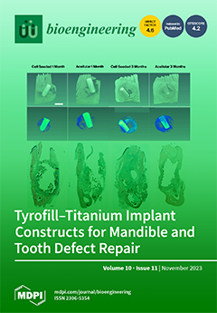
Current methods to repair CMF bone and tooth defects use a multi-step approach consisting of bone repair followed by dental implant placement. Here, we describe a novel CMF defect repair treatment consisting of TyroFill [E1001(1K)/dicalcium phosphate dihydrate (DCPD)] scaffolds supporting titanium dental implants. Human DPSC/HUVEC seeded constructs were grown in a critical-sized rabbit mandible defect for 1 or 3 months.  Micro-CT and histological/IHC analyses demonstrated that cell-seeded TyroFill constructs showed significant new bone formation around the implant, indicating the potential use of cell-seeded TyroFill scaffolds for coordinated bone-tooth defect repair. View this paper
  • Issues are regarded as officially published after their release is announced to the table of contents alert mailing list.
  • You may sign up for e-mail alerts to receive table of contents of newly released issues.
  • PDF is the official format for papers published in both, html and pdf forms. To view the papers in pdf format, click on the "PDF Full-text" link, and use the free Adobe Reader to open them.
Order results
Result details
Section
Select all
Export citation of selected articles as:
11 pages, 968 KiB  
Article
Improving the Accuracy of Otitis Media with Effusion Diagnosis in Pediatric Patients Using Deep Learning
by Jae-Hyuk Shim, Woongsang Sunwoo, Byung Yoon Choi, Kwang Gi Kim and Young Jae Kim
Bioengineering 2023, 10(11), 1337; https://doi.org/10.3390/bioengineering10111337 - 20 Nov 2023
Viewed by 1026
Abstract
Otitis media with effusion (OME), primarily seen in children aged 2 years and younger, is characterized by the presence of fluid in the middle ear, often resulting in hearing loss and aural fullness. While deep learning networks have been explored to aid OME [...] Read more.
Otitis media with effusion (OME), primarily seen in children aged 2 years and younger, is characterized by the presence of fluid in the middle ear, often resulting in hearing loss and aural fullness. While deep learning networks have been explored to aid OME diagnosis, prior work did not often specify if pediatric images were used for training, causing uncertainties about their clinical relevance, especially due to important distinctions between the tympanic membranes of small children and adults. We trained cross-validated ResNet50, DenseNet201, InceptionV3, and InceptionResNetV2 models on 1150 pediatric tympanic membrane images from otoendoscopes to classify OME. When assessed using a separate dataset of 100 pediatric tympanic membrane images, the models achieved mean accuracies of 92.9% (ResNet50), 97.2% (DenseNet201), 96.0% (InceptionV3), and 94.8% (InceptionResNetV2), compared to the seven otolaryngologists that achieved accuracies between 84.0% and 69.0%. The results showed that even the worst-performing model trained on fold 3 of InceptionResNetV2 with an accuracy of 88.0% exceeded the accuracy of the highest-performing otolaryngologist at 84.0%. Our findings suggest that these specifically trained deep learning models can potentially enhance the clinical diagnosis of OME using pediatric otoendoscopic tympanic membrane images. Full article
Show Figures

Figure 1

16 pages, 3491 KiB  
Article
Unilateral Mitochondrial–Hemodynamic Coupling and Bilateral Connectivity in the Prefrontal Cortices of Young and Older Healthy Adults
by Claire Sissons, Fiza Saeed, Caroline Carter, Kathy Lee, Kristen Kerr, Sadra Shahdadian and Hanli Liu
Bioengineering 2023, 10(11), 1336; https://doi.org/10.3390/bioengineering10111336 - 20 Nov 2023
Viewed by 924
Abstract
A recent study demonstrated that noninvasive measurements of cortical hemodynamics and metabolism in the resting human prefrontal cortex can facilitate quantitative metrics of unilateral mitochondrial–hemodynamic coupling and bilateral connectivity in infraslow oscillation frequencies in young adults. The infraslow oscillation includes three distinct vasomotions [...] Read more.
A recent study demonstrated that noninvasive measurements of cortical hemodynamics and metabolism in the resting human prefrontal cortex can facilitate quantitative metrics of unilateral mitochondrial–hemodynamic coupling and bilateral connectivity in infraslow oscillation frequencies in young adults. The infraslow oscillation includes three distinct vasomotions with endogenic (E), neurogenic (N), and myogenic (M) frequency bands. The goal of this study was to prove the hypothesis that there are significant differences between young and older adults in the unilateral coupling (uCOP) and bilateral connectivity (bCON) in the prefrontal cortex. Accordingly, we performed measurements from 24 older adults (67.2 ± 5.9 years of age) using the same two-channel broadband near-infrared spectroscopy (bbNIRS) setup and resting-state experimental protocol as those in the recent study. After quantification of uCOP and bCON in three E/N/M frequencies and statistical analysis, we demonstrated that older adults had significantly weaker bilateral hemodynamic connectivity but significantly stronger bilateral metabolic connectivity than young adults in the M band. Furthermore, older adults exhibited significantly stronger unilateral coupling on both prefrontal sides in all E/N/M bands, particularly with a very large effect size in the M band (>1.9). These age-related results clearly support our hypothesis and were well interpreted following neurophysiological principles. The key finding of this paper is that the neurophysiological metrics of uCOP and bCON are highly associated with age and may have the potential to become meaningful features for human brain health and be translatable for future clinical applications, such as the early detection of Alzheimer’s disease. Full article
Show Figures

Figure 1

11 pages, 761 KiB  
Article
Diagnostic Validation of the Screening Corneal Objective Risk of Ectasia Analyzer Evaluated by Swept Source Optical Coherence Tomography for Keratoconus in an Asian Population
by Kookyoung Kim, Kyungmin Koh, Seongjun Lee and Yongwoo Lee
Bioengineering 2023, 10(11), 1335; https://doi.org/10.3390/bioengineering10111335 - 20 Nov 2023
Viewed by 1136
Abstract
We aimed to investigate the diagnostic accuracy of Screening Corneal Objective Risk of Ectasia (SCORE) Analyzer software using ANTERION, a swept-source optical coherence tomography device, for keratoconus diagnosis in an Asian population. A total of 151 eyes of 151 patients were included in [...] Read more.
We aimed to investigate the diagnostic accuracy of Screening Corneal Objective Risk of Ectasia (SCORE) Analyzer software using ANTERION, a swept-source optical coherence tomography device, for keratoconus diagnosis in an Asian population. A total of 151 eyes of 151 patients were included in this retrospective study as follows: 60, 45, and 46 keratoconus, keratoconus suspects, and normal control eyes, respectively. Parameters in the SCORE calculation, including six indices, were compared for the three groups. The receiver operating characteristic curve analysis and cut-off value were estimated to assess the diagnostic ability to differentiate keratoconus and keratoconus suspect eyes from the normal group. The SCORE value and six indices were significantly correlated—“AntK max” (R = 0.864), “AntK oppoK” (R = 0.866), “Ant inf supK” (R = 0.943), “Ant irre 3mm” (R = 0.741), “post elevation at the thinnest point” (R = 0.943), and “minimum corneal thickness” (R = −0.750). The SCORE value showed high explanatory power (98.1%), sensitivity of 81.9%, and specificity of 78.3% (cut-off value: 0.25) in diagnosing normal eyes from the keratoconus suspect and keratoconus eyes. The SCORE Analyzer was found to be valid and consistent, showing good sensitivity and specificity for keratoconus detection in an Asian population. Full article
Show Figures

Figure 1

12 pages, 2978 KiB  
Article
Evaluating the Effectiveness of 2D and 3D CT Image Features for Predicting Tumor Response to Chemotherapy
by Neman Abdoli, Ke Zhang, Patrik Gilley, Xuxin Chen, Youkabed Sadri, Theresa Thai, Lauren Dockery, Kathleen Moore, Robert Mannel and Yuchen Qiu
Bioengineering 2023, 10(11), 1334; https://doi.org/10.3390/bioengineering10111334 - 20 Nov 2023
Cited by 1 | Viewed by 1045
Abstract
Background and Objective: 2D and 3D tumor features are widely used in a variety of medical image analysis tasks. However, for chemotherapy response prediction, the effectiveness between different kinds of 2D and 3D features are not comprehensively assessed, especially in ovarian-cancer-related applications. This [...] Read more.
Background and Objective: 2D and 3D tumor features are widely used in a variety of medical image analysis tasks. However, for chemotherapy response prediction, the effectiveness between different kinds of 2D and 3D features are not comprehensively assessed, especially in ovarian-cancer-related applications. This investigation aims to accomplish such a comprehensive evaluation. Methods: For this purpose, CT images were collected retrospectively from 188 advanced-stage ovarian cancer patients. All the metastatic tumors that occurred in each patient were segmented and then processed by a set of six filters. Next, three categories of features, namely geometric, density, and texture features, were calculated from both the filtered results and the original segmented tumors, generating a total of 1403 and 1595 features for the 2D and 3D tumors, respectively. In addition to the conventional single-slice 2D and full-volume 3D tumor features, we also computed the incomplete-3D tumor features, which were achieved by sequentially adding one individual CT slice and calculating the corresponding features. Support vector machine (SVM)-based prediction models were developed and optimized for each feature set. Five-fold cross-validation was used to assess the performance of each individual model. Results: The results show that the 2D feature-based model achieved an AUC (area under the ROC curve (receiver operating characteristic)) of 0.84 ± 0.02. When adding more slices, the AUC first increased to reach the maximum and then gradually decreased to 0.86 ± 0.02. The maximum AUC was yielded when adding two adjacent slices, with a value of 0.91 ± 0.01. Conclusions: This initial result provides meaningful information for optimizing machine learning-based decision-making support tools in the future. Full article
Show Figures

Figure 1

11 pages, 3728 KiB  
Article
SpineHRformer: A Transformer-Based Deep Learning Model for Automatic Spine Deformity Assessment with Prospective Validation
by Moxin Zhao, Nan Meng, Jason Pui Yin Cheung, Chenxi Yu, Pengyu Lu and Teng Zhang
Bioengineering 2023, 10(11), 1333; https://doi.org/10.3390/bioengineering10111333 - 20 Nov 2023
Viewed by 1100
Abstract
The Cobb angle (CA) serves as the principal method for assessing spinal deformity, but manual measurements of the CA are time-consuming and susceptible to inter- and intra-observer variability. While learning-based methods, such as SpineHRNet+, have demonstrated potential in automating CA measurement, their accuracy [...] Read more.
The Cobb angle (CA) serves as the principal method for assessing spinal deformity, but manual measurements of the CA are time-consuming and susceptible to inter- and intra-observer variability. While learning-based methods, such as SpineHRNet+, have demonstrated potential in automating CA measurement, their accuracy can be influenced by the severity of spinal deformity, image quality, relative position of rib and vertebrae, etc. Our aim is to create a reliable learning-based approach that provides consistent and highly accurate measurements of the CA from posteroanterior (PA) X-rays, surpassing the state-of-the-art method. To accomplish this, we introduce SpineHRformer, which identifies anatomical landmarks, including the vertices of endplates from the 7th cervical vertebra (C7) to the 5th lumbar vertebra (L5) and the end vertebrae with different output heads, enabling the calculation of CAs. Within our SpineHRformer, a backbone HRNet first extracts multi-scale features from the input X-ray, while transformer blocks extract local and global features from the HRNet outputs. Subsequently, an output head to generate heatmaps of the endplate landmarks or end vertebra landmarks facilitates the computation of CAs. We used a dataset of 1934 PA X-rays with diverse degrees of spinal deformity and image quality, following an 8:2 ratio to train and test the model. The experimental results indicate that SpineHRformer outperforms SpineHRNet+ in landmark detection (Mean Euclidean Distance: 2.47 pixels vs. 2.74 pixels), CA prediction (Pearson correlation coefficient: 0.86 vs. 0.83), and severity grading (sensitivity: normal-mild; 0.93 vs. 0.74, moderate; 0.74 vs. 0.77, severe; 0.74 vs. 0.7). Our approach demonstrates greater robustness and accuracy compared to SpineHRNet+, offering substantial potential for improving the efficiency and reliability of CA measurements in clinical settings. Full article
(This article belongs to the Special Issue Artificial Intelligence in Auto-Diagnosis and Clinical Applications)
Show Figures

Figure 1

18 pages, 3655 KiB  
Article
Enhancing the Super-Resolution of Medical Images: Introducing the Deep Residual Feature Distillation Channel Attention Network for Optimized Performance and Efficiency
by Sabina Umirzakova, Sevara Mardieva, Shakhnoza Muksimova, Shabir Ahmad and Taegkeun Whangbo
Bioengineering 2023, 10(11), 1332; https://doi.org/10.3390/bioengineering10111332 - 19 Nov 2023
Cited by 2 | Viewed by 1517
Abstract
In the advancement of medical image super-resolution (SR), the Deep Residual Feature Distillation Channel Attention Network (DRFDCAN) marks a significant step forward. This work presents DRFDCAN, a model that innovates traditional SR approaches by introducing a channel attention block that is tailored for [...] Read more.
In the advancement of medical image super-resolution (SR), the Deep Residual Feature Distillation Channel Attention Network (DRFDCAN) marks a significant step forward. This work presents DRFDCAN, a model that innovates traditional SR approaches by introducing a channel attention block that is tailored for high-frequency features—crucial for the nuanced details in medical diagnostics—while streamlining the network structure for enhanced computational efficiency. DRFDCAN’s architecture adopts a residual-within-residual design to facilitate faster inference and reduce memory demands without compromising the integrity of the image reconstruction. This design strategy, combined with an innovative feature extraction method that emphasizes the utility of the initial layer features, allows for improved image clarity and is particularly effective in optimizing the peak signal-to-noise ratio (PSNR). The proposed work redefines efficiency in SR models, outperforming established frameworks like RFDN by improving model compactness and accelerating inference. The meticulous crafting of a feature extractor that effectively captures edge and texture information exemplifies the model’s capacity to render detailed images, necessary for accurate medical analysis. The implications of this study are two-fold: it presents a viable solution for deploying SR technology in real-time medical applications, and it sets a precedent for future models that address the delicate balance between computational efficiency and high-fidelity image reconstruction. This balance is paramount in medical applications where the clarity of images can significantly influence diagnostic outcomes. The DRFDCAN model thus stands as a transformative contribution to the field of medical image super-resolution. Full article
(This article belongs to the Special Issue Artificial Intelligence (AI) for Medical Image Processing)
Show Figures

Figure 1

11 pages, 1637 KiB  
Article
Evaluation of the Impact of Calcium Silicate-Based Sealer Insertion Technique on Root Canal Obturation Quality: A Micro-Computed Tomography Study
by Germain Sfeir, Frédéric Bukiet, Marc Krikor Kaloustian, Naji Kharouf, Lotfi Slimani, Baptiste Casel and Carla Zogheib
Bioengineering 2023, 10(11), 1331; https://doi.org/10.3390/bioengineering10111331 - 19 Nov 2023
Cited by 1 | Viewed by 1010
Abstract
Background: Calcium silicate-based sealers have gained in popularity over time due to their physicochemical/biological properties and their possible use with single-cone obturation. The single cone technique is a sealer-based obturation and there is still a knowledge gap regarding the potential impact of the [...] Read more.
Background: Calcium silicate-based sealers have gained in popularity over time due to their physicochemical/biological properties and their possible use with single-cone obturation. The single cone technique is a sealer-based obturation and there is still a knowledge gap regarding the potential impact of the sealer insertion method on the root canal-filling quality. Therefore, the aim of this micro-CT study was to assess the impact of the calcium silicate-based sealer insertion technique on void occurrence and on the sealer extrusion following single-cone obturation. Methods: Thirty-six single-rooted mandibular premolars with one canal were shaped with Reciproc® R25 (VDW, Munich, Germany) then divided randomly into four groups of nine canals, each depending on the TotalFill® BC Sealer insertion technique used with single cone obturation: injection in the coronal two-thirds (group A); injection in the coronal two-thirds followed by direct sonic activation (group B); injection in the coronal two-thirds followed by indirect ultrasonic activation on tweezers (group C); sealer applied only on the master-cone (control group D). Samples were then scanned using micro-CT for voids and sealer extrusion calculation. Data were statistically analyzed using v.26 IBM; Results: No statistically significant differences were noted between the four groups in terms of voids; nevertheless, sonic activation (group B) followed by ultrasonic activation on the tweezers (group C) showed the best results (p = 0.066). Group D showed significantly less sealer extrusion when compared with group C (p = 0.044), with no statistically significant differences between groups D, A and B (p > 0.05). Conclusions: Despite no significant differences observed between the different sealer placement techniques, the use of sonic and ultrasonic activation might be promising to reduce void occurrence. Further investigations are needed to demonstrate the potential benefit of calcium silicate-based sealer activation especially in wide and oval root canals in order to improve the quality of the single-cone obturation. Full article
(This article belongs to the Section Regenerative Engineering)
Show Figures

Figure 1

12 pages, 9407 KiB  
Article
Functional Load Capacity of Teeth with Reduced Periodontal Support: A Finite Element Analysis
by Marco Dederichs, Paul Joedecke, Christian-Toralf Weber and Arndt Guentsch
Bioengineering 2023, 10(11), 1330; https://doi.org/10.3390/bioengineering10111330 - 18 Nov 2023
Cited by 1 | Viewed by 1290
Abstract
The purpose of this study was to investigate the functional load capacity of the periodontal ligament (PDL) in a full arch maxilla and mandible model using a numerical simulation. The goal was to determine the functional load pattern in multi- and single-rooted teeth [...] Read more.
The purpose of this study was to investigate the functional load capacity of the periodontal ligament (PDL) in a full arch maxilla and mandible model using a numerical simulation. The goal was to determine the functional load pattern in multi- and single-rooted teeth with full and reduced periodontal support. CBCT data were used to create 3D models of a maxilla and mandible. The DICOM dataset was used to create a CAD model. For a precise description of the surfaces of each structure (enamel, dentin, cementum, pulp, PDL, gingiva, bone), each tooth was segmented separately, and the biomechanical characteristics were considered. Finite Element Analysis (FEA) software computed the biomechanical behavior of the stepwise increased force of 700 N in the cranial and 350 N in the ventral direction of the muscle approach of the masseter muscle. The periodontal attachment (cementum–PDL–bone contact) was subsequently reduced in 1 mm increments, and the simulation was repeated. Quantitative (pressure, tension, and deformation) and qualitative (color-coded images) data were recorded and descriptively analyzed. The teeth with the highest load capacities were the upper and lower molars (0.4–0.6 MPa), followed by the premolars (0.4–0.5 MPa) and canines (0.3–0.4 MPa) when vertically loaded. Qualitative data showed that the areas with the highest stress in the PDL were single-rooted teeth in the cervical and apical area and molars in the cervical and apical area in addition to the furcation roof. In both single- and multi-rooted teeth, the gradual reduction in bone levels caused an increase in the load on the remaining PDL. Cervical and apical areas, as well as the furcation roof, are the zones with the highest functional stress. The greater the bone loss, the higher the mechanical load on the residual periodontal supporting structures. Full article
(This article belongs to the Special Issue Computational Biomechanics, Volume II)
Show Figures

Figure 1

18 pages, 3009 KiB  
Article
Genomic Insights on the Carbon-Negative Workhorse: Systematical Comparative Genomic Analysis on 56 Synechococcus Strains
by Meiwen Qian, Xiao Han, Jiongqin Liu, Ping Xu and Fei Tao
Bioengineering 2023, 10(11), 1329; https://doi.org/10.3390/bioengineering10111329 - 18 Nov 2023
Viewed by 1444
Abstract
Synechococcus, a type of ancient photosynthetic cyanobacteria, is crucial in modern carbon-negative synthetic biology due to its potential for producing bioenergy and high-value products. With its high biomass, fast growth rate, and established genetic manipulation tools, Synechococcus has become a research focus [...] Read more.
Synechococcus, a type of ancient photosynthetic cyanobacteria, is crucial in modern carbon-negative synthetic biology due to its potential for producing bioenergy and high-value products. With its high biomass, fast growth rate, and established genetic manipulation tools, Synechococcus has become a research focus in recent years. Abundant germplasm resources have been accumulated from various habitats, including temperature and salinity conditions relevant to industrialization. In this study, a comprehensive analysis of complete genomes of the 56 Synechococcus strains currently available in public databases was performed, clarifying genetic relationships, the adaptability of Synechococcus to the environment, and its reflection at the genomic level. This was carried out via pan-genome analysis and a detailed comparison of the functional gene groups. The results revealed an open-genome pattern, with 275 core genes and variable genome sizes within these strains. The KEGG annotation and orthology composition comparisons unveiled that the cold and thermophile strains have 32 and 84 unique KO functional units in their shared core gene functional units, respectively. Each KO functional unit reflects unique gene families and pathways. In terms of salt tolerance and comparative genomics, there are 65 unique KO functional units in freshwater-adapted strains and 154 in strictly marine strains. By delving into these aspects, our understanding of the metabolic potential of Synechococcus was deepened, promoting the development and industrial application of cyanobacterial biotechnology. Full article
(This article belongs to the Section Biochemical Engineering)
Show Figures

Figure 1

31 pages, 8260 KiB  
Review
Exo Supportive Devices: Summary of Technical Aspects
by António Diogo André and Pedro Martins
Bioengineering 2023, 10(11), 1328; https://doi.org/10.3390/bioengineering10111328 - 17 Nov 2023
Viewed by 1355
Abstract
Human societies have been trying to mitigate the suffering of individuals with physical impairments, with a special effort in the last century. In the 1950s, a new concept arose, finding similarities between animal exoskeletons, and with the goal of medically aiding human movement [...] Read more.
Human societies have been trying to mitigate the suffering of individuals with physical impairments, with a special effort in the last century. In the 1950s, a new concept arose, finding similarities between animal exoskeletons, and with the goal of medically aiding human movement (for rehabilitation applications). There have been several studies on using exosuits with this purpose in mind. So, the current review offers a critical perspective and a detailed analysis of the steps and key decisions involved in the conception of an exoskeleton. Choices such as design aspects, base materials (structure), actuators (force and motion), energy sources (actuation), and control systems will be discussed, pointing out their advantages and disadvantages. Moreover, examples of exosuits (full-body, upper-body, and lower-body devices) will be presented and described, including their use cases and outcomes. The future of exoskeletons as possible assisted movement solutions will be discussed—pointing to the best options for rehabilitation. Full article
(This article belongs to the Special Issue Bioengineering for Physical Rehabilitation)
Show Figures

Graphical abstract

19 pages, 7438 KiB  
Article
Evaluation of Cytocompatibility of PEEK-Based Composites as a Function of Manufacturing Processes
by Jorge Gil-Albarova, María José Martínez-Morlanes, José Miguel Fernández, Pere Castell, Luis Gracia and José Antonio Puértolas
Bioengineering 2023, 10(11), 1327; https://doi.org/10.3390/bioengineering10111327 - 17 Nov 2023
Viewed by 890
Abstract
The biocompatible polymer polyetheretherketone (PEEK) is a suitable candidate to be part of potential all-polymer total joint replacements, provided its use is associated with better osseointegration, mechanical performance, and wear resistance. Seeking to meet the aforementioned requirements, respectively, we have manufactured a PEEK [...] Read more.
The biocompatible polymer polyetheretherketone (PEEK) is a suitable candidate to be part of potential all-polymer total joint replacements, provided its use is associated with better osseointegration, mechanical performance, and wear resistance. Seeking to meet the aforementioned requirements, respectively, we have manufactured a PEEK composite with different fillers: carbon fibers (CF), hydroxyapatite particles (HA) and graphene platelets (GNP). The mechanical outcomes of the composites with combinations of 0, 1.5, 3.0 wt% GNP, 5 and 15 wt% HA and 30% of wt% CF concentrations pointed out that one of the best filler combinations to achieve the previous objectives was 30 wt% CF, 8 wt% HA and 2 wt% of GNP. The study compares the bioactivity of human osteoblasts on this composite prepared by injection molding with that on the material manufactured by the Fused Filament Fabrication 3D additive technique. The results indicate that the surface adhesion and proliferation of human osteoblasts over time are better with the composite obtained by injection molding than that obtained by 3D printing. This result is more closely correlated with morphological parameters of the composite surface than its wettability behavior. Full article
(This article belongs to the Section Biomedical Engineering and Biomaterials)
Show Figures

Graphical abstract

15 pages, 8210 KiB  
Article
Novel Procedure for Automatic Registration between Cone-Beam Computed Tomography and Intraoral Scan Data Supported with 3D Segmentation
by Yoon-Ji Kim, Jang-Hoon Ahn, Hyun-Kyo Lim, Thong Phi Nguyen, Nayansi Jha, Ami Kim and Jonghun Yoon
Bioengineering 2023, 10(11), 1326; https://doi.org/10.3390/bioengineering10111326 - 17 Nov 2023
Viewed by 1078
Abstract
In contemporary practice, intraoral scans and cone-beam computed tomography (CBCT) are widely adopted techniques for tooth localization and the acquisition of comprehensive three-dimensional models. Despite their utility, each dataset presents inherent merits and limitations, prompting the pursuit of an amalgamated solution for optimization. [...] Read more.
In contemporary practice, intraoral scans and cone-beam computed tomography (CBCT) are widely adopted techniques for tooth localization and the acquisition of comprehensive three-dimensional models. Despite their utility, each dataset presents inherent merits and limitations, prompting the pursuit of an amalgamated solution for optimization. Thus, this research introduces a novel 3D registration approach aimed at harmonizing these distinct datasets to offer a holistic perspective. In the pre-processing phase, a retrained Mask-RCNN is deployed on both sagittal and panoramic projections to partition upper and lower teeth from the encompassing CBCT raw data. Simultaneously, a chromatic classification model is proposed for segregating gingival tissue from tooth structures in intraoral scan data. Subsequently, the segregated datasets are aligned based on dental crowns, employing the robust RANSAC and ICP algorithms. To assess the proposed methodology’s efficacy, the Euclidean distance between corresponding points is statistically evaluated. Additionally, dental experts, including two orthodontists and an experienced general dentist, evaluate the clinical potential by measuring distances between landmarks on tooth surfaces. The computed error in corresponding point distances between intraoral scan data and CBCT data in the automatically registered datasets utilizing the proposed technique is quantified at 0.234 ± 0.019 mm, which is significantly below the 0.3 mm CBCT voxel size. Moreover, the average measurement discrepancy among expert-identified landmarks ranges from 0.368 to 1.079 mm, underscoring the promise of the proposed method. Full article
(This article belongs to the Section Biomedical Engineering and Biomaterials)
Show Figures

Figure 1

12 pages, 2823 KiB  
Article
Speech Perception Improvement Algorithm Based on a Dual-Path Long Short-Term Memory Network
by Hyeong Il Koh, Sungdae Na and Myoung Nam Kim
Bioengineering 2023, 10(11), 1325; https://doi.org/10.3390/bioengineering10111325 - 16 Nov 2023
Viewed by 890
Abstract
Current deep learning-based speech enhancement methods focus on enhancing the time–frequency representation of the signal. However, conventional methods can lead to speech damage due to resolution mismatch problems that emphasize only specific information in the time or frequency domain. To address these challenges, [...] Read more.
Current deep learning-based speech enhancement methods focus on enhancing the time–frequency representation of the signal. However, conventional methods can lead to speech damage due to resolution mismatch problems that emphasize only specific information in the time or frequency domain. To address these challenges, this paper introduces a speech enhancement model designed with a dual-path structure that identifies key speech characteristics in both the time and time–frequency domains. Specifically, the time path aims to model semantic features hidden in the waveform, while the time–frequency path attempts to compensate for the spectral details via a spectral extension block. These two paths enhance temporal and spectral features via mask functions modeled as LSTM, respectively, offering a comprehensive approach to speech enhancement. Experimental results show that the proposed dual-path LSTM network consistently outperforms conventional single-domain speech enhancement methods in terms of speech quality and intelligibility. Full article
Show Figures

Figure 1

18 pages, 4633 KiB  
Article
Gesture Classification in Electromyography Signals for Real-Time Prosthetic Hand Control Using a Convolutional Neural Network-Enhanced Channel Attention Model
by Guangjie Yu, Ziting Deng, Zhenchen Bao, Yue Zhang and Bingwei He
Bioengineering 2023, 10(11), 1324; https://doi.org/10.3390/bioengineering10111324 - 16 Nov 2023
Viewed by 1302
Abstract
Accurate and real-time gesture recognition is required for the autonomous operation of prosthetic hand devices. This study employs a convolutional neural network-enhanced channel attention (CNN-ECA) model to provide a unique approach for surface electromyography (sEMG) gesture recognition. The introduction of the ECA module [...] Read more.
Accurate and real-time gesture recognition is required for the autonomous operation of prosthetic hand devices. This study employs a convolutional neural network-enhanced channel attention (CNN-ECA) model to provide a unique approach for surface electromyography (sEMG) gesture recognition. The introduction of the ECA module improves the model’s capacity to extract features and focus on critical information in the sEMG data, thus simultaneously equipping the sEMG-controlled prosthetic hand systems with the characteristics of accurate gesture detection and real-time control. Furthermore, we suggest a preprocessing strategy for extracting envelope signals that incorporates Butterworth low-pass filtering and the fast Hilbert transform (FHT), which can successfully reduce noise interference and capture essential physiological information. Finally, the majority voting window technique is adopted to enhance the prediction results, further improving the accuracy and stability of the model. Overall, our multi-layered convolutional neural network model, in conjunction with envelope signal extraction and attention mechanisms, offers a promising and innovative approach for real-time control systems in prosthetic hands, allowing for precise fine motor actions. Full article
Show Figures

Graphical abstract

21 pages, 371 KiB  
Review
Effects of Plasma Treatment on the Strength of Bonding to Ceramic Surfaces in Orthodontics—A Comprehensive Review
by Elizabeth Gershater, Olivia Griswold, Brooke E. Talsania, Yu Zhang, Chun-Hsi Chung, Zhong Zheng and Chenshuang Li
Bioengineering 2023, 10(11), 1323; https://doi.org/10.3390/bioengineering10111323 - 16 Nov 2023
Viewed by 931
Abstract
Over the past several decades, orthodontic treatment has been increasingly sought out by adults, many of whom have undergone restorative dental procedures that cover enamel. Because the characteristics of restorative materials differ from those of enamel, typical bonding techniques do not yield excellent [...] Read more.
Over the past several decades, orthodontic treatment has been increasingly sought out by adults, many of whom have undergone restorative dental procedures that cover enamel. Because the characteristics of restorative materials differ from those of enamel, typical bonding techniques do not yield excellent restoration–bracket bonding strengths. Plasma treatment is an emerging surface treatment that could potentially improve bonding properties. The purpose of this paper is to evaluate currently available studies assessing the effect of plasma treatment on the shear bond strength (SBS) and failure mode of resin cement/composite on the surface of ceramic materials. PubMed and Google Scholar databases were searched for relevant studies, which were categorized by restorative material and plasma treatment types that were evaluated. It was determined that cold atmospheric plasma (CAP) treatment using helium and H2O gas was effective at raising the SBS of feldspathic porcelain to a bonding agent, while CAP treatment using helium gas might also be a potential treatment method for zirconia and other types of ceramics. More importantly, CAP treatment using helium has the potential for being carried out chairside due to its non-toxicity, low temperature, and short treatment time. However, because all the studies were conducted in vitro and not tested in an orthodontic setting, further research must be conducted to ascertain the effectiveness of specific plasma treatments in comparison to current orthodontic bonding treatments in vivo. Full article
(This article belongs to the Special Issue Application of Bioengineering to Clinical Orthodontics)
12 pages, 7066 KiB  
Article
Artificial Intelligence Algorithms for Benign vs. Malignant Dermoscopic Skin Lesion Image Classification
by Francesca Brutti, Federica La Rosa, Linda Lazzeri, Chiara Benvenuti, Giovanni Bagnoni, Daniela Massi and Marco Laurino
Bioengineering 2023, 10(11), 1322; https://doi.org/10.3390/bioengineering10111322 - 16 Nov 2023
Viewed by 1081
Abstract
In recent decades, the incidence of melanoma has grown rapidly. Hence, early diagnosis is crucial to improving clinical outcomes. Here, we propose and compare a classical image analysis-based machine learning method with a deep learning one to automatically classify benign vs. malignant dermoscopic [...] Read more.
In recent decades, the incidence of melanoma has grown rapidly. Hence, early diagnosis is crucial to improving clinical outcomes. Here, we propose and compare a classical image analysis-based machine learning method with a deep learning one to automatically classify benign vs. malignant dermoscopic skin lesion images. The same dataset of 25,122 publicly available dermoscopic images was used to train both models, while a disjointed test set of 200 images was used for the evaluation phase. The training dataset was randomly divided into 10 datasets of 19,932 images to obtain an equal distribution between the two classes. By testing both models on the disjoint set, the deep learning-based method returned accuracy of 85.4 ± 3.2% and specificity of 75.5 ± 7.6%, while the machine learning one showed accuracy and specificity of 73.8 ± 1.1% and 44.5 ± 4.7%, respectively. Although both approaches performed well in the validation phase, the convolutional neural network outperformed the ensemble boosted tree classifier on the disjoint test set, showing better generalization ability. The integration of new melanoma detection algorithms with digital dermoscopic devices could enable a faster screening of the population, improve patient management, and achieve better survival rates. Full article
Show Figures

Figure 1

14 pages, 842 KiB  
Systematic Review
Non-Invasive Wearable Devices for Monitoring Vital Signs in Patients with Type 2 Diabetes Mellitus: A Systematic Review
by Artur Piet, Lennart Jablonski, Jennifer I. Daniel Onwuchekwa, Steffen Unkel, Christian Weber, Marcin Grzegorzek, Jan P. Ehlers, Olaf Gaus and Thomas Neumann
Bioengineering 2023, 10(11), 1321; https://doi.org/10.3390/bioengineering10111321 - 16 Nov 2023
Cited by 1 | Viewed by 1695
Abstract
Type 2 diabetes mellitus (T2D) poses a significant global health challenge and demands effective self-management strategies, including continuous blood glucose monitoring (CGM) and lifestyle adaptations. While CGM offers real-time glucose level assessment, the quest for minimizing trauma and enhancing convenience has spurred the [...] Read more.
Type 2 diabetes mellitus (T2D) poses a significant global health challenge and demands effective self-management strategies, including continuous blood glucose monitoring (CGM) and lifestyle adaptations. While CGM offers real-time glucose level assessment, the quest for minimizing trauma and enhancing convenience has spurred the need to explore non-invasive alternatives for monitoring vital signs in patients with T2D. Objective: This systematic review is the first that explores the current literature and critically evaluates the use and reporting of non-invasive wearable devices for monitoring vital signs in patients with T2D. Methods: Employing the PRISMA and PICOS guidelines, we conducted a comprehensive search to incorporate evidence from relevant studies, focusing on randomized controlled trials (RCTs), systematic reviews, and meta-analyses published since 2017. Of the 437 publications identified, seven were selected based on predetermined criteria. Results: The seven studies included in this review used various sensing technologies, such as heart rate monitors, accelerometers, and other wearable devices. Primary health outcomes included blood pressure measurements, heart rate, body fat percentage, and cardiorespiratory endurance. Non-invasive wearable devices demonstrated potential for aiding T2D management, albeit with variations in efficacy across studies. Conclusions: Based on the low number of studies with higher evidence levels (i.e., RCTs) that we were able to find and the significant differences in design between these studies, we conclude that further evidence is required to validate the application, efficacy, and real-world impact of these wearable devices. Emphasizing transparency in bias reporting and conducting in-depth research is crucial for fully understanding the implications and benefits of wearable devices in T2D management. Full article
(This article belongs to the Special Issue Monitoring and Analysis of Human Biosignals, Volume II)
Show Figures

Figure 1

14 pages, 661 KiB  
Review
Mathematical and Machine Learning Models of Renal Cell Carcinoma: A Review
by Dilruba Sofia, Qilu Zhou and Leili Shahriyari
Bioengineering 2023, 10(11), 1320; https://doi.org/10.3390/bioengineering10111320 - 16 Nov 2023
Viewed by 995
Abstract
This review explores the multifaceted landscape of renal cell carcinoma (RCC) by delving into both mechanistic and machine learning models. While machine learning models leverage patients’ gene expression and clinical data through a variety of techniques to predict patients’ outcomes, mechanistic models focus [...] Read more.
This review explores the multifaceted landscape of renal cell carcinoma (RCC) by delving into both mechanistic and machine learning models. While machine learning models leverage patients’ gene expression and clinical data through a variety of techniques to predict patients’ outcomes, mechanistic models focus on investigating cells’ and molecules’ interactions within RCC tumors. These interactions are notably centered around immune cells, cytokines, tumor cells, and the development of lung metastases. The insights gained from both machine learning and mechanistic models encompass critical aspects such as signature gene identification, sensitive interactions in the tumors’ microenvironments, metastasis development in other organs, and the assessment of survival probabilities. By reviewing the models of RCC, this study aims to shed light on opportunities for the integration of machine learning and mechanistic modeling approaches for treatment optimization and the identification of specific targets, all of which are essential for enhancing patient outcomes. Full article
(This article belongs to the Special Issue Mathematical and Computational Modeling of Cancer Progression)
Show Figures

Figure 1

15 pages, 3645 KiB  
Article
Automatic Segmentation and Assessment of Valvular Regurgitations with Color Doppler Echocardiography Images: A VABC-UNet-Based Framework
by Jun Huang, Aiyue Huang, Ruqin Xu, Musheng Wu, Peng Wang and Qing Wang
Bioengineering 2023, 10(11), 1319; https://doi.org/10.3390/bioengineering10111319 - 16 Nov 2023
Viewed by 934
Abstract
This study investigated the automatic segmentation and classification of mitral regurgitation (MR) and tricuspid regurgitation (TR) using a deep learning-based method, aiming to improve the efficiency and accuracy of diagnosis of valvular regurgitations. A VABC-UNet model was proposed consisting of VGG16 encoder, U-Net [...] Read more.
This study investigated the automatic segmentation and classification of mitral regurgitation (MR) and tricuspid regurgitation (TR) using a deep learning-based method, aiming to improve the efficiency and accuracy of diagnosis of valvular regurgitations. A VABC-UNet model was proposed consisting of VGG16 encoder, U-Net decoder, batch normalization, attention block and deepened convolution layer based on the U-Net backbone. Then, a VABC-UNet-based assessment framework was established for automatic segmentation, classification, and evaluation of valvular regurgitations. A total of 315 color Doppler echocardiography images of MR and/or TR in an apical four-chamber view were collected, including 35 images in the test dataset and 280 images in the training dataset. In comparison with the classic U-Net and VGG16-UNet models, the segmentation performance of the VABC-UNet model was evaluated via four metrics: Dice, Jaccard, Precision, and Recall. According to the features of regurgitation jet and atrium, the regurgitation could automatically be classified into MR or TR, and evaluated to mild, moderate, moderate–severe, or severe grade by the framework. The results show that the VABC-UNet model has a superior performance in the segmentation of valvular regurgitation jets and atria to the other two models and consequently a higher accuracy of classification and evaluation. There were fewer pseudo- and over-segmentations by the VABC-UNet model and the values of the metrics significantly improved (p < 0.05). The proposed VABC-UNet-based framework achieves automatic segmentation, classification, and evaluation of MR and TR, having potential to assist radiologists in clinical decision making of the regurgitations in valvular heart diseases. Full article
(This article belongs to the Special Issue Artificial Intelligence in Auto-Diagnosis and Clinical Applications)
Show Figures

Graphical abstract

15 pages, 3212 KiB  
Article
Enhancing COVID-19 Classification Accuracy with a Hybrid SVM-LR Model
by Noor Ilanie Nordin, Wan Azani Mustafa, Muhamad Safiih Lola, Elissa Nadia Madi, Anton Abdulbasah Kamil, Marah Doly Nasution, Abdul Aziz K. Abdul Hamid, Nurul Hila Zainuddin, Elayaraja Aruchunan and Mohd Tajuddin Abdullah
Bioengineering 2023, 10(11), 1318; https://doi.org/10.3390/bioengineering10111318 - 15 Nov 2023
Viewed by 1220
Abstract
Support ector achine (SVM) is a newer machine learning algorithm for classification, while logistic regression (LR) is an older statistical classification method. Despite the numerous studies contrasting SVM and LR, new improvements such as bagging and ensemble have been applied to them since [...] Read more.
Support ector achine (SVM) is a newer machine learning algorithm for classification, while logistic regression (LR) is an older statistical classification method. Despite the numerous studies contrasting SVM and LR, new improvements such as bagging and ensemble have been applied to them since these comparisons were made. This study proposes a new hybrid model based on SVM and LR for predicting small events per variable (EPV). The performance of the hybrid, SVM, and LR models with different EPV values was evaluated using COVID-19 data from December 2019 to May 2020 provided by the WHO. The study found that the hybrid model had better classification performance than SVM and LR in terms of accuracy, mean squared error (MSE), and root mean squared error (RMSE) for different EPV values. This hybrid model is particularly important for medical authorities and practitioners working in the face of future pandemics. Full article
(This article belongs to the Section Biosignal Processing)
Show Figures

Figure 1

14 pages, 3678 KiB  
Article
Quantitative Evaluation of Caries and Calculus with Ultrahigh-Resolution Optical Coherence Tomography
by Tai-Ang Wang, Nguyễn Hoàng Trung, Hsiang-Chieh Lee, Cheng-Kuang Lee, Meng-Tsan Tsai and Yen-Li Wang
Bioengineering 2023, 10(11), 1317; https://doi.org/10.3390/bioengineering10111317 - 15 Nov 2023
Viewed by 1184
Abstract
Dental caries on the crown’s surface is caused by the interaction of bacteria and carbohydrates, which then gradually alter the tooth’s structure. In addition, calculus is the root of periodontal disease. Optical coherence tomography (OCT) has been considered to be a promising tool [...] Read more.
Dental caries on the crown’s surface is caused by the interaction of bacteria and carbohydrates, which then gradually alter the tooth’s structure. In addition, calculus is the root of periodontal disease. Optical coherence tomography (OCT) has been considered to be a promising tool for identifying dental caries; however, diagnosing dental caries in the early stage still remains challenging. In this study, we proposed an ultrahigh-resolution OCT (UHR-OCT) system with axial and transverse resolutions of 2.6 and 1.8 μm for differentiating the early-stage dental caries and calculus. The same teeth were also scanned by a conventional spectral-domain OCT (SD-OCT) system with an axial resolution of 7 μm. The results indicated that early-stage carious structures such as small cavities can be observed using UHR-OCT; however, the SD-OCT system with a lower resolution had difficulty identifying it. Moreover, the estimated surface roughness and the scattering coefficient of enamel were proposed for quantitatively differentiating the different stages of caries. Furthermore, the thickness of the calculus can be estimated from the UHR-OCT results. The results have demonstrated that UHR-OCT can detect caries and calculus in their early stages, showing that the proposed method for the quantitative evaluation of caries and calculus is potentially promising. Full article
(This article belongs to the Special Issue Optical Techniques for Biomedical Engineering)
Show Figures

Graphical abstract

19 pages, 3452 KiB  
Article
Towards a Corpus (and Language)-Independent Screening of Parkinson’s Disease from Voice and Speech through Domain Adaptation
by Emiro J. Ibarra, Julián D. Arias-Londoño, Matías Zañartu and Juan I. Godino-Llorente
Bioengineering 2023, 10(11), 1316; https://doi.org/10.3390/bioengineering10111316 - 15 Nov 2023
Cited by 1 | Viewed by 1395
Abstract
End-to-end deep learning models have shown promising results for the automatic screening of Parkinson’s disease by voice and speech. However, these models often suffer degradation in their performance when applied to scenarios involving multiple corpora. In addition, they also show corpus-dependent clusterings. These [...] Read more.
End-to-end deep learning models have shown promising results for the automatic screening of Parkinson’s disease by voice and speech. However, these models often suffer degradation in their performance when applied to scenarios involving multiple corpora. In addition, they also show corpus-dependent clusterings. These facts indicate a lack of generalisation or the presence of certain shortcuts in the decision, and also suggest the need for developing new corpus-independent models. In this respect, this work explores the use of domain adversarial training as a viable strategy to develop models that retain their discriminative capacity to detect Parkinson’s disease across diverse datasets. The paper presents three deep learning architectures and their domain adversarial counterparts. The models were evaluated with sustained vowels and diadochokinetic recordings extracted from four corpora with different demographics, dialects or languages, and recording conditions. The results showed that the space distribution of the embedding features extracted by the domain adversarial networks exhibits a higher intra-class cohesion. This behaviour is supported by a decrease in the variability and inter-domain divergence computed within each class. The findings suggest that domain adversarial networks are able to learn the common characteristics present in Parkinsonian voice and speech, which are supposed to be corpus, and consequently, language independent. Overall, this effort provides evidence that domain adaptation techniques refine the existing end-to-end deep learning approaches for Parkinson’s disease detection from voice and speech, achieving more generalizable models. Full article
(This article belongs to the Special Issue Models and Analysis of Vocal Emissions for Biomedical Applications)
Show Figures

Figure 1

20 pages, 7102 KiB  
Article
Controlled-Release Hydrogel Microspheres to Deliver Multipotent Stem Cells for Treatment of Knee Osteoarthritis
by Megan Hamilton, Jinxi Wang, Prajnaparamita Dhar and Lisa Stehno-Bittel
Bioengineering 2023, 10(11), 1315; https://doi.org/10.3390/bioengineering10111315 - 15 Nov 2023
Cited by 2 | Viewed by 1630
Abstract
Osteoarthritis (OA) is the most common form of joint disease affecting articular cartilage and peri-articular tissues. Traditional treatments are insufficient, as they are aimed at mitigating symptoms. Multipotent Stromal Cell (MSC) therapy has been proposed as a treatment capable of both preventing cartilage [...] Read more.
Osteoarthritis (OA) is the most common form of joint disease affecting articular cartilage and peri-articular tissues. Traditional treatments are insufficient, as they are aimed at mitigating symptoms. Multipotent Stromal Cell (MSC) therapy has been proposed as a treatment capable of both preventing cartilage destruction and treating symptoms. While many studies have investigated MSCs for treating OA, therapeutic success is often inconsistent due to low MSC viability and retention in the joint. To address this, biomaterial-assisted delivery is of interest, particularly hydrogel microspheres, which can be easily injected into the joint. Microspheres composed of hyaluronic acid (HA) were created as MSC delivery vehicles. Microrheology measurements indicated that the microspheres had structural integrity alongside sufficient permeability. Additionally, encapsulated MSC viability was found to be above 70% over one week in culture. Gene expression analysis of MSC-identifying markers showed no change in CD29 levels, increased expression of CD44, and decreased expression of CD90 after one week of encapsulation. Analysis of chondrogenic markers showed increased expressions of aggrecan (ACAN) and SRY-box transcription factor 9 (SOX9), and decreased expression of osteogenic markers, runt-related transcription factor 2 (RUNX2), and alkaline phosphatase (ALPL). In vivo analysis revealed that HA microspheres remained in the joint for up to 6 weeks. Rats that had undergone destabilization of the medial meniscus and had overt OA were treated with empty HA microspheres, MSC-laden microspheres, MSCs alone, or a control vehicle. Pain measurements taken before and after the treatment illustrated temporarily decreased pain in groups treated with encapsulated cells. Finally, the histopathological scoring of each group illustrated significantly less OA damage in those treated with encapsulated cells compared to controls. Overall, these studies demonstrate the potential of using HA-based hydrogel microspheres to enhance the therapeutic efficacy of MSCs in treating OA. Full article
(This article belongs to the Section Nanotechnology Applications in Bioengineering)
Show Figures

Figure 1

16 pages, 3084 KiB  
Article
COVID-19 Detection via Ultra-Low-Dose X-ray Images Enabled by Deep Learning
by Isah Salim Ahmad, Na Li, Tangsheng Wang, Xuan Liu, Jingjing Dai, Yinping Chan, Haoyang Liu, Junming Zhu, Weibin Kong, Zefeng Lu, Yaoqin Xie and Xiaokun Liang
Bioengineering 2023, 10(11), 1314; https://doi.org/10.3390/bioengineering10111314 - 14 Nov 2023
Cited by 1 | Viewed by 1278
Abstract
The detection of Coronavirus disease 2019 (COVID-19) is crucial for controlling the spread of the virus. Current research utilizes X-ray imaging and artificial intelligence for COVID-19 diagnosis. However, conventional X-ray scans expose patients to excessive radiation, rendering repeated examinations impractical. Ultra-low-dose X-ray imaging [...] Read more.
The detection of Coronavirus disease 2019 (COVID-19) is crucial for controlling the spread of the virus. Current research utilizes X-ray imaging and artificial intelligence for COVID-19 diagnosis. However, conventional X-ray scans expose patients to excessive radiation, rendering repeated examinations impractical. Ultra-low-dose X-ray imaging technology enables rapid and accurate COVID-19 detection with minimal additional radiation exposure. In this retrospective cohort study, ULTRA-X-COVID, a deep neural network specifically designed for automatic detection of COVID-19 infections using ultra-low-dose X-ray images, is presented. The study included a multinational and multicenter dataset consisting of 30,882 X-ray images obtained from approximately 16,600 patients across 51 countries. It is important to note that there was no overlap between the training and test sets. The data analysis was conducted from 1 April 2020 to 1 January 2022. To evaluate the effectiveness of the model, various metrics such as the area under the receiver operating characteristic curve, receiver operating characteristic, accuracy, specificity, and F1 score were utilized. In the test set, the model demonstrated an AUC of 0.968 (95% CI, 0.956–0.983), accuracy of 94.3%, specificity of 88.9%, and F1 score of 99.0%. Notably, the ULTRA-X-COVID model demonstrated a performance comparable to conventional X-ray doses, with a prediction time of only 0.1 s per image. These findings suggest that the ULTRA-X-COVID model can effectively identify COVID-19 cases using ultra-low-dose X-ray scans, providing a novel alternative for COVID-19 detection. Moreover, the model exhibits potential adaptability for diagnoses of various other diseases. Full article
Show Figures

Figure 1

23 pages, 3813 KiB  
Article
Time-Dependent Fluid-Structure Interaction Simulations of a Simplified Human Soft Palate
by Peng Li, Marco Laudato and Mihai Mihaescu
Bioengineering 2023, 10(11), 1313; https://doi.org/10.3390/bioengineering10111313 - 14 Nov 2023
Viewed by 936
Abstract
Obstructive Sleep Apnea Syndrome (OSAS) is a common sleep-related disorder. It is characterized by recurrent partial or total collapse of pharyngeal upper airway accompanied by induced vibrations of the soft tissues (e.g., soft palate). The knowledge of the tissue behavior subject to a [...] Read more.
Obstructive Sleep Apnea Syndrome (OSAS) is a common sleep-related disorder. It is characterized by recurrent partial or total collapse of pharyngeal upper airway accompanied by induced vibrations of the soft tissues (e.g., soft palate). The knowledge of the tissue behavior subject to a particular airflow is relevant for realistic clinic applications. However, in-vivo measurements are usually impractical. The goal of the present study is to develop a 3D fluid-structure interaction model for the human uvulopalatal system relevant to OSA based on simplified geometries under physiological conditions. Numerical simulations are performed to assess the influence of the different breathing conditions on the vibrational dynamics of the flexible structure. Meanwhile, the fluid patterns are investigated for the coupled fluid-structure system as well. Increasing the respiratory flow rate is shown to induce larger structural deformation. Vortex shedding induced resonance is not observed due to the large discrepancy between the flow oscillatory frequency and the natural frequency of the structure. The large deformation for symmetric breathing case under intensive respiration is mainly because of the positive feedback from the pressure differences on the top and the bottom surfaces of the structure. Full article
Show Figures

Figure 1

15 pages, 4871 KiB  
Article
Antibacterial Aerogels-Based Membranes by Customized Colloidal Functionalization of TEMPO-Oxidized Cellulose Nanofibers Incorporating CuO
by Elena Usala, Eduardo Espinosa, Wasim El Arfaoui, Ramón Morcillo-Martín, Begoña Ferrari and Zoilo González
Bioengineering 2023, 10(11), 1312; https://doi.org/10.3390/bioengineering10111312 - 14 Nov 2023
Viewed by 1145
Abstract
An innovative colloidal approach is proposed here to carry out the customized functionalization of TEMPO-Oxidized Cellulose Nanofibers (CNF) incorporating non-noble inorganic nanoparticles. A heterocoagulation process is applied between the delignified CNF and as-synthetized CuO nanoparticles (CuO NPs) to formulate mixtures which are used [...] Read more.
An innovative colloidal approach is proposed here to carry out the customized functionalization of TEMPO-Oxidized Cellulose Nanofibers (CNF) incorporating non-noble inorganic nanoparticles. A heterocoagulation process is applied between the delignified CNF and as-synthetized CuO nanoparticles (CuO NPs) to formulate mixtures which are used in the preparation of aerogels with antibacterial effect, which could be used to manufacture membranes, filters, foams, etc. The involved components of formulated blending, CNF and CuO NPs, were individually obtained by using a biorefinery strategy for agricultural waste valorization, together with an optimized chemical precipitation, assisted by ultrasounds. The optimization of synthesis parameters for CuO NPs has avoided the presence of undesirable species, which usually requires later thermal treatment with associated costs. The aerogels-based structure, obtained by conventional freeze-drying, acted as 3D support for CuO NPs, providing a good dispersion within the cross-linked structure of the nanocellulose and facilitating direct contact of the antibacterial phase against undesirable microorganisms. All samples showed a positive response against Escherichia coli and Staphylococcus aureus. An increase of the antibacterial response of the aerogels, measured by agar disk diffusion test, has been observed with the increase of CuO NPs incorporated, obtaining the width of the antimicrobial “halo” (nwhalo) from 0 to 0.6 and 0.35 for S. aureus and E. coli, respectively. Furthermore, the aerogels have been able to deactivate S. aureus and E. coli in less than 5 h when the antibacterial assays have been analyzed by a broth dilution method. From CNF-50CuO samples, an overlap in the nanoparticle effect produced a decrease of the antimicrobial kinetic. Full article
(This article belongs to the Special Issue Biopolymers and Nano-Objects Applications in Bioengineering)
Show Figures

Figure 1

17 pages, 4264 KiB  
Article
Early Osteogenic-Induced Adipose-Derived Stem Cells and Canine Bone Regeneration Potential Analyzed Using Biodegradable Scaffolds
by Hyun-Ho Yun, Seong-Gon Kim, Se-Il Park, Woori Jo, Kyung-Ku Kang, Eun-Joo Lee, Dong-Kyu Kim, Hoe-Su Jung, Ji-Yoon Son, Jae-Min Park, Hyun-Sook Park, Sunray Lee, Hong-In Shin, Il-Hwa Hong and Kyu-Shik Jeong
Bioengineering 2023, 10(11), 1311; https://doi.org/10.3390/bioengineering10111311 - 13 Nov 2023
Cited by 1 | Viewed by 1086
Abstract
The complex process of bone regeneration is influenced by factors such as inflammatory responses, tissue interactions, and progenitor cells. Currently, multiple traumas can interfere with fracture healing, causing the prolonging or failure of healing. In these cases, bone grafting is the most effective [...] Read more.
The complex process of bone regeneration is influenced by factors such as inflammatory responses, tissue interactions, and progenitor cells. Currently, multiple traumas can interfere with fracture healing, causing the prolonging or failure of healing. In these cases, bone grafting is the most effective treatment. However, there are several drawbacks, such as morbidity at the donor site and availability of suitable materials. Advantages have been provided in this field by a variety of stem cell types. Adipose-derived stem cells (ASCs) show promise. In the radiological examination of this study, it was confirmed that the C/S group showed faster regeneration than the other groups, and Micro-CT also showed that the degree of bone formation in the defect area was highest in the C/S group. Compared to the control group, the change in cortical bone area in the defect area decreased in the sham group (0.874), while it slightly increased in the C/S group (1.027). An increase in relative vascularity indicates a decrease in overall bone density, but a weak depression filled with fibrous tissue was observed outside the compact bone. It was confirmed that newly formed cortical bone showed a slight difference in bone density compared to surrounding normal bone tissue due to increased distribution of cortical bone. In this study, we investigated the effect of bone regeneration by ADMSCs measured by radiation and pathological effects. These data can ultimately be applied to humans with important clinical applications in various bone diseases, regenerative, and early stages of formative differentiation. Full article
(This article belongs to the Special Issue Biomechanics and Biomaterials in Bone Tissue Engineering)
Show Figures

Figure 1

14 pages, 2728 KiB  
Article
Biomechanical Comparisons of Different Reconstructive Techniques for Scapholunate Dissociation: A Cadaveric Study
by Il-Jung Park, Seungbum Chae, Dai-Soon Kwak, Yoon-Vin Kim, Seunghun Ha and Dohyung Lim
Bioengineering 2023, 10(11), 1310; https://doi.org/10.3390/bioengineering10111310 - 13 Nov 2023
Viewed by 856
Abstract
There are many techniques for the treatment of chronic scapholunate dissociation. The three-ligament tenodesis (3LT) is used most widely, but reconstruction of the dorsal ligament alone may not provide sufficient stability. The Mark–Henry technique (MHT) compensates for the insufficient stability of 3LT by [...] Read more.
There are many techniques for the treatment of chronic scapholunate dissociation. The three-ligament tenodesis (3LT) is used most widely, but reconstruction of the dorsal ligament alone may not provide sufficient stability. The Mark–Henry technique (MHT) compensates for the insufficient stability of 3LT by additional reconstruction of the volar ligament, but the procedure is complex. The SwiveLock technique (SWT), a recently introduced method, provides stability by using autologous tendons with synthetic tapes, but lacks long-term clinical results. To perform biomechanical comparisons of different reconstructive techniques for scapholunate dissociation using a controlled laboratory cadaveric model. Eleven fresh-frozen upper-extremity cadaveric specimens were prepared. The scapholunate distance, scaphoid rotation, and lunate rotation of the specimens were measured during continuous flexion–extension and ulnar–radial deviation movements. The data were collected using a wrist simulator with a linear guide rail system (tendon load/motion-controlled system) and a motion capture system. Results were compared in five conditions: (1) intact, (2) scapholunate dissociation, (3) SWT, (4) 3LT, and (5) MHT. Paired t-test was employed to compare the biomechanical characteristics of intact wrists to those of scapholunate dissociated wrists, and to those of wrists after each of the three reconstruction methods. SWT and MHT were effective solutions for reducing the widening in scapholunate distance. According to the radioscaphoid angle, all three reconstruction techniques were effective in addressing the flexion deformity of the scaphoid. According to the radiolunate angle, only SWT was effective in addressing the extension deformity of the lunate. In terms of scapholunate angle, only the results after SWT did not differ from those of the intact wrist. The SWT technique most effectively improved distraction intensity and rotational strength for the treatment of scapholunate dissociation. Taking into account the technical complexity of 3LT and MHT, SWT may be a more efficient technique to reduce operating time and minimize complications due to multiple incisions, transosseous tunnels, and complicated shuttling. Full article
(This article belongs to the Special Issue Biomechanics of Sports Injuries)
Show Figures

Graphical abstract

13 pages, 4316 KiB  
Article
Plant Cellulose as a Substrate for 3D Neural Stem Cell Culture
by Lauren J. Couvrette, Krystal L. A. Walker, Tuan V. Bui and Andrew E. Pelling
Bioengineering 2023, 10(11), 1309; https://doi.org/10.3390/bioengineering10111309 - 13 Nov 2023
Viewed by 1459
Abstract
Neural stem cell (NSC)-based therapies are at the forefront of regenerative medicine strategies for various neural defects and injuries such as stroke, traumatic brain injury, and spinal cord injury. For several clinical applications, NSC therapies require biocompatible scaffolds to support cell survival and [...] Read more.
Neural stem cell (NSC)-based therapies are at the forefront of regenerative medicine strategies for various neural defects and injuries such as stroke, traumatic brain injury, and spinal cord injury. For several clinical applications, NSC therapies require biocompatible scaffolds to support cell survival and to direct differentiation. Here, we investigate decellularized plant tissue as a novel scaffold for three-dimensional (3D), in vitro culture of NSCs. Plant cellulose scaffolds were shown to support the attachment and proliferation of adult rat hippocampal neural stem cells (NSCs). Further, NSCs differentiated on the cellulose scaffold had significant increases in their expression of neuron-specific beta-III tubulin and glial fibrillary acidic protein compared to 2D culture on a polystyrene plate, indicating that the scaffold may enhance the differentiation of NSCs towards astrocytic and neuronal lineages. Our findings suggest that plant-derived cellulose scaffolds have the potential to be used in neural tissue engineering and can be harnessed to direct the differentiation of NSCs. Full article
(This article belongs to the Special Issue Analytical Approaches in 3D in vitro Systems)
Show Figures

Figure 1

17 pages, 1429 KiB  
Article
Emotion Recognition from Physiological Signals Collected with a Wrist Device and Emotional Recall
by Enni Mattern, Roxanne R. Jackson, Roya Doshmanziari, Marieke Dewitte, Damiano Varagnolo and Steffi Knorn
Bioengineering 2023, 10(11), 1308; https://doi.org/10.3390/bioengineering10111308 - 11 Nov 2023
Viewed by 1093
Abstract
Implementing affective engineering in real-life applications requires the ability to effectively recognize emotions using physiological measurements. Despite being a widely researched topic, there seems to be a lack of systems that translate results from data collected in a laboratory setting to higher technology [...] Read more.
Implementing affective engineering in real-life applications requires the ability to effectively recognize emotions using physiological measurements. Despite being a widely researched topic, there seems to be a lack of systems that translate results from data collected in a laboratory setting to higher technology readiness levels. In this paper, we delve into the feasibility of emotion recognition beyond controlled laboratory environments. For this reason, we create a minimally-invasive experimental setup by combining emotional recall via autobiographical emotion memory tasks with a user-friendly Empatica wristband measuring blood volume pressure, electrodermal activity, skin temperature, and acceleration. We employ standard practices of feature-based supervised learning and specifically use support vector machines to explore subject dependency through various segmentation methods. We collected data from 45 participants. After preprocessing, using a data set of 134 segments from 40 participants, the accuracy of the classifier after 10-fold cross-validation was barely better than random guessing (36% for four emotions). However, when extracting multiple segments from each emotion task per participant using 10-fold cross-validation (i.e., including subject-dependent data in the training set), the classification rate increased to up to 75% for four emotions but was still as low as 32% for leave-one-subject-out cross-validation (i.e., subject-independent training). We conclude that highly subject-dependent issues might pose emotion recognition. Full article
(This article belongs to the Special Issue Data-Driven Modeling of Biological Systems)
Show Figures

Figure 1

Previous Issue
Next Issue
Back to TopTop