Next Issue
Volume 14, July
Previous Issue
Volume 14, May


Biosensors, Volume 14, Issue 6 (June 2024) – 53 articles

Cover Story (view full-size image): 
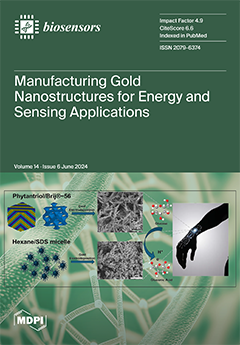
This study demonstrates two cost-effective and efficient strategies with which to manipulate the morphology of gold nanostructures, and thus their catalytic properties, for the successful manufacturing of glucose fuel cells (GFCs). GFCs hold great promise as in- or on-body energy harvesters for ultra-low-power bioelectronics to enable the effective management of long-term diseases, minimizing the risk of associated complications. We have generated gold nanostructures via gold electrodeposition assisted by two types of soft template: a lipid cubic phase Phytantriol/Brij®-56, leading to a nanofeather-like Au structure, and an emulsion hexane/SDS, leading to a nanocoral-like Au structure. Both showed great sensitivity to glucose, with a 7 μW cm−2 maximum power density and an up to 70 μA cm−2 current density. View this paper
  • Issues are regarded as officially published after their release is announced to the table of contents alert mailing list.
  • You may sign up for e-mail alerts to receive table of contents of newly released issues.
  • PDF is the official format for papers published in both, html and pdf forms. To view the papers in pdf format, click on the "PDF Full-text" link, and use the free Adobe Reader to open them.
Order results
Result details
Section
Select all
Export citation of selected articles as:
16 pages, 6549 KiB  
Article
Integrated High-Throughput Centrifugal Microfluidic Chip Device for Pathogen Detection On-Site
by Shuyu Lu, Yuanzhan Yang, Siqi Cui, Anyi Li, Cheng Qian and Xiaoqiong Li
Biosensors 2024, 14(6), 313; https://doi.org/10.3390/bios14060313 - 19 Jun 2024
Viewed by 668
Abstract
An integrated and high-throughput device for pathogen detection is crucial in point-of-care testing (POCT), especially for early diagnosis of infectious diseases and preventing the spread of infection. We developed an on-site testing platform that utilizes a centrifugal microfluidic chip and automated device to [...] Read more.
An integrated and high-throughput device for pathogen detection is crucial in point-of-care testing (POCT), especially for early diagnosis of infectious diseases and preventing the spread of infection. We developed an on-site testing platform that utilizes a centrifugal microfluidic chip and automated device to achieve high-throughput detection. The low-power (<32 W), portable (220 mm × 220 mm × 170 mm, 4 kg) device can complete bacterial lysis, nucleic acid extraction and purification, loop-mediated isothermal amplification (LAMP) reaction, and real-time fluorescence detection. Magnetic beads for nucleic acid adsorption can be mixed by applying electromagnetic fields and centrifugal forces, and the efficiency of nucleic acid extraction is improved by 60% compared to the no-mixing group. The automated nucleic acid extraction process achieves equivalent nucleic acid extraction efficiency in only 40% of the time consumed using the kit protocol. By designing the valve system and disc layout, the maximum speed required for the centrifugal microfluidic chip is reduced to 1500 rpm, greatly reducing the equipment power consumption and size. In detecting E. coli, our platform achieves a limit of detection (LOD) of 102 CFU/mL in 60 min. In summary, our active centrifugal microfluidic platform provides a solution for the integration of complex biological assays on turntables, with great potential in the application of point-of-care diagnosis. Full article
(This article belongs to the Special Issue Biosensing Technologies in Medical Diagnosis)
Show Figures

Figure 1

12 pages, 4414 KiB  
Article
Photoelectric Multi-Signal Output Sensor Based on Two-Dimensional Covalent Organic Polymer Film Modified by Novel Aggregation-Induced Emission Probes
by Yaru Song, Guoling Wu, Enbing Zhang, Guangyuan Feng, Shengbin Lei and Lingli Wu
Biosensors 2024, 14(6), 312; https://doi.org/10.3390/bios14060312 - 18 Jun 2024
Viewed by 689
Abstract
Optical sensors, especially fluorescence sensors, have been widely used because of their advantages in sensing, such as the high sensitivity, good selectivity, no radiation source, and easy operation. Here, we report an example of fluorescence sensing based on two-dimensional (2D) covalent organic polymers [...] Read more.
Optical sensors, especially fluorescence sensors, have been widely used because of their advantages in sensing, such as the high sensitivity, good selectivity, no radiation source, and easy operation. Here, we report an example of fluorescence sensing based on two-dimensional (2D) covalent organic polymers and highlight that the material can achieve a fast response and multi-signal output. This 2DPTPAK+TAPB-based sensor can quickly detect aromatic hydrocarbons and Fe3+ by the fluorescence signal or electrical resistance signal. Full article
Show Figures

Figure 1

14 pages, 2828 KiB  
Article
Development of a Portable Cell-Based Biosensor for the Ultra-Rapid Screening for Boscalid Residues in Lettuce
by Georgia Moschopoulou, Vasileios Tsekouras, Josep V. Mercader, Antonio Abad-Fuentes and Spyridon Kintzios
Biosensors 2024, 14(6), 311; https://doi.org/10.3390/bios14060311 - 18 Jun 2024
Viewed by 607
Abstract
Fungal plant pathogens have posed a significant threat to crop production. However, the large-scale application of pesticides is associated with possible risks for human health and the environment. Boscalid is a widely used fungicide, consistently implemented for the management of significant plant pathogens. [...] Read more.
Fungal plant pathogens have posed a significant threat to crop production. However, the large-scale application of pesticides is associated with possible risks for human health and the environment. Boscalid is a widely used fungicide, consistently implemented for the management of significant plant pathogens. Conventionally, the detection and determination of boscalid residues is based on chromatographic separations. In the present study, a Bioelectric Recognition Assay (BERA)-based experimental approach combined with MIME technology was used, where changes in the electric properties of the membrane-engineering cells with anti-boscalid antibodies were recorded in response to the presence of boscalid at different concentrations based on the maximum residue level (MRL) for lettuce. The membrane-engineering Vero cells with 0.5 μg/mL of antibody in their surface were selected as the best cell line in combination with the lowest antibody concentration. Furthermore, the biosensor was tested against another fungicide in order to prove its selectivity. Finally, the BERA cell-based biosensor was able to detect the boscalid residue, below and above the MRL, in spiked lettuce leaf extracts in an entirely distinct and reproducible manner. This study indicates that the BERA-based biosensor, after further development and optimization, could be used for the routine, high-throughput detection of boscalid residue in lettuce, and not only that. Full article
(This article belongs to the Special Issue Cell-Based Biosensors for Rapid Detection and Monitoring)
Show Figures

Graphical abstract

13 pages, 1614 KiB  
Article
Bismuth Film along with dsDNA-Modified Electrode Surfaces as Promising (bio)Sensors in the Analysis of Heavy Metals in Soils
by Vasiliki Keramari, Sotiria G. Papadimou, Evangelia E. Golia and Stella Girousi
Biosensors 2024, 14(6), 310; https://doi.org/10.3390/bios14060310 - 18 Jun 2024
Viewed by 750
Abstract
Heavy metals constitute pollutants that are particularly common in air, water, and soil. They are present in both urban and rural environments, on land, and in marine ecosystems, where they cause serious environmental problems since they do not degrade easily, remain almost unchanged [...] Read more.
Heavy metals constitute pollutants that are particularly common in air, water, and soil. They are present in both urban and rural environments, on land, and in marine ecosystems, where they cause serious environmental problems since they do not degrade easily, remain almost unchanged for long periods, and bioaccumulate. The detection and especially the quantification of metals require a systematic process. Regular monitoring is necessary because of seasonal variations in metal levels. Consequently, there is a significant need for rapid and low-cost metal determination methods. In this study, we compare and analytically validate absorption spectrometry with a sensitive voltammetric method, which uses a bismuth film-plated electrode surface and applies stripping voltammetry. Atomic absorption spectroscopy (AAS) represents a well-established analytical technique, while the applicability of anodic stripping voltammetry (ASV) in complicated sample matrices such as soil samples is currently unknown. This sample-handling challenge is investigated in the present study. The results show that the AAS and ASV methods were satisfactorily correlated and showed that the metal concentration in soils was lower than the limit values but with an increasing trend. Therefore, continuous monitoring of metal levels in the urban complex of a city is necessary and a matter of great importance. The limits of detection of cadmium (Cd) were lower when using the stripping voltammetry (SWASV) graphite furnace technique compared with those obtained with AAS when using the graphite furnace. However, when using flame atomic absorption spectroscopy (flame-AAS), the measurements tended to overestimate the concentration of Cd compared with the values found using SWASV. This highlights the differences in sensitivity and accuracy between these analytical methods for detecting Cd. The SWASV method has the advantage of being cheaper and faster, enabling the simultaneous determination of heavy elements across the range of concentrations that these elements can occur in Mediterranean soils. Additionally, a dsDNA biosensor is suggested for the discrimination of Cu(I) along with Cu(II) based on the oxidation peak of guanine, and adenine residues can be applied in the redox speciation analysis of copper in soil, which represents an issue of great importance. Full article
Show Figures

Figure 1

31 pages, 1674 KiB  
Review
Challenges and Advances in Biomarker Detection for Rapid and Accurate Sepsis Diagnosis: An Electrochemical Approach
by Deivasigamani Ranjith Kumar, Angelika Banaś and Katarzyna Krukiewicz
Biosensors 2024, 14(6), 309; https://doi.org/10.3390/bios14060309 - 17 Jun 2024
Viewed by 735
Abstract
Sepsis is a life-threatening condition with high mortality rates due to delayed treatment of patients. The conventional methodology for blood diagnosis takes several hours, which suspends treatment, limits early drug administration, and affects the patient’s recovery. Thus, rapid, accurate, bedside (onsite), economical, and [...] Read more.
Sepsis is a life-threatening condition with high mortality rates due to delayed treatment of patients. The conventional methodology for blood diagnosis takes several hours, which suspends treatment, limits early drug administration, and affects the patient’s recovery. Thus, rapid, accurate, bedside (onsite), economical, and reliable sepsis biomarker reading of the clinical sample is an emergent need for patient lifesaving. Electrochemical label-free biosensors are specific and rapid devices that are able to perform analysis at the patient’s bedside; thus, they are considered an attractive methodology in a clinical setting. To reveal their full diagnostic potential, electrode architecture strategies of fabrication are highly desirable, particularly those able to preserve specific antibody–antigen attraction, restrict non-specific adsorption, and exhibit high sensitivity with a low detection limit for a target biomarker. The aim of this review is to provide state-of-the-art methodologies allowing the fabrication of ultrasensitive and highly selective electrochemical sensors for sepsis biomarkers. This review focuses on different methods of label-free biomarker sensors and discusses their advantages and disadvantages. Then, it highlights effective ways of avoiding false results and the role of molecular labels and functionalization. Recent literature on electrode materials and antibody grafting strategies is discussed, and the most efficient methodology for overcoming the non-specific attraction issues is listed. Finally, we discuss the existing electrode architecture for specific biomarker readers and promising tactics for achieving quick and low detection limits for sepsis biomarkers. Full article
(This article belongs to the Special Issue Biosensors for the Analysis and Detection of Drug, Food or Disease)
Show Figures

Figure 1

13 pages, 2847 KiB  
Article
Design and Application of Microfluidic Capture Device for Physical–Magnetic Isolation of MCF-7 Circulating Tumor Cells
by Akhilesh Bendre, Derangula Somasekhara, Varalakshmi K. Nadumane, Ganesan Sriram, Ramesh S. Bilimagga and Mahaveer D. Kurkuri
Biosensors 2024, 14(6), 308; https://doi.org/10.3390/bios14060308 - 15 Jun 2024
Viewed by 742
Abstract
Circulating tumor cells (CTCs) are a type of cancer cell that spreads from the main tumor to the bloodstream, and they are often the most important among the various entities that can be isolated from the blood. For the diagnosis of cancer, conventional [...] Read more.
Circulating tumor cells (CTCs) are a type of cancer cell that spreads from the main tumor to the bloodstream, and they are often the most important among the various entities that can be isolated from the blood. For the diagnosis of cancer, conventional biopsies are often invasive and unreliable, whereas a liquid biopsy, which isolates the affected item from blood or lymph fluid, is a less invasive and effective diagnostic technique. Microfluidic technologies offer a suitable channel for conducting liquid biopsies, and this technology is utilized to extract CTCs in a microfluidic chip by physical and bio-affinity-based techniques. This effort uses functionalized magnetic nanoparticles (MNPs) in a unique microfluidic chip to collect CTCs using a hybrid (physical and bio-affinity-based/guided magnetic) capturing approach with a high capture rate. Accordingly, folic acid-functionalized Fe3O4 nanoparticles have been used to capture MCF-7 (breast cancer) CTCs with capture efficiencies reaching up to 95% at a 10 µL/min flow rate. Moreover, studies have been conducted to support this claim, including simulation and biomimetic investigations. Full article
Show Figures

Figure 1

31 pages, 7599 KiB  
Review
Advances in Nanoplasmonic Biosensors: Optimizing Performance for Exosome Detection Applications
by Devi Taufiq Nurrohman, Nan-Fu Chiu, Yu-Sheng Hsiao, Yun-Ju Lai and Himansu Sekhar Nanda
Biosensors 2024, 14(6), 307; https://doi.org/10.3390/bios14060307 - 14 Jun 2024
Viewed by 1253
Abstract
The development of sensitive and specific exosome detection tools is essential because they are believed to provide specific information that is important for early detection, screening, diagnosis, and monitoring of cancer. Among the many detection tools, surface-plasmon resonance (SPR) biosensors are analytical devices [...] Read more.
The development of sensitive and specific exosome detection tools is essential because they are believed to provide specific information that is important for early detection, screening, diagnosis, and monitoring of cancer. Among the many detection tools, surface-plasmon resonance (SPR) biosensors are analytical devices that offer advantages in sensitivity and detection speed, thereby making the sample-analysis process faster and more accurate. In addition, the penetration depth of the SPR biosensor, which is <300 nm, is comparable to the size of the exosome, making the SPR biosensor ideal for use in exosome research. On the other hand, another type of nanoplasmonic sensor, namely a localized surface-plasmon resonance (LSPR) biosensor, has a shorter penetration depth of around 6 nm. Structural optimization through the addition of supporting layers and gap control between particles is needed to strengthen the surface-plasmon field. This paper summarizes the progress of the development of SPR and LSPR biosensors for detecting exosomes. Techniques in signal amplification from two sensors will be discussed. There are three main parts to this paper. The first two parts will focus on reviewing the working principles of each sensor and introducing several methods that can be used to isolate exosomes. This article will close by explaining the various sensor systems that have been developed and the optimizations carried out to obtain sensors with better performance. To illustrate the performance improvements in each sensor system discussed, the parameters highlighted include the detection limit, dynamic range, and sensitivity. Full article
(This article belongs to the Special Issue Recent Progress in Biosensor Technologies for Detection of Exosomes)
Show Figures

Figure 1

12 pages, 3883 KiB  
Article
Integration of Image Pattern Recognition and Photon Sensor for Analyzing Cytokine Gene Expression Using πCode MicroDisc
by On-anong Juntit, Kanokporn Sornsuwan, Umpa Yasamut and Chatchai Tayapiwatana
Biosensors 2024, 14(6), 306; https://doi.org/10.3390/bios14060306 - 13 Jun 2024
Viewed by 727
Abstract
Current quantitative gene expression detection in genomic and transcriptomic research heavily relies on quantitative real-time PCR (qPCR). While existing multiplex gene detection techniques offer simultaneous analysis of multiple targets, we present an alternative assay capable of detecting gene expression simultaneously within a single [...] Read more.
Current quantitative gene expression detection in genomic and transcriptomic research heavily relies on quantitative real-time PCR (qPCR). While existing multiplex gene detection techniques offer simultaneous analysis of multiple targets, we present an alternative assay capable of detecting gene expression simultaneously within a single well. This highly sensitive method utilizes πCode MicroDiscs, featuring unique identification patterns and fluorescent detection. Our study compared this multiplex πCode platform with a qPCR platform for profiling cytokine gene expression. The πCode MicroDisc assay successfully demonstrated the expression of polymerization markers for M1- and M2-like macrophages generated from THP-1-derived macrophages in a qualitative assay. Additionally, our findings suggest a pattern agreement between the πCode assay and the qPCR assay, indicating the potential of the πCode technology for comparative gene expression analysis. Regarding the inherent sensitivity and linearity, the developed πCode assay primarily provides qualitative gene expression to discriminate the polarization of macrophages. This remarkable capability presents substantial advantages for researchers, rendering the technology highly suitable for high-throughput applications in clinical diagnosis and disease monitoring. Full article
Show Figures

Figure 1

12 pages, 4944 KiB  
Article
Enhancing Sensitivity in SARS-CoV-2 Rapid Antigen Testing through Integration of a Water-Soluble Polymer Wall
by Xiuzhen Wang, Yu Wang, Huiyang Jie, Sidi Liu, Chenguang Shen and Qian Liu
Biosensors 2024, 14(6), 305; https://doi.org/10.3390/bios14060305 - 12 Jun 2024
Viewed by 615
Abstract
Lateral flow immunoassays (LFIAs) are recognized for their practicality in homecare and point-of-care testing, owing to their simplicity, cost-efficiency, and rapid visual readouts. Despite these advantages, LFIAs typically fall short in sensitivity, particularly in detecting viruses such as SARS-CoV-2, thus limiting their broader [...] Read more.
Lateral flow immunoassays (LFIAs) are recognized for their practicality in homecare and point-of-care testing, owing to their simplicity, cost-efficiency, and rapid visual readouts. Despite these advantages, LFIAs typically fall short in sensitivity, particularly in detecting viruses such as SARS-CoV-2, thus limiting their broader application. In response to this challenge, we have innovated an approach to substantially enhance LFIA sensitivity. This involves the integration of a water-soluble dextran–methacrylate polymer wall with a 15% grafting degree positioned between the test and control lines on the LFIA strip. This novel modification significantly improved the sensitivity of the assay, achieving detection limits as low as 50 pg mL−1 and enhancing the sensitivity by 5–20-fold relative to existing LFIA kits available on the market. Furthermore, our developed LFIA kit (WSPW-LFIA) demonstrated exceptional specificity for SARS-CoV-2. Coupled with a straightforward fabrication process and robust stability, the WSPW-LFIA represents a promising advancement for real-time in vitro diagnosis across a spectrum of diseases. Full article
(This article belongs to the Special Issue Point of Care Diagnostics for Infectious Disease)
Show Figures

Graphical abstract

10 pages, 2216 KiB  
Communication
Design and Fabrication of a Biomimetic Smart Material for Electrochemical Detection of Carbendazim Pesticides in Real Samples with Enhanced Selectivity
by Francisco Franciné Maia Júnior, Rui Sales Junior, Geovani Ferreira Barbosa, Sajjad Hussain, Eduardo Jara-Cornejo and Sabir Khan
Biosensors 2024, 14(6), 304; https://doi.org/10.3390/bios14060304 - 10 Jun 2024
Viewed by 894
Abstract
Agricultural products are vitally important for sustaining life on earth and their production has notably grown over the years worldwide in general and in Brazil particularly. Elevating agricultural practices consequently leads to a proportionate increase in the usage of pesticides that are crucially [...] Read more.
Agricultural products are vitally important for sustaining life on earth and their production has notably grown over the years worldwide in general and in Brazil particularly. Elevating agricultural practices consequently leads to a proportionate increase in the usage of pesticides that are crucially important for enhanced crop yield and protection. These compounds have been employed excessively in alarming concentrations, causing the contamination of soil, water, and air. Additionally, they pose serious threats to human health. The current study introduces an innovative tool for producing appropriate materials coupled with an electrochemical sensor designed to measure carbendazim levels. The sensor is developed using a molecularly imprinted polymer (MIP) mounted on a glassy carbon electrode. This electrode is equipped with multi-walled carbon nanotubes (MWCNTs) for improved performance. The combined system demonstrates promising potential for accurately quantifying carbendazim. The morphological characteristics of the synthesized materials were investigated using field emission scanning electron microscopy (FESEM) and the Fourier-transform infrared (FTIR) technique. The analytical curve was drawn using the electrochemical method in the range of 2 to 20 ppm while for HPLC 2–12 ppm; the results are presented as the maximum adsorption capacity of the MIP (82.4%) when compared with NIP (41%) using the HPLC method. The analysis conducted using differential pulse voltammetry (DPV) yielded a limit of detection (LOD) of 1.0 ppm and a repeatability of 5.08% (n = 10). The results obtained from the analysis of selectivity demonstrated that the proposed electrochemical sensor is remarkably efficient for the quantitative assessment of carbendazim, even in the presence of another interferent. The sensor was successfully tested for river water samples for carbendazim detection, and recovery rates ranging from 94 to 101% were obtained for HPLC and 94 to 104% for the electrochemical method. The results obtained show that the proposed electrochemical technique is viable for the application and quantitative determination of carbendazim in any medium. Full article
(This article belongs to the Special Issue Molecularly Imprinted Polymers for Chemical Sensing)
Show Figures

Figure 1

23 pages, 24757 KiB  
Article
Comparing the Mechanical Properties of Rice Cells and Protoplasts under PEG6000 Drought Stress Using Double Resonator Piezoelectric Cytometry
by Yu Yan, Tiean Zhou, Yu Zhang, Zhicheng Kong, Weisong Pan and Chengfang Tan
Biosensors 2024, 14(6), 303; https://doi.org/10.3390/bios14060303 - 9 Jun 2024
Viewed by 840
Abstract
Plant cells’ ability to withstand abiotic stress is strongly linked to modifications in their mechanical characteristics. Nevertheless, the lack of a workable method for consistently tracking plant cells’ mechanical properties severely restricts our comprehension of the mechanical alterations in plant cells under stress. [...] Read more.
Plant cells’ ability to withstand abiotic stress is strongly linked to modifications in their mechanical characteristics. Nevertheless, the lack of a workable method for consistently tracking plant cells’ mechanical properties severely restricts our comprehension of the mechanical alterations in plant cells under stress. In this study, we used the Double Resonator Piezoelectric Cytometry (DRPC) method to dynamically and non-invasively track changes in the surface stress (ΔS) generated and viscoelasticity (storage modulus G′ and loss modulus G″) of protoplasts and suspension cells of rice under a drought stress of 5–25% PEG6000. The findings demonstrate that rice suspension cells and protoplasts react mechanically differently to 5–15% PEG6000 stress, implying distinct resistance mechanisms. However, neither of them can withstand 25% PEG6000 stress; they respond mechanically similarly to 25% PEG6000 stress. The results of DRPC are further corroborated by the morphological alterations of rice cells and protoplasts observed under an optical microscope. To sum up, the DRPC technique functions as a precise cellular mechanical sensor and offers novel research tools for the evaluation of plant cell adversity and differentiating between the mechanical reactions of cells and protoplasts under abiotic stress. Full article
(This article belongs to the Section Environmental Biosensors and Biosensing)
Show Figures

Figure 1

53 pages, 30849 KiB  
Review
Microbial Biofilms: Features of Formation and Potential for Use in Bioelectrochemical Devices
by Roman Perchikov, Maxim Cheliukanov, Yulia Plekhanova, Sergei Tarasov, Anna Kharkova, Denis Butusov, Vyacheslav Arlyapov, Hideaki Nakamura and Anatoly Reshetilov
Biosensors 2024, 14(6), 302; https://doi.org/10.3390/bios14060302 - 8 Jun 2024
Viewed by 796
Abstract
Microbial biofilms present one of the most widespread forms of life on Earth. The formation of microbial communities on various surfaces presents a major challenge in a variety of fields, including medicine, the food industry, shipping, etc. At the same time, this process [...] Read more.
Microbial biofilms present one of the most widespread forms of life on Earth. The formation of microbial communities on various surfaces presents a major challenge in a variety of fields, including medicine, the food industry, shipping, etc. At the same time, this process can also be used for the benefit of humans—in bioremediation, wastewater treatment, and various biotechnological processes. The main direction of using electroactive microbial biofilms is their incorporation into the composition of biosensor and biofuel cells This review examines the fundamental knowledge acquired about the structure and formation of biofilms, the properties they have when used in bioelectrochemical devices, and the characteristics of the formation of these structures on different surfaces. Special attention is given to the potential of applying the latest advances in genetic engineering in order to improve the performance of microbial biofilm-based devices and to regulate the processes that take place within them. Finally, we highlight possible ways of dealing with the drawbacks of using biofilms in the creation of highly efficient biosensors and biofuel cells. Full article
(This article belongs to the Special Issue Microbial Biosensor: From Design to Applications)
Show Figures

Figure 1

16 pages, 2389 KiB  
Review
Advancing Tissue Culture with Light-Driven 3D-Printed Microfluidic Devices
by Xiangke Li, Meng Wang, Thomas P. Davis, Liwen Zhang and Ruirui Qiao
Biosensors 2024, 14(6), 301; https://doi.org/10.3390/bios14060301 - 8 Jun 2024
Viewed by 873
Abstract
Three-dimensional (3D) printing presents a compelling alternative for fabricating microfluidic devices, circumventing certain limitations associated with traditional soft lithography methods. Microfluidics play a crucial role in the biomedical sciences, particularly in the creation of tissue spheroids and pharmaceutical research. Among the various 3D [...] Read more.
Three-dimensional (3D) printing presents a compelling alternative for fabricating microfluidic devices, circumventing certain limitations associated with traditional soft lithography methods. Microfluidics play a crucial role in the biomedical sciences, particularly in the creation of tissue spheroids and pharmaceutical research. Among the various 3D printing techniques, light-driven methods such as stereolithography (SLA), digital light processing (DLP), and photopolymer inkjet printing have gained prominence in microfluidics due to their rapid prototyping capabilities, high-resolution printing, and low processing temperatures. This review offers a comprehensive overview of light-driven 3D printing techniques used in the fabrication of advanced microfluidic devices. It explores biomedical applications for 3D-printed microfluidics and provides insights into their potential impact and functionality within the biomedical field. We further summarize three light-driven 3D printing strategies for producing biomedical microfluidic systems: direct construction of microfluidic devices for cell culture, PDMS-based microfluidic devices for tissue engineering, and a modular SLA-printed microfluidic chip to co-culture and monitor cells. Full article
(This article belongs to the Special Issue Microfluidics for Biomedical Applications (Volume II))
Show Figures

Figure 1

46 pages, 34337 KiB  
Review
Sustainable Sensing with Paper Microfluidics: Applications in Health, Environment, and Food Safety
by Sanjay Kumar, Jyoti Bala Kaushal and Heow Pueh Lee
Biosensors 2024, 14(6), 300; https://doi.org/10.3390/bios14060300 - 7 Jun 2024
Viewed by 1000
Abstract
This manuscript offers a concise overview of paper microfluidics, emphasizing its sustainable sensing applications in healthcare, environmental monitoring, and food safety. Researchers have developed innovative sensing platforms for detecting pathogens, pollutants, and contaminants by leveraging the paper’s unique properties, such as biodegradability and [...] Read more.
This manuscript offers a concise overview of paper microfluidics, emphasizing its sustainable sensing applications in healthcare, environmental monitoring, and food safety. Researchers have developed innovative sensing platforms for detecting pathogens, pollutants, and contaminants by leveraging the paper’s unique properties, such as biodegradability and affordability. These portable, low-cost sensors facilitate rapid diagnostics and on-site analysis, making them invaluable tools for resource-limited settings. This review discusses the fabrication techniques, principles, and applications of paper microfluidics, showcasing its potential to address pressing challenges and enhance human health and environmental sustainability. Full article
(This article belongs to the Special Issue Microfluidics for Biomedical Applications (Volume II))
Show Figures

Figure 1

18 pages, 4421 KiB  
Article
Colorimetric Glucose Biosensor Based on Chitosan Films and Its Application for Glucose Detection in Beverages Using a Smartphone Application
by Anastasia Skonta, Myrto G. Bellou, Theodore E. Matikas and Haralambos Stamatis
Biosensors 2024, 14(6), 299; https://doi.org/10.3390/bios14060299 - 7 Jun 2024
Viewed by 762
Abstract
Nowadays, biosensors are gaining increasing interest in foods’ and beverages’ quality control, owing to their economic production, enhanced sensitivity, specificity, and faster analysis. In particular, colorimetric biosensors can be combined with color recognition applications on smartphones for the detection of analytes, rendering the [...] Read more.
Nowadays, biosensors are gaining increasing interest in foods’ and beverages’ quality control, owing to their economic production, enhanced sensitivity, specificity, and faster analysis. In particular, colorimetric biosensors can be combined with color recognition applications on smartphones for the detection of analytes, rendering the whole procedure more applicable in everyday life. Herein, chitosan (CS) films were prepared with the deep eutectic solvent (DES) choline chloride/urea/glycerol (ChCl:U:Gly). Glucose oxidase (GOx), a widely utilized enzyme in quality control, was immobilized within CS films through glutaraldehyde (GA), leading to the formation of CS/GOx films. The optimized GOx concentration and DES content were determined for the films. Moreover, the effect of the pH and temperature of the glucose oxidation reaction on the enzymatic activity of GOx was studied. The structure, stability, and specificity of the CS/GOx films as well as the Km values of free and immobilized GOx were also determined. Finally, the analytical performance of the films was studied by using both a spectrophotometer and a color recognition application on a smartphone. The results demonstrated that the films were highly accurate, specific to glucose, and stable when stored at 4 °C for 4 weeks and when reused 10 times, without evident activity loss. Furthermore, the films displayed a good linear response range (0.1–0.8 mM) and a good limit of detection (LOD, 33 μM), thus being appropriate for the estimation of glucose concentration in real samples through a smartphone application. Full article
(This article belongs to the Special Issue Biosensing, Biosafety and Diagnosis)
Show Figures

Figure 1

12 pages, 2412 KiB  
Article
Electroanalytical Platform for Rapid E. coli O157:H7 Detection in Water Samples
by Kundan Kumar Mishra, Vikram Narayanan Dhamu, Chesna Jophy, Sriram Muthukumar and Shalini Prasad
Biosensors 2024, 14(6), 298; https://doi.org/10.3390/bios14060298 - 7 Jun 2024
Viewed by 931
Abstract
There is a pressing need to enhance early detection methods of E. coli O157:H7 to mitigate the occurrence and consequences of pathogenic contamination and associated outbreaks. This study highlights the efficacy of a portable electrochemical sensing platform that operates without faradaic processes towards [...] Read more.
There is a pressing need to enhance early detection methods of E. coli O157:H7 to mitigate the occurrence and consequences of pathogenic contamination and associated outbreaks. This study highlights the efficacy of a portable electrochemical sensing platform that operates without faradaic processes towards detecting and quantifying E. coli O157:H7. It is specifically tailored for quick identification in potable water. The assay processing time is approximately 5 min, addressing the need for swift and efficient pathogen detection. The sensing platform was constructed utilizing specific, monoclonal E. coli antibodies, based on single-capture, non-faradaic, electrochemical immunoassay principles. The E. coli sensor assay underwent testing over a wide concentration range, spanning from 10 to 105 CFU/mL, and a limit of detection (LoD) of 1 CFU/mL was demonstrated. Significantly, the sensor’s performance remained consistent across studies, with both inter- and intra-study coefficients of variation consistently below 20%. To evaluate real-world feasibility, a comparative examination was performed between laboratory-based benchtop data and data obtained from the portable device. The proposed sensing platform exhibited remarkable sensitivity and selectivity, enabling the detection of minimal E. coli concentrations in potable water. This successful advancement positions it as a promising solution for prompt on-site detection, characterized by its portability and user-friendly operation. This study presents electrochemical-based sensors as significant contributors to ensuring food safety and public health. They play a crucial role in preventing the occurrence of epidemics and enhancing the supervision of water quality. Full article
Show Figures

Graphical abstract

14 pages, 9296 KiB  
Article
Combination of an Optically Induced Dielectrophoresis (ODEP) Mechanism and a Laminar Flow Pattern in a Microfluidic System for the Continuous Size-Based Sorting and Separation of Microparticles
by Po-Yu Chu, Ai-Yun Wu, Kun-Yu Tsai, Chia-Hsun Hsieh and Min-Hsien Wu
Biosensors 2024, 14(6), 297; https://doi.org/10.3390/bios14060297 - 6 Jun 2024
Viewed by 614
Abstract
Optically induced dielectrophoresis (ODEP)-based microparticle sorting and separation is regarded as promising. However, current methods normally lack the downstream process for the transportation and collection of separated microparticles, which could limit its applications. To address this issue, an ODEP microfluidic chip encompassing three [...] Read more.
Optically induced dielectrophoresis (ODEP)-based microparticle sorting and separation is regarded as promising. However, current methods normally lack the downstream process for the transportation and collection of separated microparticles, which could limit its applications. To address this issue, an ODEP microfluidic chip encompassing three microchannels that join only at the central part of the microchannels (i.e., the working zone) was designed. During operation, three laminar flows were generated in the zone, where two dynamic light bar arrays were designed to sort and separate PS (polystyrene) microbeads of different sizes in a continuous manner. The separated PS microbeads were then continuously transported in laminar flows in a partition manner for the final collection. The results revealed that the method was capable of sorting and separating PS microbeads in a high-purity manner (e.g., the microbead purity values were 89.9 ± 3.7, 88.0 ± 2.5, and 92.8 ± 6.5% for the 5.8, 10.8, and 15.8 μm microbeads harvested, respectively). Overall, this study demonstrated the use of laminar flow and ODEP to achieve size-based sorting, separation, and collection of microparticles in a continuous and high-performance manner. Apart from the demonstration, this method can also be utilized for size-based sorting and the separation of other biological or nonbiological microparticles. Full article
(This article belongs to the Section Optical and Photonic Biosensors)
Show Figures

Figure 1

25 pages, 7207 KiB  
Review
Optical Devices for the Diagnosis and Management of Spinal Cord Injuries: A Review
by Sonika Sharma, Neeti Kalyani, Taposhree Dutta, Jesús Salvador Velázquez-González, Ignacio Llamas-Garro, Bora Ung, Joan Bas, Rakesh Dubey and Satyendra K. Mishra
Biosensors 2024, 14(6), 296; https://doi.org/10.3390/bios14060296 - 5 Jun 2024
Viewed by 1302
Abstract
Throughout the central nervous system, the spinal cord plays a very important role, namely, transmitting sensory and motor information inwardly so that it can be processed by the brain. There are many different ways this structure can be damaged, such as through traumatic [...] Read more.
Throughout the central nervous system, the spinal cord plays a very important role, namely, transmitting sensory and motor information inwardly so that it can be processed by the brain. There are many different ways this structure can be damaged, such as through traumatic injury or surgery, such as scoliosis correction, for instance. Consequently, damage may be caused to the nervous system as a result of this. There is no doubt that optical devices such as microscopes and cameras can have a significant impact on research, diagnosis, and treatment planning for patients with spinal cord injuries (SCIs). Additionally, these technologies contribute a great deal to our understanding of these injuries, and they are also essential in enhancing the quality of life of individuals with spinal cord injuries. Through increasingly powerful, accurate, and minimally invasive technologies that have been developed over the last decade or so, several new optical devices have been introduced that are capable of improving the accuracy of SCI diagnosis and treatment and promoting a better quality of life after surgery. We aim in this paper to present a timely overview of the various research fields that have been conducted on optical devices that can be used to diagnose spinal cord injuries as well as to manage the associated health complications that affected individuals may experience. Full article
(This article belongs to the Section Optical and Photonic Biosensors)
Show Figures

Figure 1

14 pages, 3413 KiB  
Article
Novel Multiparametric Bioelectronic Measurement System for Monitoring Virus-Induced Alterations in Functional Neuronal Networks
by Heinz-Georg Jahnke, Verena te Kamp, Christoph Prönnecke, Sabine Schmidt, Ronny Azendorf, Barbara Klupp, Andrea A. Robitzki and Stefan Finke
Biosensors 2024, 14(6), 295; https://doi.org/10.3390/bios14060295 - 5 Jun 2024
Viewed by 717
Abstract
Development and optimisation of bioelectronic monitoring techniques like microelectrode array-based field potential measurement and impedance spectroscopy for the functional, label-free and non-invasive monitoring of in vitro neuronal networks is widely investigated in the field of biosensors. Thus, these techniques were individually used to [...] Read more.
Development and optimisation of bioelectronic monitoring techniques like microelectrode array-based field potential measurement and impedance spectroscopy for the functional, label-free and non-invasive monitoring of in vitro neuronal networks is widely investigated in the field of biosensors. Thus, these techniques were individually used to demonstrate the capabilities of, e.g., detecting compound-induced toxicity in neuronal culture models. In contrast, extended application for investigating the effects of central nervous system infecting viruses are rarely described. In this context, we wanted to analyse the effect of herpesviruses on functional neuronal networks. Therefore, we developed a unique hybrid bioelectronic monitoring platform that allows for performing field potential monitoring and impedance spectroscopy on the same microelectrode. In the first step, a neuronal culture model based on primary hippocampal cells from neonatal rats was established with reproducible and stable synchronised electrophysiological network activity after 21 days of cultivation on microelectrode arrays. For a proof of concept, the pseudorabies model virus PrV Kaplan-ΔgG-GFP was applied and the effect on the neuronal networks was monitored by impedance spectroscopy and field potential measurement for 72 h in a multiparametric mode. Analysis of several bioelectronic parameters revealed a virus concentration-dependent degeneration of the neuronal network within 24–48 h, with a significant early change in electrophysiological activity, subsequently leading to a loss of activity and network synchronicity. In conclusion, we successfully developed a microelectrode array-based hybrid bioelectronic measurement platform for quantitative monitoring of pathologic effects of a herpesvirus on electrophysiological active neuronal networks. Full article
(This article belongs to the Special Issue Biosensors for Monitoring and Diagnostics)
Show Figures

Graphical abstract

13 pages, 2754 KiB  
Article
Long-Term Detection of Glycemic Glucose/Hypoglycemia by Microfluidic Sweat Monitoring Patch
by Wenjie Xu, Lei Lu, Yuxin He, Lin Cheng and Aiping Liu
Biosensors 2024, 14(6), 294; https://doi.org/10.3390/bios14060294 - 5 Jun 2024
Viewed by 745
Abstract
A microfluidic sweat monitoring patch that collects human sweat for a long time is designed to achieve the effect of detecting the rise and fall of human sweat glucose over a long period of time by increasing the use time of a single [...] Read more.
A microfluidic sweat monitoring patch that collects human sweat for a long time is designed to achieve the effect of detecting the rise and fall of human sweat glucose over a long period of time by increasing the use time of a single patch. Five collection pools, four serpentine channels, and two different valves are provided. Among them, the three-dimensional valve has a large burst pressure as a balance between the internal and external air pressures of the patch. The bursting pressure of the two-dimensional diverter valve is smaller than that of the three-dimensional gas valve, and its role is to control the flow direction of the liquid. Through plasma hydrophilic treatment of different durations, the optimal hydrophilic duration is obtained. The embedded chromogenic disc detects the sweat glucose value at two adjacent time intervals and compares the information of the human body to increase or reduce glucose. The patch has good flexibility and can fit well with human skin, and because polydimethylsiloxane (PDMS) has good light transmission, it reduces the measurement error caused by the color-taking process and makes the detection results more accurate. Full article
Show Figures

Figure 1

18 pages, 8646 KiB  
Article
Multifunctional Three-in-One Sensor on t-ZnO for Ultraviolet and VOC Sensing for Bioengineering Applications
by Rajat Nagpal, Cristian Lupan, Adrian Bîrnaz, Alexandr Sereacov, Erik Greve, Monja Gronenberg, Leonard Siebert, Rainer Adelung and Oleg Lupan
Biosensors 2024, 14(6), 293; https://doi.org/10.3390/bios14060293 - 5 Jun 2024
Viewed by 1018
Abstract
Zinc oxide (ZnO) is considered to be one of the most explored and reliable sensing materials for UV detection due to its excellent properties, like a wide band gap and high exciton energy. Our current study on a photodetector based on tetrapodal ZnO [...] Read more.
Zinc oxide (ZnO) is considered to be one of the most explored and reliable sensing materials for UV detection due to its excellent properties, like a wide band gap and high exciton energy. Our current study on a photodetector based on tetrapodal ZnO (t-ZnO) reported an extremely high UV response of ~9200 for 394 nm UV illumination at 25 °C. The t-ZnO network structure and morphology were investigated using XRD and SEM. The sensor showed a UV/visible ratio of ~12 at 25 °C for 394 nm UV illumination and 443 nm visible illumination. By increasing the temperature, monotonic decreases in response and recovery time were observed. By increasing the bias voltage, the response time was found to decrease while the recovery time was increased. The maximum responsivity shifted to higher wavelengths from 394 nm to 400 nm by increasing the operating temperature from 25 °C to 100 °C. The t-ZnO networks exhibited gas-sensing performances at temperatures above 250 °C, and a maximum response of ~1.35 was recorded at 350 °C with a good repeatability and fast recovery in 16 s for 100 ppm of n-butanol vapor. This study demonstrated that t-ZnO networks are good biosensors that can be used for diverse biomedical applications like the sensing of VOCs (volatile organic compounds) and ultraviolet detection under a wide range of temperatures, and may find new possibilities in biosensing applications. Full article
Show Figures

Figure 1

12 pages, 2334 KiB  
Article
Europium Nanoparticle-Based Lateral Flow Strip Biosensors for the Detection of Quinoxaline Antibiotics and Their Main Metabolites in Fish Feeds and Tissues
by Qing Mei, Biao Ma, Yun Fang, Yunfei Gong, Jiali Li and Mingzhou Zhang
Biosensors 2024, 14(6), 292; https://doi.org/10.3390/bios14060292 - 4 Jun 2024
Viewed by 791
Abstract
Olaquindox (OLA) and quinocetone (QCT) have been prohibited in aquatic products due to their significant toxicity and side effects. In this study, rapid and visual europium nanoparticle (EuNP)-based lateral flow strip biosensors (LFSBs) were developed for the simultaneous quantitative detection of OLA, QCT, [...] Read more.
Olaquindox (OLA) and quinocetone (QCT) have been prohibited in aquatic products due to their significant toxicity and side effects. In this study, rapid and visual europium nanoparticle (EuNP)-based lateral flow strip biosensors (LFSBs) were developed for the simultaneous quantitative detection of OLA, QCT, and 3-methyl-quinoxaline-2-carboxylic acid (MQCA) in fish feed and tissue. The EuNP-LFSBs enabled sensitive detection for OLA, QCT, and MQCA with a limit of detection of 0.067, 0.017, and 0.099 ng/mL (R2 ≥ 0.9776) within 10 min. The average recovery of the EuNP-LFSBs was 95.13%, and relative standard deviations were below 9.38%. The method was verified by high-performance liquid chromatography (HPLC), and the test results were consistent. Therefore, the proposed LFSBs serve as a powerful tool to monitor quinoxalines in fish feeds and their residues in fish tissues. Full article
(This article belongs to the Special Issue Immunoassays and Biosensing)
Show Figures

Figure 1

18 pages, 7513 KiB  
Article
Fabrication of Curcumin-Based Electrochemical Nanosensors for the Detection of Environmental Pollutants: 1,4-Dioxane and Hydrazine
by Renjith Kumar Rasal, Iffath Badsha, Muthaiah Shellaiah, Kumaran Subramanian, Abinaya Gayathri, Abdurahman Hajinur Hirad, Kumaravel Kaliaperumal and Thiyagarajan Devasena
Biosensors 2024, 14(6), 291; https://doi.org/10.3390/bios14060291 - 4 Jun 2024
Cited by 1 | Viewed by 813
Abstract
This work reports the development of novel curcuminoid-based electrochemical sensors for the detection of environmental pollutants from water. In this study, the first set of electrochemical experiments was carried out using curcumin-conjugated multi-walled carbon nanotubes (MWCNT–CM) for 1,4-dioxane detection. The MWCNT–CM/GCE showed good [...] Read more.
This work reports the development of novel curcuminoid-based electrochemical sensors for the detection of environmental pollutants from water. In this study, the first set of electrochemical experiments was carried out using curcumin-conjugated multi-walled carbon nanotubes (MWCNT–CM) for 1,4-dioxane detection. The MWCNT–CM/GCE showed good sensitivity (103.25 nA nM−1 cm−2 in the linear range 1 nM to 1 µM), with LOD of 35.71 pM and LOQ of 108.21 pM. The second set of electrochemical experiments was carried out with bisdemethoxy curcumin analog quantum dots (BDMCAQD) for hydrazine detection. The BDMCAQD/GCE exhibited good sensitivity (74.96 nA nM−1 cm−2 in the linear range 100 nM to 1 µM), with LOD of 10 nM and LOQ of 44.93 nM. Thus, this work will serve as a reference for the fabrication of metal-free electrochemical sensors using curcuminoids as the redox mediator for the enhanced detection of environmental pollutants. Full article
Show Figures

Figure 1

17 pages, 7116 KiB  
Article
A Study on the Mechanism and Properties of a Self-Powered H2O2 Electrochemical Sensor Based on a Fuel Cell Configuration with FePc and Graphene Cathode Catalyst Materials
by Yunong Zhang, Andreas Offenhäusser and Yulia Mourzina
Biosensors 2024, 14(6), 290; https://doi.org/10.3390/bios14060290 - 4 Jun 2024
Viewed by 3153
Abstract
Conventional electrochemical sensors use voltammetric and amperometric methods with external power supply and modulation systems, which hinder the flexibility and application of the sensors. To avoid the use of an external power system and to minimize the number of electrochemical cell components, a [...] Read more.
Conventional electrochemical sensors use voltammetric and amperometric methods with external power supply and modulation systems, which hinder the flexibility and application of the sensors. To avoid the use of an external power system and to minimize the number of electrochemical cell components, a self-powered electrochemical sensor (SPES) for hydrogen peroxide was investigated here. Iron phthalocyanine, an enzyme mimetic material, and Ni were used as a cathode catalyst and an anode material, respectively. The properties of the iron phthalocyanine catalyst modified by graphene nanoplatelets (GNPs) were investigated. Open circuit potential tests demonstrated the feasibility of this system. The GNP-modulated interface helped to solve the problems of aggregation and poor conductivity of iron phthalocyanine and allowed for the achievement of the best analytical characteristics of the self-powered H2O2 sensor with a low detection limit of 0.6 µM and significantly higher sensitivity of 0.198 A/(M·cm2) due to the enhanced electrochemical properties. The SPES demonstrated the best performance at pH 3.0 compared to pH 7.4 and 12.0. The sensor characteristics under the control of external variable load resistances are discussed and the cell showed the highest power density of 65.9 μW/cm2 with a 20 kOhm resistor. The practical applicability of this method was verified by the determination of H2O2 in blood serum. Full article
Show Figures

Graphical abstract

14 pages, 2533 KiB  
Article
Soft-Template-Based Manufacturing of Gold Nanostructures for Energy and Sensing Applications
by Tushar Kanti Maiti, Wanli Liu, Asghar Niyazi, Adam M. Squires, Sujay Chattpoadhyay and Mirella Di Lorenzo
Biosensors 2024, 14(6), 289; https://doi.org/10.3390/bios14060289 - 3 Jun 2024
Viewed by 519
Abstract
Implantable and wearable bioelectronic systems can enable tailored therapies for the effective management of long-term diseases, thus minimising the risk of associated complications. In this context, glucose fuel cells hold great promise as in- or on-body energy harvesters for ultra-low-power bioelectronics and as [...] Read more.
Implantable and wearable bioelectronic systems can enable tailored therapies for the effective management of long-term diseases, thus minimising the risk of associated complications. In this context, glucose fuel cells hold great promise as in- or on-body energy harvesters for ultra-low-power bioelectronics and as self-powered glucose sensors. We report here the generation of gold nanostructures through a gold electrodeposition method in a soft template for the abiotic electrocatalysis of glucose in glucose fuel cells. Two different types of soft template were used: a lipid cubic phase-based soft template composed of Phytantriol and Brij®-56, and an emulsion-based soft template composed of hexane and sodium dodecyl sulphate (SDS). The resulting gold structures were first characterised by SAXS, SEM and TEM to elucidate their structure, and then their electrocatalytic activity towards glucose was compared in both a three-electrode set-up and in a fuel cell set-up. The Phytantriol/Brij®-56 template led to a nanofeather-like Au structure, while the hexane/SDS template led to a nanocoral-like Au structure. These templated electrodes exhibited similar electrochemical active surface areas (0.446 cm2 with a roughness factor (RF) of 14.2 for Phytantriol/Brij®-56 templated nanostructures and 0.421 cm2 with an RF of 13.4 for hexane/SDS templated nanostructures), and a sensitivity towards glucose of over 7 μA mM−1 cm−2. When tested as the anode of an abiotic glucose fuel cell (in a phosphate-buffered solution with a glucose concentration of 6 mM), a maximum power density of 7 μW cm−2 was reached; however the current density in the case of the fuel cell with the Phytantriol/Brij®-56 templated anode was approximately two times higher, reaching the value of 70 μA cm−2. Overall, this study demonstrates two simple, cost-effective and efficient strategies to manipulate the morphology of gold nanostructures, and thus their catalytic property, paving the way for the successful manufacturing of functional abiotic glucose fuel cells. Full article
(This article belongs to the Special Issue Feature Paper in Biosensor and Bioelectronic Devices 2024)
Show Figures

Figure 1

12 pages, 1119 KiB  
Article
Development of High Surface Area Organosilicate Nanoparticulate Thin Films for Use in Sensing Hydrophobic Compounds in Sediment and Water
by Sangho Bok, Venumadhav R. Korampally, Jacob K. Stanley, Keshab Gangopadhyay, Shubhra Gangopadhyay and Jeffery A. Steevens
Biosensors 2024, 14(6), 288; https://doi.org/10.3390/bios14060288 - 3 Jun 2024
Viewed by 619
Abstract
The scope of this study was to apply advances in materials science, specifically the use of organosilicate nanoparticles as a high surface area platform for passive sampling of chemicals or pre-concentration for active sensing in multiple-phase complex environmental media. We have developed a [...] Read more.
The scope of this study was to apply advances in materials science, specifically the use of organosilicate nanoparticles as a high surface area platform for passive sampling of chemicals or pre-concentration for active sensing in multiple-phase complex environmental media. We have developed a novel nanoporous organosilicate (NPO) film as an extraction phase and proof of concept for application in adsorbing hydrophobic compounds in water and sediment. We characterized the NPO film properties and provided optimization for synthesis and coatings in order to apply the technology in environmental media. NPO films in this study had a very high surface area, up to 1325 m2/g due to the high level of mesoporosity in the film. The potential application of the NPO film as a sorbent phase for sensors or passive samplers was evaluated using a model hydrophobic chemical, polychlorinated biphenyls (PCB), in water and sediment. Sorption of PCB to this porous high surface area nanoparticle platform was highly correlated with the bioavailable fraction of PCB measured using whole sediment chemistry, porewater chemistry determined by solid-phase microextraction fiber methods, and the Lumbriculus variegatus bioaccumulation bioassay. The surface-modified NPO films in this study were found to highly sorb chemicals with a log octanol-water partition coefficient (Kow) greater than four; however, surface modification of these particles would be required for application to other chemicals. Full article
(This article belongs to the Section Environmental Biosensors and Biosensing)
Show Figures

Figure 1

15 pages, 3162 KiB  
Article
Design and Characterization of a Dual-Protein Strategy for an Early-Stage Assay of Ovarian Cancer Biomarker Lysophosphatidic Acid
by Katharina Davoudian, Sandro Spagnolo, Navina Lotay, Monika Satkauskas, Gábor Mészáros, Tibor Hianik, Zsófia Keresztes, Gilbert Walker and Michael Thompson
Biosensors 2024, 14(6), 287; https://doi.org/10.3390/bios14060287 - 2 Jun 2024
Viewed by 747
Abstract
The overall 5-year survival rate of ovarian cancer (OC) is generally low as the disease is often diagnosed at an advanced stage of progression. To save lives, OC must be identified in its early stages when treatment is most effective. Early-stage OC causes [...] Read more.
The overall 5-year survival rate of ovarian cancer (OC) is generally low as the disease is often diagnosed at an advanced stage of progression. To save lives, OC must be identified in its early stages when treatment is most effective. Early-stage OC causes the upregulation of lysophosphatidic acid (LPA), making the molecule a promising biomarker for early-stage detection. An LPA assay can additionally stage the disease since LPA levels increase with OC progression. This work presents two methods that demonstrate the prospective application for detecting LPA: the electromagnetic piezoelectric acoustic sensor (EMPAS) and a chemiluminescence-based iron oxide nanoparticle (IONP) approach. Both methods incorporate the protein complex gelsolin–actin, which enables testing for detection of the biomarker as the binding of LPA to the complex results in the separation of gelsolin from actin. The EMPAS was characterized with contact angle goniometry and atomic force microscopy, while gelsolin–actin-functionalized IONPs were characterized with transmission electron microscopy and Fourier transform infrared spectroscopy. In addition to characterization, LPA detection was demonstrated as a proof-of-concept in Milli-Q water, buffer, or human serum, highlighting various LPA assays that can be developed for the early-stage detection of OC. Full article
(This article belongs to the Special Issue Development of Biosensors for Detection/ Diagnostic Purposes)
Show Figures

Figure 1

18 pages, 11054 KiB  
Article
Development of a Tetherless Bioimpedance Device That Uses Morphologic Changes to Predict Blood Flow Restrictions Mimicking Peripheral Artery Disease Progression
by Sungcheol Hong and Gerard Coté
Biosensors 2024, 14(6), 286; https://doi.org/10.3390/bios14060286 - 1 Jun 2024
Cited by 1 | Viewed by 974
Abstract
A tetherless multi-targeted bioimpedance device was designed, modeled, built, and tested for measuring arterial pulse and, using morphological analysis, its potential for monitoring blood flow restrictions that mimic Peripheral Artery Disease (PAD) was assessed across multiple peripheral arteries. Specifically, we first developed a [...] Read more.
A tetherless multi-targeted bioimpedance device was designed, modeled, built, and tested for measuring arterial pulse and, using morphological analysis, its potential for monitoring blood flow restrictions that mimic Peripheral Artery Disease (PAD) was assessed across multiple peripheral arteries. Specifically, we first developed a small form factor, tetherless, bioimpedance device, based on high-frequency structure simulator (HFSS) simulations. After designing and building the device we then tested it in vivo on human subjects on multiple arteries and found that we did not need to modify the gain on the device compared to the bench top system. Further, it was found that changes in the morphology of the bioimpedance signal over time, depicted through the ratio of the first and second harmonic in the signal frequency, could be used to predict blood flow restrictions that mimic peripheral artery disease (PAD). The HFSS simulations helped guide the modulation frequency selection and the placement of the bioimpedance electrodes. We built the device and compared it to two commercially available bioimpedance devices and it was shown to demonstrate a distinct advantage in its multi-target capability, enabling more accurate pulse measurements from different arteries without the need for tuning the circuit for each artery. Comparing the ratio of the 1st and 2nd harmonics as a function of the blood flow restriction, the two commercial devices showed a maximum error across arteries of between 22% and 27% depending on the measurement location, whereas our system consistently displayed a stable value of just below 4%. With this system, there is the potential for comprehensive and personalized medical examinations for PAD at the point of care (POC). Full article
(This article belongs to the Section Biosensors and Healthcare)
Show Figures

Figure 1

12 pages, 7242 KiB  
Article
A Novel Ratiometric Photoelectrochemical Biosensor Based on Front and Back Illumination for Sensitive and Accurate Glutathione Sensing
by Jie Huang, Florian Ion Tiberiu Petrescu, Bing Li, Likui Wang, Haiyan Zhu and Ying Li
Biosensors 2024, 14(6), 285; https://doi.org/10.3390/bios14060285 - 1 Jun 2024
Viewed by 481
Abstract
The ratiometric detection method has a strong attraction for photoelectrochemical bioanalysis due to its high reliability and real-time calibration. However, its implementation typically depends on the spatial resolution of equipment and the pairing of wavelength/potential with photoactive materials. In this paper, a novel [...] Read more.
The ratiometric detection method has a strong attraction for photoelectrochemical bioanalysis due to its high reliability and real-time calibration. However, its implementation typically depends on the spatial resolution of equipment and the pairing of wavelength/potential with photoactive materials. In this paper, a novel ratiometric photoelectrochemical biosensor based on front and back illumination was prepared for the detection of glutathione (GSH). Unlike traditional ratio methods, this ratiometric biosensor does not require voltage and wavelength modulation, thereby avoiding potential crosstalk caused by voltage and wavelength modulation. Additionally, the formation of a heterojunction between mTiO2 and Ag2S is conducive to enhancing light absorption and promoting charge separation, thereby boosting the photocurrent signal. Apart from forming a heterojunction with TiO2, Ag2S also shows a specific affinity towards GSH, thus enhancing the selectivity of the mTiO2/Ag2S ratiometric photoelectrochemical biosensor. The results demonstrate that the ratiometric photoelectrochemical biosensor exhibits a good detection range and a low detection limit for GSH, while also possessing significant interference elimination capability. The GSH detection range is 0.01–10 mmol L−1 with a detection limit of 6.39 × 10⁻³ mmol·L−1. The relative standard deviation of 20 repeated detections is 0.664%. Impressively, the proposed novel ratiometric PEC biosensor demonstrates enviable universality, providing new insights for the design and construction of PEC ratiometric sensing platforms. Full article
(This article belongs to the Special Issue New Biosensors and Nanosensors)
Show Figures

Figure 1

12 pages, 4085 KiB  
Article
D-Glucose-Mediated Gold Nanoparticle Fabrication for Colorimetric Detection of Foodborne Pathogens
by Seo Yeon Park, Rajamanickam Sivakumar and Nae Yoon Lee
Biosensors 2024, 14(6), 284; https://doi.org/10.3390/bios14060284 - 1 Jun 2024
Viewed by 580
Abstract
Gold nanoparticle (AuNP) fabrication via the oxidation of D-glucose is applied for detecting two foodborne pathogens, Enterococcus faecium (E. faecium) and Staphylococcus aureus (S. aureus). D-glucose is used as a reducing agent due to its oxidation to gluconic acid [...] Read more.
Gold nanoparticle (AuNP) fabrication via the oxidation of D-glucose is applied for detecting two foodborne pathogens, Enterococcus faecium (E. faecium) and Staphylococcus aureus (S. aureus). D-glucose is used as a reducing agent due to its oxidation to gluconic acid by sodium hydroxide (NaOH), resulting in the formation of AuNPs. Based on this mechanism, we develop AuNP-based colorimetric detection in conjunction with loop-mediated isothermal amplification (LAMP) for accurately identifying the infectious bacteria. Here, Au+ ions bind to the base of double-stranded DNA. In the presence of D-glucose and NaOH, the LAMP amplicon-Au+ complex maintains its bound state at 65 °C for 10 min while it is reduced to AuNPs in a dispersed form, exhibiting a red color. We aimed to pre-mix D-glucose with LAMP reagents before amplification and induce successful colorimetry without inhibiting amplification to simplify the experimental process and decrease the reaction time. Therefore, the entire process, including LAMP and colorimetric detection, is accomplished in approximately 1 h. The limit of detection of E. faecium and S. aureus is confirmed using the introduced method as 101 CFU/mL and 100 fg/μL, respectively. We expect that colorimetric detection using D-glucose-mediated AuNP synthesis offers an application for simple and immediate molecular diagnosis. Full article
(This article belongs to the Special Issue Microfluidic Systems for Single Cell Analysis)
Show Figures

Figure 1

Previous Issue
Next Issue
Back to TopTop