


Biosensors, Volume 14, Issue 5 (May 2024) – 51 articles
Cover Story (view full-size image):
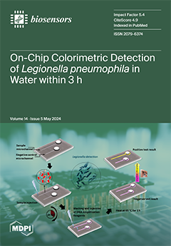
Legionella pneumophila bacterium accounts for more than 95% of the cases of Legionnaires’ disease. Bacterium detection by classical microbiology may take more than 10 days. Therefore, methods for its rapid and point-of-need detection are instrumental in preventing outbreaks. Α simple and compact chip is presented for the detection of L. pneumophila in water samples, integrating, for the first time, sample preparation and naked-eye qualitative or image analysis-based semiquantitative detection within 3 h. The achieved analysis time is reduced by 99% compared to that required by the gold-standard (bacteria culturing) method, while the simplified assay enables performing the test at the point of need. The method showed high sensitivity in drinking and drilling water. View this paper
- Issues are regarded as officially published after their release is announced to the table of contents alert mailing list.
- You may sign up for e-mail alerts to receive table of contents of newly released issues.
- PDF is the official format for papers published in both, html and pdf forms. To view the papers in pdf format, click on the "PDF Full-text" link, and use the free Adobe Reader to open them.
Previous Issue
Next Issue