Next Issue
Volume 11, March
Previous Issue
Volume 10, September


J. Funct. Biomater., Volume 10, Issue 4 (December 2019) – 13 articles

Cover Story (view full-size image): 
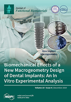
Alterations in the implant macrogeometry may improve the osseointegration process. New concepts about bone tissue compression during implant installation are being established, especially in order to generate as little stress as possible during the insertion of implants in this tissue. Thus, the elaboration of the healing chambers in the implant body (macrogeometry) was an excellent alternative for less compression of bone tissue without losing the fundamental characteristics necessary for the primary stability of these implants. View this paper.
  • Issues are regarded as officially published after their release is announced to the table of contents alert mailing list.
  • You may sign up for e-mail alerts to receive table of contents of newly released issues.
  • PDF is the official format for papers published in both, html and pdf forms. To view the papers in pdf format, click on the "PDF Full-text" link, and use the free Adobe Reader to open them.
Order results
Result details
Section
Select all
Export citation of selected articles as:
16 pages, 1845 KiB  
Article
Understanding the Role of Shape and Composition of Star-Shaped Polymers and their Ability to Both Bind and Prevent Bacteria Attachment on Oral Relevant Surfaces
by Hamid Mortazavian, Guillaume A. Picquet, Jānis Lejnieks, Lynette A. Zaidel, Carl P. Myers and Kenichi Kuroda
J. Funct. Biomater. 2019, 10(4), 56; https://doi.org/10.3390/jfb10040056 - 17 Dec 2019
Cited by 9 | Viewed by 4867
Abstract
In this study, we have prepared a series of 4- and 6-arm star-shaped polymers with varying molecular weight and hydrophobicity in order to provide insight into the role and relationship that shape and composition have on the binding and protecting of oral relevant [...] Read more.
In this study, we have prepared a series of 4- and 6-arm star-shaped polymers with varying molecular weight and hydrophobicity in order to provide insight into the role and relationship that shape and composition have on the binding and protecting of oral relevant surfaces (hydroxyapatite, HAP) from bacteria colonization. Star-shaped acrylic acid polymers were prepared by free-radical polymerization in the presence of chain transfer agents with thiol groups, and their binding to the HAP surfaces and subsequent bacteria repulsion was measured. We observed that binding was dependent on both polymer shape and hydrophobicity (star vs. linear), but their relative efficacy to reduce oral bacteria attachment from surfaces was dependent on their hydrophobicity only. We further measured the macroscopic effects of these materials to modify the mucin-coated HAP surfaces through contact angle experiments; the degree of angle change was dependent on the relative hydrophobicity of the materials suggesting future in vivo efficacy. The results from this study highlight that star-shaped polymers represent a new material platform for the development of dental applications to control bacterial adhesion which can lead to tooth decay, with various compositional and structural aspects of materials being vital to effectively design oral care products. Full article
(This article belongs to the Special Issue Bacterial Interactions with Dental and Medical Materials)
Show Figures

Figure 1

17 pages, 8075 KiB  
Article
Double-Emulsion Copolyester Microcapsules for Sustained Intraperitoneal Release of Carboplatin
by Aneta Cymbaluk-Płoska, Peter Sobolewski, Anita Chudecka-Głaz, Ewa Wiśniewska, Joanna Łapczuk, Marcin Frankowski, Marek Droździk and Miroslawa El Fray
J. Funct. Biomater. 2019, 10(4), 55; https://doi.org/10.3390/jfb10040055 - 06 Dec 2019
Cited by 2 | Viewed by 4238
Abstract
Despite on-going medical advances, ovarian cancer survival rates have stagnated. In order to improve IP delivery of platinum-based antineoplastics, we aimed to develop a sustained drug delivery system for carboplatin (CPt). Toward this aim, we pursued a double emulsion process for obtaining CPt-loaded [...] Read more.
Despite on-going medical advances, ovarian cancer survival rates have stagnated. In order to improve IP delivery of platinum-based antineoplastics, we aimed to develop a sustained drug delivery system for carboplatin (CPt). Toward this aim, we pursued a double emulsion process for obtaining CPt-loaded microcapsules composed of poly(ethylene terephthalate-ethylene dilinoleate) (PET-DLA) copolymer. We were able to obtain PET-DLA microspheres in the targeted size range of 10–25 µm (median: 18.5 µm), to reduce intraperitoneal clearance by phagocytosis and lymphoid transit. Empty microspheres showed the lack of toxicity in vitro. The double emulsion process yielded 2.5% w/w CPt loading and obtained microcapsules exhibited sustained (>20 day) zero-order release. The encapsulated CPt was confirmed to be bioavailable, as the microcapsules demonstrated efficacy against human ovarian adenocarcinoma (SK-OV-3) cells in vitro. Following intraperitoneal injection in mice, we did not observe adhesions, only mild, clinically-insignificant, local inflammatory response. Tissue platinum levels, monitored over 14 days using atomic absorption spectroscopy, revealed low burst and reduced systemic uptake (plasma, kidney), as compared to neat carboplatin injection. Overall, the results demonstrate the potential of the developed microencapsulation system for long-term intraperitoneal sustained release of carboplatin for the treatment of ovarian cancer. Full article
Show Figures

Graphical abstract

20 pages, 4520 KiB  
Article
Phosphoserine Functionalized Cements Preserve Metastable Phases, and Reprecipitate Octacalcium Phosphate, Hydroxyapatite, Dicalcium Phosphate, and Amorphous Calcium Phosphate, during Degradation, In Vitro
by Joseph Lazraq Bystrom and Michael Pujari-Palmer
J. Funct. Biomater. 2019, 10(4), 54; https://doi.org/10.3390/jfb10040054 - 27 Nov 2019
Cited by 8 | Viewed by 6891
Abstract
Phosphoserine modified cements (PMC) exhibit unique properties, including strong adhesion to tissues and biomaterials. While TTCP-PMCs remodel into bone in vivo, little is known regarding the bioactivity and physiochemical changes that occur during resorption. In the present study, changes in the mechanical strength [...] Read more.
Phosphoserine modified cements (PMC) exhibit unique properties, including strong adhesion to tissues and biomaterials. While TTCP-PMCs remodel into bone in vivo, little is known regarding the bioactivity and physiochemical changes that occur during resorption. In the present study, changes in the mechanical strength and composition were evaluated for 28 days, for three formulations of αTCP based PMCs. PMCs were significantly stronger than unmodified cement (38–49 MPa vs. 10 MPa). Inclusion of wollastonite in PMCs appeared to accelerate the conversion to hydroxyapatite, coincident with slight decrease in strength. In non-wollastonite PMCs the initial compressive strength did not change after 28 days in PBS (p > 0.99). Dissolution/degradation of PMC was evaluated in acidic (pH 2.7, pH 4.0), and supersaturated fluids (simulated body fluid (SBF)). PMCs exhibited comparable mass loss (<15%) after 14 days, regardless of pH and ionic concentration. Electron microscopy, infrared spectroscopy, and X-ray analysis revealed that significant amounts of brushite, octacalcium phosphate, and hydroxyapatite reprecipitated, following dissolution in acidic conditions (pH 2.7), while amorphous calcium phosphate formed in SBF. In conclusion, PMC surfaces remodel into metastable precursors to hydroxyapatite, in both acidic and neutral environments. By tuning the composition of PMCs, durable strength in fluids, and rapid transformation can be obtained. Full article
(This article belongs to the Special Issue Functionalized Biomimetic Calcium Phosphates)
Show Figures

Graphical abstract

16 pages, 4827 KiB  
Article
Simulated Performance of a Xenohybrid Bone Graft (SmartBone®) in the Treatment of Acetabular Prosthetic Reconstruction
by Carlo Francesco Grottoli, Alberto Cingolani, Fabio Zambon, Riccardo Ferracini, Tomaso Villa and Giuseppe Perale
J. Funct. Biomater. 2019, 10(4), 53; https://doi.org/10.3390/jfb10040053 - 22 Nov 2019
Cited by 4 | Viewed by 5134
Abstract
Total hip arthroplasty (THA) is a surgical procedure for the replacement of hip joints with artificial prostheses. Several approaches are currently employed in the treatment of this kind of defect. Overall, the most common method involves using a quite invasive metallic support (a [...] Read more.
Total hip arthroplasty (THA) is a surgical procedure for the replacement of hip joints with artificial prostheses. Several approaches are currently employed in the treatment of this kind of defect. Overall, the most common method involves using a quite invasive metallic support (a Burch–Schneider ring). Moreover, valid alternatives and less invasive techniques still need to be supported by novel material development. In this work, we evaluated the performance of SmartBone®, a xenohybrid bone graft composed of a bovine bone matrix reinforced with biodegradable polymers and collagen, as an effective support in acetabular prosthesis reconstruction. Specifically, the material’s mechanical properties were experimentally determined (E = ~1.25 GPa, Ef = ~0.34 GPa, and Et = ~0.49 GPa) and used for simulation of the hip joint system with a SmartBone® insert. Moreover, a comparison with a similar case treated with a Burch–Schneider ring was also conducted. It was found that it is possible to perform THA revision surgeries without the insertion of an invasive metal support and it can be nicely combined with SmartBone®’s osteointegration characteristics. The material can withstand the loads independently (σmax = ~12 MPa) or be supported by a thinner titanium plate in contact with the bone in the worst cases. This way, improved bone regeneration can be achieved. Full article
(This article belongs to the Special Issue Application of Biomechanical Model on Tissue Engineering)
Show Figures

Figure 1

19 pages, 4812 KiB  
Article
Intrinsic Cellular Responses of Human Wharton’s Jelly Mesenchymal Stem Cells Influenced by O2-Plasma-Modified and Unmodified Surface of Alkaline-Hydrolyzed 2D and 3D PCL Scaffolds
by Kewalin Inthanon, Wanida Janvikul, Siriwan Ongchai and Siriwadee Chomdej
J. Funct. Biomater. 2019, 10(4), 52; https://doi.org/10.3390/jfb10040052 - 18 Nov 2019
Cited by 1 | Viewed by 4319
Abstract
Polycaprolactone (PCL), a hydrophobic-degradable polyester, has been widely investigated and extensively developed, to increase the biocompatibility for tissue engineering. This research was the first trial to evaluate the intrinsic biological responses of human Wharton’s Jelly Mesenchymal Stem Cells (hWJMSCs) cultured on alkaline hydrolysis [...] Read more.
Polycaprolactone (PCL), a hydrophobic-degradable polyester, has been widely investigated and extensively developed, to increase the biocompatibility for tissue engineering. This research was the first trial to evaluate the intrinsic biological responses of human Wharton’s Jelly Mesenchymal Stem Cells (hWJMSCs) cultured on alkaline hydrolysis and low-pressure oxygen plasma modified 2D and 3D PCL scaffolds, without adding any differentiation inducers; this has not been reported before. Four types of the substrate were newly established: 2D plasma-treated PCL (2D-TP), 2D non-plasma-treated PCL (2D-NP), 3D plasma-treated PCL (3D-TP), and 3D non-plasma-treated PCL (3D-NP). Physicochemical characterization revealed that only plasma-treated PCL scaffolds significantly increased the hydrophilicity and % oxygen/carbon ratio on the surfaces. The RMS roughness of 3D was higher than 2D conformation, whilst the plasma-treated surfaces were rougher than the non-plasma treated ones. The cytocompatibility test demonstrated that the 2D PCLs enhanced the initial cell attachment in comparison to the 3Ds, indicated by a higher expression of focal adhesion kinase. Meanwhile, the 3Ds promoted cell proliferation and migration as evidence of higher cyclin-A expression and filopodial protrusion, respectively. The 3Ds potentially protected the cell from apoptosis/necrosis but also altered the pluripotency/differentiation-related gene expression. In summary, the different configuration and surface properties of PCL scaffolds displayed the significant potential and effectiveness for facilitating stem cell growth and differentiation in vitro. The cell–substrate interactions on modified surface PCL may provide some information which could be further applied in substrate architecture for stem cell accommodation in cell delivery system for tissue repair. Full article
Show Figures

Figure 1

13 pages, 2477 KiB  
Article
Biomechanical Properties and Biocompatibility of a Non-Absorbable Elastic Thread
by Yeji Choi, Moonseok Kang, Moon Seop Choi, Jennifer Kim Song, Eugene Lih, Deahyung Lee and Hong-Hee Jung
J. Funct. Biomater. 2019, 10(4), 51; https://doi.org/10.3390/jfb10040051 - 16 Nov 2019
Cited by 4 | Viewed by 5989
Abstract
To date, extensive studies have been conducted to assess diverse types of sutures. But there is a paucity of data regarding biomechanical properties of commonly used suture materials. In the current experiment, we compared biomechanical properties and biocompatibility, such as tensile strength and [...] Read more.
To date, extensive studies have been conducted to assess diverse types of sutures. But there is a paucity of data regarding biomechanical properties of commonly used suture materials. In the current experiment, we compared biomechanical properties and biocompatibility, such as tensile strength and elongation, the degree of bovine serum albumin (BSA) release, in vitro cytotoxicity and ex vivo frictional properties, between a non-absorbable elastic thread (NAT; HansBiomed Co. Ltd., Seoul, Korea) (NAT-R: NAT with a rough surface, NAT-S: NAT with a smooth surface) and the Elasticum® (Korpo SRL, Genova, Italy). The degree of tensile strength and elongation of Si threads was significantly higher in both the NAT-R and -S as compared with the Elasticum® (p < 0.05). Moreover, the degree of tensile strength and elongation of PET threads was significantly lower in both NAT-R and -S as compared with the Elasticum® (p < 0.05). Furthermore, the degree of tensile strength and elongation of braided Si/PET threads was significantly lower in NAT-S as compared with NAT-R and Elasticum® (p < 0.05). The degree of BSA release was significantly higher in the NAT-R as compared with Elasticum® and NAT-S throughout a 2-h period in the descending order (p < 0.05). The degree of cell viability was significantly higher in both NAT-R and -S as compared with Elasticum® (p < 0.05). The degree of coefficient of friction as well as the frictional force and strength was significantly higher in NAT-R as compared with NAT-S and Elasticum® (p < 0.05). NAT had a higher degree of biomechanical properties and biocompatibility as compared with Elasticum®. But further experimental and clinical studies are warranted to compare the efficacy, safety, and potential role as a carrier for drug delivery between NAT and Elasticum®. Full article
Show Figures

Figure 1

15 pages, 2924 KiB  
Communication
In Situ Crosslinking Bionanocomposite Hydrogels with Potential for Wound Healing Applications
by Federica Leone, Melike Firlak, Kirsty Challen, Wayne Bonnefin, Barbara Onida, Karen L. Wright and John G. Hardy
J. Funct. Biomater. 2019, 10(4), 50; https://doi.org/10.3390/jfb10040050 - 14 Nov 2019
Cited by 7 | Viewed by 6023
Abstract
In situ forming hydrogels are a class of biomaterials that can fulfil a variety of important biomedically relevant functions and hold promise for the emerging field of patient-specific treatments (e.g., cell therapy, drug delivery). Here we report the results of our investigations on [...] Read more.
In situ forming hydrogels are a class of biomaterials that can fulfil a variety of important biomedically relevant functions and hold promise for the emerging field of patient-specific treatments (e.g., cell therapy, drug delivery). Here we report the results of our investigations on the generation of in situ forming hydrogels with potential for wound healing applications (e.g., complex blast injuries). The combination of polysaccharides that were oxidized to display aldehydes, amine displaying chitosan and nanostructured ZnO yields in situ forming bionanocomposite hydrogels. The physicochemical properties of the components, their cytotoxicity towards HaCat cells and the in vitro release of zinc ions on synthetic skin were studied. The in situ gel formation process was complete within minutes, the components were non-toxic towards HaCat cells at functional levels, Zn2+ was released from the gels, and such materials may facilitate wound healing. Full article
Show Figures

Graphical abstract

17 pages, 5220 KiB  
Article
Design of Silk-Elastin-Like Protein Nanoparticle Systems with Mucoadhesive Properties
by Rachael N. Parker, Wenyao A. Wu, Tina B. McKay, Qiaobing Xu and David L. Kaplan
J. Funct. Biomater. 2019, 10(4), 49; https://doi.org/10.3390/jfb10040049 - 12 Nov 2019
Cited by 13 | Viewed by 5734
Abstract
Transmucosal drug delivery is a promising avenue to improve therapeutic efficacy through localized therapeutic administration. Drug delivery systems that increase retention in the mucosal layer are needed to improve efficiency of such transmucosal platforms. However, the applicability of such systems is often limited [...] Read more.
Transmucosal drug delivery is a promising avenue to improve therapeutic efficacy through localized therapeutic administration. Drug delivery systems that increase retention in the mucosal layer are needed to improve efficiency of such transmucosal platforms. However, the applicability of such systems is often limited by the range of chemistries and properties that can be achieved. Here we present the design and implementation of silk-elastin-like proteins (SELPs) with mucoadhesive properties. SELP-based micellar-like nanoparticles provide a system to tailor chemical and physical properties through genetic engineering of the SELP sequence, which enables the fabrication of nanoparticles with specific chemical and physical features. Analysis of the adhesion of four different SELP-based nanoparticle systems in an artificial mucus system, as well as in in vitro cellular assays indicates that addition of mucoadhesive chemical features on the SELP systems increases retention of the particles in mucosal environments. The results indicated that SELP-based nanoparticles provide a useful approach to study and develop transmucosal protein drug delivery system with unique mucoadhesive properties. Future studies will serve to further expand the range of achievable properties, as well as the utilization of SELPs to fabricate mucoadhesive materials for in vivo testing. Full article
Show Figures

Figure 1

13 pages, 5676 KiB  
Article
Regulation of Osteogenic Markers at Late Stage of Osteoblast Differentiation in Silicon and Zinc Doped Porous TCP
by Gary A. Fielding, Naboneeta Sarkar, Sahar Vahabzadeh and Susmita Bose
J. Funct. Biomater. 2019, 10(4), 48; https://doi.org/10.3390/jfb10040048 - 05 Nov 2019
Cited by 17 | Viewed by 5312
Abstract
Calcium phosphates (CaPs) are one of the most widely used synthetic materials for bone grafting applications in the orthopedic industry. Recent trends in synthetic bone graft applications have shifted towards the incorporation of metal trace elements that extend the performance of CaPs to [...] Read more.
Calcium phosphates (CaPs) are one of the most widely used synthetic materials for bone grafting applications in the orthopedic industry. Recent trends in synthetic bone graft applications have shifted towards the incorporation of metal trace elements that extend the performance of CaPs to have osteoinductive properties. The objective of this study is to investigate the effects of silicon (Si) and zinc (Zn) dopants in highly porous tricalcium phosphate (TCP) scaffolds on late-stage osteoblast cell differentiation markers. In this study, an oil emulsion method is utilized to fabricate highly porous SiO2 doped β-TCP (Si-TCP) and ZnO doped β-TCP (Zn-TCP) scaffolds through the incorporation of 0.5 wt.% SiO2 and 0.25 wt.% ZnO, respectively, to the β-TCP scaffold. Reverse transcriptase quantitative polymerase chain reaction (RT-qPCR) is utilized to analyze the mRNA expression of osteoprotegerin (OPG), receptor activator of nuclear kappa beta ligand (RANKL), bone morphogenetic protein 2 (BMP2), and runt-related transcription factor 2 (Runx2) at the later stage of osteoblast differentiation, day 21 and day 28. Results show that the addition of Si and Zn to the β-TCP structure inhibited the β to α-TCP phase transformation and enhance the density without affecting the dissolution properties. Normal BMP-2 and Runx2 transcriptions are observed in both Si-TCP and Zn-TCP scaffolds at the initial time point, as demonstrated by RT-qPCR. Moreover, the addition of both Si and Zn positively regulate the osteoprotegerin: receptor activator of nuclear factor k-β ligand (OPG:RANKL) ratio at 21-days for Si-TCP and Zn-TCP scaffolds. These results demonstrate the effects of Si and Zn doped porous β-TCP scaffolds on the upregulation of osteoblast marker gene expression including OPG, RANKL, BMP-2, and Runx2, indicating the role of trace elements on the effective regulation of late-stage osteoblast cell differentiation markers. Full article
(This article belongs to the Special Issue Functionalized Biomimetic Calcium Phosphates)
Show Figures

Figure 1

14 pages, 4082 KiB  
Article
Biomechanical Effects of a New Macrogeometry Design of Dental Implants: An In Vitro Experimental Analysis
by Sergio Alexandre Gehrke, Leticia Pérez-Díaz, Patricia Mazón and Piedad N. De Aza
J. Funct. Biomater. 2019, 10(4), 47; https://doi.org/10.3390/jfb10040047 - 25 Oct 2019
Cited by 17 | Viewed by 5228
Abstract
The purpose of the present study was to measure and compare the insertion torque, removal torque, and the implant stability quotient by resonance frequency analysis in different polyurethane block densities of two implant macrogeometries. Four different polyurethane synthetic bone blocks were used with [...] Read more.
The purpose of the present study was to measure and compare the insertion torque, removal torque, and the implant stability quotient by resonance frequency analysis in different polyurethane block densities of two implant macrogeometries. Four different polyurethane synthetic bone blocks were used with three cortical thickness: Bone 1 with a cortical thickness of 1 mm, Bone 2 with a cortical thickness of 2 mm, Bone 3 with a cortical thickness of 3 mm, and Bone 4, which was totally cortical. Four groups were created in accordance with the implant macrogeometry (n = 10 per group) and surface treatment: G1—regular implant design without surface treatment; G2—regular implant design with surface treatment; G3—new implant design without surface treatment; G4—new implant design with surface treatment. All implants used were 4 mm in diameter and 10 mm in length and manufactured in commercially pure titanium (grade IV) by Implacil De Bortoli (São Paulo, Brazil). The implants were installed using a computed torque machine, and following installation of the implant, the stability quotient (implant stability quotient, ISQ) values were measured in two directions using Osstell devices. The data were analyzed by considering the 5% level of significance. All implant groups showed similar mean ISQ values without statistical differences (p > 0.05), for the same synthetic bone block: for Bone 1, the value was 57.7 ± 3.0; for Bone 2, it was 58.6 ± 2.2; for Bone 3, it was 60.6 ± 2.3; and for Bone 4, it was 68.5 ± 2.8. However, the insertion torque showed similar higher values for the regular macrogeometry (G1 and G2 groups) in comparison with the new implant macrogeometry (G3 and G4 groups). The analysis of the results found that primary stability does not simply depend on the insertion torque but also on the bone quality. In comparison with the regular implant macrogeometry, the new implant macrogeometry decreased the insertion torque without affecting the implant stability quotient values. Full article
Show Figures

Figure 1

15 pages, 2741 KiB  
Article
Impact of Bi2O3 and ZrO2 Radiopacifiers on the Early Hydration and C–S–H Gel Structure of White Portland Cement
by Qiu Li and Nichola J. Coleman
J. Funct. Biomater. 2019, 10(4), 46; https://doi.org/10.3390/jfb10040046 - 18 Oct 2019
Cited by 17 | Viewed by 5352
Abstract
Bismuth oxide (monoclinic α-Bi2O3) and zirconium oxide (monoclinic ZrO2) are the most popular radiopacifiers in commercial Portland cement-based endodontic restoratives, yet their effects on the setting and hydration reactions are not fully understood. This study compares the [...] Read more.
Bismuth oxide (monoclinic α-Bi2O3) and zirconium oxide (monoclinic ZrO2) are the most popular radiopacifiers in commercial Portland cement-based endodontic restoratives, yet their effects on the setting and hydration reactions are not fully understood. This study compares the impact of 20 wt.% of Bi2O3 or ZrO2 on the early hydration reactions and C–S–H gel structure of white Portland cement (WPC). Cement paste samples were hydrated at 37.5 °C prior to analysis by 29Si and 27Al magic angle spinning nuclear magnetic resonance spectroscopy at 3 h and 24 h, and transmission electron microscopy at 3 h. Initial and final setting times were determined using a Vicat apparatus and reaction kinetics were monitored by isothermal conduction calorimetry. Bi2O3 was found to prolong initial and final setting times and retard the degree of hydration by 32% at 24 h. Heat evolution during the acceleration and deceleration phases of the hydration process was reduced and the exotherm arising from renewed ettringite formation was delayed and diminished in the presence of Bi2O3. Conversely, ZrO2 had no significant impact on either setting time; although, it accelerated hydration by 23% within 24 h. Increases in the mean silicate chain length and the extent of aluminum substitution in the C–S–H gel were observed in the presence of both radiopacifying agents after 24 h relative to those of the unblended WPC. The Bi2O3 and ZrO2 particles remained intact within the cement matrix and neither bismuth nor zirconium was chemically incorporated in the hydration products. Full article
(This article belongs to the Special Issue Endodontic Biomaterials)
Show Figures

Figure 1

17 pages, 5752 KiB  
Article
Cell-Penetrating Peptides as a Tool for the Cellular Uptake of a Genetically Modified Nitroreductase for use in Directed Enzyme Prodrug Therapy
by Simon D. Anderson, Robert J. Hobbs, Vanessa V. Gwenin, Patrick Ball, Lindsey A. Bennie, Jonathan A. Coulter and Chris D. Gwenin
J. Funct. Biomater. 2019, 10(4), 45; https://doi.org/10.3390/jfb10040045 - 01 Oct 2019
Cited by 4 | Viewed by 5221
Abstract
Directed enzyme prodrug therapy (DEPT) involves the delivery of a prodrug-activating enzyme to a solid tumour site, followed by the subsequent activation of an administered prodrug. One of the most studied enzyme–prodrug combinations is the nitroreductase from Escherichia coli (NfnB) with the prodrug [...] Read more.
Directed enzyme prodrug therapy (DEPT) involves the delivery of a prodrug-activating enzyme to a solid tumour site, followed by the subsequent activation of an administered prodrug. One of the most studied enzyme–prodrug combinations is the nitroreductase from Escherichia coli (NfnB) with the prodrug CB1954 [5-(aziridin-1-yl)-2,4-dinitro-benzamide]. One of the major issues faced by DEPT is the ability to successfully internalize the enzyme into the target cells. NfnB has previously been genetically modified to contain cysteine residues (NfnB-Cys) which bind to gold nanoparticles for a novel DEPT therapy called magnetic nanoparticle directed enzyme prodrug therapy (MNDEPT). One cellular internalisation method is the use of cell-penetrating peptides (CPPs), which aid cellular internalization of cargo. Here the cell-penetrating peptides: HR9 and Pep-1 were tested for their ability to conjugate with NfnB-Cys. The conjugates were further tested for their potential use in MNDEPT, as well as conjugating with the delivery vector intended for use in MNDEPT and tested for the vectors capability to penetrate into cells. Full article
Show Figures

Figure 1

35 pages, 1622 KiB  
Article
An Insight into Advanced Approaches for Photosensitizer Optimization in Endodontics—A Critical Review
by Patrícia Diogo, M. Amparo F. Faustino, M. Graça P. M. S. Neves, Paulo J. Palma, Isabel P. Baptista, Teresa Gonçalves and João Miguel Santos
J. Funct. Biomater. 2019, 10(4), 44; https://doi.org/10.3390/jfb10040044 - 30 Sep 2019
Cited by 35 | Viewed by 8632
Abstract
Apical periodontitis is a biofilm-mediated disease; therefore, an antimicrobial approach is essential to cure or prevent its development. In the quest for efficient strategies to achieve this objective, antimicrobial photodynamic therapy (aPDT) has emerged as an alternative to classical endodontic irrigation solutions and [...] Read more.
Apical periodontitis is a biofilm-mediated disease; therefore, an antimicrobial approach is essential to cure or prevent its development. In the quest for efficient strategies to achieve this objective, antimicrobial photodynamic therapy (aPDT) has emerged as an alternative to classical endodontic irrigation solutions and antibiotics. The aim of the present critical review is to summarize the available evidence on photosensitizers (PSs) which has been confirmed in numerous studies from diverse areas combined with several antimicrobial strategies, as well as emerging options in order to optimize their properties and effects that might be translational and useful in the near future in basic endodontic research. Published data notably support the need for continuing the search for an ideal endodontic photosensitizer, that is, one which acts as an excellent antimicrobial agent without causing toxicity to the human host cells or presenting the risk of tooth discoloration. The current literature on experimental studies mainly relies on assessment of mixed disinfection protocols, combining approaches which are already available with aPDT as an adjunct therapy. In this review, several approaches concerning aPDT efficiency are appraised, such as the use of bacteriophages, biopolymers, drug and light delivery systems, efflux pump inhibitors, negative pressure systems, and peptides. The authors also analyzed their combination with other approaches for aPDT improvement, such as sonodynamic therapy. All of the aforementioned techniques have already been tested, and we highlight the biological challenges of each formulation, predicting that the collected information may encourage the development of other effective photoactive materials, in addition to being useful in endodontic basic research. Moreover, special attention is dedicated to studies on detailed conditions, aPDT features with a focus on PS enhancer strategies, and the respective final antimicrobial outcomes. From all the mentioned approaches, the two which are most widely discussed and which show the most promising outcomes for endodontic purposes are drug delivery systems (with strong development in nanoparticles) and PS solubilizers. Full article
(This article belongs to the Special Issue Endodontic Biomaterials)
Show Figures

Figure 1

Previous Issue
Next Issue
Back to TopTop