


J. Clin. Med., Volume 14, Issue 10 (May-2 2025) – 351 articles
Cover Story (view full-size image):
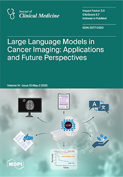
Recently, there has been tremendous interest in the use of large language models (LLMs) in radiology. LLMs have been employed for various applications in cancer imaging, including improving reporting speeds and accuracy via the generation of standardized reports, automating the classification and staging of abnormal findings, incorporating appropriate guidelines, and calculating individualized risk scores. LLMs can also improve patients’ comprehension of imaging reports and translate them into multiple languages. Other future applications of LLMs include aiding patient management and preventing and predicting adverse events. However, limitations such as hallucinations and variable performances could present obstacles to their widespread clinical implementation. This review discusses the applications and limitations of LLMs in cancer imaging. View this paper
- Issues are regarded as officially published after their release is announced to the table of contents alert mailing list.
- You may sign up for e-mail alerts to receive table of contents of newly released issues.
- PDF is the official format for papers published in both, html and pdf forms. To view the papers in pdf format, click on the "PDF Full-text" link, and use the free Adobe Reader to open them.
Previous Issue
Next Issue