Next Issue
Volume 13, April-1
Previous Issue
Volume 13, March-1


Cells, Volume 13, Issue 6 (March-2 2024) – 94 articles

Cover Story (view full-size image): 
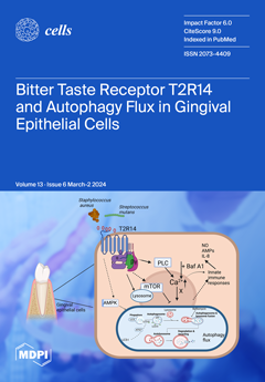
Macroautophagy (autophagy) maintains cellular homeostasis and regulates energy production and innate immunity. The role of the human bitter taste receptors (T2Rs) in autophagy is poorly understood. This study targets T2R14, the taste receptor expressed at significant levels in oral gingival epithelial cells (GECs). Our study reveals that T2R14 downregulates autophagy protein 7 independent autophagy flux in gingival epithelial cells. Further, our results show that Streptococcus mutans quorum sensing molecule (competence stimulating peptide-1) causes robust intracellular calcium release in GECs, which is both T2R14 and autophagy protein 7 dependent. These findings reveal the different mechanisms by which T2Rs influence host-microbe interactions and modulate the oral immune environment. View this paper
  • Issues are regarded as officially published after their release is announced to the table of contents alert mailing list.
  • You may sign up for e-mail alerts to receive table of contents of newly released issues.
  • PDF is the official format for papers published in both, html and pdf forms. To view the papers in pdf format, click on the "PDF Full-text" link, and use the free Adobe Reader to open them.
Order results
Result details
Section
Select all
Export citation of selected articles as:
13 pages, 8746 KiB  
Communication
Transgene-Free Cynomolgus Monkey iPSCs Generated under Chemically Defined Conditions
by Yuliia Tereshchenko, Nesil Esiyok, Enrique Garea-Rodríguez, Daniele Repetto, Rüdiger Behr and Ignacio Rodríguez-Polo
Cells 2024, 13(6), 558; https://doi.org/10.3390/cells13060558 - 21 Mar 2024
Viewed by 742
Abstract
Non-human primates (NHPs) are pivotal animal models for translating novel cell replacement therapies into clinical applications, including validating the safety and efficacy of induced pluripotent stem cell (iPSC)-derived products. Preclinical development and the testing of cell-based therapies ideally comprise xenogeneic (human stem cells [...] Read more.
Non-human primates (NHPs) are pivotal animal models for translating novel cell replacement therapies into clinical applications, including validating the safety and efficacy of induced pluripotent stem cell (iPSC)-derived products. Preclinical development and the testing of cell-based therapies ideally comprise xenogeneic (human stem cells into NHPs) and allogenic (NHP stem cells into NHPs) transplantation studies. For the allogeneic approach, it is necessary to generate NHP-iPSCs with generally equivalent quality to the human counterparts that will be used later on in patients. Here, we report the generation and characterization of transgene- and feeder-free cynomolgus monkey (Macaca fascicularis) iPSCs (Cyno-iPSCs). These novel cell lines have been generated according to a previously developed protocol for the generation of rhesus macaque, baboon, and human iPSC lines. Beyond their generation, we demonstrate the potential of the novel Cyno-iPSCs to differentiate into two clinically relevant cell types, i.e., cardiomyocytes and neurons. Overall, we provide a resource of novel iPSCs from the most frequently used NHP species in the regulatory testing of biologics and classical pharmaceutics to expand our panel of iPSC lines from NHP species with high relevance in preclinical testing and translational research. Full article
(This article belongs to the Collection Stem Cells in Tissue Engineering and Regeneration)
Show Figures

Figure 1

21 pages, 8935 KiB  
Article
MiR-23b and miR-133 Cotarget TGFβ2/NOTCH1 in Sheep Dermal Fibroblasts, Affecting Hair Follicle Development
by Junmin He, Chen Wei, Xixia Huang, Guoping Zhang, Jingyi Mao, Xue Li, Cunming Yang, Wenjing Zhang, Kechuan Tian and Guifen Liu
Cells 2024, 13(6), 557; https://doi.org/10.3390/cells13060557 - 21 Mar 2024
Viewed by 807
Abstract
Wool is produced and controlled by hair follicles (HFs). However, little is known about the mechanisms involved in HF development and regulation. Sheep dermal fibroblasts (SDFs) play a key role in the initial stage of HF development. Analyzing the molecular mechanism that regulates [...] Read more.
Wool is produced and controlled by hair follicles (HFs). However, little is known about the mechanisms involved in HF development and regulation. Sheep dermal fibroblasts (SDFs) play a key role in the initial stage of HF development. Analyzing the molecular mechanism that regulates early HF development in superfine wool sheep is of great importance for better understanding the HF morphogenesis process and for the breeding of fine wool sheep. Here, we show that two microRNAs (miRNAs) affect the development of HFs by targeting two genes that are expressed by SDFs. Meanwhile, the overexpression and inhibition of oar-miR-23b and oar-miR-133 in SDFs cells and cell proliferation, apoptosis, and migration were further detected using a CCK-8 assay, an Annexin V-FITC assay, a Transwell assay, and flow cytometry. We found that oar-miR-23b, oar-miR-133, and their cotarget genes TGFβ2 and NOTCH1 were differentially expressed during the six stages of HF development in superfine wool sheep. Oar-miR-23b and oar-miR-133 inhibited the proliferation and migration of SDFs and promoted the apoptosis of SDFs through TGFβ2 and NOTCH1. oar-miR-23b and oar-miR-133 inhibited the proliferation and migration of SDFs by jointly targeting TGFβ2 and NOTCH1, thereby inhibiting the development of superfine wool HFs. Our research provides a molecular marker that can be used to guide the breeding of ultrafine wool sheep. Full article
Show Figures

Graphical abstract

14 pages, 1556 KiB  
Review
MuSK Myasthenia Gravis—Potential Pathomechanisms and Treatment Directed against Specific Targets
by Edyta Dziadkowiak, Dagmara Baczyńska and Marta Waliszewska-Prosół
Cells 2024, 13(6), 556; https://doi.org/10.3390/cells13060556 - 21 Mar 2024
Viewed by 796
Abstract
Myasthenia gravis (MG) is an autoimmune disease in which autoantibodies target structures within the neuromuscular junction, affecting neuromuscular transmission. Muscle-specific tyrosine kinase receptor-associated MG (MuSK-MG) is a rare, often more severe, subtype of the disease with different pathogenesis and specific clinical features. It [...] Read more.
Myasthenia gravis (MG) is an autoimmune disease in which autoantibodies target structures within the neuromuscular junction, affecting neuromuscular transmission. Muscle-specific tyrosine kinase receptor-associated MG (MuSK-MG) is a rare, often more severe, subtype of the disease with different pathogenesis and specific clinical features. It is characterized by a more severe clinical course, more frequent complications, and often inadequate response to treatment. Here, we review the current state of knowledge about potential pathomechanisms of the MuSK-MG and their therapeutic implications as well as ongoing research in this field, with reference to key points of immune-mediated processes involved in the background of myasthenia gravis. Full article
Show Figures

Figure 1

13 pages, 2521 KiB  
Article
Modulating Cholesterol Metabolism via ACAT1 Knockdown Enhances Anti-B-Cell Lymphoma Activities of CD19-Specific Chimeric Antigen Receptor T Cells by Improving the Cell Activation and Proliferation
by Qiong Su, Jie Yao, Muhammad Asad Farooq, Iqra Ajmal, Yixin Duan, Cong He, Xuefei Hu and Wenzheng Jiang
Cells 2024, 13(6), 555; https://doi.org/10.3390/cells13060555 - 21 Mar 2024
Viewed by 956
Abstract
CD19-specific CAR-T immunotherapy has been extensively studied for the treatment of B-cell lymphoma. Recently, cholesterol metabolism has emerged as a modulator of T lymphocyte function and can be exploited in immunotherapy to increase the efficacy of CAR-based systems. Acetyl-CoA acetyltransferase 1 (ACAT1) is [...] Read more.
CD19-specific CAR-T immunotherapy has been extensively studied for the treatment of B-cell lymphoma. Recently, cholesterol metabolism has emerged as a modulator of T lymphocyte function and can be exploited in immunotherapy to increase the efficacy of CAR-based systems. Acetyl-CoA acetyltransferase 1 (ACAT1) is the major cholesterol esterification enzyme. ACAT1 inhibitors previously shown to modulate cardiovascular diseases are now being implicated in immunotherapy. In the present study, we achieved knockdown of ACAT1 in T cells via RNA interference technology by inserting ACAT1-shRNA into anti-CD19-CAR-T cells. Knockdown of ACAT1 led to an increased cytotoxic capacity of the anti-CD19-CAR-T cells. In addition, more CD69, IFN-γ, and GzmB were expressed in the anti-CD19-CAR-T cells. Cell proliferation was also enhanced in both antigen-independent and antigen-dependent manners. Degranulation was also improved as evidenced by an increased level of CD107a. Moreover, the knockdown of ACAT1 led to better anti-tumor efficacy of anti-CD19 CAR-T cells in the B-cell lymphoma mice model. Our study demonstrates novel CAR-T cells containing ACAT1 shRNA with improved efficacy compared to conventional anti-CD19-CAR-T cells in vitro and in vivo. Full article
(This article belongs to the Special Issue New Advances in Cellular Immunotherapy)
Show Figures

Graphical abstract

24 pages, 5018 KiB  
Article
Hematin- and Hemin-Induced Spherization and Hemolysis of Human Erythrocytes Are Independent of Extracellular Calcium Concentration
by Diana M. Mikhailova, Elisaveta Skverchinskaya, Julia Sudnitsyna, Kirill R. Butov, Ekaterina M. Koltsova, Igor V. Mindukshev and Stepan Gambaryan
Cells 2024, 13(6), 554; https://doi.org/10.3390/cells13060554 - 21 Mar 2024
Viewed by 724
Abstract
Pathologies such as malaria, hemorrhagic stroke, sickle cell disease, and thalassemia are characterized by the release of hemoglobin degradation products from damaged RBCs. Hematin (liganded with OH) and hemin (liganded with Cl)—are the oxidized forms of heme with toxic [...] Read more.
Pathologies such as malaria, hemorrhagic stroke, sickle cell disease, and thalassemia are characterized by the release of hemoglobin degradation products from damaged RBCs. Hematin (liganded with OH) and hemin (liganded with Cl)—are the oxidized forms of heme with toxic properties due to their hydrophobicity and the presence of redox-active Fe3. In the present study, using the original LaSca-TM laser particle analyzer, flow cytometry, and confocal microscopy, we showed that both hematin and hemin induce dose-dependent RBC spherization and hemolysis with ghost formation. Hematin and hemin at nanomolar concentrations increased [Ca2+]i in RBC; however, spherization and hemolysis occurred in the presence and absence of calcium, indicating that both processes are independent of [Ca2+]i. Both compounds triggered acute phosphatidylserine exposure on the membrane surface, reversible after 60 min of incubation. A comparison of hematin and hemin effects on RBCs revealed that hematin is a more reactive toxic metabolite than hemin towards human RBCs. The toxic effects of heme derivatives were reduced and even reversed in the presence of albumin, indicating the presence in RBCs of the own recovery system against the toxic effects of heme derivatives. Full article
(This article belongs to the Special Issue Highlights in Red Blood Cell Research)
Show Figures

Figure 1

20 pages, 4275 KiB  
Review
Variation of Structure and Cellular Functions of Type IA Topoisomerases across the Tree of Life
by Kemin Tan and Yuk-Ching Tse-Dinh
Cells 2024, 13(6), 553; https://doi.org/10.3390/cells13060553 - 21 Mar 2024
Viewed by 794
Abstract
Topoisomerases regulate the topological state of cellular genomes to prevent impediments to vital cellular processes, including replication and transcription from suboptimal supercoiling of double-stranded DNA, and to untangle topological barriers generated as replication or recombination intermediates. The subfamily of type IA topoisomerases are [...] Read more.
Topoisomerases regulate the topological state of cellular genomes to prevent impediments to vital cellular processes, including replication and transcription from suboptimal supercoiling of double-stranded DNA, and to untangle topological barriers generated as replication or recombination intermediates. The subfamily of type IA topoisomerases are the only topoisomerases that can alter the interlinking of both DNA and RNA. In this article, we provide a review of the mechanisms by which four highly conserved N-terminal protein domains fold into a toroidal structure, enabling cleavage and religation of a single strand of DNA or RNA. We also explore how these conserved domains can be combined with numerous non-conserved protein sequences located in the C-terminal domains to form a diverse range of type IA topoisomerases in Archaea, Bacteria, and Eukarya. There is at least one type IA topoisomerase present in nearly every free-living organism. The variation in C-terminal domain sequences and interacting partners such as helicases enable type IA topoisomerases to conduct important cellular functions that require the passage of nucleic acids through the break of a single-strand DNA or RNA that is held by the conserved N-terminal toroidal domains. In addition, this review will exam a range of human genetic disorders that have been linked to the malfunction of type IA topoisomerase. Full article
Show Figures

Figure 1

24 pages, 3790 KiB  
Review
Pros and Cons of Cryopreserving Allogeneic Stem Cell Products
by Caterina Giovanna Valentini, Claudio Pellegrino and Luciana Teofili
Cells 2024, 13(6), 552; https://doi.org/10.3390/cells13060552 - 21 Mar 2024
Viewed by 944
Abstract
The COVID-19 pandemic has precipitously changed the practice of transplanting fresh allografts. The safety measures adopted during the pandemic prompted the near-universal graft cryopreservation. However, the influence of cryopreserving allogeneic grafts on long-term transplant outcomes has emerged only in the most recent literature. [...] Read more.
The COVID-19 pandemic has precipitously changed the practice of transplanting fresh allografts. The safety measures adopted during the pandemic prompted the near-universal graft cryopreservation. However, the influence of cryopreserving allogeneic grafts on long-term transplant outcomes has emerged only in the most recent literature. In this review, the basic principles of cell cryopreservation are revised and the effects of cryopreservation on the different graft components are carefully reexamined. Finally, a literature revision on studies comparing transplant outcomes in patients receiving cryopreserved and fresh grafts is illustrated. Full article
Show Figures

Figure 1

18 pages, 33207 KiB  
Article
IF1 Promotes Cellular Proliferation and Inhibits Oxidative Phosphorylation in Mouse Embryonic Fibroblasts under Normoxia and Hypoxia
by Lothar Lauterboeck, Sung Wook Kang, Donnell White III, Rong Bao, Parnia Mobasheran and Qinglin Yang
Cells 2024, 13(6), 551; https://doi.org/10.3390/cells13060551 - 21 Mar 2024
Viewed by 701
Abstract
ATP synthase inhibitory factor subunit 1 (IF1) is an inhibitory subunit of mitochondrial ATP synthase, playing a crucial role in regulating mitochondrial respiration and energetics. It is well-established that IF1 interacts with the F1 sector of ATP synthase to inhibit the reversal rotation [...] Read more.
ATP synthase inhibitory factor subunit 1 (IF1) is an inhibitory subunit of mitochondrial ATP synthase, playing a crucial role in regulating mitochondrial respiration and energetics. It is well-established that IF1 interacts with the F1 sector of ATP synthase to inhibit the reversal rotation and, thus, ATP hydrolysis. Recent evidence supports that IF1 also inhibits forward rotation or the ATP synthesis activity. Adding to the complexity, IF1 may also facilitate mitophagy and cristae formation. The implications of these complex actions of IF1 for cellular function remain obscure. In the present study, we found that IF1 expression was markedly upregulated in hypoxic MEFs relative to normoxic MEFs. We investigate how IF1 affects cellular growth and function in cultured mouse embryonic fibroblasts derived from mouse lines with systemic IF1 overexpression and knockout under normoxia and hypoxia. Cell survival and proliferation analyses revealed that IF1 overexpression exerted limited effects on cellular viability but substantially increased proliferation under normoxia, whereas it facilitated both cellular viability and proliferation under hypoxia. The absence of IF1 may have a pro-survival effect but not a proliferative one in both normoxia and hypoxia. Cellular bioenergetic analyses revealed that IF1 suppressed cellular respiration when subjected to normoxia and was even more pronounced when subjected to hypoxia with increased mitochondrial ATP production. In contrast, IF1 knockout MEFs showed markedly increased cellular respiration under both normoxia and hypoxia with little change in mitochondrial ATP. Glycolytic stress assay revealed that IF1 overexpression modestly increased glycolysis in normoxia and hypoxia. Interestingly, the absence of IF1 in MEFs led to substantial increases in glycolysis. Therefore, we conclude that IF1 mainly inhibits cellular respiration and enhances cellular glycolysis to preserve mitochondrial ATP. On the other hand, IF1 deletion can significantly facilitate cellular respiration and glycolysis without leading to mitochondrial ATP deficit. Full article
Show Figures

Figure 1

32 pages, 934 KiB  
Review
The Molecular Mechanisms in Senescent Cells Induced by Natural Aging and Ionizing Radiation
by Milana Ibragimova, Assiya Kussainova, Akmaral Aripova, Rakhmetkazhi Bersimbaev and Olga Bulgakova
Cells 2024, 13(6), 550; https://doi.org/10.3390/cells13060550 - 21 Mar 2024
Viewed by 1235
Abstract
This review discusses the relationship between cellular senescence and radiation exposure. Given the wide range of ionizing radiation sources encountered by people in professional and medical spheres, as well as the influence of natural background radiation, the question of the effect of radiation [...] Read more.
This review discusses the relationship between cellular senescence and radiation exposure. Given the wide range of ionizing radiation sources encountered by people in professional and medical spheres, as well as the influence of natural background radiation, the question of the effect of radiation on biological processes, particularly on aging processes, remains highly relevant. The parallel relationship between natural and radiation-induced cellular senescence reveals the common aspects underlying these processes. Based on recent scientific data, the key points of the effects of ionizing radiation on cellular processes associated with aging, such as genome instability, mitochondrial dysfunction, altered expression of miRNAs, epigenetic profile, and manifestation of the senescence-associated secretory phenotype (SASP), are discussed. Unraveling the molecular mechanisms of cellular senescence can make a valuable contribution to the understanding of the molecular genetic basis of age-associated diseases in the context of environmental exposure. Full article
Show Figures

Graphical abstract

20 pages, 17724 KiB  
Review
The Origin and Regulation of Neuromesodermal Progenitors (NMPs) in Embryos
by Hisato Kondoh and Tatsuya Takemoto
Cells 2024, 13(6), 549; https://doi.org/10.3390/cells13060549 - 21 Mar 2024
Viewed by 767
Abstract
Neuromesodermal progenitors (NMPs), serving as the common origin of neural and paraxial mesodermal development in a large part of the trunk, have recently gained significant attention because of their critical importance in the understanding of embryonic organogenesis and the design of in vitro [...] Read more.
Neuromesodermal progenitors (NMPs), serving as the common origin of neural and paraxial mesodermal development in a large part of the trunk, have recently gained significant attention because of their critical importance in the understanding of embryonic organogenesis and the design of in vitro models of organogenesis. However, the nature of NMPs at many essential points remains only vaguely understood or even incorrectly assumed. Here, we discuss the nature of NMPs, focusing on their dynamic migratory behavior during embryogenesis and the mechanisms underlying their neural vs. mesodermal fate choice. The discussion points include the following: (1) How the sinus rhomboidals is organized; the tissue where the neural or mesodermal fate choice of NMPs occurs. (2) NMPs originating from the broad posterior epiblast are associated with Sox2 N1 enhancer activity. (3) Tbx6-dependent Sox2 repression occurs during NMP-derived paraxial mesoderm development. (4) The nephric mesenchyme, a component of the intermediate mesoderm, was newly identified as an NMP derivative. (5) The transition of embryonic tissue development from tissue-specific progenitors in the anterior part to that from NMPs occurs at the forelimb bud axial level. (6) The coexpression of Sox2 and Bra in NMPs is conditional and is not a hallmark of NMPs. (7) The ability of the NMP pool to sustain axial embryo growth depends on Wnt3a signaling in the NMP population. Current in vitro models of NMPs are also critically reviewed. Full article
(This article belongs to the Section Stem Cells)
Show Figures

Figure 1

20 pages, 7576 KiB  
Article
Eye Drop with Fas-Blocking Peptide Attenuates Age-Related Macular Degeneration
by Yujong Yi, Seon-Hong Pyun, Chae-Yeon Kim, Gyeongju Yun, Eunhwa Kang, Seoyoun Heo, Irfan Ullah and Sang-Kyung Lee
Cells 2024, 13(6), 548; https://doi.org/10.3390/cells13060548 - 20 Mar 2024
Viewed by 774
Abstract
Age-related macular degeneration (AMD), characterized by macular retinal degeneration, poses a significant health concern due to the lack of effective treatments for prevalent dry AMD. The progression of AMD is closely linked to reactive oxygen species and Fas signaling, emphasizing the need for [...] Read more.
Age-related macular degeneration (AMD), characterized by macular retinal degeneration, poses a significant health concern due to the lack of effective treatments for prevalent dry AMD. The progression of AMD is closely linked to reactive oxygen species and Fas signaling, emphasizing the need for targeted interventions. In this study, we utilized a NaIO3-induced retinal degeneration mouse model to assess the efficacy of Fas-blocking peptide (FBP). Intravitreal administration of FBP successfully suppressed Fas-mediated inflammation and apoptosis, effectively arresting AMD progression in mice. We developed a 6R-conjugated FBP (6R-FBP) for eye drop administration. 6R-FBP, administered as an eye drop, reached the retinal region, attenuating degeneration by modulating the expression of inflammatory cytokines and blocking Fas-mediated apoptosis in rodent and rabbit NaIO3-induced retinal degeneration models to address practical concerns. Intravitreal FBP and 6R-FBP eye drops effectively reduced retinal degeneration and improved retinal thickness in rodent and rabbit models. This study highlights the therapeutic potential of FBP, particularly 6R-FBP as an eye drop, in inhibiting Fas-mediated cell signaling and protecting against retinal cell death and inflammation in dry AMD. Future investigations should explore the translational prospects of this approach in primates with eye structures comparable to those of humans. Full article
(This article belongs to the Special Issue Recent Advancements in the Regulated Cell Death)
Show Figures

Graphical abstract

11 pages, 12740 KiB  
Brief Report
Generation of Highly Functional Hepatocyte-like Organoids from Human Adipose-Derived Mesenchymal Stem Cells Cultured with Endothelial Cells
by Shuhai Chen, Yu Saito, Yuhei Waki, Tetsuya Ikemoto, Hiroki Teraoku, Shinichiro Yamada, Yuji Morine and Mitsuo Shimada
Cells 2024, 13(6), 547; https://doi.org/10.3390/cells13060547 - 20 Mar 2024
Viewed by 734
Abstract
Previously, we successfully established a highly functional, three-dimensional hepatocyte-like cell (3D-HLC) model from adipose-derived mesenchymal stem cells (ADSCs) via a three-step differentiation protocol. The aim of the present study was to investigate whether generating hepatocyte-like organoids (H-organoids) by adding endothelial cells further improved [...] Read more.
Previously, we successfully established a highly functional, three-dimensional hepatocyte-like cell (3D-HLC) model from adipose-derived mesenchymal stem cells (ADSCs) via a three-step differentiation protocol. The aim of the present study was to investigate whether generating hepatocyte-like organoids (H-organoids) by adding endothelial cells further improved the liver-like functionality of 3D-HLCs and to assess H-organoids’ immunogenicity properties. Genes representing liver maturation and function were detected by quantitative reverse transcription–PCR analysis. The expression of hepatic maturation proteins was measured using immunofluorescence staining. Cytochrome P (CYP)450 metabolism activity and ammonia metabolism tests were used to assess liver function. H-organoids were successfully established by adding human umbilical vein endothelial cells at the beginning of the definitive endoderm stage in our 3D differentiation protocol. The gene expression of alpha-1 antitrypsin, carbamoyl–phosphate synthase 1, and apolipoprotein E, which represent liver maturation state and function, was higher in H-organoids than non-organoid 3D-HLCs. H-organoids possessed higher CYP3A4 metabolism activity and comparable ammonia metabolism capacity than 3D-HLCs. Moreover, although H-organoids expressed human leukocyte antigen class I, they expressed little human leukocyte antigen class II, cluster of differentiation (CD)40, CD80, CD86, and programmed cell death ligand 1, suggesting their immunogenicity properties were not significantly upregulated during differentiation from ADSCs. In conclusion, we successfully established an H-organoid model with higher liver-like functionality than previously established 3D-HLCs and comparable immunogenicity to ADSCs. Full article
(This article belongs to the Special Issue 3D Stem Cell Culture—Series 2)
Show Figures

Figure 1

13 pages, 2979 KiB  
Article
Comparison of Minced Cartilage Implantation with Autologous Chondrocyte Transplantation in an In Vitro Inflammation Model
by Robert Ossendorff, Lisa Grede, Sebastian Scheidt, Andreas C. Strauss, Christof Burger, Dieter C. Wirtz, Gian M. Salzmann and Frank A. Schildberg
Cells 2024, 13(6), 546; https://doi.org/10.3390/cells13060546 - 20 Mar 2024
Viewed by 726
Abstract
The current gold standard to treat large cartilage defects is autologous chondrocyte transplantation (ACT). As a new surgical method of cartilage regeneration, minced cartilage implantation (MCI) is increasingly coming into focus. The aim of this study is to investigate the influence of chondrogenesis [...] Read more.
The current gold standard to treat large cartilage defects is autologous chondrocyte transplantation (ACT). As a new surgical method of cartilage regeneration, minced cartilage implantation (MCI) is increasingly coming into focus. The aim of this study is to investigate the influence of chondrogenesis between isolated and cultured chondrocytes compared to cartilage chips in a standardized inflammation model with the proinflammatory cytokine TNFα. Articular chondrocytes from bovine cartilage were cultured according to the ACT method to passage 3 and transferred to spheroid culture. At the same time, cartilage was fragmented (<1 mm3) to produce cartilage chips. TNFα (20 ng/mL) was supplemented to simulate an inflammatory process. TNFα had a stronger influence on the passaged chondrocytes compared to the non-passaged ones, affecting gene expression profiles differently between isolated chondrocytes and cartilage chips. MCI showed less susceptibility to TNFα, with reduced IL-6 release and less impact on inflammation markers. Biochemical and histological analyses supported these findings, showing a greater negative influence of TNFα on the passaged pellet cultures compared to the unpassaged cells and MCI constructs. This study demonstrated the negative influence of TNFα on chondrogenesis in a chondrocyte spheroid culture and cartilage fragment model. Passaged chondrocytes are more sensitive to cytokine influences compared to non-passaged cells and chondrons. This suggests that MCI may have superior regeneration potential in osteoarthritic conditions compared to ACT. Further investigations are necessary for the translation of these findings into clinical practice. Full article
(This article belongs to the Special Issue Gene and Cell Therapy in Regenerative Medicine—Second Edition)
Show Figures

Graphical abstract

18 pages, 924 KiB  
Review
Harnessing Extracellular microRNAs for Diagnostics and Therapeutics in Acute Systemic Inflammation
by Russell Hollis, Monowar Aziz, Asha Jacob and Ping Wang
Cells 2024, 13(6), 545; https://doi.org/10.3390/cells13060545 - 19 Mar 2024
Viewed by 758
Abstract
Micro-ribonucleic acids (miRNAs) are small sequences of genetic materials that are primarily transcribed from the intronic regions of deoxyribonucleic acid (DNAs), and they are pivotal in regulating messenger RNA (mRNA) expression. miRNAs were first discovered to regulate mRNAs of the same cell in [...] Read more.
Micro-ribonucleic acids (miRNAs) are small sequences of genetic materials that are primarily transcribed from the intronic regions of deoxyribonucleic acid (DNAs), and they are pivotal in regulating messenger RNA (mRNA) expression. miRNAs were first discovered to regulate mRNAs of the same cell in which they were transcribed. Recent studies have unveiled their ability to traverse cells, either encapsulated in vesicles or freely bound to proteins, influencing distant recipient cells. Activities of extracellular miRNAs have been observed during acute inflammation in clinically relevant pathologies, such as sepsis, shock, trauma, and ischemia/reperfusion (I/R) injuries. This review comprehensively explores the activity of miRNAs during acute inflammation as well as the mechanisms of their extracellular transport and activity. Evaluating the potential of extracellular miRNAs as diagnostic biomarkers and therapeutic targets in acute inflammation represents a critical aspect of this review. Finally, this review concludes with novel concepts of miRNA activity in the context of alleviating inflammation, delivering potential future directions to advance the field of miRNA therapeutics. Full article
(This article belongs to the Special Issue RNA in Focus)
Show Figures

Figure 1

15 pages, 3435 KiB  
Article
Grafted Sertoli Cells Exert Immunomodulatory Non-Immunosuppressive Effects in Preclinical Models of Infection and Cancer
by Sara Chiappalupi, Laura Salvadori, Monica Borghi, Francesca Mancuso, Marilena Pariano, Francesca Riuzzi, Giovanni Luca, Luigina Romani, Iva Arato and Guglielmo Sorci
Cells 2024, 13(6), 544; https://doi.org/10.3390/cells13060544 - 19 Mar 2024
Viewed by 718
Abstract
The Sertoli cells (SeCs) of the seminiferous tubules secrete a multitude of immunoregulatory and trophic factors to provide immune protection and assist in the orderly development of germ cells. Grafts of naked or encapsulated SeCs have been proved to represent an interesting therapeutic [...] Read more.
The Sertoli cells (SeCs) of the seminiferous tubules secrete a multitude of immunoregulatory and trophic factors to provide immune protection and assist in the orderly development of germ cells. Grafts of naked or encapsulated SeCs have been proved to represent an interesting therapeutic option in a plethora of experimental models of diseases. However, whether SeCs have immunosuppressive or immunomodulatory effects, which is imperative for their clinical translatability, has not been demonstrated. We directly assessed the immunopotential of intraperitoneally grafted microencapsulated porcine SeCs (MC-SeCs) in murine models of fungal infection (Aspergillus fumigatus or Candida albicans) or cancer (Lewis lung carcinoma/LLC or B16 melanoma cells). We found that MC-SeCs (i) provide antifungal resistance with minimum inflammatory pathology through the activation of the tolerogenic aryl hydrocarbon receptor/indoleamine 2,3-dioxygenase pathway; (ii) do not affect tumor growth in vivo; and (iii) reduce the LLC cell metastatic cancer spread associated with restricted Vegfr2 expression in primary tumors. Our results point to the fine immunoregulation of SeCs in the relative absence of overt immunosuppression in both infection and cancer conditions, providing additional support for the potential therapeutic use of SeC grafts in human patients. Full article
(This article belongs to the Section Cellular Immunology)
Show Figures

Graphical abstract

19 pages, 2458 KiB  
Article
All but Small: miRNAs from Wharton’s Jelly-Mesenchymal Stromal Cell Small Extracellular Vesicles Rescue Premature White Matter Injury after Intranasal Administration
by Vera Tscherrig, Marel Steinfort, Valérie Haesler, Daniel Surbek, Andreina Schoeberlein and Marianne Simone Joerger-Messerli
Cells 2024, 13(6), 543; https://doi.org/10.3390/cells13060543 - 19 Mar 2024
Viewed by 633
Abstract
White matter injury (WMI) is a common neurological issue in premature-born neonates, often causing long-term disabilities. We recently demonstrated a key beneficial role of Wharton’s jelly mesenchymal stromal cell-derived small extracellular vesicles (WJ-MSC-sEVs) microRNAs (miRNAs) in WMI-related processes in vitro. Here, we studied [...] Read more.
White matter injury (WMI) is a common neurological issue in premature-born neonates, often causing long-term disabilities. We recently demonstrated a key beneficial role of Wharton’s jelly mesenchymal stromal cell-derived small extracellular vesicles (WJ-MSC-sEVs) microRNAs (miRNAs) in WMI-related processes in vitro. Here, we studied the functions of WJ-MSC-sEV miRNAs in vivo using a preclinical rat model of premature WMI. Premature WMI was induced in rat pups through inflammation and hypoxia-ischemia. Small EVs were purified from the culture supernatant of human WJ-MSCs. The capacity of WJ-MSC-sEV-derived miRNAs to decrease microglia activation and promote oligodendrocyte maturation was evaluated by knocking down (k.d) DROSHA in WJ-MSCs, releasing sEVs containing significantly less mature miRNAs. Wharton’s jelly MSC-sEVs intranasally administrated 24 h upon injury reached the brain within 1 h, remained detectable for at least 24 h, significantly reduced microglial activation, and promoted oligodendrocyte maturation. The DROSHA k.d in WJ-MSCs lowered the therapeutic capabilities of sEVs in experimental premature WMI. Our results strongly indicate the relevance of miRNAs in the therapeutic abilities of WJ-MSC-sEVs in premature WMI in vivo, opening the path to clinical application. Full article
(This article belongs to the Collection Extracellular Vesicles and Nucleic Acids in Health and Disease)
Show Figures

Graphical abstract

16 pages, 2137 KiB  
Article
In Vitro Effects of Charged and Zwitterionic Liposomes on Human Spermatozoa and Supplementation with Liposomes and Chlorogenic Acid during Sperm Freezing
by Elena Moretti, Claudia Bonechi, Cinzia Signorini, Roberta Corsaro, Lucia Micheli, Laura Liguori, Gabriele Centini and Giulia Collodel
Cells 2024, 13(6), 542; https://doi.org/10.3390/cells13060542 - 19 Mar 2024
Viewed by 641
Abstract
Semen handling and cryopreservation induce oxidative stress that should be minimized. In this study, human semen was supplemented during cryopreservation with formulations of handmade liposomes and chlorogenic acid (CGA), an antioxidant compound. Zwitterionic (ZL), anionic (AL), and cationic (CL) liposomes were synthesized and [...] Read more.
Semen handling and cryopreservation induce oxidative stress that should be minimized. In this study, human semen was supplemented during cryopreservation with formulations of handmade liposomes and chlorogenic acid (CGA), an antioxidant compound. Zwitterionic (ZL), anionic (AL), and cationic (CL) liposomes were synthesized and characterized. Three aliquots of swim-up-selected sperm were incubated with ZL, AL, and CL (1:10,000), respectively. The percentages of sperm with progressive motility, high mitochondrial membrane potential (MMP; JC-1), double-stranded DNA (dsDNA acridine orange), and acrosome integrity (Pisum sativum agglutinin) were assessed. Then, human semen was frozen using both 1:10,000 ZL and CGA as follows: freezing medium/empty ZL (EL), freezing medium/empty ZL/CGA in the medium (CGA + EL), freezing medium/CGA loaded ZL (CGA), freezing medium (CTR). The same sperm endpoints were evaluated. ZL were the most tolerated and used for semen cryopreservation protocols. All the supplemented samples showed better endpoints versus CTR (p < 0.001). In particular, spermatozoa from the CGA and CGA + EL A samples showed increased motility, dsDNA, and acrosome integrity versus CTR and EL (p < 0.001; motility EL vs. CGA + EL p < 0.05). ZL and CGA can improve post-thaw sperm quality, acting on both cold shock effect management and oxidative stress. These findings open new perspectives on human and animal reproduction. Full article
(This article belongs to the Section Reproductive Cells and Development)
Show Figures

Figure 1

22 pages, 1328 KiB  
Review
EZH2-Myc Hallmark in Oncovirus/Cytomegalovirus Infections and Cytomegalovirus’ Resemblance to Oncoviruses
by Ranim El Baba and Georges Herbein
Cells 2024, 13(6), 541; https://doi.org/10.3390/cells13060541 - 19 Mar 2024
Viewed by 752
Abstract
Approximately 15–20% of global cancer cases are attributed to virus infections. Oncoviruses employ various molecular strategies to enhance replication and persistence. Human cytomegalovirus (HCMV), acting as an initiator or promoter, enables immune evasion, supporting tumor growth. HCMV activates pro-oncogenic pathways within infected cells [...] Read more.
Approximately 15–20% of global cancer cases are attributed to virus infections. Oncoviruses employ various molecular strategies to enhance replication and persistence. Human cytomegalovirus (HCMV), acting as an initiator or promoter, enables immune evasion, supporting tumor growth. HCMV activates pro-oncogenic pathways within infected cells and direct cellular transformation. Thus, HCMV demonstrates characteristics reminiscent of oncoviruses. Cumulative evidence emphasizes the crucial roles of EZH2 and Myc in oncogenesis and stemness. EZH2 and Myc, pivotal regulators of cellular processes, gain significance in the context of oncoviruses and HCMV infections. This axis becomes a central focus for comprehending the mechanisms driving virus-induced oncogenesis. Elevated EZH2 expression is evident in various cancers, making it a prospective target for cancer therapy. On the other hand, Myc, deregulated in over 50% of human cancers, serves as a potent transcription factor governing cellular processes and contributing to tumorigenesis; Myc activates EZH2 expression and induces global gene expression. The Myc/EZH2 axis plays a critical role in promoting tumor growth in oncoviruses. Considering that HCMV has been shown to manipulate the Myc/EZH2 axis, there is emerging evidence suggesting that HCMV could be regarded as a potential oncovirus due to its ability to exploit this critical pathway implicated in tumorigenesis. Full article
(This article belongs to the Section Cellular Pathology)
Show Figures

Figure 1

16 pages, 9542 KiB  
Article
Analyses of Conditional Knockout Mice for Pogz, a Gene Responsible for Neurodevelopmental Disorders in Excitatory and Inhibitory Neurons in the Brain
by Nanako Hamada, Takuma Nishijo, Ikuko Iwamoto, Sagiv Shifman and Koh-ichi Nagata
Cells 2024, 13(6), 540; https://doi.org/10.3390/cells13060540 - 19 Mar 2024
Viewed by 672
Abstract
POGZ (Pogo transposable element derived with ZNF domain) is known to function as a regulator of gene expression. While variations in the POGZ gene have been associated with intellectual disabilities and developmental delays in humans, the exact pathophysiological mechanisms remain unclear. To shed [...] Read more.
POGZ (Pogo transposable element derived with ZNF domain) is known to function as a regulator of gene expression. While variations in the POGZ gene have been associated with intellectual disabilities and developmental delays in humans, the exact pathophysiological mechanisms remain unclear. To shed light on this, we created two lines of conditional knockout mice for Pogz, one specific to excitatory neurons (Emx1-Pogz mice) and the other to inhibitory neurons (Gad2-Pogz mice) in the brain. Emx1-Pogz mice showed a decrease in body weight, similar to total Pogz knockout mice. Although the two lines did not display significant morphological abnormalities in the telencephalon, impaired POGZ function affected the electrophysiological properties of both excitatory and inhibitory neurons differently. These findings suggest that these mouse lines could be useful tools for clarifying the precise pathophysiological mechanisms of neurodevelopmental disorders associated with POGZ gene abnormalities. Full article
(This article belongs to the Section Cells of the Nervous System)
Show Figures

Graphical abstract

0 pages, 6109 KiB  
Article
Foot-and-Mouth Disease Virus Capsid Protein VP1 Antagonizes Type I Interferon Signaling via Degradation of Histone Deacetylase 5
by Qing Gong, Shanhui Ren, Yongxi Dou, Berihun Afera Tadele, Tao Hu, Luoyi Zhou, Tao Wang, Kaishen Yao, Jian Xu, Xiangping Yin and Yuefeng Sun
Cells 2024, 13(6), 539; https://doi.org/10.3390/cells13060539 - 19 Mar 2024
Viewed by 731
Abstract
Foot-and-mouth disease (FMD) is a highly contagious and economically important disease of cloven-hoofed animals that hampers trade and production. To ensure effective infection, the foot-and-mouth disease virus (FMDV) evades host antiviral pathways in different ways. Although the effect of histone deacetylase 5 (HDAC5) [...] Read more.
Foot-and-mouth disease (FMD) is a highly contagious and economically important disease of cloven-hoofed animals that hampers trade and production. To ensure effective infection, the foot-and-mouth disease virus (FMDV) evades host antiviral pathways in different ways. Although the effect of histone deacetylase 5 (HDAC5) on the innate immune response has previously been documented, the precise molecular mechanism underlying HDAC5-mediated FMDV infection is not yet clearly understood. In this study, we found that silencing or knockout of HDAC5 promoted FMDV replication, whereas HDAC5 overexpression significantly inhibited FMDV propagation. IFN-β and IFN-stimulated response element (ISRE) activity was strongly activated through the overexpression of HDAC5. The silencing and knockout of HDAC5 led to an increase in viral replication, which was evident by decreased IFN-β, ISG15, and ISG56 production, as well as a noticeable reduction in IRF3 phosphorylation. Moreover, the results showed that the FMDV capsid protein VP1 targets HDAC5 and facilitates its degradation via the proteasomal pathway. In conclusion, this study highlights that HDAC5 acts as a positive modulator of IFN-β production during viral infection, while FMDV capsid protein VP1 antagonizes the HDAC5-mediated antiviral immune response by degrading HDAC5 to facilitate viral replication. Full article
(This article belongs to the Special Issue Innate Immunity in Health and Disease)
Show Figures

Figure 1

19 pages, 3121 KiB  
Article
Tofogliflozin Delays Portal Hypertension and Hepatic Fibrosis by Inhibiting Sinusoidal Capillarization in Cirrhotic Rats
by Shohei Asada, Kosuke Kaji, Norihisa Nishimura, Aritoshi Koizumi, Takuya Matsuda, Misako Tanaka, Nobuyuki Yorioka, Shinya Sato, Koh Kitagawa, Tadashi Namisaki, Takemi Akahane and Hitoshi Yoshiji
Cells 2024, 13(6), 538; https://doi.org/10.3390/cells13060538 - 19 Mar 2024
Viewed by 881
Abstract
Background: Liver cirrhosis leads to portal hypertension (PH) with capillarization of liver sinusoidal endothelial cells (LSECs), although drug treatment options for PH are currently limited. Sodium glucose transporter 2 inhibitors, which are antidiabetic agents, have been shown to improve endothelial dysfunction. We aimed [...] Read more.
Background: Liver cirrhosis leads to portal hypertension (PH) with capillarization of liver sinusoidal endothelial cells (LSECs), although drug treatment options for PH are currently limited. Sodium glucose transporter 2 inhibitors, which are antidiabetic agents, have been shown to improve endothelial dysfunction. We aimed to elucidate the effect of tofogliflozin on PH and liver fibrosis in a rat cirrhosis model. Methods: Male-F344/NSlc rats repeatedly received carbon tetrachloride (CCl4) intraperitoneally to induce PH and liver cirrhosis alongside tofogliflozin (10 or 20 mg/kg). Portal hemodynamics and hepatic phenotypes were assessed after 14 weeks. An in vitro study investigated the effects of tofogliflozin on the crosstalk between LSEC and activated hepatic stellate cells (Ac-HSC), which are relevant to PH development. Results: Tofogliflozin prevented PH with attenuated intrahepatic vasoconstriction, sinusoidal capillarization, and remodeling independent of glycemic status in CCl4-treated rats. Hepatic macrophage infiltration, proinflammatory response, and fibrogenesis were suppressed by treatment with tofogliflozin. In vitro assays showed that tofogliflozin suppressed Ac-HSC-stimulated capillarization and vasoconstriction in LSECs by enhancing the antioxidant capacity, as well as inhibited the capilliarized LSEC-stimulated contractive, profibrogenic, and proliferative activities of Ac-HSCs. Conclusions: Our study provides strong support for tofogliflozin in the prevention of liver cirrhosis-related PH. Full article
(This article belongs to the Special Issue Emerging Therapeutic Approaches for Chronic Liver Diseases)
Show Figures

Figure 1

26 pages, 2423 KiB  
Review
Protein Stability Regulation in Osteosarcoma: The Ubiquitin-like Modifications and Glycosylation as Mediators of Tumor Growth and as Targets for Therapy
by Jacopo Di Gregorio, Laura Di Giuseppe, Sara Terreri, Michela Rossi, Giulia Battafarano, Olivia Pagliarosi, Vincenzo Flati and Andrea Del Fattore
Cells 2024, 13(6), 537; https://doi.org/10.3390/cells13060537 - 18 Mar 2024
Viewed by 744
Abstract
The identification of new therapeutic targets and the development of innovative therapeutic approaches are the most important challenges for osteosarcoma treatment. In fact, despite being relatively rare, recurrence and metastatic potential, particularly to the lungs, make osteosarcoma a deadly form of cancer. In [...] Read more.
The identification of new therapeutic targets and the development of innovative therapeutic approaches are the most important challenges for osteosarcoma treatment. In fact, despite being relatively rare, recurrence and metastatic potential, particularly to the lungs, make osteosarcoma a deadly form of cancer. In fact, although current treatments, including surgery and chemotherapy, have improved survival rates, the disease’s recurrence and metastasis are still unresolved complications. Insights for analyzing the still unclear molecular mechanisms of osteosarcoma development, and for finding new therapeutic targets, may arise from the study of post-translational protein modifications. Indeed, they can influence and alter protein structure, stability and function, and cellular interactions. Among all the post-translational modifications, ubiquitin-like modifications (ubiquitination, deubiquitination, SUMOylation, and NEDDylation), as well as glycosylation, are the most important for regulating protein stability, which is frequently altered in cancers including osteosarcoma. This review summarizes the relevance of ubiquitin-like modifications and glycosylation in osteosarcoma progression, providing an overview of protein stability regulation, as well as highlighting the molecular mediators of these processes in the context of osteosarcoma and their possible targeting for much-needed novel therapy. Full article
Show Figures

Graphical abstract

0 pages, 6620 KiB  
Article
Fluvastatin Converts Human Macrophages into Foam Cells with Increased Inflammatory Response to Inactivated Mycobacterium tuberculosis H37Ra
by María Teresa Montero-Vega, Joaquín Matilla, Eulalia Bazán, Diana Reimers, Ana De Andrés-Martín, Rafael Gonzalo-Gobernado, Carlos Correa, Francisco Urbano and Diego Gómez-Coronado
Cells 2024, 13(6), 536; https://doi.org/10.3390/cells13060536 - 18 Mar 2024
Viewed by 989
Abstract
Cholesterol biosynthesis inhibitors (statins) protect hypercholesterolemic patients against developing active tuberculosis, suggesting that these drugs could help the host to control the pathogen at the initial stages of the disease. This work studies the effect of fluvastatin on the early response of healthy [...] Read more.
Cholesterol biosynthesis inhibitors (statins) protect hypercholesterolemic patients against developing active tuberculosis, suggesting that these drugs could help the host to control the pathogen at the initial stages of the disease. This work studies the effect of fluvastatin on the early response of healthy peripheral blood mononuclear cells (PBMCs) to inactivated Mycobacterium tuberculosis (Mtb) H37Ra. We found that in fluvastatin-treated PBMCs, most monocytes/macrophages became foamy cells that overproduced NLRP3 inflammasome components in the absence of immune stimulation, evidencing important cholesterol metabolism/immunity connections. When both fluvastatin-treated and untreated PBMCs were exposed to Mtb H37Ra, a small subset of macrophages captured large amounts of bacilli and died, concentrating the bacteria in necrotic areas. In fluvastatin-untreated cultures, most of the remaining macrophages became epithelioid cells that isolated these areas of cell death in granulomatous structures that barely produced IFNγ. By contrast, in fluvastatin-treated cultures, foamy macrophages surrounded the accumulated bacteria, degraded them, markedly activated caspase-1 and elicited a potent IFNγ/cytotoxic response. In rabbits immunized with the same bacteria, fluvastatin increased the tuberculin test response. We conclude that statins may enhance macrophage efficacy to control Mtb, with the help of adaptive immunity, offering a promising tool in the design of alternative therapies to fight tuberculosis. Full article
(This article belongs to the Special Issue Tuberculosis: From Pathogenesis to Targeted Therapies)
Show Figures

Figure 1

20 pages, 4412 KiB  
Article
Disulfiram Inhibits Opsonin-Independent Phagocytosis and Migration of Human Long-Lived In Vitro Cultured Phagocytes from Multiple Inflammatory Diseases
by Chen Li, Julian M. Schneider and E. Marion Schneider
Cells 2024, 13(6), 535; https://doi.org/10.3390/cells13060535 - 18 Mar 2024
Viewed by 764
Abstract
Disulfiram (DSF), an anti-alcoholism medicine, exerts treatment effects in patients suffering from persistent Borreliosis and also exhibits anti-cancer effects through its copper chelating derivatives and induction of oxidative stress in mitochondria. Since chronic/persistent borreliosis is characterized by increased amounts of pro-inflammatory macrophages, this [...] Read more.
Disulfiram (DSF), an anti-alcoholism medicine, exerts treatment effects in patients suffering from persistent Borreliosis and also exhibits anti-cancer effects through its copper chelating derivatives and induction of oxidative stress in mitochondria. Since chronic/persistent borreliosis is characterized by increased amounts of pro-inflammatory macrophages, this study investigated opsonin-independent phagocytosis, migration, and surface marker expression of in vivo activated and in vitro cultured human monocyte-derived phagocytes (macrophages and dendritic cells) with and without DSF treatment. Phagocytosis of non-opsonized Dynabeads® M-450 and migration of macrophages and dendritic cells were monitored using live cell analyzer Juli™ Br for 24 h, imaging every 3.5 min. To simultaneously monitor phagocyte function, results were analyzed by a newly developed software based on the differential phase contrast images of cells before and after ingestion of Dynabeads. DSF decreased the phagocytic capacities exhibited by in vitro enriched and long-lived phagocytes. Although no chemotactic gradient was applied to the test system, vigorous spontaneous migration was observed. We therefore set up an algorithm to monitor and quantify both phagocytosis and migration simultaneously. DSF not only reduced phagocytosis in a majority of these long-lived phagocytes but also impaired their migration. Despite these selective effects by DSF, we found that DSF reduced the expression densities of surface antigens CD45 and CD14 in all of our long-lived phagocytes. In cells with a high metabolic activity and high mitochondrial contents, DSF led to cell death corresponding to mitochondrial oxidative stress, whereas metabolically inactive phagocytes survived our DSF treatment protocol. In conclusion, DSF affects the viability of metabolically active phagocytes by inducing mitochondrial stress and secondly attenuates phagocytosis and migration in some long-lived phagocytes. Full article
Show Figures

Figure 1

14 pages, 15582 KiB  
Article
Deep Learning Powered Identification of Differentiated Early Mesoderm Cells from Pluripotent Stem Cells
by Sakib Mohammad, Arpan Roy, Andreas Karatzas, Sydney L. Sarver, Iraklis Anagnostopoulos and Farhan Chowdhury
Cells 2024, 13(6), 534; https://doi.org/10.3390/cells13060534 - 18 Mar 2024
Viewed by 733
Abstract
Pluripotent stem cells can be differentiated into all three germ-layers including ecto-, endo-, and mesoderm in vitro. However, the early identification and rapid characterization of each germ-layer in response to chemical and physical induction of differentiation is limited. This is a long-standing issue [...] Read more.
Pluripotent stem cells can be differentiated into all three germ-layers including ecto-, endo-, and mesoderm in vitro. However, the early identification and rapid characterization of each germ-layer in response to chemical and physical induction of differentiation is limited. This is a long-standing issue for rapid and high-throughput screening to determine lineage specification efficiency. Here, we present deep learning (DL) methodologies for predicting and classifying early mesoderm cells differentiated from embryoid bodies (EBs) based on cellular and nuclear morphologies. Using a transgenic murine embryonic stem cell (mESC) line, namely OGTR1, we validated the upregulation of mesodermal genes (Brachyury (T): DsRed) in cells derived from EBs for the deep learning model training. Cells were classified into mesodermal and non-mesodermal (representing endo- and ectoderm) classes using a convolutional neural network (CNN) model called InceptionV3 which achieved a very high classification accuracy of 97% for phase images and 90% for nuclei images. In addition, we also performed image segmentation using an Attention U-Net CNN and obtained a mean intersection over union of 61% and 69% for phase-contrast and nuclear images, respectively. This work highlights the potential of integrating cell culture, imaging technologies, and deep learning methodologies in identifying lineage specification, thus contributing to the advancements in regenerative medicine. Collectively, our trained deep learning models can predict the mesoderm cells with high accuracy based on cellular and nuclear morphologies. Full article
(This article belongs to the Special Issue Pluripotent Stem Cells: Current Applications and Future Directions)
Show Figures

Figure 1

12 pages, 1426 KiB  
Article
Granular Insights: Neutrophil Predominance and Elastase Release in Severe Asthma Exacerbations in a Pediatric Cohort
by Kirstin Henley, Erin Tresselt, Jessica S. Hook, Parth A. Patel, Michelle A. Gill and Jessica G. Moreland
Cells 2024, 13(6), 533; https://doi.org/10.3390/cells13060533 - 18 Mar 2024
Viewed by 675
Abstract
The chronic inflammatory component of asthma is propagated by granulocytes, including neutrophils and eosinophils, in the peripheral circulation and airway. Previous studies have suggested that these cells have an altered expression of adhesion-related molecules and a propensity for the release of granule contents [...] Read more.
The chronic inflammatory component of asthma is propagated by granulocytes, including neutrophils and eosinophils, in the peripheral circulation and airway. Previous studies have suggested that these cells have an altered expression of adhesion-related molecules and a propensity for the release of granule contents that may contribute to tissue damage and enhance inflammatory complications in patients with status asthmaticus. The goal of this prospective cohort study at a tertiary care pediatric hospital with a large population of asthma patients was to assess the role of granulocyte-based inflammation in the development of asthma exacerbation. Subjects were enrolled from two patient populations: those with mild-to-moderate asthma exacerbations seen in the emergency department and those with severe asthma admitted to the intensive care unit (PICU). Clinical data were collected, and blood was drawn. Granulocytes were immediately purified, and the phenotype was assessed, including the expression of cell surface markers, elastase release, and cytokine production. Severe asthmatics admitted to the PICU displayed a significantly higher total neutrophil count when compared with healthy donors. Moreover, little to no eosinophils were found in granulocyte preparations from severe asthmatics. Circulating neutrophils from severe asthmatics admitted to the PICU displayed significantly increased elastase release ex vivo when compared with the PMN from healthy donors. These data suggest that the neutrophil-based activation and release of inflammatory products displayed by severe asthmatics may contribute to the propagation of asthma exacerbations. Full article
Show Figures

Figure 1

16 pages, 6456 KiB  
Article
Effects of Diabetes Mellitus on Corneal Immune Cell Activation and the Development of Keratopathy
by Pier Luigi Surico, Akitomo Narimatsu, Katayoon Forouzanfar, Rohan Bir Singh, Sara Shoushtari, Reza Dana and Tomas Blanco
Cells 2024, 13(6), 532; https://doi.org/10.3390/cells13060532 - 18 Mar 2024
Viewed by 1317
Abstract
Diabetes mellitus (DM) is one of the most prevalent diseases globally, and its prevalence is rapidly increasing. Most patients with a long-term history of DM present with some degree of keratopathy (DK). Despite its high incidence, the underlying inflammatory mechanism of DK has [...] Read more.
Diabetes mellitus (DM) is one of the most prevalent diseases globally, and its prevalence is rapidly increasing. Most patients with a long-term history of DM present with some degree of keratopathy (DK). Despite its high incidence, the underlying inflammatory mechanism of DK has not been elucidated yet. For further insights into the underlying immunopathologic processes, we utilized streptozotocin-induced mice to model type 1 DM (T1D) and B6.Cg-Lepob/J mice to model type 2 DM (T2D). We evaluated the animals for the development of clinical manifestations of DK. Four weeks post-induction, the total frequencies of corneal CD45+CD11b+Ly-6G myeloid cells, with enhanced gene and protein expression levels for the proinflammatory cytokines TNF-α and IL-1β, were higher in both T1D and T2D animals. Additionally, the frequencies of myeloid cells/mm2 in the sub-basal neural plexus (SBNP) were significantly higher in T1D and T2D compared to non-diabetic mice. DK clinical manifestations were observed four weeks post-induction, including significantly lower tear production, corneal sensitivity, and epitheliopathy. Nerve density in the SBNP and intraepithelial terminal endings per 40x field were lower in both models compared to the normal controls. The findings of this study indicate that DM alters the immune quiescent state of the cornea during disease onset, which may be associated with the progressive development of the clinical manifestations of DK. Full article
(This article belongs to the Special Issue Molecular Insights into Corneal Wound Healing and Inflammation)
Show Figures

Graphical abstract

15 pages, 2894 KiB  
Article
Bitter Taste Receptor T2R14 and Autophagy Flux in Gingival Epithelial Cells
by Nisha Singh, Ben Ulmer, Manoj Reddy Medapati, Christine Zhang, Robert J. Schroth, Saeid Ghavami and Prashen Chelikani
Cells 2024, 13(6), 531; https://doi.org/10.3390/cells13060531 - 17 Mar 2024
Viewed by 876
Abstract
Macroautophagy (hereafter autophagy) is a lysosomal degradation pathway that functions in nutrient recycling and as a mechanism of innate immunity. Previously, we reported a novel host–bacteria interaction between cariogenic S. mutans and bitter taste receptor (T2R14) in gingival epithelial cells (GECs), leading to [...] Read more.
Macroautophagy (hereafter autophagy) is a lysosomal degradation pathway that functions in nutrient recycling and as a mechanism of innate immunity. Previously, we reported a novel host–bacteria interaction between cariogenic S. mutans and bitter taste receptor (T2R14) in gingival epithelial cells (GECs), leading to an innate immune response. Further, S. mutans might be using the host immune system to inhibit other Gram-positive bacteria, such as S. aureus. To determine whether these bacteria exploit the autophagic machinery of GEC, it is first necessary to evaluate the role of T2R14 in modulating autophagic flux. So far, the role of T2R14 in the regulation of autophagy is not well characterized. Therefore, in this study, for the first time, we report that T2R14 downregulates autophagy flux in GECs, and T2R14 knockout increases acidic vacuoles. However, the treatments of GEC WT with a T2R14 agonist and antagonist did not lead to a significant change in acidic vacuole formation. Transmission electron microscopy morphometric results also suggested an increased number of autophagic vesicles in T2R14-knockout GEC. Further, our results suggest that S. mutans competence stimulating peptide CSP-1 showed robust intracellular calcium release and this effect is both T2R14- and autophagy protein 7-dependent. In this study, we provide the first evidence that T2R14 modulates autophagy flux in GEC. The results of the current study could help in identifying the impact of T2R in regulation of the immuno-microenvironment of GEC and subsequently oral health. Full article
(This article belongs to the Special Issue Autophagy in Cell Survival and Growth—2nd Edition)
Show Figures

Graphical abstract

27 pages, 11107 KiB  
Article
Autologous T-Cell-Free Antigen Presentation System Unveils hCMV-Specific NK Cell Response
by Maria O. Ustiuzhanina, Maria A. Streltsova, Nikita D. Timofeev, Maxim A. Kryukov, Dmitriy M. Chudakov and Elena I. Kovalenko
Cells 2024, 13(6), 530; https://doi.org/10.3390/cells13060530 - 17 Mar 2024
Viewed by 1048
Abstract
NK cells play a decisive role in controlling hCMV infection by combining innate and adaptive-like immune reactions. The hCMV-derived VMAPRTLFL (LFL) peptide is a potent activator of NKG2C+ NK cells. Proposed here is an autologous system of LFL stimulation without T lymphocytes [...] Read more.
NK cells play a decisive role in controlling hCMV infection by combining innate and adaptive-like immune reactions. The hCMV-derived VMAPRTLFL (LFL) peptide is a potent activator of NKG2C+ NK cells. Proposed here is an autologous system of LFL stimulation without T lymphocytes and exogenous cytokines that allows us to evaluate NK-cell hCMV-specific responses in more native settings. In this model, we evaluated LFL-induced IFNγ production, focusing on signaling pathways and the degranulation and proliferation of NK cells orchestrated by microenvironment cytokine production and analyzed the transcriptome of expanded NK cells. NK cells of individuals having high anti-hCMV-IgG levels, in contrast to NK cells of hCMV-seronegative and low-positive donors, displayed increased IFNγ production and degranulation and activation levels and enhanced proliferation upon LFL stimulation. Cytokine profiles of these LFL-stimulated cultures demonstrated a proinflammatory shift. LFL-induced NK-cell IFNγ production was dependent on the PI3K and Ras/Raf/Mek signaling pathways, independently of cytokines. In hCMV-seropositive individuals, this model allowed obtaining NK-cell antigen-specific populations proliferating in response to LFL. The transcriptomic profile of these expanded NK cells showed increased adaptive gene expression and metabolic activation. The results complement the existing knowledge about hCMV-specific NK-cell response. This model may be further exploited for the identification and characterization of antigen-specific NK cells. Full article
(This article belongs to the Special Issue Untangling the Cross-Talk between Immune Responses and Infection)
Show Figures

Graphical abstract

20 pages, 4917 KiB  
Article
Approaching Thrombospondin-1 as a Potential Target for Mesenchymal Stromal Cells to Support Liver Regeneration after Partial Hepatectomy in Mouse and Humans
by Lysann Tietze, Madlen Christ, Jiyeon Yu, Peggy Stock, Sandra Nickel, Annelie Schulze, Michael Bartels, Hans-Michael Tautenhahn and Bruno Christ
Cells 2024, 13(6), 529; https://doi.org/10.3390/cells13060529 - 17 Mar 2024
Viewed by 763
Abstract
Extended liver resection carries the risk of post-surgery liver failure involving thrombospondin-1-mediated aggravation of hepatic epithelial plasticity and function. Mesenchymal stromal cells (MSCs), by interfering with thrombospondin-1 (THBS1), counteract hepatic dysfunction, though the mechanisms involved remain unknown. Herein, two-thirds partial hepatectomy in mice [...] Read more.
Extended liver resection carries the risk of post-surgery liver failure involving thrombospondin-1-mediated aggravation of hepatic epithelial plasticity and function. Mesenchymal stromal cells (MSCs), by interfering with thrombospondin-1 (THBS1), counteract hepatic dysfunction, though the mechanisms involved remain unknown. Herein, two-thirds partial hepatectomy in mice increased hepatic THBS1, downstream transforming growth factor-β3, and perturbation of liver tissue homeostasis. All these events were ameliorated by hepatic transfusion of human bone marrow-derived MSCs. Treatment attenuated platelet and macrophage recruitment to the liver, both major sources of THBS1. By mitigating THBS1, MSCs muted surgery-induced tissue deterioration and dysfunction, and thus supported post-hepatectomy regeneration. After liver surgery, patients displayed increased tissue THBS1, which is associated with functional impairment and may indicate a higher risk of post-surgery complications. Since liver dysfunction involving THBS1 improves with MSC treatment in various animal models, it seems feasible to also modulate THBS1 in humans to impede post-surgery acute liver failure. Full article
(This article belongs to the Section Stem Cells)
Show Figures

Graphical abstract

Previous Issue
Next Issue
Back to TopTop