Next Issue
Volume 14, May
Previous Issue
Volume 14, March


Toxins, Volume 14, Issue 4 (April 2022) – 61 articles

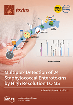
Cover Story (view full-size image): Staphylococcus enterotoxins (SEs) are potent gastrointestinal exotoxins produced by coagulase-positive staphylococci (CPS). Among the 27 SEs described in the literature, only a few can be detected using immuno-enzymatic based methods that are strongly dependent on the availability of antibodies. In this work, liquid chromatography coupled to mass spectrometry (LC–MS) was developed for the detection of 24 SEs. A robust database of specific signature peptides and LC–MS parameters was established using sequences from 24 SEs, including their 162 variants. The LC–MS method showed high performance when applied to 49 enterotoxin-producing strains. Results showed that CPS strains simultaneously produce several types of SEs. However, the concentrations expressed depended strongly on the type of SE. View this paper
  • Issues are regarded as officially published after their release is announced to the table of contents alert mailing list.
  • You may sign up for e-mail alerts to receive table of contents of newly released issues.
  • PDF is the official format for papers published in both, html and pdf forms. To view the papers in pdf format, click on the "PDF Full-text" link, and use the free Adobe Reader to open them.
Order results
Result details
Section
Select all
Export citation of selected articles as:
16 pages, 600 KiB  
Article
Detection of Enterotoxigenic Psychrotrophic Presumptive Bacillus cereus and Cereulide Producers in Food Products and Ingredients
by Jelena Jovanovic, Svitlana Tretiak, Katrien Begyn and Andreja Rajkovic
Toxins 2022, 14(4), 289; https://doi.org/10.3390/toxins14040289 - 16 Apr 2022
Cited by 10 | Viewed by 3746
Abstract
In the last decade, foodborne outbreaks and individual cases caused by bacterial toxins showed an increasing trend. The major contributors are enterotoxins and cereulide produced by Bacillus cereus, which can cause a diarrheal and emetic form of the disease, respectively. These diseases [...] Read more.
In the last decade, foodborne outbreaks and individual cases caused by bacterial toxins showed an increasing trend. The major contributors are enterotoxins and cereulide produced by Bacillus cereus, which can cause a diarrheal and emetic form of the disease, respectively. These diseases usually induce relatively mild symptoms; however, fatal cases have been reported. With the aim to detected potential toxin producers that are able to grow at refrigerator temperatures and subsequently produce cereulide, we screened the prevalence of enterotoxin and cereulide toxin gene carriers and the psychrotrophic capacity of presumptive B. cereus obtained from 250 food products (cereal products, including rice and seeds/pulses, dairy-based products, dried vegetables, mixed food, herbs, and spices). Of tested food products, 226/250 (90.4%) contained presumptive B. cereus, which communities were further tested for the presence of nheA, hblA, cytK-1, and ces genes. Food products were mainly contaminated with the nheA B. cereus carriers (77.9%), followed by hblA (64.8%), ces (23.2%), and cytK-1 (4.4%). Toxigenic B. cereus communities were further subjected to refrigerated (4 and 7 °C) and mild abuse temperatures (10 °C). Overall, 77% (94/121), 86% (104/121), and 100% (121/121) were able to grow at 4, 7, and 10 °C, respectively. Enterotoxin and cereulide potential producers were detected in 81% of psychrotrophic presumptive B. cereus. Toxin encoding genes nheA, hblA, and ces gene were found in 77.2, 55, and 11.7% of tested samples, respectively. None of the psychrotrophic presumptive B. cereus were carriers of the cytotoxin K-1 encoding gene (cytK-1). Nearly half of emetic psychrotrophic B. cereus were able to produce cereulide in optimal conditions. At 4 °C none of the examined psychrotrophs produced cereulide. The results of this research highlight the high prevalence of B. cereus and the omnipresence of toxin gene harboring presumptive B. cereus that can grow at refrigerator temperatures, with a focus on cereulide producers. Full article
Show Figures

Figure 1

19 pages, 4575 KiB  
Article
New Mutants of Epsilon Toxin from Clostridium perfringens with an Altered Receptor-Binding Site and Cell-Type Specificity
by Jonatan Dorca-Arévalo, Inmaculada Gómez de Aranda and Juan Blasi
Toxins 2022, 14(4), 288; https://doi.org/10.3390/toxins14040288 - 16 Apr 2022
Cited by 5 | Viewed by 2876
Abstract
Epsilon toxin (Etx) from Clostridium perfringens is the third most potent toxin after the botulinum and tetanus toxins. Etx is the main agent of enterotoxemia in ruminants and is produced by Clostridium perfringens toxinotypes B and D, causing great economic losses. Etx selectively [...] Read more.
Epsilon toxin (Etx) from Clostridium perfringens is the third most potent toxin after the botulinum and tetanus toxins. Etx is the main agent of enterotoxemia in ruminants and is produced by Clostridium perfringens toxinotypes B and D, causing great economic losses. Etx selectively binds to target cells, oligomerizes and inserts into the plasma membrane, and forms pores. A series of mutants have been previously generated to understand the cellular and molecular mechanisms of the toxin and to obtain valid molecular tools for effective vaccination protocols. Here, two new non-toxic Etx mutants were generated by selective deletions in the binding (Etx-ΔS188-F196) or insertion (Etx-ΔV108-F135) domains of the toxin. As expected, our results showed that Etx-ΔS188-F196 did not exhibit the usual Etx binding pattern but surprisingly recognized specifically an O-glycoprotein present in the proximal tubules of the kidneys in a wide range of animals, including ruminants. Although diminished, Etx-ΔV108-F135 maintained the capacity for binding and even oligomerization, indicating that the mutation particularly affected the pore-forming ability of the toxin. Full article
(This article belongs to the Section Bacterial Toxins)
Show Figures

Figure 1

10 pages, 448 KiB  
Article
Serum P-Cresyl Sulfate Level Is an Independent Marker of Peripheral Arterial Stiffness as Assessed Using Brachial-Ankle Pulse Wave Velocity in Patients with Non-Dialysis Chronic Kidney Disease Stage 3 to 5
by Yu-Chi Chang, Yu-Li Lin, Yu-Hsien Lai, Chih-Hsien Wang and Bang-Gee Hsu
Toxins 2022, 14(4), 287; https://doi.org/10.3390/toxins14040287 - 16 Apr 2022
Cited by 1 | Viewed by 2308
Abstract
p-Cresyl sulfate (PCS) is a uremic toxin that causes cardiovascular injury and progression in patients with chronic kidney disease (CKD). Peripheral arterial stiffness (PAS) as measured using the brachial-ankle pulse wave velocity (baPWV) is considered a valuable predictor of cardiovascular event risk [...] Read more.
p-Cresyl sulfate (PCS) is a uremic toxin that causes cardiovascular injury and progression in patients with chronic kidney disease (CKD). Peripheral arterial stiffness (PAS) as measured using the brachial-ankle pulse wave velocity (baPWV) is considered a valuable predictor of cardiovascular event risk in the general population. The study investigated the correlation between serum PCS levels and PAS (baPWV > 18.0 m/s) in 160 patients with stage 3–5 CKD. Liquid chromatography–mass spectrometry was used to assay serum PCS levels. PAS was detected in 54 patients (33.8%), and it was linked to older age, a higher prevalence of hypertension, higher systolic and diastolic blood pressure, higher serum calcium–phosphorus product and PCS levels, and lower height and body weight. Multivariable logistic regression analysis for independent factors associated with PAS illustrated that, in addition to age and diastolic blood pressure, serum PCS levels exhibited an odds ratio (OR) of 1.098 (95% confidence interval = 1.029–1.171, p = 0.005). These findings demonstrated that serum PCS levels were associated with PAS among patients with stage 3–5 CKD. Full article
(This article belongs to the Special Issue Uremic Toxins and Organ Damage)
Show Figures

Figure 1

10 pages, 998 KiB  
Article
Effectiveness and Safety of Intracavernosal IncobotulinumtoxinA (Xeomin®) 100 U as an Add-on Therapy to Standard Pharmacological Treatment for Difficult-to-Treat Erectile Dysfunction: A Case Series
by Francois Giuliano, Pierre Denys and Charles Joussain
Toxins 2022, 14(4), 286; https://doi.org/10.3390/toxins14040286 - 16 Apr 2022
Cited by 5 | Viewed by 3204
Abstract
Registered pharmacological treatments are insufficiently effective for erectile dysfunction (ED) in around 30% of affected men. Intracavernosal injection (ICI) of ona- and abobotulinumtoxinA can reduce ED in insufficient responders. We aimed to assess the safety and effectiveness of incobotulinumtoxinA ICI as an add-on [...] Read more.
Registered pharmacological treatments are insufficiently effective for erectile dysfunction (ED) in around 30% of affected men. Intracavernosal injection (ICI) of ona- and abobotulinumtoxinA can reduce ED in insufficient responders. We aimed to assess the safety and effectiveness of incobotulinumtoxinA ICI as an add-on therapy to phosphodiesterase-type 5 inhibitors (PDE5-Is) or prostaglandinE1 ICIs (PGE1 ICIs) to treat ED that did not respond sufficiently to this treatment alone. We retrospectively analyzed data from 66 men with difficult to treat ED treated with single or repeated incobotulinumtoxinA 100U ICI as an add-on therapy. Response rate (increase in International Index of Erectile Function-Erectile Function domain score ≥ the minimum clinically important difference) was 52% (median (1st–3rd quartile) 43.5 (34–71) days post-incobotulinumtoxinA ICI). ED etiology (except spinal cord injury) and severity did not influence effectiveness. Only a clinically significant response to the 1st injection predicted a request for a 2nd injection (OR = 5.6, 95%, CI 1.6–19.4). Three men reported mild penile pain during the injection. These results provide preliminary evidence for the effectiveness and safety of incobotulinumtoxinA ICI as an add-on therapy to treat ED that is insufficiently responsive to standard care and provides support for the multicenter randomized clinical trial NCT05196308. Full article
Show Figures

Figure 1

12 pages, 3101 KiB  
Article
Development and Characterization of Anti-Naja ashei Three-Finger Toxins (3FTxs)-Specific Monoclonal Antibodies and Evaluation of Their In Vitro Inhibition Activity
by Ernest Z. Manson, Mutinda C. Kyama, Josephine Kimani, Aleksandra Bocian, Konrad K. Hus, Vladimír Petrilla, Jaroslav Legáth and James H. Kimotho
Toxins 2022, 14(4), 285; https://doi.org/10.3390/toxins14040285 - 16 Apr 2022
Cited by 5 | Viewed by 3350
Abstract
Antivenom immunotherapy is the mainstay of treatment for snakebite envenoming. Most parts of the world affected by snakebite envenoming depend on broad-spectrum polyspecific antivenoms that are known to contain a low content of case-specific efficacious immunoglobulins. Thus, advances in toxin-specific antibodies production hold [...] Read more.
Antivenom immunotherapy is the mainstay of treatment for snakebite envenoming. Most parts of the world affected by snakebite envenoming depend on broad-spectrum polyspecific antivenoms that are known to contain a low content of case-specific efficacious immunoglobulins. Thus, advances in toxin-specific antibodies production hold much promise in future therapeutic strategies of snakebite envenoming. We report anti-3FTxs monoclonal antibodies developed against N. ashei venom in mice. All the three test mAbs (P4G6a, P6D9a, and P6D9b) were found to be IgG antibodies, isotyped as IgG1. SDS-PAGE analysis of the test mAbs showed two major bands at approximately 55 and 29 kDa, suggestive of immunoglobulin heavy and light chain composition, respectively. The immunoaffinity-purified test mAbs demonstrated higher binding efficacy to the target antigen compared to negative control. Similarly, a cocktail of the test mAbs was found to induce a significantly higher inhibition (p-value < 0.0001) compared to two leading commercial brands of antivenoms on the Kenyan market, implying a higher specificity for the target antigen. Both the test mAbs and 3FTxs polyclonal antibodies induced comparable inhibition (p-value = 0.9029). The inhibition induced by the 3FTxs polyclonal antibodies was significantly different from the two antivenoms (p-value < 0.0001). Our results demonstrate the prospects of developing toxin-specific monoclonal-based antivenoms for snakebite immunotherapy. Full article
Show Figures

Graphical abstract

13 pages, 2825 KiB  
Article
Allergen Content of Therapeutic Preparations for Allergen-Specific Immunotherapy of European Paper Wasp Venom Allergy
by Johannes Grosch, Antoine Lesur, Stéphanie Kler, François Bernardin, Gunnar Dittmar, Elisabetta Francescato, Simon J. Hewings, Constanze A. Jakwerth, Ulrich M. Zissler, Matthew D. Heath, Markus Ollert, Matthias F. Kramer, Christiane Hilger, Maria Beatrice Bilò, Carsten B. Schmidt-Weber and Simon Blank
Toxins 2022, 14(4), 284; https://doi.org/10.3390/toxins14040284 - 15 Apr 2022
Cited by 7 | Viewed by 3914
Abstract
Allergy to Polistes dominula (European paper wasp) venom is of particular relevance in Southern Europe, potentially becoming a threat in other regions in the near future, and can be effectively cured by venom immunotherapy (VIT). As allergen content in extracts may vary and [...] Read more.
Allergy to Polistes dominula (European paper wasp) venom is of particular relevance in Southern Europe, potentially becoming a threat in other regions in the near future, and can be effectively cured by venom immunotherapy (VIT). As allergen content in extracts may vary and have an impact on diagnostic and therapeutic approaches, the aim was to compare five therapeutic preparations for VIT of P. dominula venom allergy available in Spain. Products from five different suppliers were analyzed by SDS-PAGE and LC-MS/MS and compared with a reference venom sample. Three products with P. dominula venom and one product with a venom mixture of American Polistes species showed a comparable band pattern in SDS-PAGE as the reference sample and the bands of the major allergens phospholipase A1 and antigen 5 were assignable. The other product, which consists of a mixture of American Polistes species, exhibited the typical band pattern in one, but not in another sample from a second batch. All annotated P. dominula allergens were detected at comparable levels in LC-MS/MS analysis of products containing P. dominula venom. Due to a lack of genomic information on the American Polistes species, the remaining products were not analyzed by this method. The major Polistes allergens were present in comparable amounts in the majority, but not in all investigated samples of venom preparations for VIT of P. dominula venom allergy. Full article
(This article belongs to the Special Issue Venom Allergy: General Concepts, Allergens, Diagnosis and Treatment)
Show Figures

Figure 1

3 pages, 242 KiB  
Editorial
Toxinologic and Pharmacological Investigation of Venomous Arthropods
by Gandhi Rádis-Baptista and Katsuhiro Konno
Toxins 2022, 14(4), 283; https://doi.org/10.3390/toxins14040283 - 15 Apr 2022
Cited by 1 | Viewed by 2365
Abstract
Arthropods comprise the largest group of living animals, including thousands of species that inhabit marine and terrestrial niches in the biosphere [...] Full article
(This article belongs to the Special Issue Toxinologic and Pharmacological Investigation of Venomous Arthropods)
32 pages, 3377 KiB  
Review
Botulinum Toxin Therapy for Oromandibular Dystonia and Other Movement Disorders in the Stomatognathic System
by Kazuya Yoshida
Toxins 2022, 14(4), 282; https://doi.org/10.3390/toxins14040282 - 14 Apr 2022
Cited by 21 | Viewed by 10611
Abstract
Various movement disorders, such as oromandibular dystonia, oral dyskinesia, bruxism, functional (psychogenic) movement disorder, and tremors, exist in the stomatognathic system. Most patients experiencing involuntary movements due to these disorders visit dentists or oral surgeons, who may be the first healthcare providers. However, [...] Read more.
Various movement disorders, such as oromandibular dystonia, oral dyskinesia, bruxism, functional (psychogenic) movement disorder, and tremors, exist in the stomatognathic system. Most patients experiencing involuntary movements due to these disorders visit dentists or oral surgeons, who may be the first healthcare providers. However, differential diagnoses require neurological and dental knowledge. This study aimed to review scientific advances in botulinum toxin therapy for these conditions. The results indicated that botulinum toxin injection is effective and safe, with few side effects in most cases when properly administered by an experienced clinician. The diagnosis and treatment of movement disorders in the stomatognathic system require both neurological and dental or oral surgical knowledge and skills, and well-designed multicenter trials with a multidisciplinary team approach must be necessary to ensure accurate diagnosis and proper treatment. Full article
(This article belongs to the Special Issue Botulinum Toxin in the Movement Disorders Clinic: State of the Art)
Show Figures

Graphical abstract

13 pages, 1436 KiB  
Article
High Cell Density Cultivation Process for the Expression of Botulinum Neurotoxin a Receptor Binding Domain
by Alon Ben David, Yoel Papir, Ophir Hazan, Moses Redelman, Eran Diamant, Ada Barnea, Amram Torgeman and Ran Zichel
Toxins 2022, 14(4), 281; https://doi.org/10.3390/toxins14040281 - 14 Apr 2022
Cited by 1 | Viewed by 2780
Abstract
The receptor-binding domain of botulinum neurotoxin (HC fragment), is a promising botulism vaccine candidate. In the current study, fermentation strategies were evaluated to upscale HC fragment expression. A simple translation of the growth conditions from shake flasks to a batch fermentation [...] Read more.
The receptor-binding domain of botulinum neurotoxin (HC fragment), is a promising botulism vaccine candidate. In the current study, fermentation strategies were evaluated to upscale HC fragment expression. A simple translation of the growth conditions from shake flasks to a batch fermentation process resulted in limited culture growth and protein expression (OD of 11 and volumetric protein yields of 123 mg/L). Conducting fed-batch fermentation with rich media and continuous nutrient supplementation significantly improved culture growth (OD of 40.3) and protein expression (1093 mg/L). A further increase in HC fragment yield was achieved by high cell density cultivation (HCDC). The bacterium was grown in a defined medium and with a combined bolus/continuous feed of nutrients to maintain desired oxygen levels and prevent acetate accumulation. The final OD of the process was 260, and the volumetric yield of the HC fragment was 2065 mg/L, which reflects improvement by an order of magnitude. Purified HC fragments, produced by HCDC, exhibited typical biochemical and protective characteristics in mice. Taken together, the advancements achieved in this study promote large-scale production of the HC fragment in E. coli for use in anti-botulism vaccines. Full article
(This article belongs to the Section Bacterial Toxins)
Show Figures

Figure 1

24 pages, 555 KiB  
Review
Uremic Toxins and Cardiovascular Risk in Chronic Kidney Disease: What Have We Learned Recently beyond the Past Findings?
by Carolla El Chamieh, Sophie Liabeuf and Ziad Massy
Toxins 2022, 14(4), 280; https://doi.org/10.3390/toxins14040280 - 14 Apr 2022
Cited by 25 | Viewed by 8930
Abstract
Patients with chronic kidney disease (CKD) have an elevated prevalence of atheromatous (ATH) and/or non-atheromatous (non-ATH) cardiovascular disease (CVD) due to an array of CKD-related risk factors, such as uremic toxins (UTs). Indeed, UTs have a major role in the emergence of a [...] Read more.
Patients with chronic kidney disease (CKD) have an elevated prevalence of atheromatous (ATH) and/or non-atheromatous (non-ATH) cardiovascular disease (CVD) due to an array of CKD-related risk factors, such as uremic toxins (UTs). Indeed, UTs have a major role in the emergence of a spectrum of CVDs, which constitute the leading cause of death in patients with end-stage renal disease. The European Uremic Toxin Work Group has identified over 100 UTs, more than 25 of which are dietary or gut-derived. Even though relationships between UTs and CVDs have been described in the literature, there are few reviews on the involvement of the most toxic compounds and the corresponding physiopathologic mechanisms. Here, we review the scientific literature on the dietary and gut-derived UTs with the greatest toxicity in vitro and in vivo. A better understanding of these toxins’ roles in the elevated prevalence of CVDs among CKD patients might facilitate the development of targeted treatments. Hence, we review (i) ATH and non-ATH CVDs and the respective levels of risk in patients with CKD and (ii) the mechanisms that underlie the influence of dietary and gut-derived UTs on CVDs. Full article
(This article belongs to the Section Uremic Toxins)
Show Figures

Figure 1

21 pages, 1574 KiB  
Article
Providing Biological Plausibility for Exposure–Health Relationships for the Mycotoxins Deoxynivalenol (DON) and Fumonisin B1 (FB1) in Humans Using the AOP Framework
by Annick D. van den Brand, Lola Bajard, Inger-Lise Steffensen, Anne Lise Brantsæter, Hubert A. A. M. Dirven, Jochem Louisse, Ad Peijnenburg, Sophie Ndaw, Alberto Mantovani, Barbara De Santis and Marcel J. B. Mengelers
Toxins 2022, 14(4), 279; https://doi.org/10.3390/toxins14040279 - 13 Apr 2022
Cited by 8 | Viewed by 4710
Abstract
Humans are chronically exposed to the mycotoxins deoxynivalenol (DON) and fumonisin B1 (FB1), as indicated by their widespread presence in foods and occasional exposure in the workplace. This exposure is confirmed by human biomonitoring (HBM) studies on (metabolites of) these mycotoxins in human [...] Read more.
Humans are chronically exposed to the mycotoxins deoxynivalenol (DON) and fumonisin B1 (FB1), as indicated by their widespread presence in foods and occasional exposure in the workplace. This exposure is confirmed by human biomonitoring (HBM) studies on (metabolites of) these mycotoxins in human matrices. We evaluated the exposure–health relationship of the mycotoxins in humans by reviewing the available literature. Since human studies did not allow the identification of unequivocal chronic health effects upon exposure to DON and FB1, the adverse outcome pathway (AOP) framework was used to structure additional mechanistic evidence from in vitro and animal studies on the identified adverse effects. In addition to a preliminary AOP for DON resulting in the adverse outcome (AO) ‘reduced body weight gain’, we developed a more elaborated AOP for FB1, from the molecular initiating event (MIE) ‘inhibition of ceramide synthases’ leading to the AO ‘neural tube defects’. The mechanistic evidence from AOPs can be used to support the limited evidence from human studies, to focus FB1- and DON-related research in humans to identify related early biomarkers of effect. In order to establish additional human exposure–health relationships in the future, recommendations are given to maximize the information that can be obtained from HBM. Full article
(This article belongs to the Special Issue Human Biomonitoring and Risk Assessment of Mycotoxins)
Show Figures

Figure 1

25 pages, 1770 KiB  
Review
Cocaine: An Updated Overview on Chemistry, Detection, Biokinetics, and Pharmacotoxicological Aspects including Abuse Pattern
by Rita Roque Bravo, Ana Carolina Faria, Andreia Machado Brito-da-Costa, Helena Carmo, Přemysl Mladěnka, Diana Dias da Silva, Fernando Remião and on behalf of The OEMONOM Researchers
Toxins 2022, 14(4), 278; https://doi.org/10.3390/toxins14040278 - 13 Apr 2022
Cited by 54 | Viewed by 33148
Abstract
Cocaine is one of the most consumed stimulants throughout the world, as official sources report. It is a naturally occurring sympathomimetic tropane alkaloid derived from the leaves of Erythroxylon coca, which has been used by South American locals for millennia. Cocaine can [...] Read more.
Cocaine is one of the most consumed stimulants throughout the world, as official sources report. It is a naturally occurring sympathomimetic tropane alkaloid derived from the leaves of Erythroxylon coca, which has been used by South American locals for millennia. Cocaine can usually be found in two forms, cocaine hydrochloride, a white powder, or ‘crack’ cocaine, the free base. While the first is commonly administered by insufflation (‘snorting’) or intravenously, the second is adapted for inhalation (smoking). Cocaine can exert local anaesthetic action by inhibiting voltage-gated sodium channels, thus halting electrical impulse propagation; cocaine also impacts neurotransmission by hindering monoamine reuptake, particularly dopamine, from the synaptic cleft. The excess of available dopamine for postsynaptic activation mediates the pleasurable effects reported by users and contributes to the addictive potential and toxic effects of the drug. Cocaine is metabolised (mostly hepatically) into two main metabolites, ecgonine methyl ester and benzoylecgonine. Other metabolites include, for example, norcocaine and cocaethylene, both displaying pharmacological action, and the last one constituting a biomarker for co-consumption of cocaine with alcohol. This review provides a brief overview of cocaine’s prevalence and patterns of use, its physical-chemical properties and methods for analysis, pharmacokinetics, pharmacodynamics, and multi-level toxicity. Full article
(This article belongs to the Special Issue Toxicity of Plant Derived Substances)
Show Figures

Figure 1

10 pages, 1961 KiB  
Article
The Effect of Australian and Asian Commercial Antivenoms in Reversing the Post-Synaptic Neurotoxicity of O. hannah, N. naja and N. kaouthia Venoms In Vitro
by Tam M. Huynh, Wayne C. Hodgson, Geoffrey K. Isbister and Anjana Silva
Toxins 2022, 14(4), 277; https://doi.org/10.3390/toxins14040277 - 13 Apr 2022
Cited by 3 | Viewed by 3190
Abstract
Despite antivenoms being the only established specific treatment for neuromuscular paralysis arising from snake envenoming, their ability to reverse the post-synaptic neurotoxicity in snake envenoming is poorly understood. We investigated the ability of five commercial antivenoms i.e., King cobra monovalent, Thai cobra monovalent, [...] Read more.
Despite antivenoms being the only established specific treatment for neuromuscular paralysis arising from snake envenoming, their ability to reverse the post-synaptic neurotoxicity in snake envenoming is poorly understood. We investigated the ability of five commercial antivenoms i.e., King cobra monovalent, Thai cobra monovalent, Thai neuro polyvalent, Indian polyvalent and Australian polyvalent antivenoms to reverse neurotoxicity induced by the venoms of King cobra (Ophiophagus hannah, 3 µg/mL), Indian cobra (Naja naja, 5 µg/mL) and Thai cobra (Naja kaouthia, 3 µg/mL) using the in vitro chick-biventer cervicis nerve–muscle preparation. All three venoms displayed post-synaptic neurotoxicity, which was prevented by all tested antivenoms (40 µL/mL) added to the bath prior to venom. All antivenoms partially reversed the established post-synaptic neuromuscular block after the addition of the three venoms during a 180 min observation period, but to varying degrees and at different rates. The neurotoxic effects of O. hannah venom recovered to a greater magnitude (based on twitch height restoration) and faster than the neurotoxicity of N. kaouthia venom, which recovered to a lower magnitude more slowly. The recovery of post-synaptic neurotoxicity by N. naja venom was hindered due to the likely presence of cytotoxins in the venom, which cause direct muscle damage. The observations made in this study provide further evidence that the commercial antivenoms are likely to actively reverse established α-neurotoxin-mediated neuromuscular paralysis in snake envenoming, and there is cross-neutralisation with different antivenoms. Full article
(This article belongs to the Special Issue Toxinology and Pharmacology of Snake Venoms)
Show Figures

Figure 1

11 pages, 713 KiB  
Article
Aflatoxin M1 in Raw Milk, Pasteurized Milk and Cottage Cheese Collected along Value Chain Actors from Three Regions of Ethiopia
by Haftom Zebib, Dawit Abate and Ashagrie Zewdu Woldegiorgis
Toxins 2022, 14(4), 276; https://doi.org/10.3390/toxins14040276 - 12 Apr 2022
Cited by 10 | Viewed by 2928
Abstract
Milk is a highly nutritious and perfect natural food for humans. However, when lactating animals feed on Aflatoxin B1 (AFB1)-containing feed, the hydroxyl metabolite aflatoxin M1 (AFM1) contaminates the milk and dairy products. The objective of the [...] Read more.
Milk is a highly nutritious and perfect natural food for humans. However, when lactating animals feed on Aflatoxin B1 (AFB1)-containing feed, the hydroxyl metabolite aflatoxin M1 (AFM1) contaminates the milk and dairy products. The objective of the current study was to assess the level of AFM1 in raw milk, normally pasteurized milk and Ethiopian cottage cheese collected from value chain actors (producers, collectors, processors and retailers). Cross-sectional study and simple random techniques were used to collect primary samples. A total of 160 composite samples was collected; raw milk (n = 64), pasteurized milk (n = 64) and cheese (n = 32) was analyzed. Quantitative analysis of AFM1 was conducted using enzyme-linked immunosorbent assay (ELISA). The results indicate that AFM1 was detected in all milk products. Results along value chains show that the concentration of AFM1 in raw milk from collectors was significantly higher than from producers, and in pasteurized milk from processors and retailers (p < 0.05). However, no significant (p > 0.05) difference was observed in cottage cheese value-chain actors in all regions. Comparison of AFM1 mean values among all dairy products shows that raw milk had a significantly higher concentration of AFM1 followed by pasteurized milk and cottage cheese. However, there was no significant difference between raw and pasteurized milk (p > 0.05). The mean AFM1 contamination in milk products ranged from 0.137 to 0.319 µg/L (mean value 0.285 µg/L). The contamination percentages of AFM1 in raw milk (62.50%), pasteurized milk (67.20%) and cottage cheese (25%) were above the regulatory limit set by the European Union (EU) (0.05 µg/L). According to USA/Ethiopian Standard (US/ES) (0.50 µg/L), 21.87%, 25% and 1% exceeded the regulatory limit for the above products, respectively. The overall prevalence (56.88%) was above the EU regulatory limit and 19.38% over US/ES regulations. Therefore, to provide accurate information about the health risk to consumers, there is a need to conduct risk assessment studies in consumers of milk and dairy products at different age groups. Full article
Show Figures

Figure 1

15 pages, 17058 KiB  
Article
Protective Effects of Ferulic Acid on Deoxynivalenol-Induced Toxicity in IPEC-J2 Cells
by Xiangyi Meng, Wenyan Yu, Nuo Duan, Zhouping Wang, Yingbin Shen and Shijia Wu
Toxins 2022, 14(4), 275; https://doi.org/10.3390/toxins14040275 - 12 Apr 2022
Cited by 17 | Viewed by 2921
Abstract
Deoxynivalenol (DON), a mycotoxin that contaminates crops such as wheat and corn, can cause severe acute or chronic injury when ingested by animals or humans. This study investigated the protective effect of ferulic acid (FA), a polyphenolic substance, on alleviating the toxicity induced [...] Read more.
Deoxynivalenol (DON), a mycotoxin that contaminates crops such as wheat and corn, can cause severe acute or chronic injury when ingested by animals or humans. This study investigated the protective effect of ferulic acid (FA), a polyphenolic substance, on alleviating the toxicity induced by DON (40 μM) in IPEC-J2 cells. The experiments results showed that FA not only alleviated the decrease in cell viability caused by DON (p < 0.05), but increased the level of superoxide dismutase (SOD) (p < 0.01), glutathione peroxidase (GSH-Px), (catalase) CAT and glutathione (GSH) (p < 0.05) through the nuclear factor erythroid 2-related factor 2 (Nrf2)-epoxy chloropropane Kelch sample related protein-1 (keap1) pathway, and then decreased the levels of intracellular oxidative stress. Additionally, FA could alleviate DON-induced inflammation through mitogen-activated protein kinases (MAPKs) and nuclear factor kappa-B (NF-κB) pathways, down-regulated the secretion of interleukin-6 (IL-6) (p < 0.0001), interleukin-8 (IL-8) (p < 0.05), interleukin-1β (IL-1β), interferon-γ (IFN-γ) and further attenuated the DON-induced intracellular apoptosis (10.7% to 6.84%) by regulating the expression of Bcl2-associated X protein (Bax) (p < 0.0001), B-cell lymphoma-2 (Bcl-2) (p < 0.0001), and caspase-3 (p < 0.0001). All these results indicate that FA exhibits a significantly protective effect against DON-induced toxicity. Full article
(This article belongs to the Special Issue Mycotoxins Study: Identification and Control)
Show Figures

Graphical abstract

9 pages, 1594 KiB  
Review
The Evolving View of Uremic Toxicity
by Bjorn Meijers and Jerome Lowenstein
Toxins 2022, 14(4), 274; https://doi.org/10.3390/toxins14040274 - 12 Apr 2022
Cited by 2 | Viewed by 2625
Abstract
Indoxyl sulfate, closely related to indigo, a dye valued for it binding to cloth, has been recognized as a protein-bound solute bound to albumin, present in increased concentration in the serum of patients with impaired glomerular filtration (13). The early studies of Niwa [...] Read more.
Indoxyl sulfate, closely related to indigo, a dye valued for it binding to cloth, has been recognized as a protein-bound solute bound to albumin, present in increased concentration in the serum of patients with impaired glomerular filtration (13). The early studies of Niwa identified indoxyl sulfate as a toxin capable of accelerating the rate of renal damage in subtotal nephrectomized rats (18). Over the past decade other protein-bound solutes have been identified in the plasma of patients with impaired glomerular filtration. Although the early studies, focused on the kidney, identified indoxyl sulfate as a toxic waste product dependent on the kidney for its removal, subsequent observations have identified organic anion transporters on many non-renal tissue, leading to the view that indoxyl sulfate is part of a systemic signaling system. Full article
Show Figures

Figure 1

15 pages, 24355 KiB  
Article
Engineering and Structural Insights of a Novel BBI-like Protease Inhibitor Livisin from the Frog Skin Secretion
by Jie Yang, Chengliang Tong, Junmei Qi, Xiaoying Liao, Xiaokun Li, Xu Zhang, Mei Zhou, Lei Wang, Chengbang Ma, Xinping Xi, Tianbao Chen, Yitian Gao and Di Wu
Toxins 2022, 14(4), 273; https://doi.org/10.3390/toxins14040273 - 12 Apr 2022
Cited by 4 | Viewed by 2658
Abstract
The Bowman–Birk protease inhibitor (BBI) family is a prototype group found mainly in plants, particularly grasses and legumes, which have been subjected to decades of study. Recently, the discovery of attenuated peptides containing the canonical Bowman–Birk protease inhibitory motif has been detected in [...] Read more.
The Bowman–Birk protease inhibitor (BBI) family is a prototype group found mainly in plants, particularly grasses and legumes, which have been subjected to decades of study. Recently, the discovery of attenuated peptides containing the canonical Bowman–Birk protease inhibitory motif has been detected in the skin secretions of amphibians, mainly from Ranidae family members. The roles of these peptides in amphibian defense have been proposed to work cooperatively with antimicrobial peptides and reduce peptide degradation. A novel trypsin inhibitory peptide, named livisin, was found in the skin secretion of the green cascade frog, Odorrana livida. The cDNA encoding the precursor of livisin was cloned, and the predicted mature peptide was characterized. The mature peptide was found to act as a potent inhibitor against several serine proteases. A comparative activity study among the native peptide and its engineered analogs was performed, and the influence of the P1 and P2′ positions, as well as the C-terminal amidation on the structure–activity relationship for livisin, was illustrated. The findings demonstrated that livisin might serve as a potential drug discovery/development tool. Full article
(This article belongs to the Section Animal Venoms)
Show Figures

Figure 1

10 pages, 2086 KiB  
Article
Fullerenol Quantum Dots-Based Highly Sensitive Fluorescence Aptasensor for Patulin in Apple Juice
by Hua Pang, Hui Li, Wen Zhang, Jin Mao, Liangxiao Zhang, Zhaowei Zhang, Qi Zhang, Du Wang, Jun Jiang and Peiwu Li
Toxins 2022, 14(4), 272; https://doi.org/10.3390/toxins14040272 - 12 Apr 2022
Cited by 11 | Viewed by 2431
Abstract
A highly selective and sensitive aptasensor for detecting patulin (PAT) was constructed based on the fluorescence quenching of fullerenol quantum dots (FOQDs) towards carboxytetramethylrhodamine (TAMRA) through PET mechanism. The π-π stacking interaction between PAT aptamer and FOQDs closed the distance between TAMRA and [...] Read more.
A highly selective and sensitive aptasensor for detecting patulin (PAT) was constructed based on the fluorescence quenching of fullerenol quantum dots (FOQDs) towards carboxytetramethylrhodamine (TAMRA) through PET mechanism. The π-π stacking interaction between PAT aptamer and FOQDs closed the distance between TAMRA and FOQDs and the fluorescence of TAMRA was quenched with maximum quenching efficiency reaching 85%. There was no non-specific fluorescence quenching caused by FOQDs. In the presence of PAT, the PAT aptamer was inclined to bind with PAT and its conformation was changed. Resulting in the weak π-π stacking interaction between PAT aptamer and FOQDs. Therefore, the fluorescence of TAMRA recovered and was linearly correlated to the concentration of PAT in the range of 0.02–1 ng/mL with a detection limit of 0.01 ng/mL. This PAT aptasensor also performed well in apple juice with linear dynamic range from 0.05–1 ng/mL. The homogeneous fluorescence aptasensor shows broad application prospect in the detection of various food pollutants. Full article
Show Figures

Figure 1

9 pages, 1840 KiB  
Article
Intramuscular Neural Distribution of the Serratus Anterior Muscle: Regarding Botulinum Neurotoxin Injection for Treating Myofascial Pain Syndrome
by Kyu-Ho Yi, Ji-Hyun Lee and Hee-Jin Kim
Toxins 2022, 14(4), 271; https://doi.org/10.3390/toxins14040271 - 11 Apr 2022
Cited by 17 | Viewed by 5307
Abstract
The serratus anterior muscle is commonly involved in myofascial pain syndrome and is treated with many different injective methods. Currently, there is no definite injection point for the muscle. This study provides a suggestion for injection points for the serratus anterior muscle considering [...] Read more.
The serratus anterior muscle is commonly involved in myofascial pain syndrome and is treated with many different injective methods. Currently, there is no definite injection point for the muscle. This study provides a suggestion for injection points for the serratus anterior muscle considering the intramuscular neural distribution using the whole-mount staining method. A modified Sihler method was applied to the serratus anterior muscles (15 specimens). The intramuscular arborization areas were identified in terms of the anterior (100%), middle (50%), and posterior axillary line (0%), and from the first to the ninth ribs. The intramuscular neural distribution for the serratus anterior muscle had the largest arborization patterns in the fifth to the ninth rib portion of between 50% and 70%, and the first to the fourth rib portion had between 20% and 40%. These intramuscular neural distribution-based injection sites are in relation to the external anatomical line for the frequently injected muscles to facilitate the efficiency of botulinum neurotoxin injections. Lastly, the intramuscular neural distribution of serratus anterior muscle should be considered in order to practice more accurately without the harmful side effects of trigger-point injections and botulinum neurotoxin injections. Full article
(This article belongs to the Special Issue Botulinum Toxins: New Uses in the Treatment of Diseases)
Show Figures

Figure 1

15 pages, 357 KiB  
Article
Resistance Allele Frequency to Cry1Ab and Vip3Aa20 in Helicoverpa zea (Boddie) (Lepidoptera: Noctuidae) in Louisiana and Three Other Southeastern U.S. States
by Shucong Lin, Isaac Oyediran, Ying Niu, Sebe Brown, Don Cook, Xinzhi Ni, Yan Zhang, Francis P. F. Reay-Jones, Jeng Shong Chen, Zhimou Wen, Marcelo Dimase and Fangneng Huang
Toxins 2022, 14(4), 270; https://doi.org/10.3390/toxins14040270 - 11 Apr 2022
Cited by 13 | Viewed by 2574
Abstract
The corn earworm/bollworm, Helicoverpa zea (Boddie), is a pest species that is targeted by both Bacillus thuringiensis (Bt) maize and cotton in the United States. Cry1Ab and Vip3Aa20 are two common Bt toxins that are expressed in transgenic maize. The objective of this [...] Read more.
The corn earworm/bollworm, Helicoverpa zea (Boddie), is a pest species that is targeted by both Bacillus thuringiensis (Bt) maize and cotton in the United States. Cry1Ab and Vip3Aa20 are two common Bt toxins that are expressed in transgenic maize. The objective of this study was to determine the resistance allele frequency (RAF) to Cry1Ab and Vip3Aa20 in H. zea populations that were collected during 2018 and 2019 from four southeastern U.S. states: Louisiana, Mississippi, Georgia, and South Carolina. By using a group-mating approach, 104 F2 iso-lines of H. zea were established from field collections with most iso-lines (85) from Louisiana. These F2 iso-lines were screened for resistance alleles to Cry1Ab and Vip3Aa20, respectively. There was no correlation in larval survivorship between Cry1Ab and Vip3Aa20 when the iso-lines were exposed to these two toxins. RAF to Cry1Ab maize was high (0.256) and the RAFs were similar between Louisiana and the other three states and between the two sampling years. In contrast, no functional major resistance allele (RA) that allowed resistant insects to survive on Vip3Aa20 maize was detected and the expected RAF of major RAs with 95% probability was estimated to 0 to 0.0073. However, functional minor RAs to Vip3Aa20 maize were not uncommon; the estimated RAF for minor alleles was 0.028. The results provide further evidence that field resistance to Cry1Ab maize in H. zea has widely occurred, while major RAs to Vip3Aa20 maize are uncommon in the southeastern U.S. region. Information that was generated from this study should be useful in resistance monitoring and refinement of resistance management strategies to preserve Vip3A susceptibility in H. zea. Full article
(This article belongs to the Special Issue Insecticidal Toxins from Bacillus thuringiensis 2021–2022)
12 pages, 2657 KiB  
Article
Proteomic Studies of the Mechanism of Cytotoxicity, Induced by Palytoxin on HaCaT Cells
by Dingyuan Cheng, Bowen Deng, Qiling Tong, Siyi Gao, Boyi Xiao, Mengxuan Zhu, Ziyu Ren, Lianghua Wang and Mingjuan Sun
Toxins 2022, 14(4), 269; https://doi.org/10.3390/toxins14040269 - 10 Apr 2022
Cited by 2 | Viewed by 3507
Abstract
Palytoxin (PLTX) is a polyether marine toxin isolated from sea anemones. It is one of the most toxic nonprotein substances, causing many people to be poisoned every year and to die in severe cases. Despite its known impact on Na+,K+ [...] Read more.
Palytoxin (PLTX) is a polyether marine toxin isolated from sea anemones. It is one of the most toxic nonprotein substances, causing many people to be poisoned every year and to die in severe cases. Despite its known impact on Na+,K+-ATPase, much still remains unclear about PLTX’s mechanism of action. Here, we tested different concentrations of PLTX on HaCaT cells and studied its distributions in cells, its impact on gene expression, and the associated pathways via proteomics combined with bioinformatics tools. We found that PLTX could cause ferroptosis in HaCaT cells, a new type of programmed cell death, by up-regulating the expression of VDAC3, ACSL4 and NCOA4, which lead to the occurrence of ferroptosis. PLTX also acts on the MAPK pathway, which is related to cell apoptosis, proliferation, division and differentiation. Different from its effect on ferroptosis, PLTX down-regulates the expression of ERK, and, as a result, the expressions of MAPK1, MAP2K1 and MAP2K2 are also lower, affecting cell proliferation. The genes from these two mechanisms showed interactions, but we did not find overlap genes between the two. Both ferroptosis and MAPK pathways can be used as anticancer targets, so PLTX may become an anticancer drug with appropriate modification. Full article
(This article belongs to the Special Issue Marine Biotoxins: Predicting and Cumulative Risk Assessment)
Show Figures

Figure 1

8 pages, 1994 KiB  
Review
Anatomical Proposal for Botulinum Neurotoxin Injection for Glabellar Frown Lines
by Kyu-Ho Yi, Ji-Hyun Lee, Hye-Won Hu and Hee-Jin Kim
Toxins 2022, 14(4), 268; https://doi.org/10.3390/toxins14040268 - 10 Apr 2022
Cited by 35 | Viewed by 9594
Abstract
Botulinum neurotoxin injection for treating glabellar frown lines is a commonly used method; however, side effects, such as ptosis and samurai eyebrow, have been reported due to a lack of comprehensive anatomical knowledge. The anatomical factors important for the injection of the botulinum [...] Read more.
Botulinum neurotoxin injection for treating glabellar frown lines is a commonly used method; however, side effects, such as ptosis and samurai eyebrow, have been reported due to a lack of comprehensive anatomical knowledge. The anatomical factors important for the injection of the botulinum neurotoxin into the corrugator supercilii muscle has been reviewed in this study. Current understanding on the localization of the botulinum neurotoxin injection point from newer anatomy examination was evaluated. We observed that for the glabellar-frown-line-related muscles, the injection point could be more accurately demarcated. We propose the injection method and the best possible injection sites for the corrugator supercilii muscle. We propose the optimal injection sites using external anatomical landmarks for the frequently injected muscles of the face to accelerate effective glabellar frown line removal. Moreover, these instructions would support a more accurate procedure without adverse events. Full article
(This article belongs to the Special Issue Clinical Application of Botulinum Toxin)
Show Figures

Figure 1

14 pages, 1760 KiB  
Article
Lipopolysaccharide from the Cyanobacterium Geitlerinema sp. Induces Neutrophil Infiltration and Lung Inflammation
by Julie A. Swartzendruber, Rosalinda Monroy Del Toro, Ryan Incrocci, Nessa Seangmany, Joshua R. Gurr, Alejandro M. S. Mayer, Philip G. Williams and Michelle Swanson-Mungerson
Toxins 2022, 14(4), 267; https://doi.org/10.3390/toxins14040267 - 9 Apr 2022
Cited by 6 | Viewed by 4081
Abstract
Glucocorticoid-resistant asthma, which predominates with neutrophils instead of eosinophils, is an increasing health concern. One potential source for the induction of neutrophil-predominant asthma is aerosolized lipopolysaccharide (LPS). Cyanobacteria have recently caused significant tidal blooms, and aerosolized cyanobacterial LPS has been detected near the [...] Read more.
Glucocorticoid-resistant asthma, which predominates with neutrophils instead of eosinophils, is an increasing health concern. One potential source for the induction of neutrophil-predominant asthma is aerosolized lipopolysaccharide (LPS). Cyanobacteria have recently caused significant tidal blooms, and aerosolized cyanobacterial LPS has been detected near the cyanobacterial overgrowth. We hypothesized that cyanobacterial LPS contributes to lung inflammation by increasing factors that promote lung inflammation and neutrophil recruitment. To test this hypothesis, c57Bl/6 mice were exposed intranasally to LPS from the cyanobacterium member, Geitlerinema sp., in vivo to assess neutrophil infiltration and the production of pro-inflammatory cytokines and chemokines from the bronchoalveolar fluid by ELISA. Additionally, we exposed the airway epithelial cell line, A549, to Geitlerinema sp. LPS in vitro to confirm that airway epithelial cells were stimulated by this LPS to increase cytokine production and the expression of the adhesion molecule, ICAM-1. Our data demonstrate that Geitlerinema sp. LPS induces lung neutrophil infiltration, the production of pro-inflammatory cytokines such as Interleukin (IL)-6, Tumor necrosis factor-alpha, and Interferongamma as well as the chemokines IL-8 and RANTES. Additionally, we demonstrate that Geitlerinema sp. LPS directly activates airway epithelial cells to produce pro-inflammatory cytokines and the adhesion molecule, Intercellular Adhesion Molecule-1 (ICAM-1), in vitro using the airway epithelial cell line, A549. Based on our findings that use Geitlerinema sp. LPS as a model system, the data indicate that cyanobacteria LPS may contribute to the development of glucocorticoid-resistant asthma seen near water sources that contain high levels of cyanobacteria. Full article
(This article belongs to the Special Issue Ecology and Toxicology of Cyanobacteria and Cyanotoxins)
Show Figures

Figure 1

12 pages, 1533 KiB  
Article
Biodegradation of Fumonisins by the Consecutive Action of a Fusion Enzyme
by Kailin Li, Song Yu, Dianzhen Yu, Huikang Lin, Na Liu and Aibo Wu
Toxins 2022, 14(4), 266; https://doi.org/10.3390/toxins14040266 - 9 Apr 2022
Cited by 9 | Viewed by 2846
Abstract
Fumonisins (FBs) are toxic mycotoxins that commonly exist in food and feed. FBs can induce many aspects of toxicity, leading to adverse effects on human and animal health; therefore, investigating methods to reduce fumonisin contamination is necessary. In our study, we generated a [...] Read more.
Fumonisins (FBs) are toxic mycotoxins that commonly exist in food and feed. FBs can induce many aspects of toxicity, leading to adverse effects on human and animal health; therefore, investigating methods to reduce fumonisin contamination is necessary. In our study, we generated a recombinant fusion enzyme called FUMDI by linking the carboxylesterase gene (fumD) and the aminotransferase gene (fumI) by overlapping polymerase chain reaction (PCR). The fusion enzyme FUMDI was successfully, secretively expressed in the host Pichia pastoris (P. pastoris) GS115, and its expression was optimized. Our results demonstrated that the fusion enzyme FUMDI had high biodegradation activity of fumonisin B1 (FB1) and other common FBs, such as fumonisin B2 (FB2) and fumonisin B3 (FB3), and almost completely degraded 5 μg/mL of each toxin within 24 h. We also found that FUMDI enzyme and its reaction products had no negative effect on cell viability and did not induce cell apoptosis, oxidative stress, or endoplasmic reticulum (ER) stress in a human gastric epithelial cell line (GES-1). The results indicated that these FBs degradation products cannot have adverse effects in a cell model. In conclusion, a safe and efficient fumonisin-degrading enzyme was discovered, which could be a new a technical method for hazard control of FBs in the future. Full article
(This article belongs to the Special Issue Mycotoxins Study: Identification and Control)
Show Figures

Graphical abstract

10 pages, 3379 KiB  
Article
Anti-Neurotoxins from Micrurus mipartitus in the Development of Coral Snake Antivenoms
by Ana Cardona-Ruda, Paola Rey-Suárez and Vitelbina Núñez
Toxins 2022, 14(4), 265; https://doi.org/10.3390/toxins14040265 - 9 Apr 2022
Cited by 6 | Viewed by 2646
Abstract
In Colombia, the genus Micrurus includes 30 species, of which M. mipartitus and M. dumerilii are the most widely distributed. Micrurus causes less than 3% of the approximately 5000 cases of snakebite per year. The elapid envenomation caused by the snakes from the [...] Read more.
In Colombia, the genus Micrurus includes 30 species, of which M. mipartitus and M. dumerilii are the most widely distributed. Micrurus causes less than 3% of the approximately 5000 cases of snakebite per year. The elapid envenomation caused by the snakes from the Micrurus genus, are characterized by the severity of their clinical manifestations, due to the venom neurotoxic components such as three-finger toxins (3FTx) and phospholipases (PLA2). The treatment for snakebites is the administration of specific antivenoms, however, some of them have limitations in their neutralizing ability. A strategy proposed to improve antivenoms is to produce antibodies against the main components of the venom. The aim of this work was to produce an antivenom, using an immunization protocol including the main 3FTx and PLA2 responsible for M. mipartitus lethality. The antibody titers were determined by ELISA in rabbits’ serum. The immunized animals elicited a response against toxins and whole venom. The Immunoglobulin G (IgGs) obtained were able to neutralize the lethal effect of their homologous toxins. A combination of antivenom from M. mipartitus with antitoxins improved their neutralizing ability. In the same way, a mixture of anti 3FTx and PLA2 protected the mice from a 1.5 median lethal dose (LD50) of M. mipartitus venom. The results showed that this might be a way to improve antibody titers specificity against the relevant toxins in M. mipartitus venom and indicated that there is a possibility to develop and use recombinant 3FTx and PLA2 toxins as immunogens to produce antivenoms. Additionally, this represents an alternative to reduce the amount of venom used in anti-coral antivenom production. Full article
Show Figures

Figure 1

4 pages, 227 KiB  
Editorial
Cyanotoxins in Bloom: Ever-Increasing Occurrence and Global Distribution of Freshwater Cyanotoxins from Planktic and Benthic Cyanobacteria
by Triantafyllos Kaloudis, Anastasia Hiskia and Theodoros M. Triantis
Toxins 2022, 14(4), 264; https://doi.org/10.3390/toxins14040264 - 8 Apr 2022
Cited by 12 | Viewed by 3230
Abstract
Toxic cyanobacteria in freshwater bodies constitute a major threat to public health and aquatic ecosystems [...] Full article
14 pages, 1552 KiB  
Article
Methylglyoxal Induces Inflammation, Metabolic Modulation and Oxidative Stress in Myoblast Cells
by Sota Todoriki, Yui Hosoda, Tae Yamamoto, Mayu Watanabe, Akiyo Sekimoto, Hiroshi Sato, Takefumi Mori, Mariko Miyazaki, Nobuyuki Takahashi and Emiko Sato
Toxins 2022, 14(4), 263; https://doi.org/10.3390/toxins14040263 - 7 Apr 2022
Cited by 12 | Viewed by 4873
Abstract
Uremic sarcopenia is a serious clinical problem associated with physical disability and increased morbidity and mortality. Methylglyoxal (MG) is a highly reactive, dicarbonyl uremic toxin that accumulates in the circulatory system in patients with chronic kidney disease (CKD) and is related to the [...] Read more.
Uremic sarcopenia is a serious clinical problem associated with physical disability and increased morbidity and mortality. Methylglyoxal (MG) is a highly reactive, dicarbonyl uremic toxin that accumulates in the circulatory system in patients with chronic kidney disease (CKD) and is related to the pathology of uremic sarcopenia. The pathophysiology of uremic sarcopenia is multifactorial; however, the details remain unknown. We investigated the mechanisms of MG-induced muscle atrophy using mouse myoblast C2C12 cells, focusing on intracellular metabolism and mitochondrial injury. We found that one of the causative pathological mechanisms of uremic sarcopenia is metabolic flow change to fatty acid synthesis with MG-induced ATP shortage in myoblasts. Evaluation of cell viability revealed that MG showed toxic effects only in myoblast cells, but not in myotube cells. Expression of mRNA or protein analysis revealed that MG induces muscle atrophy, inflammation, fibrosis, and oxidative stress in myoblast cells. Target metabolomics revealed that MG induces metabolic alterations, such as a reduction in tricarboxylic acid cycle metabolites. In addition, MG induces mitochondrial morphological abnormalities in myoblasts. These changes resulted in the reduction of ATP derived from the mitochondria of myoblast cells. Our results indicate that MG is a pathogenic factor in sarcopenia in CKD. Full article
(This article belongs to the Special Issue The Functional Analysis of Uremic Toxins by Metabolomics 2.0)
Show Figures

Figure 1

16 pages, 1576 KiB  
Article
Cytotoxicity and Antiviral Properties of Alkaloids Isolated from Pancratium maritimum
by Marco Masi, Roberta Di Lecce, Natacha Mérindol, Marie-Pierre Girard, Lionel Berthoux, Isabel Desgagné-Penix, Viola Calabrò and Antonio Evidente
Toxins 2022, 14(4), 262; https://doi.org/10.3390/toxins14040262 - 7 Apr 2022
Cited by 12 | Viewed by 4388
Abstract
Ten Amaryllidaceae alkaloids (AAs) were isolated for the first time from Pancratium maritimum collected in Calabria region, Italy. They belong to different subgroups of this family and were identified as lycorine, which is the main alkaloid, 9-O-demethyllycorine, haemanthidine, haemanthamine, 11-hydroxyvittatine, homolycorine, [...] Read more.
Ten Amaryllidaceae alkaloids (AAs) were isolated for the first time from Pancratium maritimum collected in Calabria region, Italy. They belong to different subgroups of this family and were identified as lycorine, which is the main alkaloid, 9-O-demethyllycorine, haemanthidine, haemanthamine, 11-hydroxyvittatine, homolycorine, pancracine, obliquine, tazettine and vittatine. Haemanthidine was isolated as a scalar mixture of two 6-epimers, as already known also for other 6-hydroxycrinine alkaloids, but for the first time they were separated as 6,11-O,O′-di-p-bromobenzoyl esters. The evaluation of the cytotoxic and antiviral potentials of all isolated compounds was undertaken. Lycorine and haemanthidine showed cytotoxic activity on Hacat cells and A431 and AGS cancer cells while, pancracine exhibited selective cytotoxicity against A431 cells. We uncovered that in addition to lycorine and haemanthidine, haemanthamine and pancracine also possess antiretroviral abilities, inhibiting pseudotyped human immunodeficiency virus (HIV)−1 with EC50 of 25.3 µM and 18.5 µM respectively. Strikingly, all the AAs isolated from P. maritimum were able to impede dengue virus (DENV) replication (EC50 ranged from 0.34–73.59 µM) at low to non-cytotoxic concentrations (CC50 ranged from 6.25 µM to >100 µM). Haemanthamine (EC50 = 337 nM), pancracine (EC50 = 357 nM) and haemanthidine (EC50 = 476 nM) were the most potent anti-DENV inhibitors. Thus, this study uncovered new antiviral properties of P. maritimum isolated alkaloids, a significant finding that could lead to the development of new therapeutic strategies to fight viral infectious diseases. Full article
Show Figures

Figure 1

12 pages, 2624 KiB  
Article
A Sea Anemone Lebrunia neglecta Venom Fraction Decreases Boar Sperm Cells Capacitation: Possible Involvement of HVA Calcium Channels
by Fernando Lazcano-Pérez, Karina Bermeo, Héctor Castro, Zayil Salazar Campos, Isabel Arenas, Ariana Zavala-Moreno, Sheila Narayán Chávez-Villela, Irma Jiménez, Roberto Arreguín-Espinosa, Reyna Fierro, Humberto González-Márquez, David E. Garcia and Judith Sánchez-Rodríguez
Toxins 2022, 14(4), 261; https://doi.org/10.3390/toxins14040261 - 7 Apr 2022
Cited by 2 | Viewed by 2396
Abstract
Sea anemones produce venoms characterized by a complex mixture of low molecular weight compounds, proteins and peptides acting on voltage-gated ion channels. Mammal sperm cells, like neurons, are characterized by their ion channels. Calcium channels seem to be implicated in pivotal roles such [...] Read more.
Sea anemones produce venoms characterized by a complex mixture of low molecular weight compounds, proteins and peptides acting on voltage-gated ion channels. Mammal sperm cells, like neurons, are characterized by their ion channels. Calcium channels seem to be implicated in pivotal roles such as motility and capacitation. In this study, we evaluated the effect of a low molecular weight fraction from the venom of the sea anemone Lebrunia neglecta on boar sperm cells and in HVA calcium channels from rat chromaffin cells. Spermatozoa viability seemed unaffected by the fraction whereas motility and sperm capacitation were notoriously impaired. The sea anemone fraction inhibited the HVA calcium current with partial recovery and no changes in chromaffin cells’ current kinetics and current–voltage relationship. These findings might be relevant to the pharmacological characterization of cnidarian venoms and toxins on voltage-gated calcium channels. Full article
(This article belongs to the Special Issue Cnidarian Venom)
Show Figures

Figure 1

8 pages, 1494 KiB  
Article
Ultrasound-Guided Compression Method Effectively Counteracts Russell’s Viper Bite-Induced Pseudoaneurysm
by Subramanian Senthilkumaran, Stephen W. Miller, Harry F. Williams, Rajendran Vaiyapuri, Ravi Savania, Namasivayam Elangovan, Ponniah Thirumalaikolundusubramanian, Ketan Patel and Sakthivel Vaiyapuri
Toxins 2022, 14(4), 260; https://doi.org/10.3390/toxins14040260 - 6 Apr 2022
Cited by 8 | Viewed by 5608
Abstract
Russell’s viper (Daboia russelii), one of the ‘Big Four’ venomous snakes in India, is responsible for the majority of snakebite-induced deaths and permanent disabilities. Russell’s viper bites are known to induce bleeding/clotting abnormalities, as well as myotoxic, nephrotoxic, cytotoxic and neurotoxic [...] Read more.
Russell’s viper (Daboia russelii), one of the ‘Big Four’ venomous snakes in India, is responsible for the majority of snakebite-induced deaths and permanent disabilities. Russell’s viper bites are known to induce bleeding/clotting abnormalities, as well as myotoxic, nephrotoxic, cytotoxic and neurotoxic envenomation effects. In addition, they have been reported to induce rare envenomation effects such as priapism, sialolithiasis and splenic rupture. However, Russell’s viper bite-induced pseudoaneurysm (PA) has not been previously reported. PA or false aneurysm is a rare phenomenon that occurs in arteries following traumatic injuries including some animal bites, and it can become a life-threatening condition if not treated promptly. Here, we document two clinical cases of Russell’s viper bites where PA has developed, despite antivenom treatment. Notably, a non-surgical procedure, ultrasound-guided compression (USGC), either alone, or in combination with thrombin was effectively used in both the cases to treat the PA. Following this procedure and additional measures, the patients made complete recoveries without the recurrence of PA which were confirmed by subsequent examination and ultrasound scans. These data demonstrate the development of PA as a rare complication following Russell’s viper bites and the effective use of a simple, non-surgical procedure, USGC for the successful treatment of PA. These results will create awareness among healthcare professionals on the development of PA and the use of USGC in snakebite victims following bites from Russell’s vipers, as well as other viper bites. Full article
(This article belongs to the Special Issue Venom-Induced Tissue Damage)
Show Figures

Figure 1

Previous Issue
Next Issue
Back to TopTop