


Cells 2026, 15(11), 964; https://doi.org/10.3390/cells15110964 (registering DOI) - 22 May 2026
Abstract
Reactive astrocytes are a hallmark of several neurological diseases in multiple sclerosis and experimental demyelination models. Their morphological alterations are commonly assessed by qualitative histopathology, yet quantitative tools are required to better capture astrocytic heterogeneity and to allow correlations with imaging-derived biomarkers. Here,
[...] Read more.
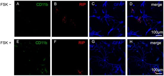
Reactive astrocytes are a hallmark of several neurological diseases in multiple sclerosis and experimental demyelination models. Their morphological alterations are commonly assessed by qualitative histopathology, yet quantitative tools are required to better capture astrocytic heterogeneity and to allow correlations with imaging-derived biomarkers. Here, we present a workflow for the quantitative analysis of Glial Fibrillary Acidic Protein (GFAP) network remodeling in astrocytes in the cuprizone model of demyelination. C57BL/6 mice were intoxicated with cuprizone for 3 or 5 weeks to induce progressive demyelination, microglial activation, and reactive astrogliosis. Brain sections were processed for anti-GFAP immunohistochemistry, and individual astrocytes from the stratum oriens of the hippocampus were digitally reconstructed. Diverse parameters of GFAP topology, including soma size, process length, branching order, convex hull area, and ramification index, were extracted using either the commercial Neurolucida® 360 software or the open-source Simple Neurite Tracer (SNT) plugin in ImageJ. Principal component analysis revealed clear differences between control astrocytes and astrocytes in cuprizone-intoxicated animals, with reactive astrocytes displaying increased numbers of primary processes, enhanced bifurcation, and process complexity. Comparative evaluation of Neurolucida® 360 and SNT demonstrated that both tools are suitable for astrocyte reconstruction, although Neurolucida® 360 enabled faster and more detailed tracing. This protocol provides a reproducible pipeline for the quantitative assessment of astrocyte morphology under control and pathological conditions, thereby supporting future efforts to link cellular remodeling to functional outcomes in neuroinflammatory disease models.
Full article
(This article belongs to the Special Issue Advanced Technology for Cellular Imaging)
►
Show Figures

Figure 1


