Next Issue
Volume 6, February
Previous Issue
Volume 5, September


Prosthesis, Volume 5, Issue 4 (December 2023) – 28 articles

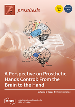
Cover Story (view full-size image): This work explores how the brain controls the human hand and how to design artificial hands that can interact with the brain. The study reveals how different neural pathways process and integrate sensory information from vision, proprioception, and touch, and how different interfaces can obtain the user’s intention to control the artificial hand. Moreover, how the user can adapt to changes in sensory inputs or outputs, using different learning mechanisms, such as reinforcement learning, motor adaptation, and internal models is discussed. Unfortunately, there are still challenges in hand and grasp control research and open questions. How can we respond to them? View this paper
  • Issues are regarded as officially published after their release is announced to the table of contents alert mailing list.
  • You may sign up for e-mail alerts to receive table of contents of newly released issues.
  • PDF is the official format for papers published in both, html and pdf forms. To view the papers in pdf format, click on the "PDF Full-text" link, and use the free Adobe Reader to open them.
Order results
Result details
Section
Select all
Export citation of selected articles as:
11 pages, 2262 KiB  
Article
Low-Cost Smartphone Photogrammetry Accurately Digitises Positive Socket and Limb Casts
by Sean Cullen, Ruth Mackay, Amir Mohagheghi and Xinli Du
Prosthesis 2023, 5(4), 1382-1392; https://doi.org/10.3390/prosthesis5040095 - 18 Dec 2023
Viewed by 647
Abstract
Digitising prosthetic sockets and moulds is critical for advanced fabrication techniques enabling reduced lead times, advanced computer modelling, and personalised design history. Current 3D scanners are expensive (>GBP 5000) and difficult to use, restricting their use by prosthetists. In this paper, we explore [...] Read more.
Digitising prosthetic sockets and moulds is critical for advanced fabrication techniques enabling reduced lead times, advanced computer modelling, and personalised design history. Current 3D scanners are expensive (>GBP 5000) and difficult to use, restricting their use by prosthetists. In this paper, we explore the use and accuracy of smartphone photogrammetry (<GBP 1000) as an accessible means of digitising rectified socket moulds. A reversed digital twin method was used for evaluating accuracy, in addition to simplified genetic algorithms to identify an optimal technique. The identified method achieved an accuracy of 99.65% and 99.13% for surface area and volume, respectively, with an interclass coefficient of 0.81. The method presented is simple, requiring less than ten minutes to capture using twenty-six photos. However, image processing time can take hours, depending on the software used. This method falls within clinical limits for accuracy, requires minimal training, and is non-destructive; thus, it can be integrated into existing workflows. This technique could bridge the gap between digital and physical workflows, helping to revolutionise the prosthetics fitting process and supporting the inclusion of additive manufactured sockets. Full article
Show Figures

Figure 1

13 pages, 6784 KiB  
Technical Note
Emergence Profile Creation with CAD Technology on Vertical Edgeless Preparation (VEP)
by Gaetano Noè, Andrea Toffoli, Roberto Bonfiglioli, Edoardo Foce, Edoardo Bianchi, Guido Maria Macaluso and Edoardo Manfredi
Prosthesis 2023, 5(4), 1369-1381; https://doi.org/10.3390/prosthesis5040094 - 18 Dec 2023
Viewed by 637
Abstract
This article proposes a digital procedure to create the emergence profile of prosthetic crowns for teeth prepared with the Vertical Edgeless Preparation (VEP) technique. This technique extends the tooth preparation below the gingival margin on the root surface of teeth with reduced periodontal [...] Read more.
This article proposes a digital procedure to create the emergence profile of prosthetic crowns for teeth prepared with the Vertical Edgeless Preparation (VEP) technique. This technique extends the tooth preparation below the gingival margin on the root surface of teeth with reduced periodontal attachment. The tooth prepared according to this technique does not present a defined prosthetic margin or the relative finishing line. Still, it shows an edgeless axial wall that emerges from the gingival sulcus. The digital procedure for creating a prosthetic emergence profile, although representing a novelty in absolute terms, is based on traditional concepts, as they have been known and described in the literature for a long time. This article will explain and review the various clinical and laboratory steps necessary to produce, through an entirely digital procedure, a fixed partial denture composed of three zirconia elements, which are going to be subsequently veneered in a traditional way on the vestibular side to improve the esthetic appearance. Full article
(This article belongs to the Special Issue Digital Technologies, Materials and Telemedicine in Dentistry)
Show Figures

Figure 1

12 pages, 1061 KiB  
Article
Unlocking the Secrets of Post-Surgical Flexion: The Vital Role of Rehabilitation in Total Knee Arthroplasty Recovery
by Gelu Florin Murvai, Calin Tudor Hozan, Timea Claudia Ghitea and Simona Cavalu
Prosthesis 2023, 5(4), 1357-1368; https://doi.org/10.3390/prosthesis5040093 - 14 Dec 2023
Cited by 1 | Viewed by 892
Abstract
This article delves into the critical aspect of postoperative flexion, particularly in the context of total knee arthroplasty (TKA), commonly known as total knee replacement. Postoperative flexion serves as a pivotal metric for assessing the success of the procedure and a patient’s ability [...] Read more.
This article delves into the critical aspect of postoperative flexion, particularly in the context of total knee arthroplasty (TKA), commonly known as total knee replacement. Postoperative flexion serves as a pivotal metric for assessing the success of the procedure and a patient’s ability to regain functional knee movement. The exploration encompasses the desired range of post-surgery flexion, the surgical factors influencing it, and the indispensable role of rehabilitation in facilitating patients in achieving functional flexion. The study tracks the progress of 713 patients who underwent total knee arthroplasty utilizing the cemented technique, categorizing them based on whether they received non-steroidal anti-inflammatory drugs for postoperative treatment. The monitoring of prosthetic and knee complications, along with the evaluation of the Knee Association Score (KSS) for functional assessment, revealed postoperative complications in approximately 18.23% of the patient cohort. These complications were predominantly associated with a restricted range of motion (ROM < 90°) and patellar clunk syndrome. Significantly, the KSS scale exhibited notable enhancements in the quality of life at 12 months post-surgery compared to preoperative and 6-month assessments. The majority of patients achieved scores classified as good or excellent, underlining the positive impact of the surgical approach and postoperative management on functional outcomes and overall patient well-being. Full article
(This article belongs to the Special Issue State of Art in Hip and Knee Replacement)
Show Figures

Figure 1

14 pages, 1737 KiB  
Systematic Review
Salvage Hip Arthroplasty in Nail Failure: A Systematic Review
by Salvatore Pantè, Lorenzo Braconi, Umberto Cottino, Federico Dettoni and Roberto Rossi
Prosthesis 2023, 5(4), 1343-1356; https://doi.org/10.3390/prosthesis5040092 - 13 Dec 2023
Viewed by 604
Abstract
Background: Femoral nailing is a largely widespread procedure in the elderly population, and the number of these surgeries is rising. Hip arthroplasty is a salvage procedure performed to improve function of the hip after femoral nail failure. The aim of the study is [...] Read more.
Background: Femoral nailing is a largely widespread procedure in the elderly population, and the number of these surgeries is rising. Hip arthroplasty is a salvage procedure performed to improve function of the hip after femoral nail failure. The aim of the study is to evaluate functional outcomes, complications and survivorship in hip arthroplasty after femoral nail failure. Methods: A systematic review of three databases (Pubmed, Embase and Cochrane) was performed using the PRISMA 2020 guidelines. After selection, four studies met the inclusion criteria, and 483 treated hips have been analyzed (476 total hip arthroplasties, 7 partial hip arthroplasties). Results: The median value of Harris Hip Score (HHS) after salvage treatment was 86.1. The main indications for salvage treatment were osteoarthrosis, avascular necrosis of the femoral head and instability of the hip. Complications are more frequent than in primary total hip arthroplasty, in particular aseptic loosening and dislocation. Good outcomes have also been achieved using revision-type stems and proximal femoral replacements (PFR). Conclusions: Conversion total hip arthroplasty is confirmed as the optimal treatment for femoral nail failure in the elderly population. Cemented or hybrid total hip arthroplasties have better outcomes than uncemented total hip arthroplasties, and the use of different types of implants widens the possible approaches to surgery in restoring the biomechanics of the hip and increases the satisfaction of patients. Full article
(This article belongs to the Special Issue State of Art in Hip and Knee Replacement)
Show Figures

Figure 1

21 pages, 6394 KiB  
Article
Five-Year Follow-Up of 8 and 6 mm Locking-Taper Implants Treated with a Reconstructive Surgical Protocol for Peri-Implantitis: A Retrospective Evaluation
by Giorgio Lombardo, Annarita Signoriello, Mauro Marincola, Estevam Augusto Bonfante, Antonio Díaz-Caballero, Nicolò Tomizioli, Alessia Pardo and Alessandro Zangani
Prosthesis 2023, 5(4), 1322-1342; https://doi.org/10.3390/prosthesis5040091 - 12 Dec 2023
Viewed by 820
Abstract
Peri-implant infections, in the absence of adequate treatment, can finally lead to premature loss of the implant. Among targeted protocols recently proposed for the treatment of peri-implant bone defects, and in the case of short implants, reconstructive surgery represents a recommended option. The [...] Read more.
Peri-implant infections, in the absence of adequate treatment, can finally lead to premature loss of the implant. Among targeted protocols recently proposed for the treatment of peri-implant bone defects, and in the case of short implants, reconstructive surgery represents a recommended option. The purpose of this study was to evaluate the outcomes, in terms of maintenance, of a reconstructive treatment for peri-implantitis in locking-taper plateau-design single-crown implants, followed for 5 years after surgery. A retrospective evaluation was conducted in 20 patients treated with access flap surgery, concomitant chemical and mechanical surface decontamination, and bone grafting (using a self-hardening mixture of bone substitutes and biphasic calcium sulfate without the use of membranes). Of the 21 implants assessed, 9 were 8 mm-length, and 12 were 6 mm-length. Implant loss and treatment success were, respectively, 0% and 80.95% after 5 years from surgery. All parameters related to bone levels and soft tissue conditions significantly improved after 3 years and remained stable at the 5-year follow-up. The proposed protocol, followed by an effective supporting periodontal therapy, demonstrated the maintenance of the function of all implants, providing adequate stability during the healing process after surgery and limiting the onset of disease recurrence. Full article
(This article belongs to the Special Issue Minimally Invasive Protocols for Prosthetic Implant Maintenance)
Show Figures

Figure 1

10 pages, 3685 KiB  
Article
Humeral Resurfacing Arthroplasty: Clinical, Functional and Radiological Assessment at a Mean Follow-Up of 11 Years
by Stefano Giannotti, Elisa Troiano, Marco Saviori, Nicholas Crippa Orlandi, Tommaso Greco, Carlo Perisano, Giovanni Battista Colasanti and Nicola Mondanelli
Prosthesis 2023, 5(4), 1312-1321; https://doi.org/10.3390/prosthesis5040090 - 1 Dec 2023
Viewed by 739
Abstract
Humeral resurfacing arthroplasty (HRA) entails the substitution of the articular surface alone with a prosthetic cap without a humeral stem. It is a more conservative procedure which can be easily converted in a total (anatomical or reverse) arthroplasty, if necessary. The present study [...] Read more.
Humeral resurfacing arthroplasty (HRA) entails the substitution of the articular surface alone with a prosthetic cap without a humeral stem. It is a more conservative procedure which can be easily converted in a total (anatomical or reverse) arthroplasty, if necessary. The present study aimed to evaluate the clinical and radiographical outcomes in a series of patients treated with HRA by a single surgeon. Thirty-three patients with a mean follow-up of 11 years were clinically (Constant score; disability of the arm, shoulder and hand score, DASH) and radiographically assessed before and after surgery. The Constant and DASH scores improved significantly after surgery, and only two cases needed revision surgery. HRA represents a valid therapeutic option in selected cases to improve the quality of life and prevents the need for more invasive procedures. Full article
(This article belongs to the Section Orthopedics and Rehabilitation)
Show Figures

Figure 1

11 pages, 2172 KiB  
Article
Comparison between Bone-Level and Tissue-Level Implants in Immediate-Loading Full-Arch Rehabilitations: A Retrospective Multi-Center 1-Year Follow-Up Study
by Francesco Pera, Massimo Carossa, Francesco Bagnasco, Armando Crupi, Giulia Ambrogio, Gaetano Isola, Maria Menini and Paolo Pesce
Prosthesis 2023, 5(4), 1301-1311; https://doi.org/10.3390/prosthesis5040089 - 30 Nov 2023
Cited by 1 | Viewed by 1064
Abstract
The objective of the present retrospective multi-center study was to analyze the outcomes of bone-level (BL) implants and tissue-level (TL) implants in immediate-loading full-arch rehabilitations. Patients who were previously rehabilitated with full-arch immediate-loading rehabilitations with either BL or TL implants were considered. Data [...] Read more.
The objective of the present retrospective multi-center study was to analyze the outcomes of bone-level (BL) implants and tissue-level (TL) implants in immediate-loading full-arch rehabilitations. Patients who were previously rehabilitated with full-arch immediate-loading rehabilitations with either BL or TL implants were considered. Data regarding implant survival rate, marginal bone loss (MBL), peri-implant probing depth (PPD), plaque index (PI), and bleeding on probing (BOP) were recorded, and the 1-year follow-up data were statistically analyzed between the two groups. In total, 38 patients were evaluated for a total implant number of 156 (n = 80 TL implants and n = 76 BL implants). An implant survival rate of 97.37% was recoded for the BL group while an implant survival rate of 100% was noted for the TL group. A total MBL of 1.324 ± 0.64 mm was recorded for BL implants, while a total MBL of 1.194 ± 0.30 mm was recorded for TL implants. A statistically significant difference was highlighted regarding MBL at the mesial aspect (p = 0.01552) of the implants, with BL implants presenting with higher MBL. Within the range of acceptable healthy values, a statistically significant difference was also highlighted regarding BOP (p < 0.00001), with TL implants presenting higher values. No statistically significant difference (p > 0.05) was recorded for any of the other variables analyzed. Within the limitations of the present retrospective study, both TL and BL implants seem to provide good clinical outcomes after a 12-month observational period when employed in immediate-loading full-arch rehabilitation. Full article
Show Figures

Figure 1

14 pages, 4495 KiB  
Article
Myo Transformer Signal Classification for an Anthropomorphic Robotic Hand
by Bolivar Núñez Montoya, Edwin Valarezo Añazco, Sara Guerrero, Mauricio Valarezo-Añazco, Daniela Espin-Ramos and Carlos Jiménez Farfán
Prosthesis 2023, 5(4), 1287-1300; https://doi.org/10.3390/prosthesis5040088 - 28 Nov 2023
Viewed by 1197
Abstract
The evolution of anthropomorphic robotic hands (ARH) in recent years has been sizable, employing control techniques based on machine learning classifiers for myoelectric signal processing. This work introduces an innovative multi-channel bio-signal transformer (MuCBiT) for surface electromyography (EMG) signal recognition and classification. The [...] Read more.
The evolution of anthropomorphic robotic hands (ARH) in recent years has been sizable, employing control techniques based on machine learning classifiers for myoelectric signal processing. This work introduces an innovative multi-channel bio-signal transformer (MuCBiT) for surface electromyography (EMG) signal recognition and classification. The proposed MuCBiT is an artificial neural network based on fully connected layers and transformer architecture. The MuCBiT recognizes and classifies EMG signals sensed from electrodes patched over the arm’s surface. The MuCBiT classifier was trained and validated using a collected dataset of four hand gestures across ten users. Despite the smaller size of the dataset, the MuCBiT achieved a prediction accuracy of 86.25%, outperforming traditional machine learning models and other transformer-based classifiers for EMG signal classification. This integrative transformer-based gesture recognition promises notable advancements for ARH development, underscoring prospective improvements in prosthetics and human–robot interaction. Full article
(This article belongs to the Special Issue Innovations in the Control and Assessment of Prosthetic Arms)
Show Figures

Figure 1

12 pages, 3639 KiB  
Article
Medial Ball-in-Socket Posterior Cruciate-Sacrificing Total Knee Arthroplasty: Clinical, Functional and Radiographic Evaluation of 100 Consecutive Implants
by Stefano Giannotti, Nicholas Crippa Orlandi, Elisa Troiano, Matteo Cacioppo, Tiziano Giacché, Tommaso Greco, Carlo Perisano and Nicola Mondanelli
Prosthesis 2023, 5(4), 1275-1286; https://doi.org/10.3390/prosthesis5040087 - 22 Nov 2023
Viewed by 666
Abstract
The number of performed total knee arthroplasty (TKA) operations is constantly growing. This study proposes an evaluation of a series of patients undergoing medial pivot (MP) TKA surgery from a subjective, clinical and biomechanical point of view. A consecutive series of 100 TKAs [...] Read more.
The number of performed total knee arthroplasty (TKA) operations is constantly growing. This study proposes an evaluation of a series of patients undergoing medial pivot (MP) TKA surgery from a subjective, clinical and biomechanical point of view. A consecutive series of 100 TKAs implanted in a single centre, by the same surgeon, with a medial parapatellar approach with the sacrifice of the posterior cruciate ligament and cemented components were evaluated. All patients underwent standardized radiographic and functional clinical evaluation, with standing antero-posterior, lateral and patellar axial views; pre-operatively and post-operatively at 1, 3, 6 and 12 months; and then annually. Results were evaluated using three different patient-related outcome measurement scores (PROMs): the Knee Osteoarthritis Outcome Score (KOOS), the new Knee Society Score (nKSS) and the Short Form Health Survey 36 (SF-36). Excellent results in all treated knees were documented using the PROMs: the mean nKSS was 199.8, the mean KOOS was good to excellent in every subscale, and the mean was SF-36 82%. There were no cases of septic or aseptic loosening, vascular damage, neurological damage, or revision surgery for any reason. According to the experience gained, MP implants demonstrated excellent results, being clinically functional in both objective and subjective terms as well as radiographic evaluations, thus resulting in a winning strategy for obtaining a TKA that makes the patient satisfied and able to perform their daily life activities. Full article
(This article belongs to the Special Issue State of Art in Hip and Knee Replacement)
Show Figures

Figure 1

18 pages, 5427 KiB  
Review
Robotic Systems in Knee Surgery: Current Concepts and Future Perspectives
by Antonino Cantivalli, Umberto Cottino, Davide Edoardo Bonasia, Federica Rosso and Roberto Rossi
Prosthesis 2023, 5(4), 1257-1274; https://doi.org/10.3390/prosthesis5040086 - 21 Nov 2023
Cited by 1 | Viewed by 2027
Abstract
Total knee arthroplasty (TKA) is a successful and highly effective procedure in most patients with tricompartmental knee arthritis. Despite the innovations in surgical techniques due to planning software and technological innovations, patients’ dissatisfaction after TKA is still high, at up to 20%. Robotic-assisted [...] Read more.
Total knee arthroplasty (TKA) is a successful and highly effective procedure in most patients with tricompartmental knee arthritis. Despite the innovations in surgical techniques due to planning software and technological innovations, patients’ dissatisfaction after TKA is still high, at up to 20%. Robotic-assisted surgery (RAS) could be considered as a future option for improving outcomes due to its higher accuracy, precision, and reliability. Robotic systems can be classified as fully active, semi-active, or passive depending on the surgeon’s involvement during the procedure, and as imageless or image-based according to the necessity of radiological exams for the pre-operative planning. Three of the most well-known robotic systems for knee surgery are MAKO® (Stryker Ltd., Kalamazoo, MI, USA), NAVIO® (Smith & Nephew, Andover, TX, USA), and ROSA® (Zimmer Inc., Warsaw, IN, USA). These systems show differences in terms of surgeon involvement, the use of CT scans or X-rays for pre-operative planning, the possibility to perform both unicompartmental knee arthroplasty (UKA) and TKA (or even total hip arthroplasty THA), and in the different kinds of knee prosthesis that can be implanted. This article aims to describe the features of the most used robotic systems for knee arthroplasty, to examine their outcomes and analyze their cost-effectiveness, and to evaluate future perspectives. Full article
(This article belongs to the Special Issue State of Art in Hip and Knee Replacement)
Show Figures

Figure 1

25 pages, 3580 KiB  
Article
EMG and IMU Data Fusion for Locomotion Mode Classification in Transtibial Amputees
by Omar A. Gonzales-Huisa, Gonzalo Oshiro, Victoria E. Abarca, Jorge G. Chavez-Echajaya and Dante A. Elias
Prosthesis 2023, 5(4), 1232-1256; https://doi.org/10.3390/prosthesis5040085 - 21 Nov 2023
Cited by 1 | Viewed by 1472
Abstract
Despite recent advancements in prosthetic technology, lower-limb amputees often remain limited to passive prostheses, which leads to an asymmetric gait and increased energy expenditure. Developing active prostheses with effective control systems is important to improve mobility for these individuals. This study presents a [...] Read more.
Despite recent advancements in prosthetic technology, lower-limb amputees often remain limited to passive prostheses, which leads to an asymmetric gait and increased energy expenditure. Developing active prostheses with effective control systems is important to improve mobility for these individuals. This study presents a machine-learning-based approach to classify five distinct locomotion tasks: ground-level walking (GWL), ramp ascent (RPA), ramp descent (RPD), stairs ascent (SSA), and stairs descent (SSD). The dataset comprises fused electromyographic (EMG) and inertial measurement unit (IMU) signals from twenty non-amputated and five transtibial amputated participants. EMG sensors were strategically positioned on the thigh muscles, while IMU sensors were placed on various leg segments. The performance of two classification algorithms, support vector machine (SVM) and long short-term memory (LSTM), were evaluated on segmented data. The results indicate that SVM models outperform LSTM models in accuracy, precision, and F1 score in the individual evaluation of amputee and non-amputee datasets for 80–20 and 50–50 data distributions. In the 80–20 distribution, an accuracy of 95.46% and 95.35% was obtained with SVM for non-amputees and amputees, respectively. An accuracy of 93.33% and 93.30% was obtained for non-amputees and amputees by using LSTM, respectively. LSTM models show more robustness and inter-population generalizability than SVM models when applying domain-adaptation techniques. Furthermore, the average classification latency for SVM and LSTM models was 19.84 ms and 37.07 ms, respectively, within acceptable limits for real-time applications. This study contributes to the field by comprehensively comparing SVM and LSTM classifiers for locomotion tasks, laying the foundation for the future development of real-time control systems for active transtibial prostheses. Full article
(This article belongs to the Special Issue Recent Advances in Foot Prosthesis and Orthosis)
Show Figures

Figure 1

26 pages, 16399 KiB  
Technical Note
Controlling a Below-the-Elbow Prosthetic Arm Using the Infinity Foot Controller
by Peter L. Bishay, Jack Wilgus, RunRun Chen, Diego Valenzuela, Victor Medina, Calvin Tan, Taylor Ittner, Miguel Caldera, Cristina Rubalcava, Shaghik Safarian, Gerbert Funes Alfaro, Alfredo Gonzalez-Martinez, Matthew Gosparini, Jose Fuentes-Perez, Andy Lima, Jonnathan Villalobos and Abrahan Solis
Prosthesis 2023, 5(4), 1206-1231; https://doi.org/10.3390/prosthesis5040084 - 20 Nov 2023
Viewed by 1746
Abstract
Nowadays there are various prosthetic arm designs in the literature, the market, and CAD design websites, with different shapes, sizes, and degrees of freedom. Only limited options are available for controlling such prostheses. Prosthetic arm users reported muscle fatigue and unreliability when using [...] Read more.
Nowadays there are various prosthetic arm designs in the literature, the market, and CAD design websites, with different shapes, sizes, and degrees of freedom. Only limited options are available for controlling such prostheses. Prosthetic arm users reported muscle fatigue and unreliability when using the market-dominated myoelectric sensors. This work presents the “Infinity Foot Controller” as a new approach to control a five-finger below-the-elbow prosthetic arm with wrist rotation and bending capabilities. This foot control system receives user input from a custom insole and a sensor-controller unit placed alongside the user’s shoe to perform various hand grips, gestures, and/or rotations. To demonstrate the new foot controller, a design of a 3D-printed below-the-elbow prosthetic arm, called the “Infinity Arm”, is presented. This arm is suitable for gripping relatively lightweight objects and making hand gestures. It includes a wrist actuation system that permits 120° wrist rotation and 70° wrist extension and flexion. It also includes a haptic feedback system that utilizes fingertip force sensors to relay a vibratory response in an armband placed on the user’s arm, giving the user a sense of touch. A proof-of-concept model was built to demonstrate the system and a testing procedure was proposed. Full article
(This article belongs to the Special Issue Innovations in the Control and Assessment of Prosthetic Arms)
Show Figures

Graphical abstract

22 pages, 3023 KiB  
Perspective
A Perspective on Prosthetic Hands Control: From the Brain to the Hand
by Cosimo Gentile and Emanuele Gruppioni
Prosthesis 2023, 5(4), 1184-1205; https://doi.org/10.3390/prosthesis5040083 - 16 Nov 2023
Viewed by 2337
Abstract
The human hand is a complex and versatile organ that enables humans to interact with the environment, communicate, create, and use tools. The control of the hand by the brain is a crucial aspect of human cognition and behaviour, but also a challenging [...] Read more.
The human hand is a complex and versatile organ that enables humans to interact with the environment, communicate, create, and use tools. The control of the hand by the brain is a crucial aspect of human cognition and behaviour, but also a challenging problem for both neuroscience and engineering. The aim of this study is to review the current state of the art in hand and grasp control from a neuroscientific perspective, focusing on the brain mechanisms that underlie sensory integration for hand control and the engineering implications for developing artificial hands that can mimic and interface with the human brain. The brain controls the hand by processing and integrating sensory information from vision, proprioception, and touch, using different neural pathways. The user’s intention can be obtained to control the artificial hand by using different interfaces, such as electromyography, electroneurography, and electroencephalography. This and other sensory information can be exploited by different learning mechanisms that can help the user adapt to changes in sensory inputs or outputs, such as reinforcement learning, motor adaptation, and internal models. This work summarizes the main findings and challenges of each aspect of hand and grasp control research and highlights the gaps and limitations of the current approaches. In the last part, some open questions and future directions for hand and grasp control research are suggested by emphasizing the need for a neuroscientific approach that can bridge the gap between the brain and the hand. Full article
(This article belongs to the Special Issue Innovations in the Control and Assessment of Prosthetic Arms)
Show Figures

Figure 1

13 pages, 822 KiB  
Review
The Management of Bone Defects in Revision Knee Arthroplasty: The Role of Porous Metal Cones and 3D-Printed Cones
by Giuseppe Marongiu, Antonio Capone, Marco Verona, Gianluca Piovan, Claudio Zorzi, Daniel Kendoff and Marta Bandino
Prosthesis 2023, 5(4), 1171-1183; https://doi.org/10.3390/prosthesis5040082 - 16 Nov 2023
Cited by 1 | Viewed by 1094
Abstract
Worldwide, the number of revisions to total knee arthroplasty procedures is increasing. Revision surgery is a challenging procedure, required for the management of bone loss after removal of the first implant. Although further long-term follow-up studies are needed, the use of cones in [...] Read more.
Worldwide, the number of revisions to total knee arthroplasty procedures is increasing. Revision surgery is a challenging procedure, required for the management of bone loss after removal of the first implant. Although further long-term follow-up studies are needed, the use of cones in revisions of total knee arthroplasty yields reliability in fixation and stability to restore joint lines, especially in challenging surgeries with poor bone stock. The introduction of 3D-printed cones in revision surgery seems to be advantageous for AORI type III bone defects, especially in reducing intraoperative complications and procedure times. The aim of this study is to review the currently available literature to analyse clinical outcomes, complications, and radiographical results with the use of metaphyseal tantalum cones and new 3D-printed cones for the management of bone defects in the revision of total knee arthroplasty. Full article
(This article belongs to the Special Issue State of Art in Hip and Knee Replacement)
Show Figures

Figure 1

18 pages, 12540 KiB  
Protocol
Presenting a Protocol for Dental Implant Restorations
by Gaetano Iannello, Luca Fiorillo, Cesare D’Amico, Giuseppe Minervini, Antonella Terranova and Gabriele Cervino
Prosthesis 2023, 5(4), 1153-1170; https://doi.org/10.3390/prosthesis5040081 - 10 Nov 2023
Cited by 1 | Viewed by 1041
Abstract
This case report focuses on applying a protocol for dental implant restoration in aesthetic zones, specifically the anterior sectors. The protocol aims to achieve optimal results with minimal complexity, reducing the number of sessions and impressions required for transferring clinical information to the [...] Read more.
This case report focuses on applying a protocol for dental implant restoration in aesthetic zones, specifically the anterior sectors. The protocol aims to achieve optimal results with minimal complexity, reducing the number of sessions and impressions required for transferring clinical information to the laboratory. The historical development of dental implantology and the evolution of dental implant restoration workflows are discussed, highlighting the significance of osseointegration and advancements in materials, surgical techniques, and prosthetic components. The case report describes rehabilitating congenitally missing lateral incisors using fibre-reinforced composite Maryland bridges for provisional restoration and Osstem TSIII (Osstem®, Seoul, South Korea) implants, zirconia abutments, and disilicate lithium crowns for definitive repair. The discussion covers essential aspects of aesthetic dental implant restoration, including materials, types of dental implants, surgical techniques for soft tissue enhancement, and prosthetic and impression techniques. The study emphasises the importance of selecting appropriate materials, employing advanced surgical procedures, and utilising accurate prosthetic and impression techniques to enhance the aesthetic outcomes of dental implant restorations. A multidisciplinary approach and patient-centred care are essential for successful aesthetic dental implant restorations. The methodology presented in this manuscript demonstrates its effectiveness in achieving optimal outcomes while minimising stress on soft tissues and improving patient comfort and satisfaction. This case report contributes to dental implant restoration, providing evidence-based techniques for achieving aesthetic and functional success. Full article
Show Figures

Figure 1

14 pages, 487 KiB  
Systematic Review
Systematic Review of In Vitro Studies on Distortion Generated by Intraoral Scanning Systems for Oral Rehabilitations with More Than Three Implants
by Luminita Oancea, Ioana Luca, Sergiu Radulescu, Andrei Macris and Toma Ciocan
Prosthesis 2023, 5(4), 1139-1152; https://doi.org/10.3390/prosthesis5040080 - 8 Nov 2023
Cited by 1 | Viewed by 1258
Abstract
(1) Background: This systematic review intends to answer the following Patient–Intervention–Comparison–Outcome (PICO) question: Do digital impression systems generate significant errors during scanning in extensive implant restorative treatments? (2) Methods: Following the PRISMA protocol and according to predefined inclusion criteria, two trained investigators searched [...] Read more.
(1) Background: This systematic review intends to answer the following Patient–Intervention–Comparison–Outcome (PICO) question: Do digital impression systems generate significant errors during scanning in extensive implant restorative treatments? (2) Methods: Following the PRISMA protocol and according to predefined inclusion criteria, two trained investigators searched for relevant articles in the PubMed database and related sources using a standard keyword sequence. The investigators were responsible for selecting studies and performing quality analysis. (3) Results: From 78 titles, only 9 studies were selected. An analysis of registration distortion variations was conducted for each potential influencing factor in terms of accuracy: interimplant distance, implant angulation, scanner type, and scanning body type. The results showed repeatable differences in accuracy between types of scanning technologies and techniques, and a positive correlation between interimplant distance and the amplitude of deviations detected in comparative analysis, with the highest error levels in total edentulous arch recording. There was no consensus on the error level owing to implant angulation, and statistically significant differences were found between the types of scan bodies used. (4) Conclusions: Digital impression systems generate significant errors during scanning in extensive implant restorative treatments, influenced by scanning technology, interimplant distance, and scanning body type. Full article
(This article belongs to the Collection Oral Implantology: Current Aspects and Future Perspectives)
Show Figures

Figure 1

10 pages, 2252 KiB  
Case Report
Pharyngeal Obturator Prosthesis Ideal for Orthodontic Appliances: A Case Series
by Francisco Vale, Catarina Nunes, Joana Reis, Raquel Travassos, Madalena Ribeiro, Filipa Marques, Anabela Pedroso, Carlos Miguel Marto, Anabela Baptista Paula and Inês Francisco
Prosthesis 2023, 5(4), 1129-1138; https://doi.org/10.3390/prosthesis5040079 - 3 Nov 2023
Viewed by 976
Abstract
(1) Background: Cleft lip and palate is the most common congenital malformation of the head and neck. After surgical closure, velopharyngeal dysfunction can arise, which has implications for phonation, sucking, swallowing, middle ear function, and interpersonal well-being. This case series aimed to present [...] Read more.
(1) Background: Cleft lip and palate is the most common congenital malformation of the head and neck. After surgical closure, velopharyngeal dysfunction can arise, which has implications for phonation, sucking, swallowing, middle ear function, and interpersonal well-being. This case series aimed to present an adaptation of the conventional pharyngeal obturator design in order to allow its use with fixed orthodontic appliances. (2) Methods: A new custom-made pharyngeal obturator device was built in order to enable a correct function of the velopharyngeal valve. The fabrication of the plate was made by altering the conventional Hawley retainer, replacing the Adams hooks with 0.9 mm spherical hooks and removing the buccal arch. (3) Results: The new pharyngeal obturator design was used in six cleft patients with fixed orthodontic appliances. The appliance was well tolerated and there was a slight improvement in auditory-perceptive evaluations. (4) Conclusions: The new pharyngeal bulb design proved to have good retention during fixed orthodontic treatment. Moreover, despite the short-term follow-up, it also showed a reduction in the severity of the hypernasality sentences. Full article
(This article belongs to the Section Prosthodontics)
Show Figures

Figure 1

9 pages, 1090 KiB  
Article
An Experimental Method to Add New Prosthetic Teeth in the Removable Partial Denture Framework: TIG Cold Welding and Preformed Pins
by Enzo Cumbo, Pietro Messina, Giuseppe Gallina and Giuseppe Alessandro Scardina
Prosthesis 2023, 5(4), 1120-1128; https://doi.org/10.3390/prosthesis5040078 - 31 Oct 2023
Cited by 1 | Viewed by 772
Abstract
The need to modify removable partial dentures equipped with a metal framework in order to add other prosthetic teeth to replace natural teeth lost by the patient could lead to laboratory procedures so complex as to require the creation of new prostheses with [...] Read more.
The need to modify removable partial dentures equipped with a metal framework in order to add other prosthetic teeth to replace natural teeth lost by the patient could lead to laboratory procedures so complex as to require the creation of new prostheses with a heavy economic burden. The creation of preformed metal pins to be welded using the economical TIG cold welding method could represent a valid alternative solution with the aim of modifying the prostheses using a reinforced resin capable of adequately resisting masticatory loads. This study evaluates and compares the mechanical robustness and the clinical reliability of these modified prostheses in cases of junctions of one or two contiguous prosthetic teeth. The 6-month follow-up demonstrated the total validity of the method via the absence of significant breakages or detachments in all of the patients analyzed; on the other hand, the prostheses modified using the traditional method and used as controls showed a high incidence of fractures. Full article
(This article belongs to the Special Issue Innovative Prosthetic Devices Applied to the Human Body)
Show Figures

Figure 1

9 pages, 716 KiB  
Article
Patient-Specific Instrumentation with Laser-Guide-Navigated THA: Clinical and CT Evaluation of the First 100 Cases
by Leonardo Previ, Edoardo Viglietta, Veronica Giuliani, Federico Corsetti, Andrea Redler, Attilio Speranza, Angelo De Carli and Raffaele Iorio
Prosthesis 2023, 5(4), 1111-1119; https://doi.org/10.3390/prosthesis5040077 - 24 Oct 2023
Viewed by 784
Abstract
Obtaining a proper position for total hip arthroplasty components is a crucial aspect of implant performance and consequently of patient outcomes. Restoring the original hip center and maintaining the limb length are key factors in reaching the optimal implant positioning. The aim of [...] Read more.
Obtaining a proper position for total hip arthroplasty components is a crucial aspect of implant performance and consequently of patient outcomes. Restoring the original hip center and maintaining the limb length are key factors in reaching the optimal implant positioning. The aim of this study was to assess the accuracy and safety of a computed dynamic analysis system that, through patient-specific guides, tries to improve implant positioning and functional orientation according to patients’ spinopelvic mobility and anatomy. A total of 100 consecutive patients were prospectively enrolled. All patients received an Optimized Positioning System dynamic hip preoperative planning schedule. A CT scan protocol follow-up analysis was performed 6 months after surgery. The mean deviations from the planned acetabular inclination and anteversion were 4.3° and 3.8°, respectively. In total, 98% of cases were within ± 10° of the Lewinnek safe zone, both for inclination and anteversion. The height of osteotomy deviated, on average, 1.6 mm. In total, 100% of cases were included within 4 mm of osteotomy. Patient-specific and laser-guided instrumentation was found to be safe and accurately reproduced dynamic planning in terms of the component orientation, osteotomy level, leg length and offset. Full article
(This article belongs to the Special Issue State of Art in Hip and Knee Replacement)
Show Figures

Figure 1

18 pages, 4362 KiB  
Case Report
A Combined Use of Custom-Made Partial Pelvic Replacement and Proximal Femur Megaprosthesis in the Treatment of Severe Bone Loss after Multiple Total Hip Arthroplasty Revisions
by Michele Fiore, Azzurra Paolucci, Renato Zunarelli, Marta Bortoli, Andrea Montanari, Andrea Pace, Lorenzo Di Prinzio, Stefania Claudia Parisi, Roberto De Cristofaro, Massimiliano De Paolis and Andrea Sambri
Prosthesis 2023, 5(4), 1093-1110; https://doi.org/10.3390/prosthesis5040076 - 24 Oct 2023
Viewed by 1129
Abstract
Hip arthroplasty failures (either septic or aseptic) often require multiple revisions, thus leading to severe bone defects. The most common reconstruction methods do not allow the management of severe defects. For this reason, in recent years, techniques borrowed from surgical oncology have been [...] Read more.
Hip arthroplasty failures (either septic or aseptic) often require multiple revisions, thus leading to severe bone defects. The most common reconstruction methods do not allow the management of severe defects. For this reason, in recent years, techniques borrowed from surgical oncology have been applied in the field of revision surgery to deal with both acetabular and femoral bone losses. In this article, two cases of severe bone deficiency following multiple hip arthroplasty revisions that were treated with a custom-made hip prosthesis combined with a proximal femur megaprosthesis are presented. Both implants were silver coated. A review of the literature was conducted to analyze similar cases treated with either a custom-made prosthesis or a proximal femur megaprosthesis. At the 2-year follow-up, all prostheses were in site without clinical or radiographic signs of implant loosening. No postoperative complications occurred. At the last follow-up, both patients resumed their daily life activities with an MSTS score of 23 and 21, respectively. The combined approach of a proximal femur megaprosthesis with a custom-made partial pelvic replacement is a solution that allows severe bone deficiency cases to be tackled with good functional results. Additionally, silver coating may help prevent recurrence of infection. Full article
(This article belongs to the Special Issue State of Art in Hip and Knee Replacement)
Show Figures

Figure 1

18 pages, 7241 KiB  
Review
Radiographic Analysis of Grammont-Style and Lateralized Reverse Shoulder Arthroplasty in Gleno-Humeral Osteoarthritis
by Giovanni Merolla, Giuseppe Sircana, Antonio Padolino, Francesco Fauci, Carlo Alberto Augusti, Marco Saporito and Paolo Paladini
Prosthesis 2023, 5(4), 1075-1092; https://doi.org/10.3390/prosthesis5040075 - 19 Oct 2023
Viewed by 1233
Abstract
Reverse shoulder arthroplasty (RSA) has transformed the management of shoulder pathologies, including cuff tear arthropathy and osteoarthritis. The innovative design principles of RSA, such as the medialization and inferiorization of the joint center of rotation, distalization of the humerus, and a semi-constrained construct, [...] Read more.
Reverse shoulder arthroplasty (RSA) has transformed the management of shoulder pathologies, including cuff tear arthropathy and osteoarthritis. The innovative design principles of RSA, such as the medialization and inferiorization of the joint center of rotation, distalization of the humerus, and a semi-constrained construct, enable effective deltoid compensation for rotator cuff deficiency. The Grammont-style RSA demonstrated excellent clinical outcomes. However, complications like instability and scapular notching prompted the exploration of lateralized designs. The radiographic evaluation of RSA is paramount for understanding the biomechanics of the implant and to foresee possible complications. Radiographic assessments encompass glenoid and humeral component positions, identifying features like scapular notching, radiolucent lines, heterotopic ossifications, bone adaptations, and humeral lengthening. Lateralized designs alter muscle moment arms and improve deltoid efficiency, influencing abduction and adduction mechanics. Despite the reduction in scapular notching, lateralized RSA introduces new challenges, such as increased risk of scapular spine and acromial fractures. Understanding the radiographic features and biomechanics of lateralized RSA is crucial for optimizing patient outcomes and mitigating potential complications. Full article
(This article belongs to the Special Issue State of Art in Hip and Knee Replacement)
Show Figures

Figure 1

15 pages, 3726 KiB  
Article
Single Crown Restorations Supported by One-Piece Zirconia Dental Implants: Case Series with a Mean Follow-Up of 58 Months
by Eugenio Romeo, Riccardo Scaringi, Diego Lops and Antonino Palazzolo
Prosthesis 2023, 5(4), 1060-1074; https://doi.org/10.3390/prosthesis5040074 - 19 Oct 2023
Viewed by 1503
Abstract
The main aim of this case series was to report the clinical and radiographic outcomes of 22 one-piece zirconia dental implants positioned in 19 patients to restore single edentulisms and followed up for at least 2 years. The mean observation period was 58.18 [...] Read more.
The main aim of this case series was to report the clinical and radiographic outcomes of 22 one-piece zirconia dental implants positioned in 19 patients to restore single edentulisms and followed up for at least 2 years. The mean observation period was 58.18 months. At the last follow-up visit, no issues, such as foreign body sensation, discomfort or pain, were reported by the patients. No implant showed signs of infection with suppuration or implant mobility. Marginal bone levels (MBL) were assessed through standardized dental radiographs and a dedicated software. The mean distance between the implant head and the first detectable bone to implant contact was calculated at the mesial and distal aspect of each implant. The mean MBL at baseline was 1.82 ± 0.63 mm while the mean MBL at the last follow-up visit was 2.57 ± 0.72 mm. The results obtained in the present case series over a mean follow-up period of 58.18 months (range 27–96) showed that one-piece zirconia dental implants could be an alternative option to support single crowns in patients requiring metal-free restorations. Nevertheless, further better-controlled research featuring an adequate study design and longer follow-up is needed in order to clarify advantages and limitations which are related to this treatment modality. Full article
(This article belongs to the Collection Oral Implantology: Current Aspects and Future Perspectives)
Show Figures

Figure 1

11 pages, 4487 KiB  
Case Report
Early Treatment of Class II Division 1 Malocclusions with Prefabricated Myofunctional Appliances: A Case Report
by Çeljana Toti, Gerta Kaςani, Aida Meto, Etleva Droboniku, Almiro Gurakuqi, Olja Tanellari, Dorjan Hysi and Luca Fiorillo
Prosthesis 2023, 5(4), 1049-1059; https://doi.org/10.3390/prosthesis5040073 - 17 Oct 2023
Cited by 1 | Viewed by 1664
Abstract
Removable functional appliances (RFA) have long been employed to address Class II malocclusion, particularly in cases involving a significant overjet, a deep bite, and molar class 2 issues. Notably divergent from RFA, myofunctional appliances (PMA) offer several distinct advantages, including applicability in noncompliant [...] Read more.
Removable functional appliances (RFA) have long been employed to address Class II malocclusion, particularly in cases involving a significant overjet, a deep bite, and molar class 2 issues. Notably divergent from RFA, myofunctional appliances (PMA) offer several distinct advantages, including applicability in noncompliant patients, adaptability in cases of dental element scarcity, suitability for allergic patients, impression-free utilization, and reduced costs. Within the array of clinical cases treated with PMA, we chose to present an intricate case involving an 8-year-old girl. Our aim was to showcase the immediate effects of PMA and to track the progress over a two-year span. Following one year of PMA treatment, substantial improvements were observed in a large overjet, a deep bite, and lip incompetence—factors often associated with elevated risks of dental trauma. These improvements not only positively impacted dental aesthetics but also engendered normalized nasal respiration and diminished palatal impingement, thereby enhancing the overall quality of life. Upon reaching the two-year mark, the clinical status pertaining to Class II malocclusion remained stable. Further treatment was advised in a subsequent phase to refine imperfections in the dental arch form. Nonetheless, comprehensive data from the prolonged follow-up of patients treated with these appliances are essential to establish robust scientific evidence concerning their long-term efficacy. Full article
(This article belongs to the Special Issue Innovative Prosthetic Devices Applied to the Human Body)
Show Figures

Figure 1

12 pages, 1434 KiB  
Article
Screwed Monolithic Zirconia Crowns for Mono-Implant Posterior Rehabilitation: A Prospective Clinical Study on 41 Patients with a 7-Year Follow-Up
by Giuseppe Barile, Saverio Capodiferro, Giovanni De Rosa, Giovannino Muci, Alessandro Vanzanelli and Massimo Corsalini
Prosthesis 2023, 5(4), 1037-1048; https://doi.org/10.3390/prosthesis5040072 - 17 Oct 2023
Cited by 1 | Viewed by 881
Abstract
The usage of monolithic zirconia has been increasing in daily practice in dentistry for the last 20 years. Monolithic zirconia is mainly used for posterior sector rehabilitation, as it lacks optical properties and has good technical properties; it does not chip and reduces [...] Read more.
The usage of monolithic zirconia has been increasing in daily practice in dentistry for the last 20 years. Monolithic zirconia is mainly used for posterior sector rehabilitation, as it lacks optical properties and has good technical properties; it does not chip and reduces antagonist wear compared to metal–ceramic prostheses. However, monolithic zirconia may present some issues, mainly low-temperature degradation (LTD), also known as “aging”, which must be considered and investigated in clinical studies with prolonged follow-up periods. The aim of this study was the aesthetic and functional evaluation of single monolithic zirconia crowns that were screwed onto implants for posterior dental rehabilitation using USPHS parameters and a long follow-up period of 7 years. The results showed that the USPHS scoring reduces year by year, passing from 100% excellence between the first and fourth years of observation, to 88% excellence and 12% success in the seventh year. Screw abutment loosening was the only technical adversity reported; no implant failure, crown fracture, or irreparable damage were additionally registered. Considering the emerging results and comparing them with the data available in the literature, the authors suggest that monolithic zirconia crowns could be considered as a valid alternative to classic metal–ceramic rehabilitations for the rehabilitation of posterior sectors. Full article
(This article belongs to the Special Issue Novel Zirconia Materials Applied in Dental Prostheses)
Show Figures

Figure 1

17 pages, 1363 KiB  
Article
Is It Possible to Create an “Ideal Endoprosthesis” for an “Ideal Total Hip Replacement”?
by Valentin L. Popov, Aleksandr M. Poliakov and Vladimir I. Pakhaliuk
Prosthesis 2023, 5(4), 1020-1036; https://doi.org/10.3390/prosthesis5040071 - 13 Oct 2023
Cited by 1 | Viewed by 947
Abstract
Since the end of the 19th and the beginning of the 20th centuries, technological equipment, implant designs (endoprosthesis) and the surgical technique of total hip replacement (THR) have been constantly improved and reached a high level of functionality and quality. Therefore, at present, [...] Read more.
Since the end of the 19th and the beginning of the 20th centuries, technological equipment, implant designs (endoprosthesis) and the surgical technique of total hip replacement (THR) have been constantly improved and reached a high level of functionality and quality. Therefore, at present, THR is one of the most high-tech, reliable and popular surgical operations that allow a large number of people suffering from osteoarthritis and other serious diseases of the hip joint to return to an active lifestyle. At the same time, it is known that even operations at this level do not always guarantee the achievement of the desired result and can lead to various complications. The question arises: are there potential opportunities for creating an “ideal endoprosthesis” that allows one to perform an “ideal THR”? In this paper, based on a critical analysis of modern endoprosthesis designs for THR, technologies for their implantation and systemic postoperative complications, the most probable, according to the authors, ways of their development are formulated, which allow asymptotically approaching the “ideal”. Full article
(This article belongs to the Special Issue State of Art in Hip and Knee Replacement)
Show Figures

Graphical abstract

9 pages, 580 KiB  
Article
Perioperative Blood Management Programme in Jehovah’s Witnesses Undergoing Total Hip Arthroplasty
by Giuseppe Solarino, Giovanni Vicenti, Davide Bizzoca, Domenico Zaccari, Walter Ginestra, Davide Ferorelli, Matteo D’aprile and Biagio Moretti
Prosthesis 2023, 5(4), 1011-1019; https://doi.org/10.3390/prosthesis5040070 - 6 Oct 2023
Viewed by 909
Abstract
Total hip arthroplasties aim to improve quality of life and reduce pain in patients suffering from late-stage hip osteoarthritis. On the other hand, it may represent a risky surgical procedure in people who refuse blood products because of religious beliefs, such as Jehovah’s [...] Read more.
Total hip arthroplasties aim to improve quality of life and reduce pain in patients suffering from late-stage hip osteoarthritis. On the other hand, it may represent a risky surgical procedure in people who refuse blood products because of religious beliefs, such as Jehovah’s Witnesses (JW). Preoperative optimisation protocols of these patients allow medical professionals to perform arthroplasties in a safer manner, avoiding allogeneic blood transfusion. In our retrospective study, two groups of patients were evaluated. Group 1 included JW patients who underwent a preoperative Hb optimisation program; Group 2 included non-JW patients authorizing transfusion in case of necessity. Differences in Hb levels were as follows: before surgery (JW 14.24 ± 1.10 vs. non-JW 12.48 ± 1.00, p-value ≤ 0.05), and after surgery (day 1 Hb: JW 12.88 ± 0.90 vs. non-JW 10.04 ± 1.30, p-value ≤ 0.05; day 3 Hb: JW 14.65 ± 0.80 vs. non-JW 9.10 ± 0.90 p-value ≤ 0.05). Moreover, cost-effectiveness strategies were evaluated in both groups. Our findings support that patient blood management programs are a safe and good strategy in hip prosthetic surgery, decreasing risks and transfusion overuse. Full article
(This article belongs to the Special Issue State of Art in Hip and Knee Replacement)
Show Figures

Figure 1

9 pages, 3696 KiB  
Case Report
Clinical Protocol for Implant-Assisted Partial Removable Dental Prostheses in Kennedy Class I: A Case Report
by Irina Karakas-Stupar, Lucia K. Zaugg, Nicola U. Zitzmann, Tim Joda, Stefan Wolfart and Taskin Tuna
Prosthesis 2023, 5(4), 1002-1010; https://doi.org/10.3390/prosthesis5040069 - 27 Sep 2023
Cited by 1 | Viewed by 1445
Abstract
Patients with Kennedy Class I are usually treated with clasp-retained removable partial dentures (RPDs) as the prosthetic gold standard. For additional stabilization of the RPD, clinicians are often confronted with the question of secondary implant placement, which requires the fabrication of new prostheses. [...] Read more.
Patients with Kennedy Class I are usually treated with clasp-retained removable partial dentures (RPDs) as the prosthetic gold standard. For additional stabilization of the RPD, clinicians are often confronted with the question of secondary implant placement, which requires the fabrication of new prostheses. This case report is part of an ongoing multi-center randomized controlled study (RCT) investigating conventional RPDs without and with supplementary implants. A design of the RPD framework, including matrix housings, is crucial to enable subsequent implant retention or support. Ultra-short implants (Straumann TL 4.1 × 4 mm) offer the opportunity for additional support and retention in the edentulous posterior region, where bone availability is often reduced. This future-oriented and minimally invasive approach with virtual treatment planning and guided implant surgery offers the possibility of simplified functional and cost-effective aftercare. Full article
Show Figures

Figure 1

10 pages, 3659 KiB  
Case Report
Digital Analysis of a Novel Impression Method Named the Biological-Oriented Digital Impression Technique: A Clinical Audit
by Marco Tallarico, Manuel Cuccu, Silvio Mario Meloni, Aurea Immacolata Lumbau, Edoardo Baldoni, Milena Pisano, Luca Fiorillo and Gabriele Cervino
Prosthesis 2023, 5(4), 992-1001; https://doi.org/10.3390/prosthesis5040068 - 25 Sep 2023
Viewed by 729
Abstract
This report aims to clinically evaluate the feasibility of a novel biological-oriented digital impression method for teeth prepared with a vertical preparation technique. Something that makes this case unique is that the definitive impression is taken without cord displacement, reducing the invasiveness for [...] Read more.
This report aims to clinically evaluate the feasibility of a novel biological-oriented digital impression method for teeth prepared with a vertical preparation technique. Something that makes this case unique is that the definitive impression is taken without cord displacement, reducing the invasiveness for the patient. In addition, dental technicians can reproduce the exact emergence profile of the temporary restoration used to reshape the soft tissue contours and define the accurate finish line at the established prosthetic junction, potentially avoiding the aesthetic and biological issues of the double-cord technique. The finish line and emergence profile are transferred using the temporary restoration through a simple CAD procedure performed using the same software used to take the impression. This makes the proposed biological-oriented digital impression technique (BODIT) easy to perform, avoiding adjunctive time and costs for clinicians and patients. Conclusions: Finally, patients' satisfaction could be significantly improved. Full article
(This article belongs to the Special Issue Innovative Prosthetic Devices Applied to the Human Body)
Show Figures

Figure 1

Previous Issue
Next Issue
Back to TopTop