


Tomography, Volume 9, Issue 2 (April 2023) – 36 articles
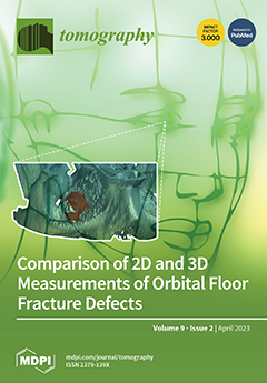
Cover Story (view full-size image):
Orbital floor fractures represent a common fracture type of the midface. The purpose of this study was to evaluate the accuracy of diagnostic measurements of isolated orbital floor fractures based on two-dimensional (2D) and three-dimensional (3D) measurement techniques. A cohort of 177 patients with an orbital floor fracture was retrospectively and multi-centrically investigated using 2D and 3D measurements. Calculated fracture areas using the 2D measurement technique revealed an average area of 287.59 mm2, while the 3D measurement showed fracture areas with a significantly larger average value of 374.16 mm2 (p < 0.001). Thus, the 3D measurements were 1.53 times larger compared to the 2D approach. Therefore, 3D-based measurement of orbital floor defects provides a more accurate estimation of fracture areas than the 2D-based technique. View this paper
- Issues are regarded as officially published after their release is announced to the table of contents alert mailing list.
- You may sign up for e-mail alerts to receive table of contents of newly released issues.
- PDF is the official format for papers published in both, html and pdf forms. To view the papers in pdf format, click on the "PDF Full-text" link, and use the free Adobe Reader to open them.
Previous Issue
Next Issue