Next Issue
Volume 13, February
Previous Issue
Volume 12, December


Bioengineering, Volume 13, Issue 1 (January 2026) – 126 articles

Cover Story (view full-size image): 
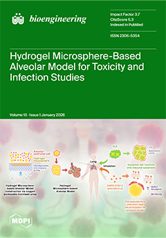
This study developed a biomimetic alveolar model using GelMA hydrogel microspheres, combined with an oxygen-permeable honeycomb microwell array. The model enabled A549 cells and primary mouse alveolar epithelial cells to form a curved monolayer structure on microspheres that mimics the native alveolar epithelium. Toxicity tests using H2S-releasing microspheres demonstrated localized cytotoxicity, downregulated SFTPC expression and upregulated apoptosis-related genes. SARS-CoV-2 pseudovirus infection experiments revealed that the 3D model required significantly higher antibody concentrations for neutralization compared to 2D cultures. This biomimetic model provides a robust platform for respiratory toxicity research, pathogen studies and drug screening. View this paper
  • Issues are regarded as officially published after their release is announced to the table of contents alert mailing list.
  • You may sign up for e-mail alerts to receive table of contents of newly released issues.
  • PDF is the official format for papers published in both, html and pdf forms. To view the papers in pdf format, click on the "PDF Full-text" link, and use the free Adobe Reader to open them.
Order results
Result details
Section
Select all
Export citation of selected articles as:
28 pages, 12315 KB  
Article
DyVarMap: Integrating Conformational Dynamics and Interpretable Machine Learning for Cancer-Associated Missense Variant Classification in FGFR2
by Yiyang Lian and Amarda Shehu
Bioengineering 2026, 13(1), 126; https://doi.org/10.3390/bioengineering13010126 - 22 Jan 2026
Viewed by 175
Abstract
Accurate interpretation of missense variants in cancer-associated genes remains a critical challenge in precision oncology, as most sequence-based predictors lack mechanistic explanations. Receptor tyrosine kinases like FGFR2 exemplify this problem: their function depends on conformational dynamics, yet most variants remain classified as variants [...] Read more.
Accurate interpretation of missense variants in cancer-associated genes remains a critical challenge in precision oncology, as most sequence-based predictors lack mechanistic explanations. Receptor tyrosine kinases like FGFR2 exemplify this problem: their function depends on conformational dynamics, yet most variants remain classified as variants of uncertain significance (VUS). In this paper we present DyVarMap, an interpretable structural-learning framework that integrates AlphaFold2-based ensemble generation with physics-driven refinement, manifold learning, and supervised classification using five biophysically motivated geometric features. Applied to FGFR2, the framework generates diverse conformational ensembles, identifies metastable states through nonlinear dimensionality reduction, and classifies pathogenicity while providing mechanistic attributions via SHAP analysis. External validation on ten kinase-domain variants yields an AUROC of 0.77 with superior calibration (Brier score = 0.108) compared to PolyPhen-2 (0.125) and AlphaMissense (0.132). Feature importance analysis consistently identifies K659–E565 salt-bridge distance and DFG motif dihedral angles as top predictors, directly linking predictions to known activation mechanisms. Case studies of borderline variants (A628T, E608K, L618F) demonstrate the framework’s ability to provide structurally coherent mechanistic explanations. DyVarMap bridges the gap between static structure prediction and dynamics-aware functional assessment, generating testable hypotheses for experimental validation and demonstrating the value of incorporating conformational dynamics into variant effect prediction for precision oncology. Full article
(This article belongs to the Special Issue Machine Learning in Precision Oncology: Innovations and Applications)
Show Figures

Graphical abstract

27 pages, 1031 KB  
Article
PMR-Q&A: Development of a Bilingual Expert-Evaluated Question–Answer Dataset for Large Language Models in Physical Medicine and Rehabilitation
by Muhammed Zahid Sahin, Fatma Betul Derdiyok, Serhan Ayberk Kilic, Kasim Serbest and Kemal Nas
Bioengineering 2026, 13(1), 125; https://doi.org/10.3390/bioengineering13010125 - 22 Jan 2026
Viewed by 201
Abstract
Objectives: This study presents the development of a bilingual, expert-evaluated question–answer (Q&A) dataset, named PMR-Q&A, designed for training large language models (LLMs) in the field of Physical Medicine and Rehabilitation (PMR). Methods: The dataset was created through a systematic and semi-automated [...] Read more.
Objectives: This study presents the development of a bilingual, expert-evaluated question–answer (Q&A) dataset, named PMR-Q&A, designed for training large language models (LLMs) in the field of Physical Medicine and Rehabilitation (PMR). Methods: The dataset was created through a systematic and semi-automated framework that converts unstructured scientific texts into structured Q&A pairs. Source materials included eight core reference books, 2310 academic publications, and 323 theses covering 15 disease categories commonly encountered in PMR clinical practice. Texts were digitized using layout-aware optical character recognition (OCR), semantically segmented, and distilled through a two-pass LLM strategy employing GPT-4.1 and GPT-4.1-mini models. Results: The resulting dataset consists of 143,712 bilingual Q&A pairs, each annotated with metadata including disease category, reference source, and keywords. A representative subset of 3000 Q&A pairs was extracted for expert validation to evaluate the dataset’s reliability and representativeness. Statistical analyses showed that the validation sample accurately reflected the thematic and linguistic structure of the full dataset, with an average score of 1.90. Conclusions: The PMR-Q&A dataset is a structured and expert-evaluated resource for developing and fine-tuning domain-specific large language models, supporting research and educational efforts in the field of physical medicine and rehabilitation. Full article
Show Figures

Figure 1

15 pages, 5052 KB  
Article
Automated Collateral Classification on CT Angiography in Acute Ischemic Stroke: Performance Trends Across Hyperparameter Combinations
by Chi-Ming Ku and Tzong-Rong Ger
Bioengineering 2026, 13(1), 124; https://doi.org/10.3390/bioengineering13010124 - 21 Jan 2026
Viewed by 196
Abstract
Collateral status is an important therapeutic indicator for acute ischemic stroke (AIS), yet visual collateral grading remains subjective and suffers from inter-observer variability. To address this limitation, this study automatically extracted binarized vascular morphological features from CTA images and developed a convolutional neural [...] Read more.
Collateral status is an important therapeutic indicator for acute ischemic stroke (AIS), yet visual collateral grading remains subjective and suffers from inter-observer variability. To address this limitation, this study automatically extracted binarized vascular morphological features from CTA images and developed a convolutional neural network (CNN) for automated collateral classification. Performance trends were systematically analyzed across diverse hyperparameter combinations to meet different clinical decision needs. A total of 157 AIS patients (median age 65 [57–74] years; 61.8% were male) were retrospectively enrolled and stratified by Menon score into good (3–5, n = 117) and poor (0–2, n = 40) collateral groups. A total of 192 architectures were established, and three representative model tendencies emerged: a sensitivity-oriented model (AUC = 0.773; sensitivity = 87.18%; specificity = 65.00%), a balanced model (AUC = 0.768; sensitivity = 72.65%; specificity = 77.50%), and a specificity-oriented model (AUC = 0.753; sensitivity = 63.25%; specificity = 85.00%). These results demonstrate that kernel size, the number of filters in the first layer, and the number of convolutional layers are key determinants of performance directionality, allowing tailored model selection depending on clinical requirements. This work highlights the feasibility of CTA-based automated collateral classification and provides a systematic framework for developing models optimized for sensitivity, specificity, or balanced decision-making. The findings may serve as a reference for clinical model deployment and have potential for integration into multi-objective AI systems for endovascular thrombectomy patient triage. Full article
Show Figures

Graphical abstract

18 pages, 2182 KB  
Article
Neuromuscular Evaluation in Orthodontic–Surgical Treatment: A Comparison Between Monomaxillary and Bimaxillary Surgery
by Lucia Giannini, Luisa Gigante, Giada Di Iasio, Giovanni Cattaneo and Cinzia Maspero
Bioengineering 2026, 13(1), 123; https://doi.org/10.3390/bioengineering13010123 - 21 Jan 2026
Viewed by 303
Abstract
Purpose: Orthognathic surgery is a cornerstone therapeutic approach for correcting dentofacial deformities; however, its Impact on neuromuscular adaptation remains incompletely understood, particularly regarding different surgical strategies. The aim of this study was to evaluate and compare neuromuscular changes in patients undergoing monomaxillary or [...] Read more.
Purpose: Orthognathic surgery is a cornerstone therapeutic approach for correcting dentofacial deformities; however, its Impact on neuromuscular adaptation remains incompletely understood, particularly regarding different surgical strategies. The aim of this study was to evaluate and compare neuromuscular changes in patients undergoing monomaxillary or bimaxillary orthognathic surgery. Methods: Eighty adult patients treated with combined orthodontic–surgical therapy were included (37 monomaxillary; 43 bimaxillary). A control group of 20 healthy adult subjects with physiological occlusion and no history of orthodontic or orthognathic treatment was included. Surface electromyography (sEMG) of the masseter and anterior temporalis muscles and mandibular kinesiography were performed using standardized protocols at five treatment phases. Electromyographic symmetry indices (Percent Overlapping Coefficient—POC), muscle activity (µV), IMPACT values, and mandibular movement parameters were analyzed. Results: During the presurgical orthodontic phase, both groups showed comparable reductions in neuromuscular activity. Postoperatively, monomaxillary patients exhibited earlier stabilization of sEMG symmetry and a faster increase in IMPACT values, approaching physiological reference ranges at the final follow-up. In contrast, bimaxillary patients showed greater variability and slower functional recovery. Mandibular opening and lateral movements improved in all patients, with more stable kinesiographic patterns observed in the monomaxillary group. Conclusions: Within the limitations of this study, neuromuscular adaptation following orthodontic–surgical treatment appears to be associated with the surgical approach adopted, rather than representing a direct effect of surgical extent. These findings support the role of functional assessment as a complementary component in the management of orthognathic patients. Full article
(This article belongs to the Section Biomedical Engineering and Biomaterials)
Show Figures

Figure 1

15 pages, 1543 KB  
Systematic Review
Performance of Artificial Intelligence Models in Radiographic Image Analysis for Predicting Hip and Knee Prosthesis Failure: A Systematic Review
by Riccardo Stuani, Marco Di Maio, Vincenzo Di Matteo, Katia Chiappetta, Guido Grappiolo and Mattia Loppini
Bioengineering 2026, 13(1), 122; https://doi.org/10.3390/bioengineering13010122 - 21 Jan 2026
Viewed by 205
Abstract
Background and objectives: The increasing volume of total hip and knee arthroplasty created a significant postoperative surveillance burden. While plain radiographs are standard, the detection of aseptic loosening is subjective. This review evaluates the state of the art regarding AI in radiographic [...] Read more.
Background and objectives: The increasing volume of total hip and knee arthroplasty created a significant postoperative surveillance burden. While plain radiographs are standard, the detection of aseptic loosening is subjective. This review evaluates the state of the art regarding AI in radiographic analysis for identifying aseptic loosening and mechanical failure in primary hip and knee prostheses. Methods: A systematic search in PubMed, Scopus, Web of Science, and Cochrane was conducted up to November 2025, following PRISMA guidelines. Peer-reviewed studies describing AI tools applied to radiographs for detecting aseptic loosening or implant failure were included. Studies focusing on infection or acute complications were excluded. Results: Ten studies published between 2020 and 2025 met the inclusion criteria. In internal testing, AI models demonstrated high diagnostic capability, with accuracies ranging from 83.9% to 97.5% and AUC values between 0.86 and 0.99. A performance drop was observed during external validation. Emerging trends include the integration of clinical variables and the use of sequential imaging. Conclusions: AI models show robust potential to match or outperform standard radiographic interpretation for detecting failure. Clinical deployment is limited by variable performance on external datasets. Future research must prioritize robust multi-institutional validation, explainability, and integration of longitudinal data. Full article
(This article belongs to the Section Biomedical Engineering and Biomaterials)
Show Figures

Figure 1

16 pages, 321 KB  
Systematic Review
Quantifying In Vivo Arterial Deformation from CT and MRI: A Systematic Review of Segmentation, Motion Tracking, and Kinematic Metrics
by Rodrigo Valente, Bernardo Henriques, André Mourato, José Xavier, Moisés Brito, Stéphane Avril, António Tomás and José Fragata
Bioengineering 2026, 13(1), 121; https://doi.org/10.3390/bioengineering13010121 - 20 Jan 2026
Viewed by 234
Abstract
This article presents a systematic review on methods for quantifying three-dimensional, time-resolved (3D+t) deformation and motion of human arteries from Computed Tomography (CT) and Magnetic Resonance Imaging (MRI). Following Preferred Reporting Items for Systematic Reviews and Meta-Analyses (PRISMA) guidelines, we searched Scopus, Web [...] Read more.
This article presents a systematic review on methods for quantifying three-dimensional, time-resolved (3D+t) deformation and motion of human arteries from Computed Tomography (CT) and Magnetic Resonance Imaging (MRI). Following Preferred Reporting Items for Systematic Reviews and Meta-Analyses (PRISMA) guidelines, we searched Scopus, Web of Science, IEEE Xplore, Google Scholar, and PubMed on 19 December 2025 for in vivo, patient-specific CT or MRI studies reporting motion or deformation of large human arteries. We included studies that quantified arterial deformation or motion tracking and excluded non-vascular tissues, in vitro or purely computational work. Thirty-five studies were included in the qualitative synthesis; most were small, single-centre observational cohorts. Articles were analysed qualitatively, and results were synthesised narratively. Across the 35 studies, the most common segmentation approaches are active contours and threshold, while temporal motion is tracked using either voxel registration or surface methods. These kinematic data are used to compute metrics such as circumferential and longitudinal strain, distensibility, and curvature. Several studies also employ inverse methods to estimate wall stiffness. The findings consistently show that arterial strain decreases with age (on the order of 20% per decade in some cases) and in the presence of disease, that stiffness correlates with geometric remodelling, and that deformation is spatially heterogeneous. However, insufficient data prevents meaningful comparison across methods. Full article
Show Figures

Figure 1

19 pages, 393 KB  
Article
HybridSense-LLM: A Structured Multimodal Framework for Large-Language-Model–Based Wellness Prediction from Wearable Sensors with Contextual Self-Reports
by Cheng-Huan Yu and Mohammad Masum
Bioengineering 2026, 13(1), 120; https://doi.org/10.3390/bioengineering13010120 - 20 Jan 2026
Viewed by 276
Abstract
Wearable sensors generate continuous physiological and behavioral data at a population scale, yet wellness prediction remains limited by noisy measurements, irregular sampling, and subjective outcomes. We introduce HybridSense, a unified framework that integrates raw wearable signals and their statistical descriptors with large language [...] Read more.
Wearable sensors generate continuous physiological and behavioral data at a population scale, yet wellness prediction remains limited by noisy measurements, irregular sampling, and subjective outcomes. We introduce HybridSense, a unified framework that integrates raw wearable signals and their statistical descriptors with large language model–based reasoning to produce accurate and interpretable estimates of stress, fatigue, readiness, and sleep quality. Using the PMData dataset, minute-level heart rate and activity logs are transformed into daily statistical features, whose relevance is ranked using a Random Forest model. These features, together with short waveform segments, are embedded into structured prompts and evaluated across seven prompting strategies using three large language model families: OpenAI 4o-mini, Gemini 2.0 Flash, and DeepSeek Chat. Bootstrap analyses demonstrate robust, task-dependent performance. Zero-shot prompting performs best for fatigue and stress, while few-shot prompting improves sleep-quality estimation. HybridSense further enhances readiness prediction by combining high-level descriptors with waveform context, and self-consistency and tree-of-thought prompting stabilize predictions for highly variable targets. All evaluated models exhibit low inference cost and practical latency. These results suggest that prompt-driven large language model reasoning, when paired with interpretable signal features, offers a scalable and transparent approach to wellness prediction from consumer wearable data. Full article
Show Figures

Figure 1

19 pages, 7416 KB  
Article
Atypical Resting-State and Task-Evoked EEG Signatures in Children with Developmental Language Disorder
by Aimin Liang, Zhijun Cui, Yang Shi, Chunyan Qu, Zhuang Wei, Hanxiao Wang, Xu Zhang, Xiaolin Ning, Xin Ni and Jiancheng Fang
Bioengineering 2026, 13(1), 119; https://doi.org/10.3390/bioengineering13010119 - 20 Jan 2026
Viewed by 233
Abstract
Developmental Language Disorder (DLD) is associated with abnormalities in both intrinsic resting-state brain networks and task-evoked neural responses, yet direct electrophysiological evidence linking these levels remains limited. This study examined multi-level EEG markers in 21 typically developing children and 15 children with DLD [...] Read more.
Developmental Language Disorder (DLD) is associated with abnormalities in both intrinsic resting-state brain networks and task-evoked neural responses, yet direct electrophysiological evidence linking these levels remains limited. This study examined multi-level EEG markers in 21 typically developing children and 15 children with DLD across resting-state, a semantic matching task, and an auditory oddball task. Resting-state analyses revealed frequency-specific connectivity imbalances, reduced stability of intrinsic microstate dynamics, and atypical transitions between microstates in the DLD group. During the semantic matching task, DLD children showed weaker occipital P1 and N2 responses (100–300 ms) and lacked the right fronto-central difference wave (500–700 ms) observed in TD children. In the auditory oddball task, DLD children exhibited high-theta/low-alpha event-related desynchronization at left frontal electrodes (400–500 ms), in contrast to TD children. A machine learning framework integrating resting-state and task-based features discriminated DLD from TD children (test-set F1 = 70.3–80.0%) but showed limited generalizability, highlighting the constraints of small clinical samples. These findings support a translational neurophysiological signature for DLD, in which atypical intrinsic network organization constrains emergent neural computations, providing a foundation for future biomarker development and targeted intervention strategies. Full article
(This article belongs to the Special Issue Machine Learning and Artificial Intelligence in Pediatric Healthcare)
Show Figures

Figure 1

23 pages, 5756 KB  
Article
MG-HGLNet: A Mixed-Grained Hierarchical Geometric-Semantic Learning Framework with Dynamic Prototypes for Coronary Artery Lesions Assessment
by Xiangxin Wang, Yangfan Chen, Yi Wu, Yujia Zhou, Yang Chen and Qianjin Feng
Bioengineering 2026, 13(1), 118; https://doi.org/10.3390/bioengineering13010118 - 20 Jan 2026
Viewed by 224
Abstract
Automated assessment of coronary artery (CA) lesions via Coronary Computed Tomography Angiography (CCTA) is essential for the diagnosis of coronary artery disease (CAD). However, current deep learning approaches confront several challenges, primarily regarding the modeling of long-range anatomical dependencies, the effective decoupling of [...] Read more.
Automated assessment of coronary artery (CA) lesions via Coronary Computed Tomography Angiography (CCTA) is essential for the diagnosis of coronary artery disease (CAD). However, current deep learning approaches confront several challenges, primarily regarding the modeling of long-range anatomical dependencies, the effective decoupling of plaque texture from stenosis geometry, and the utilization of clinically prevalent mixed-grained annotations. To address these challenges, we propose a novel mixed-grained hierarchical geometric-semantic learning network (MG-HGLNet). Specifically, we introduce a topology-aware dual-stream encoding (TDE) module, which incorporates a bidirectional vessel Mamba (BiV-Mamba) encoder to capture global hemodynamic contexts and rectify spatial distortions inherent in curved planar reformation (CPR). Furthermore, a synergistic spectral–morphological decoupling (SSD) module is designed to disentangle task-specific features; it utilizes frequency-domain analysis to extract plaque spectral fingerprints while employing a texture-guided deformable attention mechanism to refine luminal boundary. To mitigate the scarcity of fine-grained labels, we implement a mixed-grained supervision optimization (MSO) strategy, utilizing anatomy-aware dynamic prototypes and logical consistency constraints to effectively leverage coarse branch-level labels. Extensive experiments on an in-house dataset demonstrate that MG-HGLNet achieves a stenosis grading accuracy of 92.4% and a plaque classification accuracy of 91.5%. The results suggest that our framework not only outperforms state-of-the-art methods but also maintains robust performance under weakly supervised settings, offering a promising solution for label-efficient CAD diagnosis. Full article
Show Figures

Graphical abstract

29 pages, 3485 KB  
Systematic Review
Integrating Genomics, Radiomics, and Pathomics in Oncology: A Scoping Review and a Framework for AI-Enabled Surgomics
by Selma Mtoor, Niki Rashidian, Nouredin Messaoudi, Vincent Grasso, Floriane Noel, Michele Steindler, Derar Jaradat, Isabella Frigerio, Giovanni Butturini, Roland Croner, Karol Rawicz-Pruszynski, Giulia Capelli, Gaya Spolverato, Marc G. Besselink, Takeaki Ishizawa, Elie Chouillard, Mohammad Abu-Hilal, Ulf Kahlert, Ibrahim Dagher and Andrew A. Gumbs
Bioengineering 2026, 13(1), 117; https://doi.org/10.3390/bioengineering13010117 - 20 Jan 2026
Viewed by 247
Abstract
Background: Multimodal AI integration across genomics, radiomics, and pathomics is rapidly evolving in oncology, but evidence remains heterogeneous and unevenly distributed across modalities. Objective: To map empirical studies integrating two or more -omic modalities, summarize integration and validation approaches, and identify gaps informing [...] Read more.
Background: Multimodal AI integration across genomics, radiomics, and pathomics is rapidly evolving in oncology, but evidence remains heterogeneous and unevenly distributed across modalities. Objective: To map empirical studies integrating two or more -omic modalities, summarize integration and validation approaches, and identify gaps informing future directions toward surgomics. Methods: We conducted a scoping review in accordance with PRISMA-ScR, searching PubMed, Ovid, Wiley Online Library, and Google Scholar for English-language studies published from January 2020 to 5 March 2025. We charted study characteristics, modalities combined, fusion strategies, AI model categories, validation approaches, and reported performance metrics as presented by the original studies. Results: From 184 records, 11 studies met inclusion criteria (n = 1078 total participants across reported studies), most focusing on radiomics–pathomics integration; fewer incorporated genomics, and tri-modal fusion was uncommon. Studies varied widely in clinical tasks, endpoints, preprocessing, and validation, limiting direct comparability. Conclusions: The mapped evidence indicates growing methodological activity in radiopathomics and cross-scale association modeling, while tri-modal pipelines and clinically deployable multimodal workflows remain underdeveloped. Surgomics is presented as a conceptual, staged roadmap informed by these gaps rather than a current clinical capability. Full article
(This article belongs to the Special Issue AI and Data Science in Bioengineering: Innovations and Applications)
Show Figures

Figure 1

15 pages, 4670 KB  
Article
A Novel Murine Model to Study the Early Biological Events of Corticosteroid-Associated Osteonecrosis of the Femoral Head
by Issei Shinohara, Yosuke Susuki, Simon Kwoon-Ho Chow, Pierre Cheung, Abraham S. Moses, Masatoshi Murayama, Mayu Morita, Tomohiro Uno, Qi Gao, Chao Ma, Takahiro Igei, Corinne Beinat and Stuart B. Goodman
Bioengineering 2026, 13(1), 116; https://doi.org/10.3390/bioengineering13010116 - 20 Jan 2026
Viewed by 193
Abstract
This study establishes a murine model of corticosteroid-associated osteonecrosis of the femoral head (ONFH) using a sustained-release prednisolone pellet and evaluates mitochondrial stress using 18F-fluorodeoxyglucose positron emission tomography/computed tomography (PET/CT) and changes in key histologic markers of bone over a 6-week period. [...] Read more.
This study establishes a murine model of corticosteroid-associated osteonecrosis of the femoral head (ONFH) using a sustained-release prednisolone pellet and evaluates mitochondrial stress using 18F-fluorodeoxyglucose positron emission tomography/computed tomography (PET/CT) and changes in key histologic markers of bone over a 6-week period. Sixteen 12-week-old Balb/C mice were divided into two groups: a prednisolone group (PRED) and a control group (SHAM). The PRED group received a subcutaneous 60-day sustained-release pellet containing 2.5 mg of prednisolone, while the SHAM group received placebo pellets. PET/CT imaging was performed at 1, 3, and 6 weeks. Bone mineral density (BMD) measurements, and histomorphological analyses for the number of empty lacunae, osteoblasts, osteoclasts, and NADPH oxidase (NOX) 2, a marker for oxidative stress, were conducted at 4 or 6 weeks. PET/CT imaging demonstrated increased uptake in the femoral head at 3 weeks in the PRED group. This was accompanied by increased numbers of empty lacunae and osteoclasts, increased oxidative stress, and decreased alkaline phosphatase staining at 4 weeks in the PRED group. We have successfully established and validated a small murine model of ONFH. The findings of this preclinical study suggest a critical timeline for potential interventions to mitigate the early adverse effects of continuous corticosteroid exposure on bone. Full article
(This article belongs to the Special Issue Application of Bioengineering to Orthopedics)
Show Figures

Figure 1

49 pages, 8938 KB  
Review
A Review of 3D-Printed Medical Devices for Cancer Radiation Therapy
by Radiah Pinckney, Santosh Kumar Parupelli, Peter Sandwall, Sha Chang and Salil Desai
Bioengineering 2026, 13(1), 115; https://doi.org/10.3390/bioengineering13010115 - 19 Jan 2026
Viewed by 533
Abstract
This review explores the transformative role of three-dimensional (3D) printing in radiation therapy for cancer treatment, emphasizing its potential to deliver patient-specific, cost-effective, and sustainable medical devices. The integration of 3D printing enables rapid fabrication of customized boluses, compensators, immobilization devices, and GRID [...] Read more.
This review explores the transformative role of three-dimensional (3D) printing in radiation therapy for cancer treatment, emphasizing its potential to deliver patient-specific, cost-effective, and sustainable medical devices. The integration of 3D printing enables rapid fabrication of customized boluses, compensators, immobilization devices, and GRID collimators tailored to individual anatomical and clinical requirements. Comparative analysis reveals that additive manufacturing surpasses conventional machining in design flexibility, lead time reduction, and material efficiency, while offering significant cost savings and recyclability benefits. Case studies demonstrate that 3D-printed GRID collimators achieve comparable dosimetric performance to traditional devices, with peak-to-valley dose ratios optimized for spatially fractionated radiation therapy. Furthermore, emerging applications of artificial intelligence (AI) in conjunction with 3D printing promise automated treatment planning, generative device design, and real-time quality assurance, and are paving the way for adaptive and intelligent radiotherapy solutions. Regulatory considerations, including FDA guidelines for additive manufacturing, are discussed to ensure compliance and patient safety. Despite challenges such as material variability, workflow standardization, and large-scale clinical validation, evidence indicates that 3D printing significantly enhances therapeutic precision, reduces toxicity, and improves patient outcomes. This review underscores the synergy between 3D printing and AI-driven innovations as a cornerstone for next-generation radiation oncology, offering a roadmap for clinical adoption and future research. Full article
(This article belongs to the Section Biomedical Engineering and Biomaterials)
Show Figures

Figure 1

17 pages, 3130 KB  
Article
ColiFormer: A Transformer-Based Codon Optimization Model Balancing Multiple Objectives for Enhanced E. coli Gene Expression
by Saketh Baddam, Omar Emam, Abdelrahman Elfikky, Francesco Cavarretta, George Luka, Ibrahim Farag and Yasser Sanad
Bioengineering 2026, 13(1), 114; https://doi.org/10.3390/bioengineering13010114 - 19 Jan 2026
Viewed by 395
Abstract
Codon optimization is widely used to improve heterologous gene expression in Escherichia coli. However, many existing methods focus primarily on maximizing the codon adaptation index (CAI) and neglect broader aspects of biological context. In this study, we present ColiFormer, a transformer-based codon [...] Read more.
Codon optimization is widely used to improve heterologous gene expression in Escherichia coli. However, many existing methods focus primarily on maximizing the codon adaptation index (CAI) and neglect broader aspects of biological context. In this study, we present ColiFormer, a transformer-based codon optimization framework fine-tuned on 3676 high-expression E. coli genes curated from the NCBI database. Built on the CodonTransformer BigBird architecture, ColiFormer employs self-attention mechanisms and a mathematical optimization method (the augmented Lagrangian approach) to balance multiple biological objectives simultaneously, including CAI, GC content, tRNA adaptation index (tAI), RNA stability, and minimization of negative cis-regulatory elements. Based on in silico evaluations on 37,053 native E. coli genes and 80 recombinant protein targets commonly used in industrial studies, ColiFormer demonstrated significant improvements in CAI and tAI values, maintained GC content within biologically optimal ranges, and reduced inhibitory cis-regulatory motifs compared with established codon optimization approaches, while maintaining competitive runtime performance. These results represent computational predictions derived from standard in silico metrics; future experimental work is anticipated to validate these computational predictions in vivo. ColiFormer has been released as an open-source tool alongside the benchmark datasets used in this study. Full article
(This article belongs to the Section Biochemical Engineering)
Show Figures

Graphical abstract

18 pages, 840 KB  
Article
Large Language Models Evaluation of Medical Licensing Examination Using GPT-4.0, ERNIE Bot 4.0, and GPT-4o
by Luoyu Lian, Xin Luo, Kavimbi Chipusu, Muhammad Awais Ashraf, Kelvin K. L. Wong and Wenjun Zhang
Bioengineering 2026, 13(1), 113; https://doi.org/10.3390/bioengineering13010113 - 17 Jan 2026
Viewed by 416
Abstract
This study systematically evaluated the performance of three advanced large language models (LLMs)—GPT-4.0, ERNIE Bot 4.0, and GPT-4o—in the 2023 Chinese Medical Licensing Examination. Employing a dataset of 600 standardized questions, we analyzed the accuracy of each model in answering questions from three [...] Read more.
This study systematically evaluated the performance of three advanced large language models (LLMs)—GPT-4.0, ERNIE Bot 4.0, and GPT-4o—in the 2023 Chinese Medical Licensing Examination. Employing a dataset of 600 standardized questions, we analyzed the accuracy of each model in answering questions from three comprehensive sections: Basic Medical Comprehensive, Clinical Medical Comprehensive, and Humanities and Preventive Medicine Comprehensive. Our results demonstrate that both ERNIE Bot 4.0 and GPT-4o significantly outperformed GPT-4.0, achieving accuracies above the national pass mark. The study further examined the strengths and limitations of each model, providing insights into their applicability in medical education and potential areas for future improvement. These findings underscore the promise and challenges of deploying LLMs in multilingual medical education, suggesting a pathway towards integrating AI into medical training and assessment practices. Full article
(This article belongs to the Special Issue New Sights of Data Analysis and Digital Model in Biomedicine)
Show Figures

Graphical abstract

14 pages, 250 KB  
Article
Exploring an AI-First Healthcare System
by Ali Gates, Asif Ali, Scott Conard and Patrick Dunn
Bioengineering 2026, 13(1), 112; https://doi.org/10.3390/bioengineering13010112 - 17 Jan 2026
Viewed by 447
Abstract
Artificial intelligence (AI) is now embedded across many aspects of healthcare, yet most implementations remain fragmented, task-specific, and layered onto legacy workflows. This paper does not review AI applications in healthcare per se; instead, it examines what an AI-first healthcare system would look [...] Read more.
Artificial intelligence (AI) is now embedded across many aspects of healthcare, yet most implementations remain fragmented, task-specific, and layered onto legacy workflows. This paper does not review AI applications in healthcare per se; instead, it examines what an AI-first healthcare system would look like, one in which AI functions as a foundational organizing principle of care delivery rather than an adjunct technology. We synthesize evidence across ambulatory, inpatient, diagnostic, post-acute, and population health settings to assess where AI capabilities are sufficiently mature to support system-level integration and where critical gaps remain. Across domains, the literature demonstrates strong performance for narrowly defined tasks such as imaging interpretation, documentation support, predictive surveillance, and remote monitoring. However, evidence for longitudinal orchestration, cross-setting integration, and sustained impact on outcomes, costs, and equity remains limited. Key barriers include data fragmentation, workflow misalignment, algorithmic bias, insufficient governance, and lack of prospective, multi-site evaluations. We argue that advancing toward AI-first healthcare requires shifting evaluation from accuracy-centric metrics to system-level outcomes, emphasizing human-enabled AI, interoperability, continuous learning, and equity-aware design. Using hypertension management and patient journey exemplars, we illustrate how AI-first systems can enable proactive risk stratification, coordinated intervention, and continuous support across the care continuum. We further outline architectural and governance requirements, including cloud-enabled infrastructure, interoperability, operational machine learning practices, and accountability frameworks—necessary to operationalize AI-first care safely and at scale, subject to prospective validation, regulatory oversight, and post-deployment surveillance. This review contributes a system-level framework for understanding AI-first healthcare, identifies priority research and implementation gaps, and offers practical considerations for clinicians, health systems, researchers, and policymakers. By reframing AI as infrastructure rather than isolated tools, the AI-first approach provides a pathway toward more proactive, coordinated, and equitable healthcare delivery while preserving the central role of human judgment and trust. Full article
(This article belongs to the Special Issue AI and Data Science in Bioengineering: Innovations and Applications)
16 pages, 2231 KB  
Article
Evaluating Explainability: A Framework for Systematic Assessment of Explainable AI Features in Medical Imaging
by Miguel A. Lago, Ghada Zamzmi, Brandon Eich and Jana G. Delfino
Bioengineering 2026, 13(1), 111; https://doi.org/10.3390/bioengineering13010111 - 16 Jan 2026
Viewed by 367
Abstract
Explainability features are intended to provide insight into the internal mechanisms of an Artificial Intelligence (AI) device, but there is a lack of evaluation techniques for assessing the quality of provided explanations. We propose a framework to assess and report explainable AI features [...] Read more.
Explainability features are intended to provide insight into the internal mechanisms of an Artificial Intelligence (AI) device, but there is a lack of evaluation techniques for assessing the quality of provided explanations. We propose a framework to assess and report explainable AI features in medical images. Our evaluation framework for AI explainability is based on four criteria that relate to the particular needs in AI-enabled medical devices: (1) Consistency quantifies the variability of explanations to similar inputs; (2) plausibility estimates how close the explanation is to the ground truth; (3) fidelity assesses the alignment between the explanation and the model internal mechanisms; and (4) usefulness evaluates the impact on task performance of the explanation. Finally, we developed a scorecard for AI explainability methods in medical imaging that serves as a complete description and evaluation to accompany this type of device. We describe these four criteria and give examples on how they can be evaluated. As a case study, we use Ablation CAM and Eigen CAM to illustrate the evaluation of explanation heatmaps on the detection of breast lesions on synthetic mammographies. The first three criteria are evaluated for task-relevant scenarios. This framework establishes criteria through which the quality of explanations provided by medical devices can be quantified. Full article
(This article belongs to the Special Issue Explainable Artificial Intelligence (XAI) in Medical Imaging)
Show Figures

Figure 1

22 pages, 12812 KB  
Article
bFGF-Loaded PDA Microparticles Enhance Vascularization of Engineered Skin with a Concomitant Increase in Leukocyte Recruitment
by Britani N. Blackstone, Zachary W. Everett, Syed B. Alvi, Autumn C. Campbell, Emilio Alvalle, Olivia Borowski, Jennifer M. Hahn, Divya Sridharan, Dorothy M. Supp, Mahmood Khan and Heather M. Powell
Bioengineering 2026, 13(1), 110; https://doi.org/10.3390/bioengineering13010110 - 16 Jan 2026
Viewed by 362
Abstract
Engineered skin (ES) can serve as an advanced therapy for treatment of large full-thickness wounds, but delayed vascularization can cause ischemia, necrosis, and graft failure. To accelerate ES vascularization, this study assessed incorporation of polydopamine (PDA) microparticles loaded with different concentrations of basic [...] Read more.
Engineered skin (ES) can serve as an advanced therapy for treatment of large full-thickness wounds, but delayed vascularization can cause ischemia, necrosis, and graft failure. To accelerate ES vascularization, this study assessed incorporation of polydopamine (PDA) microparticles loaded with different concentrations of basic fibroblast growth factor (bFGF) into collagen scaffolds, which were subsequently seeded with human fibroblasts to create dermal templates (DTs), and then keratinocytes to create ES. DTs and ES were evaluated in vitro and following grafting to full-thickness wounds in immunodeficient mice. In vitro, metabolic activity of DTs was enhanced with PDA+bFGF, though this increase was not observed following seeding with keratinocytes to generate ES. After grafting, ES with bFGF-loaded PDA microparticles displayed dose-dependent increases in CD31-positive vessel formation vs. PDA-only controls (p < 0.001 at day 7; p < 0.05 at day 14). Interestingly, ES containing PDA+bFGF microparticles exhibited an almost 3-fold increase in water loss through the skin and a less-organized basal keratinocyte layer at day 14 post-grafting vs. controls. This was associated with significantly increased inflammatory cell infiltrate vs. controls at day 7 in vivo (p < 0.001). The results demonstrate that PDA microparticles are a viable method for delivery of growth factors in ES. However, further investigation of bFGF concentrations, and/or investigation of alternative growth factors, will be required to promote vascularization while reducing inflammation and maintaining epidermal health. Full article
Show Figures

Figure 1

22 pages, 1464 KB  
Article
Optimal Recycling Ratio of Biodried Product at 12% Enhances Digestate Valorization: Synergistic Acceleration of Drying Kinetics, Nutrient Enrichment, and Energy Recovery
by Xiandong Hou, Hangxi Liao, Bingyan Wu, Nan An, Yuanyuan Zhang and Yangyang Li
Bioengineering 2026, 13(1), 109; https://doi.org/10.3390/bioengineering13010109 - 16 Jan 2026
Viewed by 332
Abstract
Rapid urbanization in China has driven annual food waste production to 130 million tons, posing severe environmental challenges for anaerobic digestate management. To resolve trade-offs among drying efficiency, resource recovery (fertilizer/fuel), and carbon neutrality by optimizing the biodried product (BDP) recycling ratio (0–15%), [...] Read more.
Rapid urbanization in China has driven annual food waste production to 130 million tons, posing severe environmental challenges for anaerobic digestate management. To resolve trade-offs among drying efficiency, resource recovery (fertilizer/fuel), and carbon neutrality by optimizing the biodried product (BDP) recycling ratio (0–15%), six BDP treatments were tested in 60 L bioreactors. Metrics included drying kinetics, product properties, and environmental–economic trade-offs. The results showed that 12% BDP achieved a peak temperature integral (514.13 °C·d), an optimal biodrying index (3.67), and shortened the cycle to 12 days. Furthermore, 12% BDP yielded total nutrients (N + P2O5 + K2O) of 4.19%, meeting the NY 525-2021 standard in China, while ≤3% BDP maximized fuel suitability with LHV > 5000 kJ·kg−1, compliant with CEN/TC 343 RDF standards. BDP recycling reduced global warming potential by 27.3% and eliminated leachate generation, mitigating groundwater contamination risks. The RDF pathway (12% BDP) achieved the highest NPV (USD 716,725), whereas organic fertilizer required farmland subsidies (28.57/ton) to offset its low market value. A 12% BDP recycling ratio optimally balances technical feasibility, environmental safety, and economic returns, offering a closed-loop solution for global food waste valorization. Full article
(This article belongs to the Special Issue Anaerobic Digestion Advances in Biomass and Waste Treatment)
Show Figures

Graphical abstract

15 pages, 3826 KB  
Review
Artificial Authority: The Promise and Perils of LLM Judges in Healthcare
by Ariana Genovese, Lars Hegstrom, Srinivasagam Prabha, Cesar A. Gomez-Cabello, Syed Ali Haider, Bernardo Collaco, Nadia G. Wood and Antonio Jorge Forte
Bioengineering 2026, 13(1), 108; https://doi.org/10.3390/bioengineering13010108 - 16 Jan 2026
Viewed by 436
Abstract
Background: Large language models (LLMs) are increasingly integrated into clinical documentation, decision support, and patient-facing applications across healthcare, including plastic and reconstructive surgery. Yet, their evaluation remains bottlenecked by costly, time-consuming human review. This has given rise to LLM-as-a-judge, in which LLMs are [...] Read more.
Background: Large language models (LLMs) are increasingly integrated into clinical documentation, decision support, and patient-facing applications across healthcare, including plastic and reconstructive surgery. Yet, their evaluation remains bottlenecked by costly, time-consuming human review. This has given rise to LLM-as-a-judge, in which LLMs are used to evaluate the outputs of other AI systems. Methods: This review examines LLM-as-a-judge in healthcare with particular attention to judging architectures, validation strategies, and emerging applications. A narrative review of the literature was conducted, synthesizing LLM judge methodologies as well as judging paradigms, including those applied to clinical documentation, medical question-answering systems, and clinical conversation assessment. Results: Across tasks, LLM judges align most closely with clinicians on objective criteria (e.g., factuality, grammaticality, internal consistency), benefit from structured evaluation and chain-of-thought prompting, and can approach or exceed inter-clinician agreement, but remain limited for subjective or affective judgments and by dataset quality and task specificity. Conclusions: The literature indicates that LLM judges can enable efficient, standardized evaluation in controlled settings; however, their appropriate role remains supportive rather than substitutive, and their performance may not generalize to complex plastic surgery environments. Their safe use depends on rigorous human oversight and explicit governance structures. Full article
(This article belongs to the Section Biosignal Processing)
Show Figures

Figure 1

19 pages, 953 KB  
Article
Energy Measures as Biomarkers of SARS-CoV-2 Variants and Receptors
by Khawla Ghannoum Al Chawaf and Salim Lahmiri
Bioengineering 2026, 13(1), 107; https://doi.org/10.3390/bioengineering13010107 - 16 Jan 2026
Viewed by 351
Abstract
The COVID-19 outbreak has made it evident that the nature and behavior of SARS-CoV-2 requires constant research and surveillance, owing to the high mutation rates that lead to variants. This work focuses on the statistical analysis of energy measures as biomarkers of SARS-CoV-2. [...] Read more.
The COVID-19 outbreak has made it evident that the nature and behavior of SARS-CoV-2 requires constant research and surveillance, owing to the high mutation rates that lead to variants. This work focuses on the statistical analysis of energy measures as biomarkers of SARS-CoV-2. The main purpose of this study is to determine which energy measure can differentiate between SARS-CoV-2 variants, human cell receptors (GRP78 and ACE2), and their combinations. The dataset includes energy measures for different biological structures categorized by variants, receptors, and combinations, representing the sequence of variants and receptors. A multiple analysis of variance (ANOVA) test for equality of means and a Bartlett test for equality of variances are applied to energy measures. Results from multiple ANOVA show (a) the presence of significant differences in energy across variants, receptors, and combinations, (b) that average energy is significant only for receptors and combinations, but not for variants, and (c) the absence of significant differences observed for standard deviation across variants or combinations, but that there are significant differences across receptors. The results from the Bartlett tests show that (a) there is a presence of significant differences in the variances in energy across the variants and combinations, but no significant differences across receptors, (b) there is an absence of significant differences in variances across any group (variants, receptors, combinations), and (c) there is an absence of significant differences in variances for standard deviation of energy across variants, receptors, or combinations. In summary, it is concluded that energy and mean energy are the key biomarkers used to differentiate receptors and combinations. In addition, energy is the primary biomarker where variances differ across variants and combinations. These findings can help to implement tailored interventions, address the SARS-CoV-2 issue, and contribute considerably to the global fight against the pandemic. Full article
(This article belongs to the Special Issue Data Modeling and Algorithms in Biomedical Applications)
Show Figures

Figure 1

12 pages, 964 KB  
Review
Jawbone Cavitations: Current Understanding and Conceptual Introduction of Covered Socket Residuum (CSR)
by Shahram Ghanaati, Anja Heselich, Johann Lechner, Robert Sader, Jerry E. Bouquot and Sarah Al-Maawi
Bioengineering 2026, 13(1), 106; https://doi.org/10.3390/bioengineering13010106 - 16 Jan 2026
Viewed by 267
Abstract
Jawbone cavitations have been described for decades under various terminologies, including neuralgia-inducing cavitational osteonecrosis (NICO) and fatty degenerative osteolysis of the jawbone (FDOJ). Their biological nature and clinical relevance remain controversial. The present review aimed to summarize the current understanding of jawbone cavitations, [...] Read more.
Jawbone cavitations have been described for decades under various terminologies, including neuralgia-inducing cavitational osteonecrosis (NICO) and fatty degenerative osteolysis of the jawbone (FDOJ). Their biological nature and clinical relevance remain controversial. The present review aimed to summarize the current understanding of jawbone cavitations, identify relevant research gaps, and propose a unified descriptive terminology. This narrative literature review was conducted using PubMed/MEDLINE, Google Scholar, and manual searches of relevant journals. The available evidence was qualitatively synthesized. The results indicate that most published data on jawbone cavitations are derived from observational, retrospective, and cohort studies, with etiological concepts largely based on histopathological findings. Recent three-dimensional radiological analyses suggest that intraosseous non-mineralized areas frequently observed at former extraction sites may represent a physiological outcome of socket collapse and incomplete ossification rather than a pathological condition. This review introduces Covered Socket Residuum (CSR) as a radiological descriptive term and clearly distinguishes it from pathological entities such as NICO and FDOJ. Recognition of CSR is clinically relevant, particularly in dental implant planning, where unrecognized non-mineralized areas may compromise primary stability. The findings emphasize the role of three-dimensional radiological assessment for diagnosis and implant planning and discuss preventive and therapeutic strategies, including Guided Open Wound Healing (GOWHTM). Prospective controlled clinical studies are required to validate this concept and determine its clinical relevance. Full article
(This article belongs to the Section Regenerative Engineering)
Show Figures

Figure 1

14 pages, 1165 KB  
Article
Lean-NET-Based Local Brain Connectome Analysis for Autism Spectrum Disorder Classification
by Aoumria Chelef, Demet Yuksel Dal, Mahmut Ozturk, Mosab A. A. Yousif and Gokce Koc
Bioengineering 2026, 13(1), 99; https://doi.org/10.3390/bioengineering13010099 - 15 Jan 2026
Viewed by 310
Abstract
Autism spectrum disorder (ASD) is a neurodevelopmental condition characterized by impairments in social interaction and communication, along with atypical behavioral patterns. Affected individuals often seem isolated in their inner world and exhibit particular sensory reactions. The World Health Organization has indicated a persistent [...] Read more.
Autism spectrum disorder (ASD) is a neurodevelopmental condition characterized by impairments in social interaction and communication, along with atypical behavioral patterns. Affected individuals often seem isolated in their inner world and exhibit particular sensory reactions. The World Health Organization has indicated a persistent increase in the global prevalence of autism, with approximately 1 in 127 persons affected worldwide. This study contributes to the growing research effort by presenting a comprehensive analysis of functional connectivity patterns for ASD prediction using rs-fMRI datasets. A novel approach was used for ASD identification using the ABIDE II dataset, based on functional networks derived from BOLD signals. The sparse functional brain connectome (Lean-NET) model is employed to construct subject-specific connectomes, from which local graph metrics are extracted to quantify regional network properties. Statistically significant features are selected using Welch’s t-test, then subjected to False Discovery Rate (FDR) correction and classified using a Support Vector Machine (SVM). Our experimental results demonstrate that locally derived graph metrics effectively discriminate ASD from typically developing (TD) subjects and achieve accuracy ranging from 70% up to 91%, highlighting the potential of graph learning approaches for functional connectivity analysis and ASD characterization. Full article
(This article belongs to the Special Issue Neuroimaging Techniques and Applications in Neuroscience)
Show Figures

Figure 1

12 pages, 782 KB  
Article
Development of an Immersive Virtual Reality-Based Nursing Program Involving Patients with Respiratory Infections
by Eun-Joo Ji, Sang Sik Lee and Eun-Kyung Lee
Bioengineering 2026, 13(1), 98; https://doi.org/10.3390/bioengineering13010098 - 15 Jan 2026
Viewed by 286
Abstract
This study aimed to develop an immersive virtual reality (VR) program and conduct preliminary evaluation of its feasibility and learner perception for enhancing nursing students’ clinical practicum education. The VR program was designed using the ADDIE model (analysis, design, development, implementation, and evaluation) [...] Read more.
This study aimed to develop an immersive virtual reality (VR) program and conduct preliminary evaluation of its feasibility and learner perception for enhancing nursing students’ clinical practicum education. The VR program was designed using the ADDIE model (analysis, design, development, implementation, and evaluation) and implemented on the UNITY 3D platform. Expert evaluation was conducted through a VR application, and its effectiveness was further assessed among 25 fourth-year nursing students in terms of immersion, presence, and satisfaction. The expert evaluation yielded a mean score of 6.54 out of 7, indicating acceptable content validity. Among learners, evaluation demonstrated immersion at 42.28 ± 2.37 out of 50 (95% CI: 41.30–43.26), presence at 81.36 ± 7.32 out of 95 (95% CI: 78.34–84.38), and satisfaction at 13.48 ± 1.26 out of 15 (95% CI: 12.96–14.00). Overall, the developed VR program demonstrated acceptable expert validity and positive learner perceptions. These preliminary findings suggest feasibility as a supplementary practicum. However, the single-group design without control comparison and reliance on self-reported measures preclude conclusions about educational effectiveness. Full article
(This article belongs to the Section Biosignal Processing)
Show Figures

Figure 1

41 pages, 5624 KB  
Article
Tackling Imbalanced Data in Chronic Obstructive Pulmonary Disease Diagnosis: An Ensemble Learning Approach with Synthetic Data Generation
by Yi-Hsin Ko, Chuan-Sheng Hung, Chun-Hung Richard Lin, Da-Wei Wu, Chung-Hsuan Huang, Chang-Ting Lin and Jui-Hsiu Tsai
Bioengineering 2026, 13(1), 105; https://doi.org/10.3390/bioengineering13010105 - 15 Jan 2026
Viewed by 424
Abstract
Chronic obstructive pulmonary disease (COPD) is a major health burden worldwide and in Taiwan, ranking as the third leading cause of death globally, and its prevalence in Taiwan continues to rise. Readmission within 14 days is a key indicator of disease instability and [...] Read more.
Chronic obstructive pulmonary disease (COPD) is a major health burden worldwide and in Taiwan, ranking as the third leading cause of death globally, and its prevalence in Taiwan continues to rise. Readmission within 14 days is a key indicator of disease instability and care efficiency, driven jointly by patient-level physiological vulnerability (such as reduced lung function and multiple comorbidities) and healthcare system-level deficiencies in transitional care. To mitigate the growing burden and improve quality of care, it is urgently necessary to develop an AI-based prediction model for 14-day readmission. Such a model could enable early identification of high-risk patients and trigger multidisciplinary interventions, such as pulmonary rehabilitation and remote monitoring, to effectively reduce avoidable early readmissions. However, medical data are commonly characterized by severe class imbalance, which limits the ability of conventional machine learning methods to identify minority-class cases. In this study, we used real-world clinical data from multiple hospitals in Kaohsiung City to construct a prediction framework that integrates data generation and ensemble learning to forecast readmission risk among patients with chronic obstructive pulmonary disease (COPD). CTGAN and kernel density estimation (KDE) were employed to augment the minority class, and the impact of these two generation approaches on model performance was compared across different augmentation ratios. We adopted a stacking architecture composed of six base models as the core framework and conducted systematic comparisons against the baseline models XGBoost, AdaBoost, Random Forest, and LightGBM across multiple recall thresholds, different feature configurations, and alternative data generation strategies. Overall, the results show that, under high-recall targets, KDE combined with stacking achieves the most stable and superior overall performance relative to the baseline models. We further performed ablation experiments by sequentially removing each base model to evaluate and analyze its contribution. The results indicate that removing KNN yields the greatest negative impact on the stacking classifier, particularly under high-recall settings where the declines in precision and F1-score are most pronounced, suggesting that KNN is most sensitive to the distributional changes introduced by KDE-generated data. This configuration simultaneously improves precision, F1-score, and specificity, and is therefore adopted as the final recommended model setting in this study. Full article
Show Figures

Figure 1

22 pages, 4811 KB  
Article
MedSegNet10: A Publicly Accessible Network Repository for Split Federated Medical Image Segmentation
by Chamani Shiranthika, Zahra Hafezi Kafshgari, Hadi Hadizadeh and Parvaneh Saeedi
Bioengineering 2026, 13(1), 104; https://doi.org/10.3390/bioengineering13010104 - 15 Jan 2026
Viewed by 262
Abstract
Machine Learning (ML) and Deep Learning (DL) have shown significant promise in healthcare, particularly in medical image segmentation, which is crucial for accurate disease diagnosis and treatment planning. Despite their potential, challenges such as data privacy concerns, limited annotated data, and inadequate training [...] Read more.
Machine Learning (ML) and Deep Learning (DL) have shown significant promise in healthcare, particularly in medical image segmentation, which is crucial for accurate disease diagnosis and treatment planning. Despite their potential, challenges such as data privacy concerns, limited annotated data, and inadequate training data persist. Decentralized learning approaches such as federated learning (FL), split learning (SL), and split federated learning (SplitFed/SFL) address these issues effectively. This paper introduces “MedSegNet10,” a publicly accessible repository designed for medical image segmentation using split-federated learning. MedSegNet10 provides a collection of pre-trained neural network architectures optimized for various medical image types, including microscopic images of human blastocysts, dermatoscopic images of skin lesions, and endoscopic images of lesions, polyps, and ulcers. MedSegNet10 implements SplitFed versions of ten established segmentation architectures, enabling collaborative training without centralizing raw data and labels, reducing the computational load required at client sites. This repository supports researchers, practitioners, trainees, and data scientists, aiming to advance medical image segmentation while maintaining patient data privacy. Full article
(This article belongs to the Special Issue Medical Imaging Analysis: Current and Future Trends)
Show Figures

Figure 1

45 pages, 2207 KB  
Article
Integrating the Contrasting Perspectives Between the Constrained Disorder Principle and Deterministic Optical Nanoscopy: Enhancing Information Extraction from Imaging of Complex Systems
by Yaron Ilan
Bioengineering 2026, 13(1), 103; https://doi.org/10.3390/bioengineering13010103 - 15 Jan 2026
Viewed by 250
Abstract
This paper examines the contrasting yet complementary approaches of the Constrained Disorder Principle (CDP) and Stefan Hell’s deterministic optical nanoscopy for managing noise in complex systems. The CDP suggests that controlled disorder within dynamic boundaries is crucial for optimal system function, particularly in [...] Read more.
This paper examines the contrasting yet complementary approaches of the Constrained Disorder Principle (CDP) and Stefan Hell’s deterministic optical nanoscopy for managing noise in complex systems. The CDP suggests that controlled disorder within dynamic boundaries is crucial for optimal system function, particularly in biological contexts, where variability acts as an adaptive mechanism rather than being merely a measurement error. In contrast, Hell’s recent breakthrough in nanoscopy demonstrates that engineered diffraction minima can achieve sub-nanometer resolution without relying on stochastic (random) molecular switching, thereby replacing randomness with deterministic measurement precision. Philosophically, these two approaches are distinct: the CDP views noise as functionally necessary, while Hell’s method seeks to overcome noise limitations. However, both frameworks address complementary aspects of information extraction. The primary goal of microscopy is to provide information about structures, thereby facilitating a better understanding of their functionality. Noise is inherent to biological structures and functions and is part of the information in complex systems. This manuscript achieves integration through three specific contributions: (1) a mathematical framework combining CDP variability bounds with Hell’s precision measurements, validated through Monte Carlo simulations showing 15–30% precision improvements; (2) computational demonstrations with N = 10,000 trials quantifying performance under varying biological noise regimes; and (3) practical protocols for experimental implementation, including calibration procedures and real-time parameter optimization. The CDP provides a theoretical understanding of variability patterns at the system level, while Hell’s technique offers precision tools at the molecular level for validation. Integrating these approaches enables multi-scale analysis, allowing for deterministic measurements to accurately quantify the functional variability that the CDP theory predicts is vital for system health. This synthesis opens up new possibilities for adaptive imaging systems that maintain biologically meaningful noise while achieving unprecedented measurement precision. Specific applications include cancer diagnostics through chromosomal organization variability, neurodegenerative disease monitoring via protein aggregation disorder patterns, and drug screening by assessing cellular response heterogeneity. The framework comprises machine learning integration pathways for automated recognition of variability patterns and adaptive acquisition strategies. Full article
(This article belongs to the Section Biosignal Processing)
Show Figures

Figure 1

22 pages, 13863 KB  
Article
AI-Based Augmented Reality Microscope for Real-Time Sperm Detection and Tracking in Micro-TESE
by Mahmoud Mohamed, Ezaki Yuriko, Yuta Kawagoe, Kazuhiro Kawamura and Masashi Ikeuchi
Bioengineering 2026, 13(1), 102; https://doi.org/10.3390/bioengineering13010102 - 15 Jan 2026
Viewed by 439
Abstract
Non-obstructive azoospermia (NOA) is a severe male infertility condition characterized by extremely low or absent sperm production. In microdissection testicular sperm extraction (Micro-TESE) procedures for NOA, embryologists must manually search through testicular tissue under a microscope for rare sperm, a process that can [...] Read more.
Non-obstructive azoospermia (NOA) is a severe male infertility condition characterized by extremely low or absent sperm production. In microdissection testicular sperm extraction (Micro-TESE) procedures for NOA, embryologists must manually search through testicular tissue under a microscope for rare sperm, a process that can take 1.8–7.5 h and impose significant fatigue and burden. This paper presents an augmented reality (AR) microscope system with AI-based image analysis to accelerate sperm retrieval in Micro-TESE. The proposed system integrates a deep learning model (YOLOv5) for real-time sperm detection in microscope images, a multi-object tracker (DeepSORT) for continuous sperm tracking, and a velocity calculation module for sperm motility analysis. Detected sperm positions and motility metrics are overlaid in the microscope’s eyepiece view via a microdisplay, providing immediate visual guidance to the embryologist. In experiments on seminiferous tubule sample images, the YOLOv5 model achieved a precision of 0.81 and recall of 0.52, outperforming previous classical methods in accuracy and speed. The AR interface allowed an operator to find sperm faster, roughly doubling the sperm detection rate (66.9% vs. 30.8%). These results demonstrate that the AR microscope system can significantly aid embryologists by highlighting sperm in real time and potentially shorten Micro-TESE procedure times. This application of AR and AI in sperm retrieval shows promise for improving outcomes in assisted reproductive technology. Full article
(This article belongs to the Special Issue Artificial Intelligence-Based Medical Imaging Processing)
Show Figures

Graphical abstract

18 pages, 1606 KB  
Review
Biologic Augmentation for Meniscus Repair: A Narrative Review
by Tsung-Lin Lee and Scott Rodeo
Bioengineering 2026, 13(1), 101; https://doi.org/10.3390/bioengineering13010101 - 15 Jan 2026
Viewed by 310
Abstract
Meniscal preservation is increasingly recognized as a critical determinant of long-term knee joint health, yet successful repair remains challenging due to the meniscus’s limited intrinsic healing capacity. The adult meniscus is characterized by restricted vascularity, low cellularity, a dense extracellular matrix, complex biomechanical [...] Read more.
Meniscal preservation is increasingly recognized as a critical determinant of long-term knee joint health, yet successful repair remains challenging due to the meniscus’s limited intrinsic healing capacity. The adult meniscus is characterized by restricted vascularity, low cellularity, a dense extracellular matrix, complex biomechanical loading, and a hostile post-injury intra-articular inflammatory environment—factors that collectively impair meniscus healing, particularly in the avascular zones. Over the past several decades, a wide range of biologic augmentation strategies have been explored to overcome these barriers, including synovial abrasion, fibrin clot implantation, marrow stimulation, platelet-derived biologics, cell-based therapies, scaffold coverage, and emerging biologic and biophysical interventions. This review summarizes the biological basis of meniscal healing, critically evaluates current and emerging biologic augmentation techniques, and integrates these approaches within a unified framework of vascular, cellular, matrix, biomechanical, and immunologic targets. Understanding and modulating the cellular and molecular mechanisms governing meniscal degeneration and repair may enable the development of more effective, mechanism-driven strategies to improve healing outcomes and reduce the risk of post-traumatic osteoarthritis. Full article
(This article belongs to the Special Issue Novel Techniques in Meniscus Repair)
Show Figures

Figure 1

14 pages, 1368 KB  
Article
Three-Dimensional Visualization and Detection of the Pulmonary Venous–Left Atrium Connection Using Artificial Intelligence in Fetal Cardiac Ultrasound Screening
by Reina Komatsu, Masaaki Komatsu, Katsuji Takeda, Naoaki Harada, Naoki Teraya, Shohei Wakisaka, Takashi Natsume, Tomonori Taniguchi, Rina Aoyama, Mayumi Kaneko, Kazuki Iwamoto, Ryu Matsuoka, Akihiko Sekizawa and Ryuji Hamamoto
Bioengineering 2026, 13(1), 100; https://doi.org/10.3390/bioengineering13010100 - 15 Jan 2026
Viewed by 331
Abstract
Total anomalous pulmonary venous connection (TAPVC) is one of the most severe congenital heart defects; however, prenatal diagnosis remains suboptimal. A normal fetal heart has a junction between the pulmonary venous (PV) and left atrium (LA). In contrast, no junctions are observed in [...] Read more.
Total anomalous pulmonary venous connection (TAPVC) is one of the most severe congenital heart defects; however, prenatal diagnosis remains suboptimal. A normal fetal heart has a junction between the pulmonary venous (PV) and left atrium (LA). In contrast, no junctions are observed in patients with TAPVC. In the present study, we attempted to visualize and detect fetal PV-LA connections using artificial intelligence (AI) trained on the fetal cardiac ultrasound videos of 100 normal cases and six TAPVC cases. The PV-LA aggregate area was segmented using the following three-dimensional (3D) segmentation models: SegResNet, Swin UNETR, MedNeXt, and SegFormer3D. The Dice coefficient and 95% Hausdorff distance were used to evaluate segmentation performance. The mean values of the shortest PV-LA distance (PLD) and major axis angle (PLA) in each video were calculated. These methods demonstrated sufficient performance in visualizing and detecting the PV-LA connection. In terms of TAPVC screening performance, MedNeXt-PLD and SegResNet-PLA achieved mean area under the receiver operating characteristic curve values of 0.844 and 0.840, respectively. Overall, this study shows that our approach can support unskilled examiners in capturing the PV-LA connection and has the potential to improve the prenatal detection rate of TAPVC. Full article
Show Figures

Figure 1

15 pages, 3927 KB  
Article
Leaflet Lengths and Commissural Dimensions as the Primary Determinants of Orifice Area in Mitral Regurgitation: A Sobol Sensitivity Analysis
by Ashkan Bagherzadeh, Vahid Keshavarzzadeh, Patrick Hoang, Steve Kreuzer, Jiang Yao, Lik Chuan Lee, Ghassan S. Kassab and Julius Guccione
Bioengineering 2026, 13(1), 97; https://doi.org/10.3390/bioengineering13010097 - 14 Jan 2026
Viewed by 265
Abstract
Mitral valve orifice area is a key functional metric that depends on complex geometric features, motivating a systematic assessment of the relative influence of these parameters. In this study, the mitral valve geometry is parameterized using twelve geometric variables, and a global sensitivity [...] Read more.
Mitral valve orifice area is a key functional metric that depends on complex geometric features, motivating a systematic assessment of the relative influence of these parameters. In this study, the mitral valve geometry is parameterized using twelve geometric variables, and a global sensitivity analysis based on Sobol indices is performed to quantify their relative importance. Because global sensitivity analysis requires many simulations, a Gaussian Process regressor is developed to efficiently predict the orifice area from the geometric inputs. Structural simulations of the mitral valve are carried out in Abaqus, focusing exclusively on the valve mechanics. The predicted distribution of orifice areas obtained from the Gaussian Process shows strong agreement with the ground-truth simulation results, and similar agreement is observed when only the most influential geometric parameters are varied. The analysis identifies a subset of geometric parameters that dominantly govern the mitral valve orifice area and can be reliably extracted from medical imaging modalities such as echocardiography. These findings establish a direct link between echocardiographic measurements and physics-based simulations and provide a framework for patient-specific assessment of mitral valve mechanics, with potential applications in guiding interventional strategies such as MitraClip placement. Full article
(This article belongs to the Section Biomedical Engineering and Biomaterials)
Show Figures

Graphical abstract

Previous Issue
Next Issue
Back to TopTop