


Dent. J., Volume 13, Issue 9 (September 2025) – 56 articles
Cover Story (view full-size image):
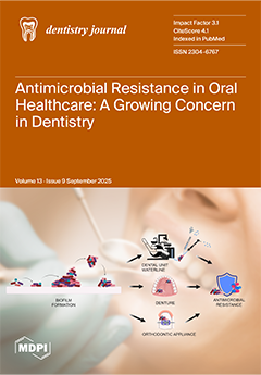
Antimicrobial resistance (AMR) is one of today’s most pressing global health challenges and dentistry contributes to this crisis. Although antibiotic misuse often draws the most attention, resistant microorganisms can be found in overlooked reservoirs within dental practice, including dental unit waterlines, dentures, and orthodontic appliances. Microbes can survive, adapt, and spread resistance within these biofilm-rich environments. This review highlights how these sources act as hidden contributors to AMR in oral healthcare and the urgent need for targeted infection control and antimicrobial stewardship. By raising awareness of these underestimated sources, this study calls for greater vigilance to protect both oral and systemic health. View this paper
- Issues are regarded as officially published after their release is announced to the table of contents alert mailing list.
- You may sign up for e-mail alerts to receive table of contents of newly released issues.
- PDF is the official format for papers published in both, html and pdf forms. To view the papers in pdf format, click on the "PDF Full-text" link, and use the free Adobe Reader to open them.
Previous Issue
Next Issue