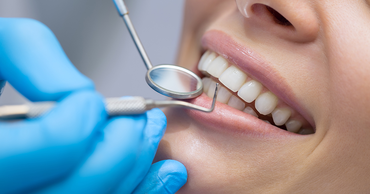
Feature Review Papers in Dentistry

A special issue of Dentistry Journal (ISSN 2304-6767).

Deadline for manuscript submissions: 10 June 2025 | Viewed by 149030

Special Issue Editors


E-Mail Website
Guest Editor
Department of Operative Dentistry, School of Dentistry, National and Kapodistrian University of Athens, 115 27 Athens, Greece
Interests: dental caries; dental materials; dentin hypersensitivity; restorative dentistry; sports dentistry
Special Issues, Collections and Topics in MDPI journals

E-Mail Website
Guest Editor
Department of Orthodontics and Dentofacial Orthopedics, School of Dental Medicine, University of Bern, CH-3010 Bern, Switzerland
Interests: orthodontics; facial esthetics; tooth agenesis; geometric morphometrics; 3D imaging; 3D superimposition
Special Issues, Collections and Topics in MDPI journals

Special Issue Information

Dear Colleagues,

This Special Issue aims to publish high-quality review papers in the research fields of dentistry. Manuscripts pertinent to basic (experimental), clinical, and epidemiological research questions will be published, following the successful fulfillment of the regular peer-review process, in case they fall within the journal’s scope. All types of reviews will be considered as long as they meet the journal’s standards. We encourage researchers from various fields to contribute review papers highlighting the latest developments in their research field or to invite relevant experts and colleagues to do so.

Feel free to contact the Managing Editor Ms. Adele Min ([email protected]) or our editorial office ([email protected]) if you have any requests.

We look forward to receiving your excellent work.

Dr. Christos Rahiotis
Prof. Dr. Nikolaos Gkantidis
Guest Editors

Manuscript Submission Information

Manuscripts should be submitted online at www.mdpi.com by registering and logging in to this website. Once you are registered, click here to go to the submission form. Manuscripts can be submitted until the deadline. All submissions that pass pre-check are peer-reviewed. Accepted papers will be published continuously in the journal (as soon as accepted) and will be listed together on the special issue website. Research articles, review articles as well as short communications are invited. For planned papers, a title and short abstract (about 100 words) can be sent to the Editorial Office for announcement on this website.

Submitted manuscripts should not have been published previously, nor be under consideration for publication elsewhere (except conference proceedings papers). All manuscripts are thoroughly refereed through a single-blind peer-review process. A guide for authors and other relevant information for submission of manuscripts is available on the Instructions for Authors page. Dentistry Journal is an international peer-reviewed open access monthly journal published by MDPI.

Please visit the Instructions for Authors page before submitting a manuscript. The Article Processing Charge (APC) for publication in this open access journal is 2000 CHF (Swiss Francs). Submitted papers should be well formatted and use good English. Authors may use MDPI's English editing service prior to publication or during author revisions.

Benefits of Publishing in a Special Issue

  • Ease of navigation: Grouping papers by topic helps scholars navigate broad scope journals more efficiently.
  • Greater discoverability: Special Issues support the reach and impact of scientific research. Articles in Special Issues are more discoverable and cited more frequently.
  • Expansion of research network: Special Issues facilitate connections among authors, fostering scientific collaborations.
  • External promotion: Articles in Special Issues are often promoted through the journal's social media, increasing their visibility.
  • e-Book format: Special Issues with more than 10 articles can be published as dedicated e-books, ensuring wide and rapid dissemination.

Further information on MDPI's Special Issue polices can be found here.

Published Papers (39 papers)

Order results
Result details
Select all
Export citation of selected articles as:

Review

Jump to: Other

11 pages, 253 KiB  
Review
Antibiotic Prophylaxis Prior to Dental Procedures
by Danica Vidović Juras, Ivana Škrinjar, Tena Križnik, Ana Andabak Rogulj, Božana Lončar Brzak, Dragana Gabrić, Marko Granić, Kristina Peroš, Ivana Šutej and Ana Ivanišević
Dent. J. 2024, 12(11), 364; https://doi.org/10.3390/dj12110364 - 15 Nov 2024
Viewed by 835
Abstract
Antibiotic prophylaxis in dentistry has been recommended for different groups of patients, such as patients with impaired immunologic function, patients at risk of developing infective endocarditis or prosthetic joint infection, patients previously exposed to high-dose irradiation of the head and neck regions, and [...] Read more.
Antibiotic prophylaxis in dentistry has been recommended for different groups of patients, such as patients with impaired immunologic function, patients at risk of developing infective endocarditis or prosthetic joint infection, patients previously exposed to high-dose irradiation of the head and neck regions, and patients receiving intravenous bisphosphonate and antiangiogenic treatment. The guidelines have been changed over the years, and the list of medical conditions requiring antibiotic prophylaxis has been shortened considerably in the context of antibiotic resistance and unnecessary antibiotic prescription. Full article
(This article belongs to the Special Issue Feature Review Papers in Dentistry)
16 pages, 406 KiB  
Review
Updates on Caries Risk Assessment—A Literature Review
by Toby Cheuk-Hang Ng, Bella Weijia Luo, Walter Yu-Hang Lam, Aylin Baysan, Chun-Hung Chu and Ollie Yiru Yu
Dent. J. 2024, 12(10), 312; https://doi.org/10.3390/dj12100312 - 29 Sep 2024
Viewed by 3214
Abstract
This narrative review aims to provide an update on caries risk assessment (CRA) and the available CRA tools. CRA can be used to monitor the status of oral care, as well as for documentation and research purposes. Caries risk is determined by the [...] Read more.
This narrative review aims to provide an update on caries risk assessment (CRA) and the available CRA tools. CRA can be used to monitor the status of oral care, as well as for documentation and research purposes. Caries risk is determined by the interplay of risk and protective factors. Common risk factors include suboptimal oral hygiene practices, previous caries experience, low plaque acidity, frequent snacking, cariogenic diet, enamel defects, reduced salivary flow, polypharmacy, and radiotherapy experience. Caries risk can be reduced through some preventive measures, such as the use of fluoride, gum chewing, dental sealants. The CRA process can be categorized as single or multifactor tools. Single-factor CRA tools include diet analysis questionnaires, cariogenic bacteria testing and saliva testing kits, whereas multifactor CRA tools encompass Caries Management by Risk Assessment (CAMBRA), Cariogram, and PreViser. Some CRA tools may quantify risk and protective factors to compute caries risk. Additionally, they can generate visual and educational presentations that foster success in oral care. Clinicians consider CRA beneficial for developing personalized care plans. However, the literature fails to reflect this and reveals limited studies supporting its use as evidence-based practice for caries management. Full article
(This article belongs to the Special Issue Feature Review Papers in Dentistry)
Show Figures

Figure 1

16 pages, 338 KiB  
Review
Multifactorial Contributors to the Longevity of Dental Restorations: An Integrated Review of Related Factors
by Maria Jacinta Moraes Coelho Santos, Elham Zare, Peter McDermott and Gildo Coelho Santos Junior
Dent. J. 2024, 12(9), 291; https://doi.org/10.3390/dj12090291 - 12 Sep 2024
Viewed by 2246
Abstract
Purpose: This integrated review aims to identify and analyze the multifactorial contributors to the longevity of direct restorations, focusing on tooth-, patient-, and dentist-related factors. Materials and Methods: A search of the literature was performed using an electronic database, PubMed/Medline, Web of Science, [...] Read more.
Purpose: This integrated review aims to identify and analyze the multifactorial contributors to the longevity of direct restorations, focusing on tooth-, patient-, and dentist-related factors. Materials and Methods: A search of the literature was performed using an electronic database, PubMed/Medline, Web of Science, and Scopus, on papers published between 1980 and 2024. The titles and abstracts of papers that evaluated aspects categorized into tooth-related, patient-related, and dentist-related factors influencing restoration failure were selected and screened. Full-text assessments were conducted, and the extracted data were compiled, summarized, and synthesized. The reference lists of the collected papers were also screened, and relevant citations were included in this review. Data were gathered from clinical and laboratorial studies, systematic reviews, and meta-analyses to provide a comprehensive understanding of restoration longevity. Results: Among the tooth-related factors, multiple-surface restorations, deep margins, tooth location, and tooth vitality significantly impact restoration survival. Patient-related factors such as medical conditions, risk predictors of caries, age, sex, parafunctional habits, smoking, periodontal health, number of restorations, and socioeconomic status all play crucial roles. Regarding dentist-related factors, the decision-making process, age, experience, and dentist manual dexterity are vital aspects. Furthermore, the technique used, including isolation methods for moisture control, as well as the type of dental practice (large group vs. small practice), notably influenced the restoration survival. Conclusions: The longevity of dental restorations is influenced by a complex interplay of tooth-related, patient-related, and dentist-related factors. Strategies to improve restoration outcomes should consider all these multifactorial contributors. Continuing professional education, diligent patient guidance on the factors that influence restoration survival, careful material selection and restorative technique, and tailored individual treatment are crucial factors to reduce failure rates and improve the lifespan of restorations. Full article
(This article belongs to the Special Issue Feature Review Papers in Dentistry)
25 pages, 1899 KiB  
Review
Conspiracy of Silence in Head and Neck Cancer Diagnosis: A Scoping Review
by Cristina Saldivia-Siracusa, Erison Santana Dos Santos, Wilfredo Alejandro González-Arriagada, Ana Carolina Prado-Ribeiro, Thaís Bianca Brandão, Adepitan Owosho, Marcio Ajudarte Lopes, Joel B. Epstein and Alan Roger Santos-Silva
Dent. J. 2024, 12(7), 214; https://doi.org/10.3390/dj12070214 - 11 Jul 2024
Viewed by 1179
Abstract
Cancer disclosure represents a complex healthcare dynamic. Physicians or caregivers may be prompted to withhold diagnosis information from patients. This study aims to comprehensively map and synthesize available evidence about diagnosis nondisclosure regarding head and neck cancer (HNC) patients. Following the Joanna Briggs [...] Read more.
Cancer disclosure represents a complex healthcare dynamic. Physicians or caregivers may be prompted to withhold diagnosis information from patients. This study aims to comprehensively map and synthesize available evidence about diagnosis nondisclosure regarding head and neck cancer (HNC) patients. Following the Joanna Briggs Institute guidelines, a scoping review was conducted across major databases without period restriction, yielding 9238 publications. After screening and selection, a descriptive synthesis was conducted. Sixteen studies were included, primarily conducted in academic settings (75%) from Europe and Asia, with a total population of 662 patients predominantly diagnosed with brain, oral, pharyngeal, or laryngeal tumors. Remarkably, 22.51% of patients were unaware of their diagnosis. Although physicians were the main source of diagnostic information (35%), they reported to often use vague terms to convey malignancy. Additionally, 13.29% of patients were aware of their diagnosis from sources other than doctors or caregivers. Caregivers (55%) supported diagnosis concealment, and physicians tended to respect family wishes. A high diagnosis-to-death interval, education, and age significantly influenced diagnosis disclosure. HNC patients expressed a desire for personalized open communication. Multiple factors influenced the decision on diagnosis disclosure. Current evidence on this topic varies significantly, and there is limited research on the consequences of nondisclosure. These findings reflect the underestimation of the patients’ outlook in the diagnosis process and highlight the need for further research, aiming to establish open communication and patient autonomy during the oncological journey. Full article
(This article belongs to the Special Issue Feature Review Papers in Dentistry)
Show Figures

Figure 1

14 pages, 285 KiB  
Review
Oral Health and Older Adults: A Narrative Review
by Martin S. Lipsky, Tejasvi Singh, Golnoush Zakeri and Man Hung
Dent. J. 2024, 12(2), 30; https://doi.org/10.3390/dj12020030 - 1 Feb 2024
Cited by 9 | Viewed by 5710
Abstract
Oral health’s association with general health, morbidity, and mortality in older adults highlights its importance for healthy aging. Poor oral health is not an inevitable consequence of aging, and a proactive, multidisciplinary approach to early recognition and treatment of common pathologies increases the [...] Read more.
Oral health’s association with general health, morbidity, and mortality in older adults highlights its importance for healthy aging. Poor oral health is not an inevitable consequence of aging, and a proactive, multidisciplinary approach to early recognition and treatment of common pathologies increases the likelihood of maintaining good oral health. Some individuals may not have regular access to a dentist, and opportunities to improve oral health may be lost if health professionals fail to appreciate the importance of oral health on overall well-being and quality of life. The authors of this narrative review examined government websites, the American Dental Association Aging and Dental Health website, and the Healthy People 2030 oral objectives and identified xerostomia, edentulism, caries, periodontitis, and oral cancer as five key topics for the non-dental provider. These conditions are associated with nutritional deficiencies, poorer quality of life, increased risk of disease development and poorer outcomes for cardiovascular disease, diabetes, and other systemic conditions prevalent among older adults. It is important to note that there is a bi-directional dimension to oral health and chronic diseases, underscoring the value of a multidisciplinary approach to maintaining oral health in older adults. Full article
(This article belongs to the Special Issue Feature Review Papers in Dentistry)
13 pages, 1899 KiB  
Review
In Vitro Models Used in the Formation of Root Caries Lesions—A Review of the Literature
by Zaid Dohan, Lara T. Friedlander, Paul R. Cooper, Kai-Chun Li, Jithendra T. Ratnayake and May L. Mei
Dent. J. 2023, 11(12), 269; https://doi.org/10.3390/dj11120269 - 26 Nov 2023
Cited by 3 | Viewed by 2234
Abstract
The management of root caries remains a challenge for clinicians due to its unique anatomical location and structure. There is increasing interest in utilising artificial root caries lesions to develop new strategies for remineralisation. An ideal protocol has not yet been agreed upon. [...] Read more.
The management of root caries remains a challenge for clinicians due to its unique anatomical location and structure. There is increasing interest in utilising artificial root caries lesions to develop new strategies for remineralisation. An ideal protocol has not yet been agreed upon. The aim of this review is to provide a structured overview of previously reported in vitro root caries models. The literature was screened and mined for information mainly on substrate selection, model systems utilised, and variables used in the models. Human roots (60%) were the most frequently used substrates, followed by bovine roots (40%). Chemical models (69%) were the most frequently utilised model systems, followed by microbiological models (27%), to form root caries lesions. Acetate buffer solution (80%), pH 5.0 or above (40%), and a demineralisation time of five days (25%) were the common variables used in the chemical systems, while mono-species biofilm was most frequently used (73%) in microbiological models and Streptococcus mutans was the most common bacterial strain utilised in these models (80%). This review highlights the variability amongst the experimental approaches, discusses the advantages and limitations of these approaches, and emphasises that standardisation of experimental conditions along with sustained research will benefit root caries research. Full article
(This article belongs to the Special Issue Feature Review Papers in Dentistry)
Show Figures

Figure 1

19 pages, 359 KiB  
Review
Emerging Applications of Nanotechnology in Dentistry
by Shiza Malik and Yasir Waheed
Dent. J. 2023, 11(11), 266; https://doi.org/10.3390/dj11110266 - 15 Nov 2023
Cited by 16 | Viewed by 8057
Abstract
Dentistry is a branch of healthcare where nanobiotechnology is reverberating in multiple ways to produce beneficial outcomes. The purpose of this review is to bring into the awareness of the readers the various practical dimensions of the nano-dental complex (nanodentistry) in healthcare and [...] Read more.
Dentistry is a branch of healthcare where nanobiotechnology is reverberating in multiple ways to produce beneficial outcomes. The purpose of this review is to bring into the awareness of the readers the various practical dimensions of the nano-dental complex (nanodentistry) in healthcare and how novelties linked with the field are revolutionizing dentistry. A methodological approach was adopted to collect the latest data on nanotechnology and dentistry from sources, including PubMed, Google Scholar, Scopus, and official websites like the WHO. Nanodentistry is an emerging field in dentistry that involves the use of nanomaterials, nanorobots, and nanotechnology to diagnose, treat, and prevent dental diseases. The results summarize the descriptive analyses of the uses of nanodentistry within orthodontics, preventive dentistry, prosthodontics, restorative dentistry, periodontics, dental surgeries, dental restoration technologies, and other areas of dentistry. The future directions of nano-industries and nano-healthcare have been included to link them with the oral healthcare sector, treatment plans, and improved medical services which could be explored in the future for advanced healthcare regulation. The major limitations to the use of dental nanoproducts are their cost-effectiveness and accessibility, especially in financially constrained countries. These data will help the readers to experience a detailed analysis and comprehensive covering of the diverse achievements of nanodentistry with past analyses, present scenarios, and future implications. Full article
(This article belongs to the Special Issue Feature Review Papers in Dentistry)
Show Figures

Graphical abstract

13 pages, 1796 KiB  
Review
Prognostic Significance of Histopathological Parameters for Salivary Gland Adenoid Cystic Carcinoma
by Everton Freitas de Morais, Hannah Gil de Farias Morais, Roseana de Almeida Freitas and Ricardo D. Coletta
Dent. J. 2023, 11(11), 262; https://doi.org/10.3390/dj11110262 - 9 Nov 2023
Cited by 3 | Viewed by 2326
Abstract
Adenoid cystic carcinoma (ACC) is a rare salivary gland tumor that accounts for approximately 1% of all head and neck cancers. Despite its initial indolent behavior, long-term survival is poor due to locoregional recurrence in approximately 40% and distant metastasis in up to [...] Read more.
Adenoid cystic carcinoma (ACC) is a rare salivary gland tumor that accounts for approximately 1% of all head and neck cancers. Despite its initial indolent behavior, long-term survival is poor due to locoregional recurrence in approximately 40% and distant metastasis in up to 60% of patients who undergo radical treatment. The histological parameters of ACC and the combination of these parameters in histopathological grading systems provide valuable prognostic information about the clinical course of the disease. Within this context, this review aims to analyze the impact of histopathological parameters, individual or combined in histopathological grading systems of malignancy, on ACC prognosis. Individual histopathological parameters such as solid pattern, presence of tumor necrosis, high-grade transformation, dominance of the epithelial component, presence of perineural and lymphovascular invasion, and positive surgical margins have negative impacts on the survival of patients with ACC. There are currently four histopathological grading systems for ACC; however, few studies have validated these systems and most of them explored small cohorts with short follow-up. Considering that the application of grading systems has been associated with ACC prognosis, a broader validation will allow not only their use for prognostic prediction but also assist in treatment planning. Full article
(This article belongs to the Special Issue Feature Review Papers in Dentistry)
Show Figures

Figure 1

25 pages, 2243 KiB  
Review
Pain Assessment in Oral Medicine through Its Different Dimensions: A Comprehensive Review
by Andrea Scribante, Matteo Pellegrini, Federica Pulicari, Martina Ghizzoni, Francesco Paolo Modugno and Francesco Spadari
Dent. J. 2023, 11(11), 246; https://doi.org/10.3390/dj11110246 - 24 Oct 2023
Cited by 4 | Viewed by 4132
Abstract
Orofacial pain is a complex experience made up of different features and involving various aspects of life. It has demonstrated a connection, especially when chronic, with conditions such as anxiety, depression, and sleep disorders, through paths that still have not been completely clarified. [...] Read more.
Orofacial pain is a complex experience made up of different features and involving various aspects of life. It has demonstrated a connection, especially when chronic, with conditions such as anxiety, depression, and sleep disorders, through paths that still have not been completely clarified. A deep understanding of orofacial pain and its impact on an individual’s life is critical for planning accurate diagnostic and therapeutic approaches. This review seeks to provide a comprehensive overview of the components constituting the pain experience, its implications in an individual’s life, the different tools for multidimensional pain assessment, and the specific applications for each tool. A comprehensive review was performed using the PubMed, Scopus, and Web of Science electronic databases. Ninety-five studies, including observational studies, clinical trials, case–control studies, and case reports, were included and analyzed in this review. Orofacial pain assessment exploits several methods, ranging from clinical evaluation to rating scales, questionnaires, and daily diaries. The choice of the correct instrument requires an evaluation of the type of pain experienced, of the patient’s characteristics and abilities to complete particular tasks, and finally, of the assessment tool features. Full article
(This article belongs to the Special Issue Feature Review Papers in Dentistry)
Show Figures

Graphical abstract

20 pages, 1274 KiB  
Review
Smartphone Applications in Dentistry: A Scoping Review
by Maurizio Pascadopoli, Paolo Zampetti, Maria Gloria Nardi, Matteo Pellegrini and Andrea Scribante
Dent. J. 2023, 11(10), 243; https://doi.org/10.3390/dj11100243 - 20 Oct 2023
Cited by 12 | Viewed by 3935
Abstract
This scoping review aims to investigate the latest literature concerning the use of smartphone applications (apps) in the prevention, management, and monitoring of oral diseases. Smartphone applications are software programs that are designed to run on smartphones. Nowadays, smartphones are regularly used by [...] Read more.
This scoping review aims to investigate the latest literature concerning the use of smartphone applications (apps) in the prevention, management, and monitoring of oral diseases. Smartphone applications are software programs that are designed to run on smartphones. Nowadays, smartphones are regularly used by people of all ages, and mobile health apps (MHAs) represent an important means of spreading information related to oral health, which is the state of the mouth and teeth, including the gums and other tissues. Several apps have been designed to promote prevention, diagnosis, and therapeutic adherence monitoring. This scoping review considered randomized clinical trials, cross-sectional studies, before–after (pre–post) studies with no control group, and observational studies. Once the inclusion and exclusion criteria had been defined, a preliminary confined search was performed on PubMed and Scopus; key terms from the collected articles were selected to design a search strategy, and then a search of all the included articles’ reference lists was run for further research. Studies were excluded if they did not fulfill the inclusion criteria. The preferred reporting items for scoping reviews (PRISMA-ScR) consensus was followed. The risk of bias was evaluated by providing a qualitative analysis of the clinical studies via the National Heart, Lung, and Blood Institute (NHLBI) Quality Assessment of Controlled Intervention Studies, Observational Cohort Studies, and Cross-Sectional Studies (NHLBI, NIH). A total of 21 studies were included in this review. As it is clear from the studies selected, the literature indicates that MHAs are effective in improving oral hygiene in adolescents and children and reducing the dental plaque index, including in patients undergoing orthodontic treatment. MHAs are also able to reduce the symptoms of patients affected by obstructive sleep apnea–hypopnea syndrome (OSAHS) and improve the swallowing-related quality of life of elderly patients. MHAs are furthermore recommended to decrease dental anxiety among patients, both during dental procedures and the post-operative period. MHAs are useful to spread knowledge about traumatic dental injuries among non-oral health professionals and to monitor dental erosion and awake bruxism. MHAs’ clinical outcomes might have been influenced by the demographic features of the subjects involved. Further studies considering a longer follow-up period and larger samples are needed. In conclusion, MHAs can be considered a useful tool to monitor oral disease and increase patients’ quality of life related to oral health. Full article
(This article belongs to the Special Issue Feature Review Papers in Dentistry)
Show Figures

Figure 1

15 pages, 677 KiB  
Review
Current Insights on Fiber Posts: A Narrative Review of Laboratory and Clinical Studies
by Dayana Campanelli de Morais, Sheila Butler and Maria Jacinta Moraes Coelho Santos
Dent. J. 2023, 11(10), 236; https://doi.org/10.3390/dj11100236 - 10 Oct 2023
Cited by 6 | Viewed by 3869
Abstract
Purpose: The aim of this study was to review the literature related to the clinical performance and laboratory findings regarding fiber posts, as well as the cementation technique employed with their use. Materials and Methods: A literature search was performed using an electronic [...] Read more.
Purpose: The aim of this study was to review the literature related to the clinical performance and laboratory findings regarding fiber posts, as well as the cementation technique employed with their use. Materials and Methods: A literature search was performed using an electronic database, PubMed/Medline, between 2010 and 2023. The terms used were “intra coronal post, fiber post, post cementation, and post length”. Titles and abstracts were initially screened, and a full-text assessment was conducted for those that fulfilled the inclusion criteria. The reference list of the collected papers was also screened for further relevant citations. Results: In this work, 135 potentially eligible studies were analyzed. Titles and abstracts of 90 studies followed the inclusion criteria and were selected for a full-text assessment, resulting in 50 studies selected. Moreover, additional studies from relevant citations were included, totaling 57 studies. Conclusion: According to the laboratory and clinical studies revised, the survival rate between fiber and prefabricated and cast metal posts was similar, and failures were mainly related to the loss of retention. The intra-canal post length of less than two-thirds of the root length presented successful results when ferrule was present. Furthermore, the ferrule increased the longevity of teeth restored with fiber posts. Additionally, the use of a surface treatment protocol for fiber posts and the adhesive cementation technique both contributed to the clinical success and longevity of the intra-canal post. Full article
(This article belongs to the Special Issue Feature Review Papers in Dentistry)
Show Figures

Figure 1

14 pages, 1113 KiB  
Review
Diet, Nutrition, and Oral Health in Older Adults: A Review of the Literature
by Alice Kit Ying Chan, Yiu Cheung Tsang, Chloe Meng Jiang, Katherine Chiu Man Leung, Edward Chin Man Lo and Chun Hung Chu
Dent. J. 2023, 11(9), 222; https://doi.org/10.3390/dj11090222 - 19 Sep 2023
Cited by 18 | Viewed by 9383
Abstract
Diet, nutrition, and oral health are closely linked. Malnutrition is a challenging health concern in older adults that is associated with physical decline affecting their daily activities and quality of life. The aim of this review is to provide an evidence-based summary of [...] Read more.
Diet, nutrition, and oral health are closely linked. Malnutrition is a challenging health concern in older adults that is associated with physical decline affecting their daily activities and quality of life. The aim of this review is to provide an evidence-based summary of the relationship between diet and nutrition and oral health in older adults and its implications. The World Health Organization has declared healthy ageing a priority of its work on ageing. The American Dental Association confirmed the bidirectional relationship between diet and nutrition and oral health. The literature shows that diet and nutrition are related to oral diseases, including dental caries, periodontal diseases, tooth wear, and even oral cancer. Insufficient nutritional intake and poor dietary habits increase the risk of oral diseases, such as dental caries, in older adults. On the other hand, in older adults, poor oral conditions such as periodontal disease may induce pain, infection, and tooth loss, affecting nutritional intake. Surveys have shown that older adults, in particular, those in disadvantaged communities, suffered from nutritional deficiencies or imbalances affecting their oral health. In addition, the current literature shows that malnutrition is associated with frailty, hospitalization, mortality, and morbidity. Good oral health and functional dentition are essential to maintain sufficient nutritional intake among older adults and reduce the risk of malnutrition. Therefore, integrating oral health into general health care service in older adults is imperative to improve their nutritional and oral health status to achieve healthy ageing. Full article
(This article belongs to the Special Issue Feature Review Papers in Dentistry)
Show Figures

Graphical abstract

24 pages, 1222 KiB  
Review
Methods for Evaluating the Effectiveness of Home Oral Hygiene Measures—A Narrative Review of Dental Biofilm Indices
by Gerarda D’Elia, William Floris, Lorenzo Marini, Denise Corridore, Mariana Andrea Rojas, Livia Ottolenghi and Andrea Pilloni
Dent. J. 2023, 11(7), 172; https://doi.org/10.3390/dj11070172 - 17 Jul 2023
Cited by 5 | Viewed by 6425
Abstract
Dental plaque is a biofilm structured in an extracellular matrix of polymers of host and microbial origin; the microorganisms can coexist in harmony with the host, thus guarantying oral health. Environmental modifications can lead to dysbiosis and onset of oral diseases; in fact, [...] Read more.
Dental plaque is a biofilm structured in an extracellular matrix of polymers of host and microbial origin; the microorganisms can coexist in harmony with the host, thus guarantying oral health. Environmental modifications can lead to dysbiosis and onset of oral diseases; in fact, plaque is the etiological agent both of periodontal disease and dental decay. The use of an effective oral hygiene index should be considered as a relevant goal for the clinicians and the researchers, and consequently, numerous plaque indices have been proposed during the years. The present literature review aims primarily to obtain a complete summary of these scores to assess plaque deposits. It is useful because the clinician/researcher will select the right scoring method for the specific situation only if he knows the available options and if he is aware of both their strengths and weaknesses. This review applies a basic classification of plaque indices that distinguishes the ones that use non-quantitative methods from the ones that use quantitative methods. Non-quantitative methods are more subjective because they are based on the ability of the clinician to point out the presence or the entity of deposits, while quantitative methods introduce objectifiable means to measure plaque deposits. Full article
(This article belongs to the Special Issue Feature Review Papers in Dentistry)
Show Figures

Figure 1

13 pages, 742 KiB  
Review
The Performance of Paralleling Technique and Bisecting Angle Technique for Taking Periapical Radiographs: A Systematic Review
by Matthew Yen and Andy Wai Kan Yeung
Dent. J. 2023, 11(7), 155; https://doi.org/10.3390/dj11070155 - 21 Jun 2023
Cited by 4 | Viewed by 12737
Abstract
Periapical radiography is a routine radiographic procedure performed by dentists on a daily basis. It can be taken with two techniques, the paralleling technique (P tech) and the bisecting angle technique (B tech). This systematic review aimed to identify the relevant literature, compare [...] Read more.
Periapical radiography is a routine radiographic procedure performed by dentists on a daily basis. It can be taken with two techniques, the paralleling technique (P tech) and the bisecting angle technique (B tech). This systematic review aimed to identify the relevant literature, compare the use of P and B techs across various dental specialties, and determine the most appropriate technique to be used for different purposes in taking periapical radiographs. In January 2023, we searched PubMed, Web of Science, Scopus, and Google Scholar to identify the studies that compared the two radiographic techniques. The search string was: (paralleling AND (“bisecting angle” OR “bisected angle”)). Manual reference tracing was also performed to identify the studies potentially missed. After screening, 26 studies were included for the qualitative review. The 26 included studies were published between 1976 and 2021. Ten of the studies were about general dentistry (dental radiology in general applications), whereas another ten studies were related to endodontics, such as working length estimation. Most studies advocated the use of the P tech for general, endodontics, implantology, and other indications. B tech was advocated for patients with a low palatal height. More future studies are needed to evaluate their performance in different scenarios with standardized equipment and radiographic positioning. Full article
(This article belongs to the Special Issue Feature Review Papers in Dentistry)
Show Figures

Graphical abstract

20 pages, 1376 KiB  
Review
Do Systemic Diseases and Medications Influence Dental Implant Osseointegration and Dental Implant Health? An Umbrella Review
by Francesco D’Ambrosio, Alessandra Amato, Andrea Chiacchio, Laura Sisalli and Francesco Giordano
Dent. J. 2023, 11(6), 146; https://doi.org/10.3390/dj11060146 - 5 Jun 2023
Cited by 20 | Viewed by 5968
Abstract
The aim of this umbrella review is to evaluate what are the most common medications and systemic diseases that can affect bone–implant integration, the success rate and survival rate of dental implants, peri-implant tissue health, and implant loss. Systematic reviews, with meta-analysis or [...] Read more.
The aim of this umbrella review is to evaluate what are the most common medications and systemic diseases that can affect bone–implant integration, the success rate and survival rate of dental implants, peri-implant tissue health, and implant loss. Systematic reviews, with meta-analysis or not, about how systemic diseases and medications influence dental implant osseointegration, survival rate, success rate, and peri-implant diseases, published only in the English language, are electronically searched across the most important scientific databases. The present umbrella review includes eight systematic reviews, and osteoporosis and diabetes are the most investigated pathologies. Systemic diseases, such as neurologic disorders, HIV, hypothyroidism, cardiovascular diseases, and drugs, such as beta blockers, anti-hypertensives, or diuretics do not show a decreased rate of implant osseointegration. It seems that drugs, such as proton-pump inhibitors (PPIs) or serotonin reuptake inhibitors (SSRIs), negatively affect implant osseointegration. Few studies compare the effects of drugs and systemic diseases on the parameters considered in this overview. It is important to underline how the results of this review need to be validated with subsequent and more reviews. Full article
(This article belongs to the Special Issue Feature Review Papers in Dentistry)
Show Figures

Graphical abstract

29 pages, 422 KiB  
Review
Treatments of COVID-19-Associated Taste and Saliva Secretory Disorders
by Hironori Tsuchiya
Dent. J. 2023, 11(6), 140; https://doi.org/10.3390/dj11060140 - 25 May 2023
Cited by 4 | Viewed by 4851
Abstract
Since the worldwide spread of severe acute respiratory syndrome coronavirus 2 (SARS-CoV-2) infection, treating taste and saliva secretory disorders associated with coronavirus disease 2019 (COVID-19) has become a critical issue. The aim of the present study was to update information on treatments applicable [...] Read more.
Since the worldwide spread of severe acute respiratory syndrome coronavirus 2 (SARS-CoV-2) infection, treating taste and saliva secretory disorders associated with coronavirus disease 2019 (COVID-19) has become a critical issue. The aim of the present study was to update information on treatments applicable to such oral symptoms and discuss their pathogenic mechanisms. The literature search indicated that different treatments using tetracycline, corticosteroids, zinc, stellate ganglion block, phytochemical curcumin, traditional herbal medicine, nutraceutical vitamin D, photobiomodulation, antiviral drugs, malic acid sialagogue, chewing gum, acupuncture, and/or moxibustion have potential effects on COVID-19-associated ageusia/dysgeusia/hypogeusia and xerostomia/dry mouth/hyposalivation. These treatments have multiple modes of action on viral cellular entry and replication, cell proliferation and differentiation, immunity, and/or SARS-CoV-2 infection-induced pathological conditions such as inflammation, cytokine storm, pyroptosis, neuropathy, zinc dyshomeostasis, and dysautonomia. An understanding of currently available treatment options is required for dental professionals because they may treat patients who were infected with SARS-CoV-2 or who recovered from COVID-19, and become aware of their abnormal taste and salivary secretion. By doing so, dentists and dental hygienists could play a crucial role in managing COVID-19 oral symptoms and contribute to improving the oral health-related quality of life of the relevant patients. Full article
(This article belongs to the Special Issue Feature Review Papers in Dentistry)
12 pages, 923 KiB  
Review
Dental Pulp Response to Silver-Containing Solutions: A Scoping Review
by Ahmed Zaeneldin, Chun-Hung Chu and Ollie Yiru Yu
Dent. J. 2023, 11(5), 114; https://doi.org/10.3390/dj11050114 - 26 Apr 2023
Cited by 1 | Viewed by 2487
Abstract
Dentists used silver-containing solutions for deep cavity disinfection before restoration. This review aims to identify the silver-containing solutions reported in the literature for deep cavity disinfection and summarize their effects on dental pulp. An extensive search was performed using the search words “(silver) [...] Read more.
Dentists used silver-containing solutions for deep cavity disinfection before restoration. This review aims to identify the silver-containing solutions reported in the literature for deep cavity disinfection and summarize their effects on dental pulp. An extensive search was performed using the search words “(silver) AND (dental pulp OR pulp)” in ProQuest, PubMed, SCOPUS, and Web of Science to identify English publications on silver-containing solutions for cavity conditioning. The pulpal response to the included silver-containing solutions was summarized. The initial search identified 4112 publications and 14 publications met the inclusion criteria. Silver fluoride, silver nitrate, silver diamine nitrate, silver diamine fluoride, and nano-silver fluoride were used in deep cavities for antimicrobial purposes. Indirect silver fluoride application induced pulp inflammation and reparative dentine in most cases, and pulp necrosis in some cases. Direct silver nitrate application caused blood clots and a wide inflammatory band in the pulp, whilst indirect silver nitrate application caused hypoplasia in shallow cavities and partial pulp necrosis in deep cavities. Direct silver diamine fluoride application induced pulp necrosis, while indirect silver diamine fluoride application induced a mild inflammatory response and reparative dentine formation. No evidence of the dental pulpal response to silver diamine nitrate or nano-silver fluoride was available in the literature. Full article
(This article belongs to the Special Issue Feature Review Papers in Dentistry)
Show Figures

Figure 1

15 pages, 2480 KiB  
Review
Medication-Related Osteonecrosis: Why the Jawbone?
by Sydney Kuehn, Rafaela Scariot and Mohammed Elsalanty
Dent. J. 2023, 11(5), 109; https://doi.org/10.3390/dj11050109 - 23 Apr 2023
Cited by 4 | Viewed by 2589
Abstract
Medication-related osteonecrosis of the jaw (MRONJ) has emerged as a complication of anti-resorptive medications. Despite its low incidence rate, this problem has gained attention in recent years due to its devastating consequences and lack of preventive strategy. The fact that MRONJ incidence has [...] Read more.
Medication-related osteonecrosis of the jaw (MRONJ) has emerged as a complication of anti-resorptive medications. Despite its low incidence rate, this problem has gained attention in recent years due to its devastating consequences and lack of preventive strategy. The fact that MRONJ incidence has been exclusive to the jawbones, despite the systemic effect of anti-resorptive medications, could be a starting point to unravel the multifactorial pathogenesis of this condition. This review aims to negotiate the question of why the jawbone is more susceptible to MRONJ than other skeletal sites. Approaching the problem from this perspective could provide new directions for the prevention of MRONJ and expand our understanding of the unique oral microenvironment. Full article
(This article belongs to the Special Issue Feature Review Papers in Dentistry)
Show Figures

Figure 1

18 pages, 4564 KiB  
Review
Morphological and Ultrastructural Collagen Defects: Impact and Implications in Dentinogenesis Imperfecta
by Lubabah S. A. Gadi, David Y. S. Chau and Susan Parekh
Dent. J. 2023, 11(4), 95; https://doi.org/10.3390/dj11040095 - 3 Apr 2023
Cited by 3 | Viewed by 2981
Abstract
Collagen is the building block for the extracellular matrix in bone, teeth and other fibrous tissues. Osteogenesis imperfecta (OI), or brittle bone disease, is a heritable disorder that results from defective collagen synthesis or metabolism, resulting in bone fragility. The dental manifestation of [...] Read more.
Collagen is the building block for the extracellular matrix in bone, teeth and other fibrous tissues. Osteogenesis imperfecta (OI), or brittle bone disease, is a heritable disorder that results from defective collagen synthesis or metabolism, resulting in bone fragility. The dental manifestation of OI is dentinogenesis imperfecta (DI), a genetic disorder that affects dentin structure and clinical appearance, with a characteristic feature of greyish-brown discolouration. The aim of this study was to conduct a systematic review to identify and/or define any ultrastructural changes in dentinal collagen in DI. Established databases were searched: Cochrane Library, OVID Embase, OVID Medline and PubMed/Medline. Search strategies included: Collagen Ultrastructure, DI and OI. Inclusion criteria were studies written in English, published after 1990, that examined human dental collagen of teeth affected by DI. A Cochrane data extraction form was modified and used for data collection. The final dataset included seventeen studies published from 1993 to 2021. The most prevalent findings on collagen in DI teeth were increased coarse collagen fibres and decreased fibre quantity. Additional findings included changes to fibre orientation (i.e., random to parallel) and differences to the fibre organisation (i.e., regular to irregular). Ultrastructural defects and anomalies included uncoiled collagen fibres and increased D-banding periodicity. Studies in collagen structure in DI reported changes to the surface topography, quantity, organisation and orientation of the fibres. Moreover, ultrastructural defects such as the packing/coiling and D-banding of the fibrils, as well as differences in the presence of other collagens are also noted. Taken together, this study provides an understanding of the changes in collagen and its impact on clinical translation, paving the way for innovative treatments in dental treatment. Full article
(This article belongs to the Special Issue Feature Review Papers in Dentistry)
Show Figures

Figure 1

14 pages, 1510 KiB  
Review
Remineralization Strategies for Teeth with Molar Incisor Hypomineralization (MIH): A Literature Review
by Joachim Enax, Bennett T. Amaechi, Rayane Farah, Jungyi Alexis Liu, Erik Schulze zur Wiesche and Frederic Meyer
Dent. J. 2023, 11(3), 80; https://doi.org/10.3390/dj11030080 - 13 Mar 2023
Cited by 14 | Viewed by 6719
Abstract
Molar incisor hypomineralization (MIH) is a highly prevalent dental developmental disorder with a significant health burden for patients and high treatment needs, yet no comprehensive review article on all remineralization systems as a non-invasive treatment approach for MIH has been published. Typical characteristics [...] Read more.
Molar incisor hypomineralization (MIH) is a highly prevalent dental developmental disorder with a significant health burden for patients and high treatment needs, yet no comprehensive review article on all remineralization systems as a non-invasive treatment approach for MIH has been published. Typical characteristics of MIH-affected teeth are a lower mineral density and lower hardness compared to healthy teeth leading to sensitivity and loss of function. Thus, the use of formulations with calcium phosphates to remineralize MIH-affected teeth is reasonable. This review presents an up-to-date overview of remineralization studies focusing on active ingredients investigated for remineralization of MIH, i.e., casein phosphopeptide amorphous calcium phosphate (CPP-ACP), casein phosphopeptide amorphous calcium fluoride phosphate (CPP-ACFP), hydroxyapatite, calcium glycerophosphate, self-assembling peptide, and fluoride. Overall, 19 studies (in vitro, in situ, and in vivo) were found. Furthermore, an additional search for studies focusing on using toothpaste/dentifrices for MIH management resulted in six studies, where three studies were on remineralization and three on reduction of sensitivity. Overall, the studies analyzed in this review showed that MIH-affected teeth could be remineralized using calcium phosphate-based approaches. In conclusion, calcium phosphates like CPP-ACP, calcium glycerophosphate, and hydroxyapatite can be used to remineralize MIH-affected teeth. In addition to MIH-remineralization, CPP-ACP and hydroxyapatite also offer relief from MIH-associated tooth sensitivity. Full article
(This article belongs to the Special Issue Feature Review Papers in Dentistry)
Show Figures

Figure 1

15 pages, 2077 KiB  
Review
Robotics in Dentistry: A Narrative Review
by Lipei Liu, Megumi Watanabe and Tetsuo Ichikawa
Dent. J. 2023, 11(3), 62; https://doi.org/10.3390/dj11030062 - 24 Feb 2023
Cited by 16 | Viewed by 5837
Abstract
Background: Robotics is progressing rapidly. The aim of this study was to provide a comprehensive overview of the basic and applied research status of robotics in dentistry and discusses its development and application prospects in several major professional fields of dentistry. Methods: A [...] Read more.
Background: Robotics is progressing rapidly. The aim of this study was to provide a comprehensive overview of the basic and applied research status of robotics in dentistry and discusses its development and application prospects in several major professional fields of dentistry. Methods: A literature search was conducted on databases: MEDLINE, IEEE and Cochrane Library, using MeSH terms: [“robotics” and “dentistry”]. Result: Forty-nine articles were eventually selected according to certain inclusion criteria. There were 12 studies on prosthodontics, reaching 24%; 11 studies were on dental implantology, accounting for 23%. Scholars from China published the most articles, followed by Japan and the United States. The number of articles published between 2011 and 2015 was the largest. Conclusions: With the advancement of science and technology, the applications of robots in dental medicine has promoted the development of intelligent, precise, and minimally invasive dental treatments. Currently, robots are used in basic and applied research in various specialized fields of dentistry. Automatic tooth-crown-preparation robots, tooth-arrangement robots, drilling robots, and orthodontic archwire-bending robots that meet clinical requirements have been developed. We believe that in the near future, robots will change the existing dental treatment model and guide new directions for further development. Full article
(This article belongs to the Special Issue Feature Review Papers in Dentistry)
Show Figures

Figure 1

18 pages, 1784 KiB  
Review
Systematic Review and Meta Analysis of the Relative Effect on Plaque Index among Pediatric Patients Using Powered (Electric) versus Manual Toothbrushes
by Andrew Graves, Troy Grahl, Mark Keiserman and Karl Kingsley
Dent. J. 2023, 11(2), 46; https://doi.org/10.3390/dj11020046 - 9 Feb 2023
Cited by 8 | Viewed by 5921
Abstract
Although many randomized controlled trials (RCT) have evaluated the efficacy of powered or electric toothbrushes compared with manual or traditional toothbrushes to remove biofilm and plaque, only one systematic review has been published for pediatric patients. The primary objective of this study was [...] Read more.
Although many randomized controlled trials (RCT) have evaluated the efficacy of powered or electric toothbrushes compared with manual or traditional toothbrushes to remove biofilm and plaque, only one systematic review has been published for pediatric patients. The primary objective of this study was to perform a systematic review and meta analysis for this population. Using the PRISMA (Preferred Reporting Items for Systematic Reviews and Meta-Analyses) protocol, N = 321 studies were initially identified. Three independent, blinded abstract reviews were completed resulting in a total of n = 38/322 or 11.8% for the final analysis (n = 27 non-orthodontic, n = 11 orthodontic studies). Meta analysis of these outcome data have revealed a strong reduction in plaque index scores among pediatric patients using electric toothbrushes of approximately 17.2% for non-orthodontic patients and 13.9% for orthodontic patients. These results provide strong clinical evidence for recommending electric toothbrushing to pediatric patients, as well as those patients undergoing orthodontic therapy and treatment. Full article
(This article belongs to the Special Issue Feature Review Papers in Dentistry)
Show Figures

Graphical abstract

25 pages, 2414 KiB  
Review
Artificial Intelligence in Periodontology: A Scoping Review
by James Scott, Alberto M. Biancardi, Oliver Jones and David Andrew
Dent. J. 2023, 11(2), 43; https://doi.org/10.3390/dj11020043 - 8 Feb 2023
Cited by 18 | Viewed by 6644
Abstract
Artificial intelligence (AI) is the development of computer systems whereby machines can mimic human actions. This is increasingly used as an assistive tool to help clinicians diagnose and treat diseases. Periodontitis is one of the most common diseases worldwide, causing the destruction and [...] Read more.
Artificial intelligence (AI) is the development of computer systems whereby machines can mimic human actions. This is increasingly used as an assistive tool to help clinicians diagnose and treat diseases. Periodontitis is one of the most common diseases worldwide, causing the destruction and loss of the supporting tissues of the teeth. This study aims to assess current literature describing the effect AI has on the diagnosis and epidemiology of this disease. Extensive searches were performed in April 2022, including studies where AI was employed as the independent variable in the assessment, diagnosis, or treatment of patients with periodontitis. A total of 401 articles were identified for abstract screening after duplicates were removed. In total, 293 texts were excluded, leaving 108 for full-text assessment with 50 included for final synthesis. A broad selection of articles was included, with the majority using visual imaging as the input data field, where the mean number of utilised images was 1666 (median 499). There has been a marked increase in the number of studies published in this field over the last decade. However, reporting outcomes remains heterogeneous because of the variety of statistical tests available for analysis. Efforts should be made to standardise methodologies and reporting in order to ensure that meaningful comparisons can be drawn. Full article
(This article belongs to the Special Issue Feature Review Papers in Dentistry)
Show Figures

Figure 1

24 pages, 692 KiB  
Review
Potential Beneficial Effects of Hydroxyapatite Nanoparticles on Caries Lesions In Vitro—A Review of the Literature
by Eisha Imran, Paul R. Cooper, Jithendra Ratnayake, Manikandan Ekambaram and May Lei Mei
Dent. J. 2023, 11(2), 40; https://doi.org/10.3390/dj11020040 - 7 Feb 2023
Cited by 15 | Viewed by 3451
Abstract
Dental caries is one of the most common human diseases which can occur in both primary and permanent dentitions throughout the life of an individual. Hydroxyapatite is the major inorganic component of human teeth, consequently, nanosized hydroxyapatite (nHAP) has recently attracted researchers’ attention [...] Read more.
Dental caries is one of the most common human diseases which can occur in both primary and permanent dentitions throughout the life of an individual. Hydroxyapatite is the major inorganic component of human teeth, consequently, nanosized hydroxyapatite (nHAP) has recently attracted researchers’ attention due to its unique properties and potential for caries management. This article provides a contemporary review of the potential beneficial effects of nHAP on caries lesions demonstrated in in vitro studies. Data showed that nHAP has potential to promote mineralization in initial caries, by being incorporated into the porous tooth structure, which resulted from the caries process, and subsequently increased mineral content and hardness. Notably, it is the particle size of nHAP which plays an important role in the mineralization process. Antimicrobial effects of nHAP can also be achieved by metal substitution in nHAP. Dual action property (mineralizing and antimicrobial) and enhanced chemical stability and bioactivity of nHAP can potentially be obtained using metal-substituted fluorhydroxyapatite nanoparticles. This provides a promising synergistic strategy which should be explored in further clinical research to enable the development of dental therapeutics for use in the treatment and management of caries. Full article
(This article belongs to the Special Issue Feature Review Papers in Dentistry)
Show Figures

Figure 1

Other

Jump to: Review

17 pages, 3792 KiB  
Systematic Review
Root Coverage Techniques: Coronally Advancement Flap vs. Tunnel Technique: A Systematic Review and Meta-Analysis
by Luis Chauca-Bajaña, Alba Pérez-Jardón, Fábio França Vieira E Silva, Mercedes Conde-Amboage, Byron Velásquez-Ron, Elena Padín-Iruegas and Mario Pérez-Sayáns
Dent. J. 2024, 12(11), 341; https://doi.org/10.3390/dj12110341 - 25 Oct 2024
Viewed by 1345
Abstract
Introduction: Gingival recession, characterized by the apical displacement of the gingival margin, presents challenges to oral health. This study compares the effectiveness of the coronally advanced flap (CAF) and the tunnel technique (TT) for treating gingival recessions. Methods: Bibliographical searches included PubMed, Embase, [...] Read more.
Introduction: Gingival recession, characterized by the apical displacement of the gingival margin, presents challenges to oral health. This study compares the effectiveness of the coronally advanced flap (CAF) and the tunnel technique (TT) for treating gingival recessions. Methods: Bibliographical searches included PubMed, Embase, Web of Science, Cochrane, Scopus, and the grey literature, with keywords “root coverage” “coronary advanced flap”, and “tunnel”. A systematic coreview was performed that included 26 studies evaluating root coverage, and 14 articles were included for the meta-analysis. Three groups were analyzed: Group 1 compared TT with connective tissue graft (CTG) versus CAF with CTG; Group 2 examined TT with CTG and/or other biomaterials versus TT with CTG alone; Group 3 compared TT with CAF, regardless of complementary biomaterials. Meta-analysis assessed mean root coverage (MRC), complete root coverage (CRC), and keratinized tissue gain (KTG). Results: In Group 1, TT with CTG demonstrated superior MRC compared with CAF with CTG (−8.68 CI95% −17.19 to −0.17; p = 0.0457). In Group 2, TT with CTG and/or other biomaterials showed similar MRC (4.17 CI95% −17.91 to 26.26; p = 0.7110) and CRC (0.37 CI95% −1.14 to 1.89; p = 0.6269) to TT with CTG alone, with variations in keratinized tissue gain. Group 3 indicated higher potential MRC for TT compared with CAF (5.73 CI95% −8.90 to 13.55; p = 0.685) but without statistically significant differences. Conclusions: This study suggests that TT with CTG might offer better root coverage than CAF with CTG; however, biomaterial selection requires consideration. Full article
(This article belongs to the Special Issue Feature Review Papers in Dentistry)
Show Figures

Figure 1

11 pages, 637 KiB  
Systematic Review
A Systematic Review of Stem Cell Applications in Maxillofacial Regeneration
by Man Hung, Mahsa Sadri, Melanie Katz, Connor Schwartz and Amir Mohajeri
Dent. J. 2024, 12(10), 315; https://doi.org/10.3390/dj12100315 - 29 Sep 2024
Viewed by 1238
Abstract
Introduction: Regenerative medicine is revolutionizing oral and maxillofacial surgeries with stem cells, particularly mesenchymal stem cells, for tissue and bone regeneration. Despite promising in-vitro results, human trials are limited. A systematic review is needed to evaluate stem cell efficacy in maxillofacial issues, aiming [...] Read more.
Introduction: Regenerative medicine is revolutionizing oral and maxillofacial surgeries with stem cells, particularly mesenchymal stem cells, for tissue and bone regeneration. Despite promising in-vitro results, human trials are limited. A systematic review is needed to evaluate stem cell efficacy in maxillofacial issues, aiming to improve surgical outcomes and patient satisfaction. Methods: Following Preferred Reporting Items for Systematic Reviews and Meta-Analyses Guidelines, this review included peer-reviewed articles (2013–2023) on stem cells in oral surgery, excluding non-English publications, abstracts, reviews, and opinion pieces. Searches were conducted in PubMed, Web of Science, OVID, Cochrane, Dentistry & Oral Sciences Source—Ebscohost, and Scopus. Two authors independently screened titles and abstracts, resolving disagreements by consensus. Full-text analysis involved extracting key data, verified by a secondary reviewer and additional quality checks. Results: From 3540 initial articles, 2528 were screened after removing duplicates, and 7 met the inclusion criteria after excluding irrelevant studies. Key themes included the safety and efficacy of stem cell therapy, and bone regeneration and quality. Studies predominantly used mesenchymal stem cells. Findings showed positive outcomes in clinical safety and effectiveness and significant potential for bone regeneration. Conclusions: This systematic review highlights the potential of stem cell therapies in maxillofacial applications, supporting their safety, efficacy, and bone regeneration capabilities. Further research is needed to standardize protocols and confirm long-term benefits. Full article
(This article belongs to the Special Issue Feature Review Papers in Dentistry)
Show Figures

Graphical abstract

13 pages, 712 KiB  
Systematic Review
Asymmetric Dimethylarginine as a Potential Mediator in the Association between Periodontitis and Cardiovascular Disease: A Systematic Review of Current Evidence
by Biagio Rapone, Francesco Inchingolo, Giulia Margherita Tartaglia, Jr., Maurizio De Francesco and Elisabetta Ferrara
Dent. J. 2024, 12(9), 297; https://doi.org/10.3390/dj12090297 - 21 Sep 2024
Viewed by 1077
Abstract
Background: Periodontitis, a chronic inflammatory disease, has been associated with an elevated risk of cardiovascular disease (CVD). Asymmetric dimethylarginine (ADMA), an endogenous inhibitor of nitric oxide synthase, has emerged as a potential biomarker linking periodontitis, endothelial dysfunction, and CVD. This systematic review aimed [...] Read more.
Background: Periodontitis, a chronic inflammatory disease, has been associated with an elevated risk of cardiovascular disease (CVD). Asymmetric dimethylarginine (ADMA), an endogenous inhibitor of nitric oxide synthase, has emerged as a potential biomarker linking periodontitis, endothelial dysfunction, and CVD. This systematic review aimed to synthesize the existing evidence on the relationship between ADMA, periodontitis, and CVD, and to evaluate ADMA’s potential as a biomarker for periodontal disease progression and its correlation with endothelial dysfunction. Methods: A comprehensive literature search was conducted in PubMed, Scopus, and Web of Science databases from their inception to March 2023. Observational and interventional studies assessing ADMA levels in patients with periodontitis and/or CVD were included. The methodological quality of the included studies was evaluated using the NIH Quality Assessment Tools. Due to the heterogeneity of the included studies, a qualitative synthesis was performed. Results: Cross-sectional studies consistently demonstrated significantly elevated ADMA levels in patients with periodontitis and CVD compared to healthy controls. The prospective cohort study indicated that successful periodontal treatment was associated with a significant reduction in ADMA levels and concomitant improvement in endothelial function. The pilot cohort study reported a significant decrease in ADMA levels following periodontal therapy in patients with chronic kidney disease. However, the randomized controlled trials did not demonstrate significant alterations in ADMA levels or endothelial function subsequent to periodontal treatment in patients with periodontitis alone. Conclusions: Periodontal treatment may effectively reduce ADMA levels and improve endothelial function, particularly in patients with comorbidities. These findings suggest that ADMA is a promising biomarker linking periodontitis, endothelial dysfunction, and CVD. However, the limitations of this study include the small number of studies, heterogeneity in the study designs, and a lack of long-term follow-up data. Further high-quality, longitudinal studies are required to confirm its clinical utility and elucidate the underlying mechanisms of these relationships. The integration of periodontal care into CVD prevention and management strategies warrants consideration, as it may contribute to mitigating the cardiovascular risk associated with periodontitis. Full article
(This article belongs to the Special Issue Feature Review Papers in Dentistry)
Show Figures

Figure 1

11 pages, 640 KiB  
Systematic Review
Osteoporosis and Apical Periodontitis Prevalence: A Systematic Review
by Natália Pestana de Vasconcelos, Isabel Silva Martins, Américo Santos Afonso, Ana Cristina Braga and Irene Pina-Vaz
Dent. J. 2024, 12(8), 272; https://doi.org/10.3390/dj12080272 - 22 Aug 2024
Viewed by 1508
Abstract
Osteoporosis is a common systemic bone disorder in the elderly, characterized by low bone mineral density and deterioration of bone structure. Apical periodontitis is an inflammatory response to the microbial infection of root canals, typically characterized by apical bone destruction surrounding the tooth’s [...] Read more.
Osteoporosis is a common systemic bone disorder in the elderly, characterized by low bone mineral density and deterioration of bone structure. Apical periodontitis is an inflammatory response to the microbial infection of root canals, typically characterized by apical bone destruction surrounding the tooth’s apex. This systematic review aimed to determine if osteoporosis affects the prevalence of apical periodontitis in adults. PRISMA guidelines have been followed. It included randomized clinical trials, cross-sectional, cohort, and case-control studies, and excluded non-relevant investigations and various secondary sources. A comprehensive search was performed in PubMed, Scopus, and Web of Science, until 13 March 2024. The Newcastle–Ottawa Scale was used to assess the quality of the three selected studies: two cross-sectional studies and one case-control study. One investigation only included post-menopausal women recruited at a dental university clinic, the other integrated data from the total hospital patients’ population, and the third selected patients referred to the university dental clinic from the university hospital. The findings varied: one study noted a marginal association between low bone mineral density and apical periodontitis, another found a significant association, and the third, with the lowest risk of bias, reported no link. The main limitations were the scarcity of eligible studies and their overall quality. The review was registered in the PROSPERO database (CRD42024523705), applied strict inclusion criteria and thorough searches by experienced and independent reviewers. There is no strong evidence that adult individuals with osteoporosis have a higher probability of developing apical periodontitis. However, clinicians should remain cautious of osteoporosis’s potential impact on apical periodontitis development. Full article
(This article belongs to the Special Issue Feature Review Papers in Dentistry)
Show Figures

Graphical abstract

14 pages, 557 KiB  
Systematic Review
Image Quality, Radiation Dose, and Patient Comfort Associated with Wireless Sensors in Digital Radiography: A Systematic Review
by Carlos M. Ardila, Annie M. Vivares-Builes and Eliana Pineda-Vélez
Dent. J. 2024, 12(8), 267; https://doi.org/10.3390/dj12080267 - 20 Aug 2024
Viewed by 1276
Abstract
Radiography facilities face challenges with the positioning of digital radiography detectors. This study evaluates the image quality, radiation dose, and patient comfort associated with wireless sensors in digital radiography. A systematic exploration was performed across PubMed/MEDLINE, EMBASE, SCOPUS, Web of Science, and SCIELO. [...] Read more.
Radiography facilities face challenges with the positioning of digital radiography detectors. This study evaluates the image quality, radiation dose, and patient comfort associated with wireless sensors in digital radiography. A systematic exploration was performed across PubMed/MEDLINE, EMBASE, SCOPUS, Web of Science, and SCIELO. Nine papers met the eligibility criteria, including three observational studies with 111 patients, four in vitro experiments with 258 extracted human teeth, and two ex vivo investigations with 16 cadaver mandibles. All studies consistently reported high-quality images produced by wireless sensors. Two studies demonstrated the superiority of wireless sensors, one found comparable accuracy with conventional radiography, and another indicated similar image quality among the sensors. Both wireless and wired sensors significantly reduced radiation doses compared to conventional X-rays. The Visual Analog Scale (VAS) did not reveal a clear superiority of wireless over wired sensors, though both were generally less comfortable than traditional film. The wireless sensors consistently produce high-quality images, comparable to or superior to other digital devices. Both wireless and wired sensors significantly reduce radiation doses compared to conventional X-rays, emphasizing their safety and efficacy. Patient comfort levels vary, with neither sensor type showing clear superiority over the other, and both being less comfortable than traditional film. Full article
(This article belongs to the Special Issue Feature Review Papers in Dentistry)
Show Figures

Graphical abstract

13 pages, 695 KiB  
Systematic Review
Efficacy and Safety of Botulinum Toxin in the Management of Temporomandibular Symptoms Associated with Sleep Bruxism: A Systematic Review
by Roxana Buzatu, Magda Mihaela Luca, Luca Castiglione and Cosmin Sinescu
Dent. J. 2024, 12(6), 156; https://doi.org/10.3390/dj12060156 - 23 May 2024
Cited by 2 | Viewed by 2263
Abstract
Sleep bruxism, characterized by involuntary grinding or clenching of teeth during sleep, poses significant challenges in management due to its potential to induce temporomandibular joint disorders (TMDs) and other related symptoms. The use of Botulinum toxin Type A (BoNT-A), also known as Botox [...] Read more.
Sleep bruxism, characterized by involuntary grinding or clenching of teeth during sleep, poses significant challenges in management due to its potential to induce temporomandibular joint disorders (TMDs) and other related symptoms. The use of Botulinum toxin Type A (BoNT-A), also known as Botox®, has been proposed as a therapeutic intervention. This systematic review aims to evaluate the efficacy and safety of BoNT-A in the management of sleep bruxism, focusing on pain reduction, improvement in jaw function, reduction in bruxism episodes, and the incidence of adverse effects. An exhaustive search was conducted across PubMed, Scopus, and Embase databases up to January 2024, adhering to the PRISMA guidelines. Nine randomized clinical trials (RCTs) involving 137 participants were analyzed for efficacy and safety outcomes. The studies demonstrated a significant reduction in mean pain scores (from 7.1 to 0.2 at 6 months and 1 year post-treatment in one study) and a notable decrease in the number of bruxism events (from 4.97/h to 1.70/h in the BoNT-A group in another study). Additionally, improvements were observed in jaw stiffness and total sleep time. Adverse effects varied but were generally mild and transient, including injection site pain in 20% of participants in one study and cosmetic changes in smile in 15.4% of patients in another. These findings suggest that BoNT-A injections may provide some benefits for treating nocturnal bruxism, potentially reducing TMD symptoms like pain and improving jaw function. However, these findings are preliminary due to variability in study designs and the absence of detailed statistical analysis. Full article
(This article belongs to the Special Issue Feature Review Papers in Dentistry)
Show Figures

Figure 1

20 pages, 1799 KiB  
Systematic Review
Effect of Bleaching Agents on Healthy Enamel, White Spots, and Carious Lesions: A Systematic Review and Meta-Analysis
by Grigoria Gkavela, Vlassios Kakouris, Eftychia Pappa and Christos Rahiotis
Dent. J. 2024, 12(5), 140; https://doi.org/10.3390/dj12050140 - 11 May 2024
Viewed by 2457
Abstract
This systematic review examines studies focusing on tooth bleaching and its effects on healthy enamel or incipient caries and bacterial adhesion. The aim is to explore the impact of different bleaching agents on incipient caries lesions and healthy enamel. Clinical studies, in vitro [...] Read more.
This systematic review examines studies focusing on tooth bleaching and its effects on healthy enamel or incipient caries and bacterial adhesion. The aim is to explore the impact of different bleaching agents on incipient caries lesions and healthy enamel. Clinical studies, in vitro studies, and observational studies that compared at least two groups were included. A search strategy was used to select studies from the MEDLINE via Pubmed and Scopus databases. Two evaluators performed data extraction, screening, and quality assessment independently. Only studies written in English were included. From 968 initial records, 28 studies were selected for a full-text evaluation. Of these, 7 studies were classified as cluster 1 (bacterial adherence on teeth), 12 studies as cluster 2 (no bacteria involved), 4 studies as cluster 3 (no teeth deployment), and 5 clinical studies were cluster 4. Of the selected studies, 6 (21.4%) supported increased bacterial attachment capacity and cariogenic dynamics, 4 (14.3%) decreased adhesion and cariogenic activity, 7 (25%) showed no difference, and 11 (39.3%) followed a different methodological approach and could not be categorized. The risk of bias appeared to be high, mainly because of the different methodologies in the studies, so we cannot reach a confident conclusion. Nevertheless, as far as carbamide peroxide bleaching is concerned, there does not seem to be a clinically significant alteration, neither in microorganism counts nor in enamel microstructure. Full article
(This article belongs to the Special Issue Feature Review Papers in Dentistry)
Show Figures

Graphical abstract

12 pages, 343 KiB  
Systematic Review
Artificial Intelligence through Wireless Sensors Applied in Restorative Dentistry: A Systematic Review
by Carlos M. Ardila and Annie Marcela Vivares-Builes
Dent. J. 2024, 12(5), 120; https://doi.org/10.3390/dj12050120 - 24 Apr 2024
Cited by 1 | Viewed by 1916
Abstract
The integration of wireless sensors with artificial intelligence could represent a transformative approach in restorative dentistry, offering a sophisticated means to enhance diagnostic precision, treatment planning, and patient outcomes. This systematic review was conducted to pinpoint and assess the efficacy of wireless sensors [...] Read more.
The integration of wireless sensors with artificial intelligence could represent a transformative approach in restorative dentistry, offering a sophisticated means to enhance diagnostic precision, treatment planning, and patient outcomes. This systematic review was conducted to pinpoint and assess the efficacy of wireless sensors in restorative dentistry. The search methodology followed the guidelines outlined by PRISMA and involved the utilization of prominent scientific databases. Following the final phase of evaluating eligibility, the systematic review included six papers. Five experiments were conducted in vitro, while one was a randomized clinical trial. The investigations focused on wireless sensors for cavity diagnosis, toothbrush forces, facial mask applications, and physiological parameter detection from dental implants. All wireless sensors demonstrated efficacy in achieving the objectives established by each study and showed the validity, accuracy, and reproducibility of this device. The investigations examined in this systematic review illustrate the potential of wireless sensors in restorative dentistry, especially in the areas of caries detection, dental implant systems, face masks, and power brushes. These technologies hold promise for enhancing patient outcomes and alleviating the workload of dental practitioners. Full article
(This article belongs to the Special Issue Feature Review Papers in Dentistry)
Show Figures

Figure 1

12 pages, 1720 KiB  
Systematic Review
The Accuracy of Bone Assessment Distal to Lower Second Molars Using Panoramic Radiography: A Systematic Review and Meta-Analysis
by Hassan Assiri, Albert Estrugo-Devesa, Xavier Roselló-Llabrés, Sonia Egido-Moreno and José López-López
Dent. J. 2024, 12(3), 73; https://doi.org/10.3390/dj12030073 - 11 Mar 2024
Viewed by 1942
Abstract
Panoramic radiography (OPG) evaluates mandibular third molar impaction (MTMI). This systematic review aimed to investigate the diagnostic accuracy of OPG in detecting bone loss distal to the lower second molars. The associated bone loss with different impaction positions and the most prevalent positions [...] Read more.
Panoramic radiography (OPG) evaluates mandibular third molar impaction (MTMI). This systematic review aimed to investigate the diagnostic accuracy of OPG in detecting bone loss distal to the lower second molars. The associated bone loss with different impaction positions and the most prevalent positions of MTMI were investigated as secondary outcomes. In January 2023, PubMed, Scopus, and Cochrane were searched to identify studies published between January 2012 and January 2023. Two examiners blindly selected the eligible studies for data extraction and quality assessment. Of 427 studies, 8 were suitable for data extraction. All studies reported bone loss distal to the second molar using OPG, ranging from 4.9 to 62.9%. The most frequent position of MTMI is mesioangular. The distal bone loss in the vertical and horizontal positions is statistically significant compared to typically positioned third molars and those that are fully erupted or impacted, but in a normal orientation (p-value 0.005 and 0.02, respectively). Bone loss was not statistically significant in the mesioangular position compared to other impacted positions (p-value 0.14). The risk of bias ranges between 66 and 88%. Despite its limitations, OPG is still considered a valuable tool to assess bone loss distal to the lower second molar in cases of an impacted mandibular third molar. Full article
(This article belongs to the Special Issue Feature Review Papers in Dentistry)
Show Figures

Graphical abstract

15 pages, 841 KiB  
Systematic Review
Mouth Breathing and Its Impact on Atypical Swallowing: A Systematic Review and Meta-Analysis
by Carmen Gómez-González, Antonio González-Mosquera, Mohammad Hamdan Alkhraisat and Eduardo Anitua
Dent. J. 2024, 12(2), 21; https://doi.org/10.3390/dj12020021 - 23 Jan 2024
Cited by 1 | Viewed by 3820
Abstract
The aim of this systematic review is the assessment of the effect of mouth breathing on the prevalence of tongue thrust. The review was performed according to the PRISMA 2020 checklist guidelines, and the protocol was registered with PROSPERO (CRD42022339527). The inclusion criteria [...] Read more.
The aim of this systematic review is the assessment of the effect of mouth breathing on the prevalence of tongue thrust. The review was performed according to the PRISMA 2020 checklist guidelines, and the protocol was registered with PROSPERO (CRD42022339527). The inclusion criteria were the following: studies of clinical trials and cross-sectional and longitudinal descriptive studies that evaluate the appearance of tongue thrust in patients with mouth breathing; healthy subjects of any age, race or sex; and studies with a minimum sample group of five cases. The exclusion criteria were the following: studies with syndromic patients, articles from case reports, and letters to the editor and/or publisher. Searches were performed in electronic databases such as The National Library of Medicine (MEDLINE via PUBMED), the Cochrane Central Register of Controlled Trials, Web of Science and Scopus, including studies published until November 2023, without a language filter. The methodological quality of the included case–control studies was assessed using the Newcastle–Ottawa Scale (NOS), and the Joanna Briggs Institute (JBI) tool was used for descriptive cross-sectional studies and cross-sectional prevalence studies. A meta-analysis was conducted on studies that provided data on patients’ classification according to mouth breathing (yes/no) as well as atypical swallowing (yes/no) using Review Manager 5.4. From 424 records, 12 articles were selected, and 4 were eligible for meta-analysis. It was shown that there is no consensus on the diagnostic methods used for mouth breathing and tongue thrust. The pooled risk ratio of atypical swallowing was significantly higher in the patients with mouth breathing (RR: 3.70; 95% CI: 2.06 to 6.66). These studies have several limitations, such as the heterogeneity among the individual studies in relation to the diagnostic tools and criteria for the assessment of mouth breathing and atypical swallowing. Considering the results, this systematic review shows that patients with mouth breathing presented higher risk ratios for atypical swallowing. Full article
(This article belongs to the Special Issue Feature Review Papers in Dentistry)
Show Figures

Figure 1

13 pages, 609 KiB  
Systematic Review
The Potential Clinical Applications of a Microfluidic Lab-on-a-Chip for the Identification and Antibiotic Susceptibility Testing of Enterococcus faecalis-Associated Endodontic Infections: A Systematic Review
by Carlos M. Ardila, Gustavo A. Jiménez-Arbeláez and Annie Marcela Vivares-Builes
Dent. J. 2024, 12(1), 5; https://doi.org/10.3390/dj12010005 - 26 Dec 2023
Cited by 4 | Viewed by 2048
Abstract
This systematic review evaluated the potential clinical use of microfluidic lab-on-a-chip (LOC) technology in the identification and antibiotic susceptibility testing of E. faecalis in endodontic infections. The search methodology employed in this review adhered to the PRISMA guidelines. Multiple scientific databases, including PubMed/MEDLINE, [...] Read more.
This systematic review evaluated the potential clinical use of microfluidic lab-on-a-chip (LOC) technology in the identification and antibiotic susceptibility testing of E. faecalis in endodontic infections. The search methodology employed in this review adhered to the PRISMA guidelines. Multiple scientific databases, including PubMed/MEDLINE, SCOPUS, and SCIELO, were utilized, along with exploration of grey literature sources. Up to September 2023, these resources were searched using specific keywords and MeSH terms. An initial comprehensive search yielded 202 articles. Ultimately, this systematic review incorporated 12 studies. Out of these, seven aimed to identify E. faecalis, while the remaining five evaluated its susceptibility to different antibiotics. All studies observed that the newly developed microfluidic chip significantly reduces detection time compared to traditional methods. This enhanced speed is accompanied by a high degree of accuracy, efficiency, and sensitivity. Most research findings indicated that the entire process took anywhere from less than an hour to five hours. It is important to note that this approach bypasses the need for minimum inhibitory concentration measurements, as it does not rely on traditional methodologies. Microfluidic devices enable the rapid identification and accurate antimicrobial susceptibility testing of E. faecalis, which are crucial for timely diagnosis and treatment in endodontic infections. Full article
(This article belongs to the Special Issue Feature Review Papers in Dentistry)
Show Figures

Figure 1

21 pages, 1137 KiB  
Systematic Review
Oligodontia in the Clinical Spectrum of Syndromes: A Systematic Review
by Natália Lopes Castilho, Kêmelly Karolliny Moreira Resende, Juliana Amorim dos Santos, Renato Assis Machado, Ricardo D. Coletta, Eliete Neves Silva Guerra, Ana Carolina Acevedo and Hercílio Martelli-Junior
Dent. J. 2023, 11(12), 279; https://doi.org/10.3390/dj11120279 - 4 Dec 2023
Cited by 1 | Viewed by 3288
Abstract
The aim of this systematic review was to describe the clinical and genetic features of syndromes showing oligodontia as a sign. The review was performed according to the PRISMA 2020 checklist guidelines, and the search was conducted using PubMed, Scopus, Lilacs, Web of [...] Read more.
The aim of this systematic review was to describe the clinical and genetic features of syndromes showing oligodontia as a sign. The review was performed according to the PRISMA 2020 checklist guidelines, and the search was conducted using PubMed, Scopus, Lilacs, Web of science, Livivo, and EMBASE and supplemented by a gray literature search on Google Scholar and ProQuest, applying key terms relevant to the research questions. The systematic review identified 47 types of syndromes in 83 studies, and the most common was hypohidrotic ectodermal dysplasia, which was reported in 24 patients in 22 studies. Other common syndromes that reported oligodontia included Axenfeld–Rieger syndrome, Witkop’s syndrome, Ellis–van Creveld syndrome, blepharocheilodontic syndrome, and oculofaciocardiodental syndrome. The X-linked mode of inheritance was the most reported (n = 13 studies), followed by the autosomal dominant (n = 13 studies). The review describes the main syndromes that may have oligodontia as a clinical sign and reinforces the need for orodental–facial examining for adequate diagnosis and treatment of the affected patients. Molecular analysis in order to better understand the occurrence of oligodontia is imperative. Full article
(This article belongs to the Special Issue Feature Review Papers in Dentistry)
Show Figures

Figure 1

11 pages, 1042 KiB  
Systematic Review
Fluorescence and Near-Infrared Light for Detection of Secondary Caries: A Systematic Review
by Dimitrios Spagopoulos, Stavroula Michou, Sotiria Gizani, Eftychia Pappa and Christos Rahiotis
Dent. J. 2023, 11(12), 271; https://doi.org/10.3390/dj11120271 - 28 Nov 2023
Viewed by 2332
Abstract
Background: Early detection of secondary caries near dental restorations is essential to prevent further complications. This systematic review seeks to evaluate the sensitivity of fluorescence and near-infrared (NIR) imaging techniques for detecting secondary caries and to provide insight into their clinical utility. Methods: [...] Read more.
Background: Early detection of secondary caries near dental restorations is essential to prevent further complications. This systematic review seeks to evaluate the sensitivity of fluorescence and near-infrared (NIR) imaging techniques for detecting secondary caries and to provide insight into their clinical utility. Methods: A comprehensive search strategy was used to select studies from seven databases, emphasizing diagnostic accuracy studies of secondary caries detection using fluorescence and NIR imaging techniques. The Quality Assessment of Diagnostic Accuracy Studies-2 (QUADAS-2) instrument assessed bias risk and practicality. Two evaluators performed data extraction, screening, and quality assessment independently. Results: From 3110 initial recordings, nine studies were selected for full-text analysis. Wide variations in sensitivity (SE) and specificity (SP) values were reported across the studies. These studies exhibited variable SE and SP values, and the findings highlighted the importance of method selection based on clinical context. This systematic review underlines the potential for fluorescence and NIR imaging to detect secondary caries. However, results from different studies vary, indicating the need to consider additional variables such as restoration materials. Conclusions: Although these technologies exhibit potential for detecting caries, our research underscores the complex procedure of identifying secondary caries lesions. It is a continuous necessity for progress in dental diagnostics to promptly identify secondary caries lesions, particularly those in proximity to tooth-colored ones. Full article
(This article belongs to the Special Issue Feature Review Papers in Dentistry)
Show Figures

Graphical abstract

21 pages, 471 KiB  
Systematic Review
Success of Xenografts in Alveolar Ridge Preservation Based on Histomorphometric Outcomes
by Rabia S. Khan, Mohsin Aslam, Cemal Ucer and Simon Wright
Dent. J. 2023, 11(9), 215; https://doi.org/10.3390/dj11090215 - 13 Sep 2023
Viewed by 2300
Abstract
Different xenograft approaches in alveolar ridge preservation (ARP) are essential to understand relative to their histomorphometric outcomes. Therefore, the aim of this study involved studying biomaterials of a xenograft nature that are used in ARP procedures, to compare the different approaches and evaluate [...] Read more.
Different xenograft approaches in alveolar ridge preservation (ARP) are essential to understand relative to their histomorphometric outcomes. Therefore, the aim of this study involved studying biomaterials of a xenograft nature that are used in ARP procedures, to compare the different approaches and evaluate their efficacy in relation to histomorphometric data. An electronic search was completed using the databases: Ovid (Medline), Google Scholar and Wiley Online Library, including a hand search for relevant articles and grey literature. Only randomised controlled trials, using xenograft biomaterials for alveolar ridge preservation procedures involving human studies, dated from 2010–2022 were included in the review. An initial search yielded 4918 articles, after application of the eligibility criteria, 18 studies were deemed eligible for inclusion in the systematic review. The two main xenograft groups found were of bovine origin and of porcine origin. The main histomorphometric outcomes evaluated included new bone percentage (N.B%) and residual graft percentage (R.G%). The mean N.B% for the bovine and porcine groups were 33.46% and 39.63% respectively and the mean R.G% for the bovine and porcine groups were 19.40% and 18.63% respectively. The current evidence suggests that the two main xenograft biomaterials used in ARP procedures after tooth extraction, which are of bovine and porcine origin, displayed effectiveness in producing new bone. Full article
(This article belongs to the Special Issue Feature Review Papers in Dentistry)
Show Figures

Figure 1

21 pages, 951 KiB  
Systematic Review
Dental, Skeletal, and Soft Tissue Changes after Bone-Borne Surgically Assisted Rapid Maxillary Expansion: A Systematic Review and Meta-Analysis
by Christina Sekertzi, Maria-Marina Koukouviti, Athina Chatzigianni and Olga-Elpis Kolokitha
Dent. J. 2023, 11(6), 143; https://doi.org/10.3390/dj11060143 - 1 Jun 2023
Cited by 1 | Viewed by 2056
Abstract
(1) Background: For non-growing patients with marked transverse maxillary deficiency, bone-borne surgically assisted rapid maxillary expansion (SARME) has been proposed as an effective treatment option. Objective: To evaluate the dental, skeletal, and soft tissue changes following bone-borne SARME. (2) Methods: An unrestricted systematic [...] Read more.
(1) Background: For non-growing patients with marked transverse maxillary deficiency, bone-borne surgically assisted rapid maxillary expansion (SARME) has been proposed as an effective treatment option. Objective: To evaluate the dental, skeletal, and soft tissue changes following bone-borne SARME. (2) Methods: An unrestricted systematic electronic search of six databases, supplemented by manual searches, was performed up to April 2023. The eligibility criteria included prospective/retrospective clinical studies with outcomes pertaining to objective measurements of dental/skeletal/soft tissue effects of bone-borne SARME in healthy patients. (3) Results: Overall, 27 studies satisfied the inclusion criteria. The risk of bias of the non-randomized trials ranged between moderate (20) and serious (4). For the two RCTs, there were some concerns of bias. Trials with outcomes measured at the same landmarks within the scope of the prespecified timeframe were deemed eligible for quantitative synthesis. Eventually, five trials were included in the meta-analysis. SARME was associated with a statistically significant lengthening of the dental arch perimeter immediately after expansion, along with a marginally significant decrease in palatal depth during the post-SARME retention period. Post-treatment SNA values exhibited no statistically significant change. (4) Conclusion: Current evidence indicates that bone-borne SARME constitutes an effective treatment option for adult patients with maxillary transverse deficiency. Further long-term randomized clinical trials with robust methodology, large sample sizes, and 3D evaluation of the outcomes are needed. Full article
(This article belongs to the Special Issue Feature Review Papers in Dentistry)
Show Figures

Figure 1

Back to TopTop