Journal Description
Dentistry Journal
Dentistry Journal
is an international, peer-reviewed, open access journal on dentistry, published monthly online by MDPI.
- Open Access— free for readers, with article processing charges (APC) paid by authors or their institutions.
- High Visibility: indexed within Scopus, ESCI (Web of Science), PubMed, PMC, and other databases.
- Journal Rank: JCR - Q1 (Dentistry, Oral Surgery and Medicine) / CiteScore - Q2 (General Dentistry)
- Rapid Publication: manuscripts are peer-reviewed and a first decision is provided to authors approximately 25.4 days after submission; acceptance to publication is undertaken in 4.6 days (median values for papers published in this journal in the second half of 2025).
- Recognition of Reviewers: reviewers who provide timely, thorough peer-review reports receive vouchers entitling them to a discount on the APC of their next publication in any MDPI journal, in appreciation of the work done.
Impact Factor:
3.1 (2024);
5-Year Impact Factor:
3.3 (2024)
Latest Articles
Evaluation of Retention and Oral Health-Related Quality of Life for Completely Edentulous Subjects Wearing Heat-Cured, 3D-Printed, and Injection-Molded Polyamide Complete Dentures: Randomized Crossover Clinical Trial
Dent. J. 2026, 14(2), 95; https://doi.org/10.3390/dj14020095 - 6 Feb 2026
Abstract
Objective: This study aims to evaluate the retentive forces and oral health-related quality of life of completely edentulous subjects wearing heat-cured, 3D-printed, and polyamide complete denture (CD) bases at different intervals. Subjects and Methods: For this crossover study, 45 CDs were
[...] Read more.
Objective: This study aims to evaluate the retentive forces and oral health-related quality of life of completely edentulous subjects wearing heat-cured, 3D-printed, and polyamide complete denture (CD) bases at different intervals. Subjects and Methods: For this crossover study, 45 CDs were constructed for 15 completely edentulous male subjects, and subjects were randomly allocated to 3 equal groups (n = 5/group, 3 CDs/subject). Each subject was randomized to receive one manufactured CD—either heat-cured, polyamide, or 3D-printed. After 3 months, subjects crossed over to the other set, with 4 weeks’ rest between each CD. The retentive force (primary outcome) was measured for each maxillary CD base at baseline, after the first and third months; however, the oral health-related quality of life (second outcome) was evaluated for each CD after the first and third months using the oral health impact profile in the completely edentulous patient (OHIP-EDENT) questionnaire. Results: There were significant differences in retention forces between the polyamide CD and the other two CDs (p < 0.05); however, no significant difference was observed between the heat-cured and 3D-printed CDs at different intervals (p > 0.05). After 3 months of follow-up, significant differences in oral health-related quality of life were observed between polyamide and both 3D-printed and heat-cured CDs (p < 0.05). Additionally, the comparison between heat-cured and 3D-printed CDs revealed no significant variation in the overall OHIP-EDENT scores (p > 0.05). Conclusions: The retention of polyamide bases was higher than that of heat-cured and 3D-printed CDs. Additionally, oral health-related quality of life with polyamide dentures was superior to that of 3D-printed and heat-cured CDs across all OHIP-EDENT measures, except for social disability. Both 3D-printed and heat-cured CD bases provide retention and patient satisfaction within acceptable clinical measures.
Full article
(This article belongs to the Special Issue 3D Printing and Restorative Dentistry)
►
Show Figures
Open AccessArticle
Comparative Analysis of Amelogenin-Derived Peptides LRAP and SP on Osteogenic Differentiation of Human Dental Pulp and Bone Marrow-Derived Stem Cells
by
Carmela Del Giudice, Giuliana La Rosa, Carmen Vito, Roberto Tiribuzi, Gianrico Spagnuolo, Ciro Menale, Carlo Rengo and Antonino Fiorino
Dent. J. 2026, 14(2), 94; https://doi.org/10.3390/dj14020094 - 6 Feb 2026
Abstract
►▼
Show Figures
Background/Objectives: This study aimed to compare the biological effects of two amelogenin-derived peptides—the leucine-rich amelogenin peptide (LRAP) and a synthetic peptide (SP)—on human dental pulp stem cells (hDPSCs) and human bone marrow–derived mesenchymal stem cells (hBMSCs). The investigation focused on cell viability,
[...] Read more.
Background/Objectives: This study aimed to compare the biological effects of two amelogenin-derived peptides—the leucine-rich amelogenin peptide (LRAP) and a synthetic peptide (SP)—on human dental pulp stem cells (hDPSCs) and human bone marrow–derived mesenchymal stem cells (hBMSCs). The investigation focused on cell viability, osteogenic differentiation, mineralization, gene expression, and β-catenin expression. Methods: hDPSCs and hBMSCs were cultured in osteogenic medium and treated with LRAP and SP at 1, 5, 10, 50, and 100 ng/mL. Cytotoxicity was assessed by MTT assay, while osteogenic differentiation was evaluated by alkaline phosphatase (ALP) activity and Alizarin Red S staining. Gene expression of RUNX2, COL1A1, OCN, MEPE, and DMP1 was quantified by qPCR. β-catenin localization was analyzed by immunofluorescence. Statistical analysis was performed using one-way ANOVA with Tukey’s post hoc test (p < 0.05). Results: Both peptides exhibited good biocompatibility with hBMSCs, while high concentrations (≥50 ng/mL) reduced hDPSC viability. In both cell types, LRAP and SP increased ALP activity and mineral deposition in a concentration-dependent manner, with the greatest effects at 10 ng/mL. LRAP significantly upregulated osteogenic (RUNX2, COL1A1, OCN) and odontogenic (MEPE, DMP1) gene expression in hDPSCs. Immunofluorescence revealed nuclear β-catenin translocation in hDPSCs and membrane-associated accumulation in hBMSCs, indicating activation of canonical and non-canonical pathways, respectively. Conclusions: LRAP and SP promote osteogenic differentiation through distinct cell-type-specific signaling mechanisms, highlighting their potential as biomimetic agents for mineralized tissue regeneration.
Full article

Graphical abstract
Open AccessArticle
Effect of a Multimedia-Assisted Microteaching Program on Oral Health Knowledge, Behavior, and Oral Hygiene Status Among Indonesian Elementary School Children: A Mixed-Methods Study
by
Selviawaty Sarifuddin Panna, Ayub Irmadani Anwar, Irfan Sugianto, Nurlindah Hamrun, Marhamah Firman Singgih and Ichlas Nanang Afandi
Dent. J. 2026, 14(2), 93; https://doi.org/10.3390/dj14020093 - 5 Feb 2026
Abstract
►▼
Show Figures
Background: Dental caries and poor oral hygiene remain major public health problems among school-aged children, particularly in low- and middle-income countries. Teachers play a strategic role in delivering sustainable school-based oral health education; however, their effectiveness depends on appropriate pedagogical training. Objective
[...] Read more.
Background: Dental caries and poor oral hygiene remain major public health problems among school-aged children, particularly in low- and middle-income countries. Teachers play a strategic role in delivering sustainable school-based oral health education; however, their effectiveness depends on appropriate pedagogical training. Objective: This study aimed to evaluate the effectiveness of a multimedia-assisted microteaching intervention for elementary school teachers in improving students’ oral health knowledge, attitudes, practices, and oral hygiene status. Methods: A mixed-methods sequential explanatory design was employed. Quantitative data were collected from 582 students and their teachers across three groups: multimedia-enhanced microteaching, multimedia-only training, and a control group. Outcomes were assessed using Knowledge–Attitude–Practice (KAP) questionnaires, the Oral Hygiene Index–Simplified (OHI-S), and the Decayed, Missing, and Filled Teeth (DMFT) index before and after a two-month implementation period. Non-parametric statistical tests were applied. Qualitative data were obtained through focus group discussions with teachers and were analyzed thematically. Results: Students in the multimedia-enhanced microteaching group demonstrated greater improvements in KAP scores and OHI-S values compared with the multimedia-only and control groups (p < 0.05). Qualitative findings indicated increased teacher confidence, improved classroom engagement, and better integration of oral health education into daily lessons. Changes in DMFT values were interpreted descriptively due to the short follow-up period. Conclusions: Multimedia-assisted microteaching appears to be a promising approach for strengthening teacher-led oral health education and improving short-term behavioral and hygiene outcomes among elementary school children. Further longitudinal studies are needed to assess long-term clinical effects.
Full article

Graphical abstract
Open AccessArticle
Comparative Analysis of Rotary Systems in Curved Root Canals: Evaluation of Wear, Transportation, and Centering Capacity
by
Siri Paulo, Pedro Zagalo, Beatriz Louro, Ricardo Jorge Teixo, José Pedro Martinho, Tiago Nóbrega, Anabela Paula, Carlos Miguel Marto, Diogo Fonseca and Manuel Marques Ferreira
Dent. J. 2026, 14(2), 92; https://doi.org/10.3390/dj14020092 - 5 Feb 2026
Abstract
Background/Objectives: Root canal instrumentation has a crucial role in the success of endodontic treatment. However, management of curved root canals remains a challenge. This study aimed to compare the performance of four rotatory file systems, ProTaper Next, TruNatomy, ProTaper Ultimate and Race Evo,
[...] Read more.
Background/Objectives: Root canal instrumentation has a crucial role in the success of endodontic treatment. However, management of curved root canals remains a challenge. This study aimed to compare the performance of four rotatory file systems, ProTaper Next, TruNatomy, ProTaper Ultimate and Race Evo, in terms of wear, transportation and centering capacity, in curved root canals. Methods: A total of 150 human tooth roots were selected, divided based on the degree of curvature, and then distributed into four experimental groups according to the rotary system used. Cone beam computed tomography images were obtained before and after instrumentation, and values were measured with ImageJ software. Results: Regarding root canal wear, the TruNatomy system displayed the lowest wear values, and the Race Evo system showed a tendency for greater wear. For transportation, TruNatomy and Race Evo had the lowest transportation, indicating a higher respect for the root canal’s original anatomy. For centering ability, Race Evo and ProTaper Ultimate displayed values closer to perfect centering compared to other systems. Conclusions: Overall, TruNatomy was confirmed as a more conservative system, Race Evo with a tendency for greater wear even though with a higher respect for root canal original anatomy. Race Evo and ProTaper Ultimate showed better centering ability.
Full article
(This article belongs to the Special Issue Endodontics and Restorative Sciences: 2nd Edition)
►▼
Show Figures

Figure 1
Open AccessArticle
Comparison of the Ultrasonic Tip with Multidirectional Angular Cutting Geometry with the Straight Dentition Cutting in Bone Osteotomies with the Piezoelectric Technique
by
Marcelo Pigatto D’Amado, Bianca Pulino, Robert Sader, Gabriele Millesi, Florian Thieringer, Geraldo Prestes de Camargo Filho and Raphael Capelli Guerra
Dent. J. 2026, 14(2), 91; https://doi.org/10.3390/dj14020091 - 5 Feb 2026
Abstract
Background: The piezoelectric saw is a technology used in osteotomies, providing precise and minimally invasive cuts, especially in areas close to vital structures. Despite its advantages, limitations such as prolonged surgical time and restrictions in use for larger bones have motivated the development
[...] Read more.
Background: The piezoelectric saw is a technology used in osteotomies, providing precise and minimally invasive cuts, especially in areas close to vital structures. Despite its advantages, limitations such as prolonged surgical time and restrictions in use for larger bones have motivated the development of ultrasonic tips with more efficient geometries. Methods: A laboratory trial was conducted with 40 ultrasonic tips (n = 40), divided into 2 groups: the test group (n = 20), with an ultrasonic tip featuring a multidirectional angular cutting-tooth geometry, and the control (n = 20), with a straight-tooth ultrasonic tip. Two operators performed standardized osteotomies on synthetic bone blocks, with monitoring of variables including cutting time (in seconds), maximum block and blade temperature (in °C), and bone mass loss (in grams). Sample randomization was block-based, and blade coding ensured operator and evaluator blinding. Results: The results showed a statistically significant reduction of approximately 26% in cutting time with the multidirectional ultrasonic tips (Test = 52.85 s; Control = 71.55 s; p < 0.001), regardless of the operator. No significant differences were observed between groups regarding maximum bone temperature (Test = 30.45 °C; Control = 29.40 °C; p = 0.337), blade temperature variation (Test = 5.30 °C; Control = 4.10 °C; p = 0.337), overall temperature variation (Test = −0.19 °C; Control = 0.06 °C; p = 0.285), or bone mass loss (Test = 0.1355 g; Control = 0.0350 g; p = 0.387). A significant interaction between operator and blade type in some variables, such as bone temperature variation (p = 0.001), reinforces the influence of technical experience on the results. Conclusions: The multidirectional angular geometry of the ultrasonic tip significantly improves cutting efficiency without compromising thermal safety, representing a promising advancement for optimizing osteotomies in surgical settings. The use of this new geometry may enhance productivity, particularly in complex procedures, and deserves future clinical investigation to expand its applicability across different surgical specialties, including orthopedics.
Full article
(This article belongs to the Topic Advances in Dental Materials)
►▼
Show Figures

Figure 1
Open AccessArticle
Oral Health Status and Dental Care Needs Among Long-Term Care Facility Residents in Warsaw: A Cross-Sectional Study
by
Julia Maria Brulińska, Aleksandra Sokołowska, Joanna Peradzyńska and Dominika Gawlak
Dent. J. 2026, 14(2), 90; https://doi.org/10.3390/dj14020090 - 4 Feb 2026
Abstract
Background: Oral health is a key component of general health and quality of life in the elderly. Residents of long-term care facilities (LTCFs) are particularly vulnerable to poor oral health due to multimorbidity, polypharmacy, and dependence on caregivers. Despite increasing awareness of this
[...] Read more.
Background: Oral health is a key component of general health and quality of life in the elderly. Residents of long-term care facilities (LTCFs) are particularly vulnerable to poor oral health due to multimorbidity, polypharmacy, and dependence on caregivers. Despite increasing awareness of this issue, dental needs in institutionalized populations remain largely unmet. Objectives: The objective of this study was to evaluate the dental treatment needs of LTCF residents in Warsaw. The analysis focused on oral health status, oral hygiene practices, difficulties with food intake, and the need for assistance in daily oral and nutritional care. Material and methods: A cross-sectional study was conducted among 29 LTCF residents. Data collection included interviews on hygiene habits and dietary difficulties, followed by clinical examination assessing oral mucosa, dentition, prosthetic status, and plaque coverage (Plaque Index). Statistical analyses were performed using GraphPad Prism with Mann–Whitney U, Fisher’s exact, and Spearman’s rank correlation tests. Results: The median number of missing teeth ranged from 22 to 24. Active caries were found in 17 residents and periodontitis in 19. Oral hygiene was poor, with plaque covering up to 100.0% of tooth surfaces. Women had significantly more missing teeth than men (p = 0.0128). Difficulties with food intake were reported by 69.0% of residents. No significant associations were found between oral hygiene products use and dental or prosthetic status. Conclusions: This study revealed severely compromised oral health among LTCF residents. Extensive tooth loss, poor hygiene, and limited access to preventive dental care indicate the need for systematic, on-site oral health programs, caregiver training, and integration of dental services into standard geriatric care.
Full article
(This article belongs to the Topic Oral Health Management and Disease Treatment)
►▼
Show Figures

Figure 1
Open AccessArticle
Influence of Chemical Composition on the Physical–Mechanical Properties of Some Experimental Titanium Alloys for Dental Implants
by
Vlad-Gabriel Vasilescu, Lucian Toma Ciocan, Andreia Cucuruz, Florin Miculescu, Alexandru Paraschiv, Gheorghe Matache, Marian Iulian Neacșu, Elisabeta Vasilescu, Marina Imre, Silviu Mirel Pițuru and Claudiu Ștefan Turculeț
Dent. J. 2026, 14(2), 89; https://doi.org/10.3390/dj14020089 - 3 Feb 2026
Abstract
Background/Objectives: The main objective of optimizing the composition of dental implants is to improve tissue compatibility for enhanced biological/biochemical performance. In this context, research on the development of new titanium alloys in dental implantology considers the careful selection of alloying elements, both in
[...] Read more.
Background/Objectives: The main objective of optimizing the composition of dental implants is to improve tissue compatibility for enhanced biological/biochemical performance. In this context, research on the development of new titanium alloys in dental implantology considers the careful selection of alloying elements, both in terms of biocompatibility (their lack of toxicity) and their potential to improve the metallurgical processing capacity (thermal and/or thermomechanical), which through controlled microstructural changes lead to the optimal combination of properties for functionality and durability of the implant. The purpose of the research is to study the influence of alloying elements on the phase composition and physical–mechanical properties of experimental titanium alloys. Methods: Four alloys with original chemical compositions were developed, coded in the experiments as follows: Ti1, Ti2, Ti3, Ti4. The characterization of the alloys was carried out by detailed analysis of the chemical composition, phase structure and by testing the physico-mechanical properties (HV hardness, tensile strength, yield strength, elongation, modulus of elasticity), by standardized modern methods. Characterization methods, such as optical microscopy, SEM, EDS and XRD were performed, followed by tensile tests based on ASTM EB/EBM-22 and EN ISO 6892-1-2009 standards. Results: The research results provide information regarding the relationship between the composition and the physico-mechanical properties (Rm, Rp, HV, A, G, E) of the experimental alloys (Ti1–Ti4). Depending on the value level of the properties, these have been highlighted: compositions in which the alloy can be indicated for conditions of intense stress (Ti3), compositions that describe highly ductile alloys, easy to process and adapt to clinical requirements (Ti4), but also alloys compositions characterized by a balanced combination of strength, plasticity/ductility (Ti1, Ti2). Conclusions: Research for the development of new titanium alloys through the optimization of chemical composition has taken into account the requirements regarding the biological/biomechanical compatibility of biomaterials. Analyzed in comparison with Cp-Ti grade 4 and Ti6A4V, the experimental alloys (Ti1–Ti4) can be characterized as follows: The mechanical strength properties (Rm and Rp) are higher than those of pure commercial titanium (Cp-Ti grade 4) for all compositions Ti1–Ti4, but slightly lower than those of alloy Ti6Al4V. The plasticity–ductility properties have values comparable to those of Cp-Ti grade 4 (Ti4 and Ti2 compositions) and Ti6Al4V (Ti1 composition), with one exception, the Ti3 alloy. All four experimental alloys have a lower modulus of elasticity than Cp-Ti grade 4 (102–104 GPa) and Ti6Al4V (113 GPa), commonly used in dental implants. An in-depth analysis, which will also consider information on corrosion behavior and cellular testing, may support the selection of some of the four experimental alloys studied. The research aims to continue the progress to a higher level of testing, through the realization of dental implants (e.g., fatigue, wear, osteointegration capacity, etc.).
Full article
(This article belongs to the Special Issue Dental Materials Design and Application)
►▼
Show Figures

Graphical abstract
Open AccessArticle
Effects of Composite Resin Teeth Versus Porcelain Teeth in Complete Dentures on Oral Health-Related Quality of Life, Masticatory Function, and Patient Satisfaction: A Randomized Controlled Trial
by
Asuka Kodama, Toshifumi Nogawa, Yoshiyuki Takayama, Kiwamu Sakaguchi and Atsuro Yokoyama
Dent. J. 2026, 14(2), 88; https://doi.org/10.3390/dj14020088 - 3 Feb 2026
Abstract
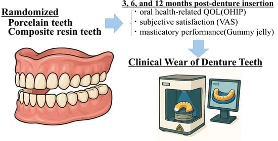
Background/Objectives: Artificial teeth in complete dentures are classified according to the materials used: porcelain (PO) or composite resin (CR). However, these materials’ effects on function, patient satisfaction, and quality of life (QOL), as well as occlusal wear, remain unclear. We compared PO
[...] Read more.
Background/Objectives: Artificial teeth in complete dentures are classified according to the materials used: porcelain (PO) or composite resin (CR). However, these materials’ effects on function, patient satisfaction, and quality of life (QOL), as well as occlusal wear, remain unclear. We compared PO and CR complete dentures in edentulous patients by assessing masticatory function, patient satisfaction, and oral health-related QOL at 3, 6, and 12 months post-insertion, as well as occlusal surface morphology owing to material differences. Methods: In this open-label, randomized, single-center, parallel-group study, participants were edentulous patients who visited our hospital and underwent treatment with new complete dentures. The outcomes were oral health-related QOL; subjective satisfaction, assessed using a visual analog scale; and masticatory performance, evaluated with gummy jelly and were assessed at baseline and 3, 6, and 12 months post-denture insertion. Occlusal surface impressions were taken twice, digitized as STL models, superimposed, and analyzed for wear. The Wilcoxon rank-sum test was used to compare between groups. Results: All evaluated items showed improvement. However, no significant differences were observed between the PO and CR groups, including between the amount of wear observed in the two groups. However, the PO group showed a tendency toward less wear. Extended observation may be required to clarify the long-term effects of artificial tooth materials. Conclusions: In the short term, the artificial tooth material did not influence masticatory function, oral health-related QOL, or patient satisfaction.
Full article
(This article belongs to the Section Dental Materials)
►▼
Show Figures
Graphical abstract
Open AccessSystematic Review
Effects of Oral Probiotics on Streptococcus mutans in Children: A Systematic Review and Meta-Analysis
by
Andrea Caiza-Rennella, Andrea Ordoñez-Balladares, Rosangela Caicedo-Quiroz, Indira Gómez-Capote and Zuilen Jiménez-Quintana
Dent. J. 2026, 14(2), 87; https://doi.org/10.3390/dj14020087 - 3 Feb 2026
Abstract
Background: Early childhood caries is closely associated with oral dysbiosis and the proliferation of Streptococcus mutans. Oral probiotics, particularly Lactobacillus reuteri and Lactobacillus rhamnosus, have been proposed as ecological modulators capable of reducing cariogenic microorganisms. Objective: To evaluate the
[...] Read more.
Background: Early childhood caries is closely associated with oral dysbiosis and the proliferation of Streptococcus mutans. Oral probiotics, particularly Lactobacillus reuteri and Lactobacillus rhamnosus, have been proposed as ecological modulators capable of reducing cariogenic microorganisms. Objective: To evaluate the efficacy of orally administered L. reuteri and L. rhamnosus in reducing salivary S. mutans levels in children aged 6 months to 12 years through a systematic review and meta-analysis. Methods: This review followed the PRISMA 2020 guidelines and was prospectively registered in PROSPERO (CRD420251086304). Searches were conducted in MEDLINE/PubMed, CENTRAL, Embase, Scopus and LILACS without language or date restrictions. Randomized controlled trials administering the target probiotic strains for ≥30 days were included. Risk of bias was assessed using RoB 2, and certainty of evidence using GRADE. Random-effects meta-analyses were performed for continuous and dichotomous outcomes. Results: Six randomized controlled trials were included (N = 1362). Only two trials reported continuous outcomes in comparable log10 CFU/mL format and could therefore be pooled for the continuous meta-analysis. This analysis showed a significant reduction in salivary S. mutans levels (MD = −0.65 log10 CFU/mL; 95% CI: −0.97 to −0.34; p < 0.0001; I2 = 19%), although the pooled estimate was largely driven by one study and should be interpreted cautiously. Four trials contributed to the dichotomous meta-analysis, which showed a non-significant trend toward risk reduction (OR = 0.73; 95% CI: 0.51–1.06; p = 0.10; I2 = 35%). Short-term interventions using high oral-retention formulations demonstrated the most consistent microbiological effects. Conclusions: Oral probiotics may significantly reduce salivary S. mutans in the short-term, especially when delivered through slow-dissolving formulations. However, their effects vary according to strain, vehicle, and intervention duration. Larger, standardized, and longer-term clinical trials are needed to determine the sustainability and clinical relevance of these effects.
Full article
(This article belongs to the Topic Oral Health Management and Disease Treatment)
►▼
Show Figures

Figure 1
Open AccessArticle
Does Palatoplasty in Patients with Cleft Palate Really Improve Otitis Media with Effusion?
by
Yosuke Kunitomi, Toshiki Hyodo, Yoshiaki Kitsukawa, Aya Koike, Yasuhiro Tsubura, Yuske Komiyama, Chonji Fukumoto, Takahiro Wakui, Hiroshi Kamioka and Hitoshi Kawamata
Dent. J. 2026, 14(2), 86; https://doi.org/10.3390/dj14020086 - 3 Feb 2026
Abstract
Background: The majority of cleft palate patients have been reported to suffer from otitis media with effusion (OME). The improvement of velopharyngeal function (VPF) after palatoplasty might be evidence for the improvement of the function of the Eustachian tube. The improvement of the
[...] Read more.
Background: The majority of cleft palate patients have been reported to suffer from otitis media with effusion (OME). The improvement of velopharyngeal function (VPF) after palatoplasty might be evidence for the improvement of the function of the Eustachian tube. The improvement of the function of Eustachian tube by palatoplasty has been reported to be effective for the treatment of OME simultaneously with the insertion of a ventilation tube into the tympanic membrane. There are only a few reports that clearly show the association between improvement of VPF and improvement of OME after palatoplasty. In this study, we discussed whether the improvement of VPF after palatoplasty in cleft palate patients with OME improved OME. Methods: Twenty-six patients with cleft palate were included in the study. We retrospectively extracted the information of cleft type, gender, surgical technique, and presence of OME risk factors from electronic medical records. We also investigated the recurrence of OME and the improvement level of VPF at 36 months postoperatively. OME was assessed based on the otolaryngologist’s findings in electronic medical records, with a good prognosis group with no symptom of OME, or a recurrence group with prolonged or recurrent OME. Results: At 36 months after palatoplasty, 19 of 23 patients (82.6%) were in the OME good prognosis group and four (17.4%) were in the OME recurrence group. The rate of patients with recurrent OME did not differ significantly by the degree of improvement of VPF. This study indicated that clear association between other risk factors for OME and OME recurrence could not be shown. Conclusion: We observed that most patients with cleft palate who underwent palatoplasty showed a good prognosis for OME at 36 months after surgery. However, further studies are needed to investigate the impact of different surgical techniques on the improvement of OME and the degree to which VPF improves, as well as the effect of each OME risk factor.
Full article
(This article belongs to the Special Issue Trends in Orofacial Cleft Research)
Open AccessArticle
The Effect of Adhesive Systems on Shade Matching of Composite Veneer
by
Fadak Al Marar, Raghad Aljarboua, Fatimah M. Alatiyyah, Shahad AlGhamdi, Faraz Ahmed Farooqi, Lama Almuhanna, Rasha AlSheikh and Abdul Samad Khan
Dent. J. 2026, 14(2), 85; https://doi.org/10.3390/dj14020085 - 3 Feb 2026
Abstract
►▼
Show Figures
Objective: This study aimed to assess the impact of different adhesive systems on the color stability of composite veneers following their exposure to various common beverages. Materials and Methods: A single layer of commercially available adhesives (4th and 7th generations) and two experimental
[...] Read more.
Objective: This study aimed to assess the impact of different adhesive systems on the color stability of composite veneers following their exposure to various common beverages. Materials and Methods: A single layer of commercially available adhesives (4th and 7th generations) and two experimental adhesives based on hydroxyapatite and bioactive glass were applied, followed by composite restoration on incisor typodonts. The typodonts were prepared with depths of 0.3, 0.5, and 0.7 mm at the cervical, middle, and incisal regions, respectively. Samples from each group were immersed in coffee, Cola, and deionized water, and color stability was analyzed on days 1 and 60. One-way and two-way analyses of variance were performed. Results: The interaction between groups and solutions was statistically significant (p = 0.001) across all tooth regions. Coffee and Cola caused significant color changes (p = 0.001). The 4th generation demonstrated better color stability than the 7th generation in the middle and cervical regions (p-values = 0.083 and 0.003, respectively). The findings showed that the bioactive glass-based bonding agent exhibited greater discoloration than the hydroxyapatite-based adhesive (p = 0.001). Conclusions: The composite thicknesses are influenced differently by adhesives with respect to shade matching. Bioactive materials-based adhesives showed more resistance towards color change than commercial adhesives.
Full article
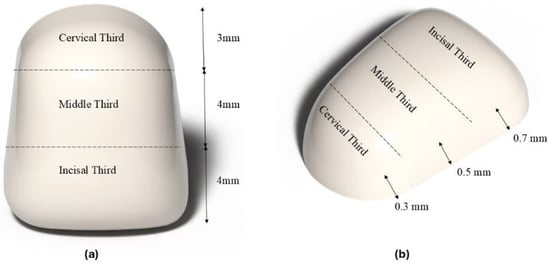
Figure 1
Open AccessArticle
Discrepancy Between Surface Wear and Subsurface Fatigue Damage in CAD/CAM Composite Crowns: A Comparative Study of Intraoral Scans and Optical Coherence Tomography
by
Julie-Jacqueline Kuhl, Maximiliane Amelie Schlenz, Bernd Wöstmann, Christin Grill, Ralf Brinkmann and Christoph Moos
Dent. J. 2026, 14(2), 84; https://doi.org/10.3390/dj14020084 - 3 Feb 2026
Abstract
Objectives: This study aimed to determine whether surface wear, identified through the superimposition of intraoral scans (IOS), can predict subsurface damage progression detected by optical coherence tomography (OCT) during fatigue testing of computer-aided design/computer-aided manufacturing (CAD/CAM) composite crowns. Methods: Monolithic CAD/CAM composite crowns
[...] Read more.
Objectives: This study aimed to determine whether surface wear, identified through the superimposition of intraoral scans (IOS), can predict subsurface damage progression detected by optical coherence tomography (OCT) during fatigue testing of computer-aided design/computer-aided manufacturing (CAD/CAM) composite crowns. Methods: Monolithic CAD/CAM composite crowns (Brilliant Crios;
(This article belongs to the Special Issue Optical Coherence Tomography (OCT) in Dentistry)
►▼
Show Figures

Graphical abstract
Open AccessArticle
Oral Side Effects of the Most Commonly Prescribed Drugs in Germany
by
Frank Halling, Rainer Lutz and Axel Meisgeier
Dent. J. 2026, 14(2), 83; https://doi.org/10.3390/dj14020083 - 2 Feb 2026
Abstract
Background: The aim of this study is to investigate the potential link between the use of specific medications and oral adverse drug reactions. Methods: The 100 most frequently prescribed drugs in Germany in 2023 were compiled using the “PharMaAnalyst” database. According to the
[...] Read more.
Background: The aim of this study is to investigate the potential link between the use of specific medications and oral adverse drug reactions. Methods: The 100 most frequently prescribed drugs in Germany in 2023 were compiled using the “PharMaAnalyst” database. According to the descriptions of adverse drug reactions (ADRs) in the patient information leaflets the ADRs were selected, analyzed and weighted with scores according to a classification system that distinguishes four groups of ADRs by frequency: ‘very common’ (4), ‘common’ (3), ‘uncommon’ (2) and ‘rare’ (1). The objective was to summarize the scores of the oral ADRs and define the ‘oral side effect score’ (OSES). Results: After accounting for duplication due to various brand names, 49 medications were reviewed. A total of 65% of the medications exhibited oral ADRs. The number of oral ADRs per medication ranged from one to seven. Xerostomia and dysgeusia were the most prevalent oral side effects, accounting for 37% of cases. Overall, 34% of side effects were classified as either ‘very common’ or ‘common’. The medication groups with the highest OSES were antidepressants, antibiotics and analgesics. Of the individual medications, azithromycin, gabapentin and pregabalin exhibited the highest OSES. Conclusions: This study provides a comprehensive overview of oral side effects associated with the 100 most frequently prescribed drugs. Patients with polypharmacy are particularly likely to experience oral side effects such as xerostomia and dysgeusia. Due to their high OSES combinations, antibiotics, analgesics or antidepressants may trigger multiple oral ADRs. It is essential that the medical community is continuously updated on pharmacological knowledge to raise awareness of oral ADRs.
Full article
(This article belongs to the Topic Oral Health Management and Disease Treatment)
►▼
Show Figures

Figure 1
Open AccessArticle
Enamel Remineralization Potential of Conventional and Biomimetic Toothpaste Formulations: A Comparative In Vitro Study
by
Cristina-Angela Ghiorghe, Ionuţ Tărăboanţă, Sorin Andrian, Galina Pancu, Corneliu Munteanu, Bogdan Istrate, Fabian Cezar Lupu, Claudia Maxim and Ana Simona Barna
Dent. J. 2026, 14(2), 82; https://doi.org/10.3390/dj14020082 - 2 Feb 2026
Abstract
Background/Objectives: Dental caries remains one of the most prevalent chronic diseases worldwide, making enamel remineralization a key objective in minimally invasive dentistry. This in vitro study compared the remineralization efficacy of five therapeutic toothpastes containing fluoride, NovaMin, CPP-ACP, nano-hydroxyapatite, arginine, and xylitol.
[...] Read more.
Background/Objectives: Dental caries remains one of the most prevalent chronic diseases worldwide, making enamel remineralization a key objective in minimally invasive dentistry. This in vitro study compared the remineralization efficacy of five therapeutic toothpastes containing fluoride, NovaMin, CPP-ACP, nano-hydroxyapatite, arginine, and xylitol. Methods: Sixty enamel specimens were prepared from extracted human posterior teeth and artificially demineralized. Samples were randomly allocated into six groups (n = 10): one negative control (C1) stored in artificial saliva and five treatment groups (P1–P5). A 28-day remineralization protocol with twice-daily applications was performed. Scanning electron microscopy (SEM) and energy-dispersive X-ray spectroscopy (EDX) were used to assess surface morphology and elemental composition (Ca, P, F, Na, O, Ca/P ratio) at days 1, 14, and 28. Vickers microhardness testing was used to evaluate changes in mechanical properties. Statistical analysis included one-way ANOVA, repeated measures ANOVA, Tukey’s post hoc test, and Kruskal–Wallis where appropriate (α = 0.05). Results: All therapeutic toothpastes produced some increase in mineral content compared to the demineralized control. At day 28, significant intergroup differences were observed for calcium, phosphorus, and fluoride (p < 0.001). The arginine–fluoride formulation (P4) and the NovaMin-based formulation (P3) showed the most consistent increases in Ca and P, with SEM revealing the formation of a continuous, compact surface layer and marked reduction in prismatic porosities. Fluoride-containing toothpastes (P1, P3, P4) showed significant fluoride incorporation (p < 0.001 vs. control). The nano-hydroxyapatite/xylitol prototype (P5) produced a delayed but progressive increase in Ca and P, with partial filling of prismatic spaces. The CPP-ACP-based toothpaste (P2) led to limited changes, with only slight differences vs. control at day 28. Vickers microhardness values increased significantly in groups P1, P3, P4, and P5 (p < 0.05), in agreement with the higher mineral levels found in these samples. Conclusions: Under the present in vitro conditions, toothpastes containing fluoride in combination with NovaMin or arginine, as well as nano-hydroxyapatite/xylitol, demonstrated the highest remineralization potential under the present in vitro conditions, both chemically and mechanically. Xylitol-based formulations without a direct mineral supply showed limited effects. The pH and active composition of the toothpaste strongly influenced enamel remineralization outcomes.
Full article
(This article belongs to the Section Preventive Dentistry)
►▼
Show Figures

Figure 1
Open AccessCase Report
Burning Mouth Syndrome as a Central Pain Disorder: A Case Study Demonstrating Response to Occipital Nerve Block Treatment
by
Shachar Zion Shemesh, Paz Kelmer and Lior Ungar
Dent. J. 2026, 14(2), 81; https://doi.org/10.3390/dj14020081 - 2 Feb 2026
Abstract
Background: Burning Mouth Syndrome (BMS) is a chronic orofacial pain condition characterized by a burning sensation in the oral cavity without identifiable lesions. It predominantly affects women (especially postmenopausal) but can occur in men. BMS is considered a multifactorial neuropathic pain disorder involving
[...] Read more.
Background: Burning Mouth Syndrome (BMS) is a chronic orofacial pain condition characterized by a burning sensation in the oral cavity without identifiable lesions. It predominantly affects women (especially postmenopausal) but can occur in men. BMS is considered a multifactorial neuropathic pain disorder involving both peripheral small-fiber neuropathy and central dysregulation, often accompanied by taste alterations (dysgusia) and xerostomia despite normal oral exams. Treatment is challenging, with modest responses to agents like clonazepam, tricyclic antidepressants, or gabapentinoids. Observations: We present a 67-year-old male with recalcitrant primary BMS who showed complete remission temporally associated with occipital nerve blockade, likely affecting central trigeminocervical pathways. Initial therapy with amitriptyline (25 mg) and gabapentin (900 mg/day) yielded ~30% pain relief. Given suspected central sensitization, greater and lesser occipital nerve (GON) blocks were administered in series. After the first, second, and third ON blocks, pain was reduced by ~50%, 80%, and 100%, respectively. Remission persisted at one-year follow-up under continued medications. A mild recurrence (~20% of baseline pain) responded fully to a fourth GON block, maintaining another year of pain-free status. Lessons: This case underscores the complex central mechanisms in BMS and illustrates that modulating central pain circuits via occipital nerve blockade, through trigeminocervical convergence mechanisms, without direct trigeminal intervention. We discuss the diagnostic challenges of BMS, the rationale of occipital neuromodulation, and how this novel therapeutic strategy compares with current literature, supporting the hypothesis of central sensitization in BMS.
Full article
Open AccessSystematic Review
Periodontitis and Oral and Oropharyngeal Cancer Risk: A Systematic Review and Meta-Analysis with Exploratory Evidence on Tumor-Associated Porphyromonas gingivalis
by
Luis Chauca-Bajaña, Bernarda Andrea Sánchez Arteaga, Andrea Ordóñez Balladares, María Isabel Romero Vasquez, Gustavo Javier Icaza Latorre, Carla Verenice Romo Olvera, Mauro Xavier Zambrano Matamoros and Byron Velásquez Ron
Dent. J. 2026, 14(2), 80; https://doi.org/10.3390/dj14020080 - 2 Feb 2026
Abstract
►▼
Show Figures
Background: Periodontitis is a chronic inflammatory condition characterized by progressive destruction of tooth-supporting tissues and sustained microbial dysbiosis. Increasing evidence suggests that chronic oral inflammation may be associated with oral and oropharyngeal carcinogenesis, although findings across epidemiological and prognostic studies remain heterogeneous. Objective:
[...] Read more.
Background: Periodontitis is a chronic inflammatory condition characterized by progressive destruction of tooth-supporting tissues and sustained microbial dysbiosis. Increasing evidence suggests that chronic oral inflammation may be associated with oral and oropharyngeal carcinogenesis, although findings across epidemiological and prognostic studies remain heterogeneous. Objective: To systematically evaluate the epidemiological association between clinically defined periodontitis and the risk of oral and/or oropharyngeal cancer, and to explore, in a distinct analytical component, the prognostic association between tumor-associated periodontal pathogens, particularly Porphyromonas gingivalis, and survival outcomes in affected patients. Methods: A systematic review and meta-analysis were conducted following PRISMA guidelines and registered in PROSPERO (CRD420251273975). Observational studies evaluating periodontitis and oral/oropharyngeal cancer risk (Arm 1) and prognostic studies assessing tumor-associated periodontal pathogens and survival outcomes (Arm 2) were identified through comprehensive database searches. Random-effects meta-analyses were performed to pool adjusted effect estimates. Risk of bias was assessed using the Newcastle–Ottawa Scale and the QUIPS tool. Results: Six observational studies were included in the epidemiological meta-analysis. Periodontitis was significantly associated with an increased risk of oral and/or oropharyngeal cancer (pooled HR = 2.14; 95% CI: 1.53–2.98), with substantial heterogeneity; trial sequential analysis supported the statistical robustness of this association. In the separate prognostic analysis, three studies evaluating intratumoral Porphyromonas gingivalis were included. A higher presence or expression of P. gingivalis was associated with poorer overall survival (HR = 2.89; 95% CI: 1.93–4.32), with no observed heterogeneity. Sensitivity and influence analyses confirmed the stability of these findings. Conclusions: This systematic review and meta-analysis demonstrate a consistent epidemiological association between periodontitis and an increased risk of oral and/or oropharyngeal cancer. In addition, exploratory prognostic evidence suggests that the presence of Porphyromonas gingivalis within tumor tissue may be associated with adverse survival outcomes. These findings should be interpreted as addressing distinct clinical and microbiological constructs, underscoring the need for further well-designed prospective and mechanistic studies.
Full article

Figure 1
Open AccessSystematic Review
Artificial Intelligence Models for the Detection and Quantification of Orthodontically Induced Root Resorption Using Cone-Beam Computed Tomography: A Systematic Review and Meta-Analysis
by
Carlos M. Ardila, Eliana Pineda-Vélez and Anny M. Vivares-Builes
Dent. J. 2026, 14(2), 79; https://doi.org/10.3390/dj14020079 - 2 Feb 2026
Abstract
Background/Objectives: Orthodontically induced root resorption (OIRR) is a well-documented but undesired consequence of orthodontic treatment. This systematic review and meta-analysis aimed to assess the diagnostic performance of artificial intelligence (AI) models applied to cone-beam computed tomography (CBCT) for detecting and quantifying OIRR
[...] Read more.
Background/Objectives: Orthodontically induced root resorption (OIRR) is a well-documented but undesired consequence of orthodontic treatment. This systematic review and meta-analysis aimed to assess the diagnostic performance of artificial intelligence (AI) models applied to cone-beam computed tomography (CBCT) for detecting and quantifying OIRR while evaluating their agreement with manual reference standards and the impact of model architecture, validation design, and quantification strategy. Methods: Comprehensive searches were conducted across PubMed/MEDLINE, Scopus, Web of Science, and EMBASE up to November 2025. Studies were included if they employed AI for OIRR diagnosis using CBCT and reported relevant performance metrics. Following PRISMA guidelines, data were extracted and a random-effect meta-analysis was performed. Subgroup analyses explored the influence of model design and validation. Results: Seven studies were included. Pooled sensitivity from three eligible studies was 0.903 (95% CI: 0.818–0.989), suggesting excellent true positive rates. Specificity ranged from 82% to 98%, and area under the receiver operating characteristic curve values reached up to 0.96 across studies using EfficientNet, U-Net, and other convolutional neural network (CNN)-based architectures. The pooled intraclass correlation coefficient for agreement with manual quantification was 1.000, reflecting near-perfect concordance. Subgroup analyzes showed slightly superior performance in CNN-only models compared to hybrid approaches, and better diagnostic metrics with internal validation. Linear assessments appeared more sensitive to early apical shortening than volumetric methods. Conclusions: AI models applied to CBCT demonstrate excellent diagnostic accuracy and high concordance with expert assessments for OIRR detection. These findings support their potential integration into clinical orthodontic workflows.
Full article
(This article belongs to the Special Issue Innovations and Trends in Modern Orthodontics)
►▼
Show Figures

Graphical abstract
Open AccessArticle
The Influence of Filler Morphology and Loading Level on the Properties of Light-Curing Dental Composites
by
Ekaterina Kuznetsova, Yaroslav Meleshkin, Oleg Yanushevich, Natella Krikheli, Elena Mendosa, Marina Bychkova and Pavel Peretyagin
Dent. J. 2026, 14(2), 78; https://doi.org/10.3390/dj14020078 - 2 Feb 2026
Abstract
►▼
Show Figures
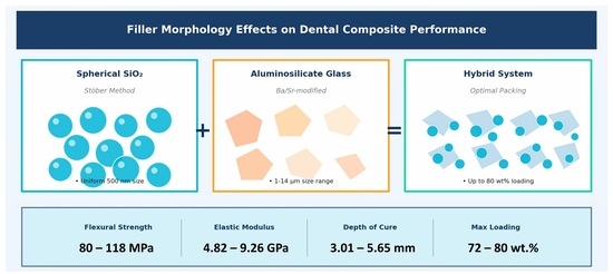
Background/Objectives: Light-curing dental resin composites remain limited by high polymerization shrinkage, inadequate wear resistance, and elevated water sorption. The combined influence of filler shape, size, and loading level on mechanical performance and hydrolytic stability remains insufficiently understood. This study aimed to systematically investigate
[...] Read more.
Background/Objectives: Light-curing dental resin composites remain limited by high polymerization shrinkage, inadequate wear resistance, and elevated water sorption. The combined influence of filler shape, size, and loading level on mechanical performance and hydrolytic stability remains insufficiently understood. This study aimed to systematically investigate the effects of filler morphology and particle size distribution on the key properties of dental composites. Methods: Spherical silica (SiO2) nanoparticles (D50 = 0.50 μm) were synthesized via the Stöber method, while irregular aluminosilicate glass was used in coarse (D50 = 3.71 μm) and fine (D50 = 1.98 μm) fractions. Three composite groups were formulated: Group 1 (72 wt.% filler with 0–30% SiO2), Group 2 (maximum filler loading 76–80 wt.% with 10–30% SiO2), and Group 3 (74.5 wt.% filler with varying coarse/fine glass ratios). Flexural strength, flexural modulus, Vickers microhardness, depth of cure, water sorption, and solubility were evaluated according to ISO 4049:2019. Results: Incorporation of spherical SiO2 nanoparticles significantly reduced composite viscosity, enabling maximum filler loading to increase from 72 to 80 wt.%. All composites exceeded ISO requirements for flexural strength (80.54–118.11 MPa), depth of cure (3.01–5.65 mm), water sorption (14.61–22.87 μg/mm3), and solubility (1.20–5.90 μg/mm3). The highest flexural strength (118.11 ± 10.54 MPa) and modulus (9.26 ± 1.12 GPa) were achieved at 78 wt.% filler loading. Bimodal glass systems (50/50 ratio) demonstrated optimal mechanical properties, while higher fine fractions reduced strength. Conclusions: Spherical SiO2 nanoparticles effectively reduce viscosity and enable higher filler loading. The optimal balance between filler loading, particle shape, and size distribution should be tailored to clinical requirements, with high-strength formulations suited for posterior restorations and bimodal formulations for universal applications.
Full article
Graphical abstract
Open AccessArticle
Evaluation of the Color Stability of Multilayer Zirconia After Exposure to Staining Solutions and Artificial Aging
by
Brunilda Koci, Alba Kamberi, Adora Shpati, Olja Tanellari, Balcos Carina and Adela Alushi
Dent. J. 2026, 14(2), 77; https://doi.org/10.3390/dj14020077 - 2 Feb 2026
Abstract
Background/Objectives: Multilayer zirconia restorations can feature a shade gradient or a strength gradient, with layers differing in color or phase composition within the same material. The aim of this in vitro study was to evaluate the color stability in all layers of multilayer
[...] Read more.
Background/Objectives: Multilayer zirconia restorations can feature a shade gradient or a strength gradient, with layers differing in color or phase composition within the same material. The aim of this in vitro study was to evaluate the color stability in all layers of multilayer zirconia after exposure to staining solutions and artificial aging. Methods: Square-shaped specimens (N = 120) of color A2 were fabricated from 4Y-PSZ and 3Y/4Y-PSZ multilayer zirconia—Katana STML, DD Cube One ML, and Katana YML—and their baseline color values (T0) were measured with a clinical spectrophotometer (VITA Easyshade V). The specimens were randomly divided into four groups (n = 10/gp) and immersed in physiologic solution, 0.2% chlorhexidine gluconate (CHX) mouth rinse, and staining coffee solution. Then, they were measured continuously for 7 (T1), 14 (T2), and 21 days (T3). The last group of specimens underwent accelerated aging in a steam autoclave at 134 °C and 2 bar pressure and measured after 1 (T1), 3 (T2), and 5 h (T3). After the immersion process and artificial aging, discoloration values (ΔE) were calculated using the formula ΔE = [(ΔL*)2 + (Δa*)2 + (Δb*)2]1/2 and analyzed with the SPSS v 23.0 software with a p value < 0.05. Results: All specimens showed significant color differences in the T3 measurements after exposure to coffee and CHX, with the highest ΔE values in the enamel layers. Katana YML showed the most significant differences in ΔE in the cervical layers after exposure to artificial aging. Conclusions: Multilayer zirconia exhibited dependent optical changes, with the enamel layers being the most affected after exposure to staining solutions. Gradient pigmentation and differences in phase composition caused differences in color to the multilayer zirconia layers after exposure to staining solutions and artificial aging.
Full article
(This article belongs to the Special Issue Advances in Esthetic Dentistry)
►▼
Show Figures
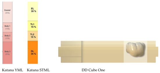
Figure 1
Open AccessArticle
Impact of Metallurgical and Geometric Features on the Cyclic Fatigue Strength of Reciprocating Endodontic Files
by
Abayomi Omokeji Baruwa, Francisco M. Braz Fernandes and Jorge N. R. Martins
Dent. J. 2026, 14(2), 76; https://doi.org/10.3390/dj14020076 - 2 Feb 2026
Abstract
Background: Nickel–titanium (NiTi) endodontic instruments have undergone significant improvements in heat treatment processing and geometric design, aimed at enhancing flexibility, cutting efficiency, and fatigue strength. Reciprocating motion was introduced to increase cyclic fatigue resistance, which remains the predominant mode of failure in NiTi
[...] Read more.
Background: Nickel–titanium (NiTi) endodontic instruments have undergone significant improvements in heat treatment processing and geometric design, aimed at enhancing flexibility, cutting efficiency, and fatigue strength. Reciprocating motion was introduced to increase cyclic fatigue resistance, which remains the predominant mode of failure in NiTi endodontic file systems. Although these instruments are widely used in both clinical practice and research, few comparative studies have integrated geometric, metallurgical and mechanical evaluations of the most commonly used reciprocating systems. Methods: In the present study, four single-file reciprocating NiTi systems (Reciproc Blue, WaveOne Gold, EdgeOne Fire, and Easy-File Flex) were evaluated for their geometric design, metallurgical composition, and cyclic fatigue strength. Stereomicroscopy and scanning electron microscopy were employed to assess active blade length, spiral configuration, and surface finish, while elemental composition and phase transformation temperatures were analyzed using energy-dispersive X-ray spectroscopy and differential scanning calorimetry. Ten instruments from each group were tested for cyclic fatigue using a standardized curved stainless-steel canal at room temperature, and the time to fracture was recorded. Fatigue data were statistically analyzed using Mood’s median test, with significance set at p < 0.05. Results: Reciproc Blue exhibited the longest active blade length, highest spiral density, and superior surface finish. R-phase start and finish temperatures were highest in WaveOne Gold and lowest in Easy-File Flex. Reciproc Blue demonstrated the higher cyclic fatigue strength, whereas Easy-File Flex showed the lowest. Conclusions: These findings suggest that the metallurgical and geometric characteristics of the Reciproc Blue file significantly enhance its strength to cyclic fatigue compared with the other instruments evaluated.
Full article
(This article belongs to the Special Issue Endodontics and Restorative Sciences: 2nd Edition)
►▼
Show Figures

Figure 1

Journal Menu
► ▼ Journal Menu-
- Dentistry Journal Home
- Aims & Scope
- Editorial Board
- Reviewer Board
- Topical Advisory Panel
- Instructions for Authors
- Special Issues
- Topics
- Sections & Collections
- Article Processing Charge
- Indexing & Archiving
- Editor’s Choice Articles
- Most Cited & Viewed
- Journal Statistics
- Journal History
- Journal Awards
- Conferences
- Editorial Office
Journal Browser
► ▼ Journal BrowserHighly Accessed Articles
Latest Books
E-Mail Alert
News
Topics
Topic in
Applied Sciences, Children, Dentistry Journal, JCM
Preventive Dentistry and Public Health
Topic Editors: Denis Bourgeois, Elena BardelliniDeadline: 30 April 2026
Topic in
Biology, JCM, Diagnostics, Dentistry Journal
Assessment of Craniofacial Morphology: Traditional Methods and Innovative Approaches
Topic Editors: Nikolaos Gkantidis, Carlalberta VernaDeadline: 1 June 2026
Topic in
Dentistry Journal, IJMS, JCM, Medicina, Applied Sciences
Oral Health Management and Disease Treatment
Topic Editors: Christos Rahiotis, Felice Lorusso, Sergio Rexhep TariDeadline: 31 July 2026
Topic in
Applied Sciences, Dentistry Journal, Polymers, Applied Biosciences, Bioengineering, Materials
Advances in Biomaterials—2nd Edition
Topic Editors: Satoshi Komasa, Yoshiro Tahara, Tohru Sekino, Hideaki Sato, Yoshiya Hashimoto, Tetsuya AdachiDeadline: 31 December 2026

Conferences
Special Issues
Special Issue in
Dentistry Journal
Endodontics: From Technique to Regeneration
Guest Editor: David E. JaramilloDeadline: 10 February 2026
Special Issue in
Dentistry Journal
Dental Public Health Landscape: Challenges, Technological Innovation and Opportunities in the 21st Century
Guest Editors: Radu Chifor, Aranka Ilea, Anca-Ștefania MesaroșDeadline: 15 February 2026
Special Issue in
Dentistry Journal
Feature Papers in Digital Dentistry
Guest Editors: Luigi Canullo, Maria Menini, Paolo PesceDeadline: 20 February 2026
Special Issue in
Dentistry Journal
Malocclusion: Treatments and Rehabilitation
Guest Editor: Teresa PinhoDeadline: 28 February 2026
Topical Collections
Topical Collection in
Dentistry Journal
Light and Laser Dentistry
Collection Editors: Samir Nammour, Aldo Brugnera Junior
Topical Collection in
Dentistry Journal
Novel Ceramic Materials in Dentistry
Collection Editors: Roberto Sorrentino, Gianrico Spagnuolo
Topical Collection in
Dentistry Journal
Bio-Logic Approaches to Implant Dentistry
Collection Editors: Luigi Canullo, Donato Antonacci, Piero Papi, Francesco Gianfreda, Bianca Di Murro, Carlo Raffone
Topical Collection in
Dentistry Journal
Dental Traumatology and Sport Dentistry
Collection Editor: Enrico Spinas


