Next Issue
Volume 12, November
Previous Issue
Volume 12, September


Biosensors, Volume 12, Issue 10 (October 2022) – 141 articles

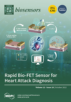
Cover Story (view full-size image): Cardiovascular disease is the number one cause of death worldwide. Heart attacks account for 85% of this high mortality. Unfortunately, a significant number of heart attack survivors die within the first year because the waiting times for a diagnosis are critical for these patients. The clinical diagnostic procedure is not yet suitable for an early diagnosis. This paper presents a field effect transistor (FET) biosensor for the rapid, sensitive quantification of heart attack biomarker detection. The developed bio-FET combines the use of an excellent semiconducting channel with a biocompatible nanosheet material, which provides a bridge between the solid-state device and the bio-recognition element. This new bio-FET demonstrated the sensitive determination of heart attack biomarkers within a few minutes using only a few drops of a sample. View this paper
  • Issues are regarded as officially published after their release is announced to the table of contents alert mailing list.
  • You may sign up for e-mail alerts to receive table of contents of newly released issues.
  • PDF is the official format for papers published in both, html and pdf forms. To view the papers in pdf format, click on the "PDF Full-text" link, and use the free Adobe Reader to open them.
Order results
Result details
Section
Select all
Export citation of selected articles as:
13 pages, 1619 KiB  
Article
Efficient Entrapment of Alpha-Synuclein Biotinylated Antibody in KCC-1-NH-CS2 and Application for the Sensitive Diagnosis of Parkinson’s Using Recognition of Biomarker: An Innovative Electrochemical Label-Free Immunosensor for the Biomedical Analysis of Neurodegenerative Diseases
by Hossein Navay Baghban, Mohammad Hasanzadeh, Yuqian Liu and Farzad Seidi
Biosensors 2022, 12(10), 911; https://doi.org/10.3390/bios12100911 - 21 Oct 2022
Cited by 4 | Viewed by 2179
Abstract
The early detection of Parkinson’s disease (PD) is a critical issue in terms of efficiency. Alpha-synuclein (α-Syn) is a biomarker in PD checks. Alpha-synuclein (α-syn) is the major constituent of Lewy bodies and a pathogenic hallmark of all synucleinopathies, including PDs, dementia with [...] Read more.
The early detection of Parkinson’s disease (PD) is a critical issue in terms of efficiency. Alpha-synuclein (α-Syn) is a biomarker in PD checks. Alpha-synuclein (α-syn) is the major constituent of Lewy bodies and a pathogenic hallmark of all synucleinopathies, including PDs, dementia with Lewy bodies, and multiple system atrophy. In this study, KCC-1-NH-CS2 was conjugated with biotinylated Ab and entrapped in P(β-CD) polymer cavities. Using this approach, a novel electrochemical label-free immunosensor was designed for the quantification of α-syn in real human samples. For this purpose, the glassy carbon electrode electropolymerized with P(β-CD) biopolymer provided an excellent matrix for entrapping of KCC-1-NH-CS2 loaded with the biotinylated antibody of α-syn. Using the chronoamperometric technique, the proposed immunosensor shows a suitable range of 0.02 to 64 ng/mL for the determination of α-syn. Additionally, a low limit of quantification of the engineered biosensor was obtained at 0.02 ng/mL. The developed immunosensor’s adequate stability, sensitivity, and selectivity, together with its ease of manufacture, make it a promising diagnostic technique for further research. This study also will pave the way for further applications of the synergetic effect of β-CD and KCC-1-NH-CS2 for biomedical analysis in the near future. Full article
(This article belongs to the Special Issue Advances in Amplification Methods for Biosensors)
Show Figures

Figure 1

78 pages, 16259 KiB  
Review
Graphene and Its Derivatives: Synthesis and Application in the Electrochemical Detection of Analytes in Sweat
by Anoop Singh, Aamir Ahmed, Asha Sharma and Sandeep Arya
Biosensors 2022, 12(10), 910; https://doi.org/10.3390/bios12100910 - 21 Oct 2022
Cited by 28 | Viewed by 6465
Abstract
Wearable sensors and invasive devices have been studied extensively in recent years as the demand for real-time human healthcare applications and seamless human–machine interaction has risen exponentially. An explosion in sensor research throughout the globe has been ignited by the unique features such [...] Read more.
Wearable sensors and invasive devices have been studied extensively in recent years as the demand for real-time human healthcare applications and seamless human–machine interaction has risen exponentially. An explosion in sensor research throughout the globe has been ignited by the unique features such as thermal, electrical, and mechanical properties of graphene. This includes wearable sensors and implants, which can detect a wide range of data, including body temperature, pulse oxygenation, blood pressure, glucose, and the other analytes present in sweat. Graphene-based sensors for real-time human health monitoring are also being developed. This review is a comprehensive discussion about the properties of graphene, routes to its synthesis, derivatives of graphene, etc. Moreover, the basic features of a biosensor along with the chemistry of sweat are also discussed in detail. The review mainly focusses on the graphene and its derivative-based wearable sensors for the detection of analytes in sweat. Graphene-based sensors for health monitoring will be examined and explained in this study as an overview of the most current innovations in sensor designs, sensing processes, technological advancements, sensor system components, and potential hurdles. The future holds great opportunities for the development of efficient and advanced graphene-based sensors for the detection of analytes in sweat. Full article
Show Figures

Graphical abstract

12 pages, 3748 KiB  
Article
Glucose Incorporated Graphite Matrix for Electroanalysis of Trimethoprim
by Rakesh R. Sawkar, Mahesh M. Shanbhag, Suresh M. Tuwar, Ravindra S. Veerapur and Nagaraj P. Shetti
Biosensors 2022, 12(10), 909; https://doi.org/10.3390/bios12100909 - 21 Oct 2022
Cited by 4 | Viewed by 1903
Abstract
The antibiotic drug trimethoprim (TMP) is used to treat bacterial infections in humans and animals, and frequently TMP is used along with sulfonamides. However, a large portion of TMP is excreted in its active state, which poses a severe problem to humans and [...] Read more.
The antibiotic drug trimethoprim (TMP) is used to treat bacterial infections in humans and animals, and frequently TMP is used along with sulfonamides. However, a large portion of TMP is excreted in its active state, which poses a severe problem to humans and the environment. A sensitive, rapid, cost-effective analytical tool is required to monitor the TMP concentration in biological and environmental samples. Hence, this study proposed an analytical methodology to analyze TMP in clinical, biological and environmental samples. The investigations were carried out using a glucose-modified carbon paste electrode (G-CPE) employing voltammetric techniques. Electrochemical behavior was examined with 0.5 mM TMP solution at optimum pH 3.4 (Phosphate Buffer Solution, I = 0.2 M). The influence of scan rate on the electro-oxidation of TMP was studied within the range of 0.05 to 0.55 V/s. The effect of pH and scan rate variations revealed proton transfer during oxidation. Moreover, diffusion phenomena governed the irreversibility of the electrode reaction. A probable and suitable electrode interaction and reaction mechanism was proposed for the electrochemical oxidation of TMP. Further, the TMP was quantitatively estimated with the differential pulse voltammetry (DPV) technique in the concentration range from 9.0 × 10⁻⁷ to 1.0 × 10⁻⁴ M. The tablet, spiked water and urine analysis demonstrated that the selected method and developed electrode were rapid, simple, sensitive, and cost-effective. Full article
(This article belongs to the Special Issue Electrochemical (Bio-) Sensors in Biological Applications)
Show Figures

Figure 1

14 pages, 3570 KiB  
Article
A Novel Activated Biochar-Based Immunosensor for Rapid Detection of E. coli O157:H7
by Abdus Sobhan, Fei Jia, Lisa Cooney Kelso, Sonatan Kumar Biswas, Kasiviswanathan Muthukumarappan, Changyong Cao, Lin Wei and Yanbin Li
Biosensors 2022, 12(10), 908; https://doi.org/10.3390/bios12100908 - 21 Oct 2022
Cited by 9 | Viewed by 2396
Abstract
E. coli O157:H7, one of the major foodborne pathogens, can cause a significant threat to the safety of foods. The aim of this research is to develop an activated biochar-based immunosensor that can rapidly detect E. coli O157:H7 cells without incubation in pure [...] Read more.
E. coli O157:H7, one of the major foodborne pathogens, can cause a significant threat to the safety of foods. The aim of this research is to develop an activated biochar-based immunosensor that can rapidly detect E. coli O157:H7 cells without incubation in pure culture. Biochar was developed from corn stalks using proprietary reactors and then activated using steam-activation treatment. The developed activated biochar presented an enhanced surface area of 830.78 m2/g. To develop the biosensor, the gold electrode of the sensor was first coated with activated biochar and then functionalized with streptavidin as a linker and further immobilized with biotin-labeled anti-E. coli polyclonal antibodies (pAbs). The optimum concentration of activated biochar for sensor development was determined to be 20 mg/mL. Binding of anti-E. coli pAbs with E. coli O157:H7 resulted in a significant increase in impedance amplitude from 3.5 to 8.5 kΩ when compared to an only activated biochar-coated electrode. The developed immunosensor was able to detect E. coli O157:H7 cells with a limit of detection of 4 log CFU/mL without incubation. Successful binding of E. coli O157:H7 onto an activated biochar-based immunosensor was observed on the microelectrode surface in scanning electron microscopy (SEM) images. Full article
(This article belongs to the Section Biosensor and Bioelectronic Devices)
Show Figures

Figure 1

14 pages, 2171 KiB  
Article
Trace Determination of Grouper Nervous Necrosis Virus in Contaminated Larvae and Pond Water Samples Using Label-Free Fiber Optic Nanoplasmonic Biosensor
by Yuan-Yu Chen, Chih-Lu Wu, Chia-Wei Hsu, Chih-Hui Wang, Chung-Rui Su, Chun-Jen Huang, Hau-Ren Chen, Lai-Kwan Chau and Shau-Chun Wang
Biosensors 2022, 12(10), 907; https://doi.org/10.3390/bios12100907 - 21 Oct 2022
Cited by 2 | Viewed by 1889
Abstract
We developed a fast (<20 min), label-free fiber optic particle plasmon resonance (FOPPR) immunosensing method to detect nervous necrosis virus (NNV), which often infects high-value economic aquatic species, such as grouper. Using spiked NNV particles in a phosphate buffer as samples, the standard [...] Read more.
We developed a fast (<20 min), label-free fiber optic particle plasmon resonance (FOPPR) immunosensing method to detect nervous necrosis virus (NNV), which often infects high-value economic aquatic species, such as grouper. Using spiked NNV particles in a phosphate buffer as samples, the standard calibration curve obtained was linear (R2 = 0.99) and the limit of detection (LOD) achieved was 2.75 × 10⁴ TCID50/mL, which is superior to that obtained using enzyme-linked immunosorbent assay (ELISA). By using an enhancement method called fiber optic nanogold-linked immunosorbent assay (FONLISA), the LOD can be further improved to <1 TCID50/mL, which is comparable to that found by the conventional qPCR method. Employing the larvae homogenate samples of NNV-infected grouper, the results obtained by the FOPPR biosensor agree with those obtained by the quantitative polymerase chain reaction (qPCR) method. We also examined pond water samples from an infected container in an indoor aquaculture facility. The lowest detectable level of NNV coat protein was found to be 0.17 μg/mL, which is one order lower than the LOD reported by ELISA. Therefore, we demonstrated the potential of the FOPPR biosensor as an outbreak surveillance tool, which is able to give warning indication even when the trend of larvae death toll increment is still not clear. Full article
(This article belongs to the Special Issue Waveguide Biosensors)
Show Figures

Figure 1

14 pages, 36650 KiB  
Communication
An Innovative Simple Electrochemical Levofloxacin Sensor Assembled from Carbon Paste Enhanced with Nano-Sized Fumed Silica
by Amany M. Fekry
Biosensors 2022, 12(10), 906; https://doi.org/10.3390/bios12100906 - 21 Oct 2022
Cited by 12 | Viewed by 2098
Abstract
A new electrochemical sensor for the detection of levofloxacin (LV) was efficiently realized. The aim was to develop a new, cheap, and simple sensor for the detection of LV, which is used in various infections due to its pharmacological importance. It consists of [...] Read more.
A new electrochemical sensor for the detection of levofloxacin (LV) was efficiently realized. The aim was to develop a new, cheap, and simple sensor for the detection of LV, which is used in various infections due to its pharmacological importance. It consists of carbon paste (CP) enhanced with nano-sized fumed silica (NFS). NFS has a very low bulk density and a large surface area. The carbon paste-enhanced NFS electrode (NFS/CPE) showed great electrocatalytic activity in the oxidation of 1.0 mM LV in Britton–Robinson buffer (BR) at pH values ranging from 3.0 to 8.0. Cyclic voltammetry (CV) and differential pulse voltammetry (DPV) were used; the peak current value (Ip) of the NFS/CPE sensor was 2.7 times that of the bare electrode, ensuring its high electrocatalytic activity. Electrochemical impedance spectroscopy (EIS) was performed at a peak potential (Ep) of +1066 mV, yielding a resistance of 10 kΩ for the designed NFS/CPE sensor compared to 2461 kΩ for the bare electrode, indicating the high conductivity of the modified sensor and verifying the data observed using the CV technique. Surface descriptions were determined by scanning electron microscopy (SEM) and energy dispersive X-ray analysis (EDX). The variation in the concentration of LV (2.0 to 1000 µM) was considered in BR buffer (pH = 5.0) at a scan rate (SR) of 10 mV/s by the NFS/CPE. The detection and quantification limits were 0.09 µM and 0.30 µM, respectively. To evaluate the application of LV in real samples, this procedure was established on Quinostarmax 500 mg tablets and human plasma samples. Reasonable results were obtained for the detection of LV. Full article
Show Figures

Figure 1

22 pages, 2405 KiB  
Review
Bacteriophage-Based Biosensors: A Platform for Detection of Foodborne Bacterial Pathogens from Food and Environment
by Rashad R. Al-Hindi, Addisu D. Teklemariam, Mona G. Alharbi, Ibrahim Alotibi, Sheren A. Azhari, Ishtiaq Qadri, Turki Alamri, Steve Harakeh, Bruce M. Applegate and Arun K. Bhunia
Biosensors 2022, 12(10), 905; https://doi.org/10.3390/bios12100905 - 21 Oct 2022
Cited by 25 | Viewed by 5814
Abstract
Foodborne microorganisms are an important cause of human illness worldwide. Two-thirds of human foodborne diseases are caused by bacterial pathogens throughout the globe, especially in developing nations. Despite enormous developments in conventional foodborne pathogen detection methods, progress is limited by the assay complexity [...] Read more.
Foodborne microorganisms are an important cause of human illness worldwide. Two-thirds of human foodborne diseases are caused by bacterial pathogens throughout the globe, especially in developing nations. Despite enormous developments in conventional foodborne pathogen detection methods, progress is limited by the assay complexity and a prolonged time-to-result. The specificity and sensitivity of assays for live pathogen detection may also depend on the nature of the samples being analyzed and the immunological or molecular reagents used. Bacteriophage-based biosensors offer several benefits, including specificity to their host organism, the detection of only live pathogens, and resistance to extreme environmental factors such as organic solvents, high temperatures, and a wide pH range. Phage-based biosensors are receiving increasing attention owing to their high degree of accuracy, specificity, and reduced assay times. These characteristics, coupled with their abundant supply, make phages a novel bio-recognition molecule in assay development, including biosensors for the detection of foodborne bacterial pathogens to ensure food safety. This review provides comprehensive information about the different types of phage-based biosensor platforms, such as magnetoelastic sensors, quartz crystal microbalance, and electrochemical and surface plasmon resonance for the detection of several foodborne bacterial pathogens from various representative food matrices and environmental samples. Full article
(This article belongs to the Special Issue Microbial Biosensors for Environmental Monitoring)
Show Figures

Figure 1

13 pages, 4305 KiB  
Article
A Paper-Based Electrochemical Sensor Based on PtNP/COFTFPB−DHzDS@rGO for Sensitive Detection of Furazolidone
by Rongfang Chen, Xia Peng, Yonghai Song and Yan Du
Biosensors 2022, 12(10), 904; https://doi.org/10.3390/bios12100904 - 21 Oct 2022
Cited by 11 | Viewed by 1938
Abstract
Herein, a paper-based electrochemical sensor based on PtNP/COFTFPB−DHzDS@rGO was developed for the sensitive detection of furazolidone. A cluster-like covalent organic framework (COFTFPB−DHzDS) was successfully grown on the surface of amino-functional reduced graphene oxide (rGO-NH2) to avoid serious [...] Read more.
Herein, a paper-based electrochemical sensor based on PtNP/COFTFPB−DHzDS@rGO was developed for the sensitive detection of furazolidone. A cluster-like covalent organic framework (COFTFPB−DHzDS) was successfully grown on the surface of amino-functional reduced graphene oxide (rGO-NH2) to avoid serious self-aggregation, which was further loaded with platinum nanoparticles (PtNPs) with high catalytic activity as nanozyme to obtain PtNP/COFTFPB−DHzDS@rGO nanocomposites. The morphology of PtNP/COFTFPB−DHzDS@rGO nanocomposites was characterized, and the results showed that the smooth rGO surface became extremely rough after the modification of COFTFPB−DHzDS. Meanwhile, ultra-small PtNPs with sizes of around 1 nm were precisely anchored on COFTFPB−DHzDS to maintain their excellent catalytic activity. The conventional electrodes were used to detect furazolidone and showed a detection limit as low as 5 nM and a linear range from 15 nM to 110 μM. In contrast, the detection limit for the paper-based electrode was 0.23 μM, and the linear range was 0.69–110 μM. The results showed that the paper-based electrode can be used to detect furazolidone. This sensor is a potential candidate for the detection of furazolidone residue in human serum and fish samples. Full article
(This article belongs to the Special Issue Paper-Based Biosensors)
Show Figures

Figure 1

10 pages, 2428 KiB  
Article
Probing Subcellular Iron Availability with Genetically Encoded Nitric Oxide Biosensors
by Gulsah Sevimli, Amy E. Alston, Felix Funk, Beat Flühmann, Roland Malli, Wolfgang F. Graier and Emrah Eroglu
Biosensors 2022, 12(10), 903; https://doi.org/10.3390/bios12100903 - 21 Oct 2022
Cited by 3 | Viewed by 3707
Abstract
Cellular iron supply is required for various biochemical processes. Measuring bioavailable iron in cells aids in obtaining a better understanding of its biochemical activities but is technically challenging. Existing techniques have several constraints that make precise localization difficult, and the lack of a [...] Read more.
Cellular iron supply is required for various biochemical processes. Measuring bioavailable iron in cells aids in obtaining a better understanding of its biochemical activities but is technically challenging. Existing techniques have several constraints that make precise localization difficult, and the lack of a functional readout makes it unclear whether the tested labile iron is available for metalloproteins. Here, we use geNOps; a ferrous iron-dependent genetically encoded fluorescent nitric oxide (NO) biosensor, to measure available iron in cellular locales. We exploited the nitrosylation-dependent fluorescence quenching of geNOps as a direct readout for cellular iron absorption, distribution, and availability. Our findings show that, in addition to ferrous iron salts, the complex of iron (III) with N,N’-bis (2-hydroxybenzyl)ethylenediamine-N,N’-diacetic acid (HBED) can activate the iron (II)-dependent NO probe within intact cells. Cell treatment for only 20 min with iron sucrose was also sufficient to activate the biosensor in the cytosol and mitochondria significantly; however, ferric carboxymaltose failed to functionalize the probe, even after 2 h of cell treatment. Our findings show that the geNOps approach detects available iron (II) in cultured cells and can be applied to assay functional iron (II) at the (sub)cellular level. Full article
(This article belongs to the Special Issue Genetically Encoded Biosensors for Biomedical Applications)
Show Figures

Figure 1

12 pages, 2796 KiB  
Article
Glycated Hemoglobin Electrochemical Immunosensor Based on Screen-Printed Electrode
by Yuliang Zhao, Hongyu Zhang, Yang Li, Xiaoai Wang, Liang Zhao, Jianghong Xu, Zhikun Zhan, Guanglie Zhang and Wen Jung Li
Biosensors 2022, 12(10), 902; https://doi.org/10.3390/bios12100902 - 21 Oct 2022
Cited by 2 | Viewed by 2770
Abstract
An electrochemical HbA1c sensor with high sensitivity and good specificity is proposed based on the electrochemical immune principle. The reproducibility and conductivity of the electrode are improved by depositing gold nanoparticles (AuNPs) on the surface of the screen-printed electrode (SPE). The HbA1c antibodies [...] Read more.
An electrochemical HbA1c sensor with high sensitivity and good specificity is proposed based on the electrochemical immune principle. The reproducibility and conductivity of the electrode are improved by depositing gold nanoparticles (AuNPs) on the surface of the screen-printed electrode (SPE). The HbA1c antibodies are immobilized on the surface of the modified electrode by adsorption to capture the HbA1c in the sample. The hindering effect of HbA1c on the electrode transfer reaction was exploited as the HbA1c detection mechanism. The electrode’s properties were characterized by electrochemical impedance spectroscopy (EIS), and the measurement properties of the electrode were analyzed using differential pulse voltammetry (DPV) and cyclic voltammetry (CV). The experimental results show that the peak current signal of the electrochemical immunosensor produced a linear response to HbA1c in the concentration range of 20–200 μg/mL, a linear relationship coefficient of 0.9812, a detection limit of 15.5 µg/mL, and a sensitivity of 0.0938 µA/µg·mL−1. The sensor delivered satisfactory repeatability, stability, and anti-interference performance. Due to its small size, high sensitivity, and wide linear detection range, it is expected to play a significant role in managing diabetes at home. Full article
(This article belongs to the Special Issue Biosensors for Bioanalytical and Healthcare Applications)
Show Figures

Figure 1

33 pages, 3655 KiB  
Systematic Review
Image Quality Improvement Techniques and Assessment Adequacy in Clinical Optoacoustic Imaging: A Systematic Review
by Ioannis Dimaridis, Patmaa Sridharan, Vasilis Ntziachristos, Angelos Karlas and Leontios Hadjileontiadis
Biosensors 2022, 12(10), 901; https://doi.org/10.3390/bios12100901 - 20 Oct 2022
Cited by 6 | Viewed by 3001
Abstract
Optoacoustic imaging relies on the detection of optically induced acoustic waves to offer new possibilities in morphological and functional imaging. As the modality matures towards clinical application, research efforts aim to address multifactorial limitations that negatively impact the resulting image quality. In an [...] Read more.
Optoacoustic imaging relies on the detection of optically induced acoustic waves to offer new possibilities in morphological and functional imaging. As the modality matures towards clinical application, research efforts aim to address multifactorial limitations that negatively impact the resulting image quality. In an endeavor to obtain a clear view on the limitations and their effects, as well as the status of this progressive refinement process, we conduct an extensive search for optoacoustic image quality improvement approaches that have been evaluated with humans in vivo, thus focusing on clinically relevant outcomes. We query six databases (PubMed, Scopus, Web of Science, IEEE Xplore, ACM Digital Library, and Google Scholar) for articles published from 1 January 2010 to 31 October 2021, and identify 45 relevant research works through a systematic screening process. We review the identified approaches, describing their primary objectives, targeted limitations, and key technical implementation details. Moreover, considering comprehensive and objective quality assessment as an essential prerequisite for the adoption of such approaches in clinical practice, we subject 36 of the 45 papers to a further in-depth analysis of the reported quality evaluation procedures, and elicit a set of criteria with the intent to capture key evaluation aspects. Through a comparative criteria-wise rating process, we seek research efforts that exhibit excellence in quality assessment of their proposed methods, and discuss features that distinguish them from works with similar objectives. Additionally, informed by the rating results, we highlight areas with improvement potential, and extract recommendations for designing quality assessment pipelines capable of providing rich evidence. Full article
(This article belongs to the Special Issue Optical Imaging and Biophotonic Sensors (OIBS))
Show Figures

Figure 1

14 pages, 3431 KiB  
Article
A Closed-Loop Approach to Fight Coronavirus: Early Detection and Subsequent Treatment
by Guoguang Rong, Yuqiao Zheng, Xi Yang, Kangjian Bao, Fen Xia, Huihui Ren, Sumin Bian, Lan Li, Bowen Zhu and Mohamad Sawan
Biosensors 2022, 12(10), 900; https://doi.org/10.3390/bios12100900 - 20 Oct 2022
Cited by 3 | Viewed by 2351
Abstract
The recent COVID-19 pandemic has caused tremendous damage to the social economy and people’s health. Some major issues fighting COVID-19 include early and accurate diagnosis and the shortage of ventilator machines for critical patients. In this manuscript, we describe a novel solution to [...] Read more.
The recent COVID-19 pandemic has caused tremendous damage to the social economy and people’s health. Some major issues fighting COVID-19 include early and accurate diagnosis and the shortage of ventilator machines for critical patients. In this manuscript, we describe a novel solution to deal with COVID-19: portable biosensing and wearable photoacoustic imaging for early and accurate diagnosis of infection and magnetic neuromodulation or minimally invasive electrical stimulation to replace traditional ventilation. The solution is a closed-loop system in that the three modules are integrated together and form a loop to cover all-phase strategies for fighting COVID-19. The proposed technique can guarantee ubiquitous and onsite detection, and an electrical hypoglossal stimulator can be more effective in helping severe patients and reducing complications caused by ventilators. Full article
Show Figures

Figure 1

13 pages, 3417 KiB  
Article
A Novel Paper-Based Electrochemical Biosensor Based on N,O-Rich Covalent Organic Frameworks for Carbaryl Detection
by Yawen Xiao, Na Wu, Li Wang and Lili Chen
Biosensors 2022, 12(10), 899; https://doi.org/10.3390/bios12100899 - 20 Oct 2022
Cited by 10 | Viewed by 2172
Abstract
A new N,O-rich covalent organic framework (COFDHNDA-BTH) was synthesized by an amine-aldehyde condensation reaction between 2,6-dialdehyde-1,5-dihydroxynaphthalene (DHNDA) and 1,3,5-phenyltriformylhydrazine (BTH) for carbaryl detection. The free NH, OH, and C=O groups of COFDHNDA-BTH not only covalently couples with acetylcholinesterase (AChE) into [...] Read more.
A new N,O-rich covalent organic framework (COFDHNDA-BTH) was synthesized by an amine-aldehyde condensation reaction between 2,6-dialdehyde-1,5-dihydroxynaphthalene (DHNDA) and 1,3,5-phenyltriformylhydrazine (BTH) for carbaryl detection. The free NH, OH, and C=O groups of COFDHNDA-BTH not only covalently couples with acetylcholinesterase (AChE) into the pores of COFDHNDA-BTH, but also greatly improves the catalytic activity of AChE in the constrained environment of COFDHNDA-BTH’s pore. Under the catalysis of AChE, the acetylthiocholine (ATCl) was decomposed into positively charged thiocholine (TCl), which was captured on the COFDHNDA-BTH modified electrode. The positive charges of TCl can attract anionic probe [Fe(CN)6]3−/4− on the COFDHNDA-BTH-modified electrode to show a good oxidation peak at 0.25 V (versus a saturated calomel electrode). The carbaryl detection can inhibit the activity of AChE, resulting in the decrease in the oxidation peak. Therefore, a turn-off electrochemical carbaryl biosensor based on a flexible carbon paper electrode loaded with COFDHNDA-BTH and AChE was constructed using the oxidation peak of an anionic probe [Fe(CN)6]3−/4− as the detection signal. The detection limit was 0.16 μM (S/N = 3), and the linear range was 0.48~35.0 μM. The sensor has good selectivity, repeatability, and stability, and has a good application prospect in pesticide detection. Full article
(This article belongs to the Special Issue Paper-Based Biosensors)
Show Figures

Figure 1

25 pages, 2733 KiB  
Review
Advances in Biosensing Technologies for Diagnosis of COVID-19
by Sulaiman Alsalameh, Khalid Alnajjar, Tariq Makhzoum, Noor Al Eman, Ismail Shakir, Tanveer Ahmad Mir, Khaled Alkattan, Raja Chinnappan and Ahmed Yaqinuddin
Biosensors 2022, 12(10), 898; https://doi.org/10.3390/bios12100898 - 20 Oct 2022
Cited by 12 | Viewed by 3635
Abstract
The COVID-19 pandemic has severely impacted normal human life worldwide. Due to its rapid community spread and high mortality statistics, the development of prompt diagnostic tests for a massive number of samples is essential. Currently used traditional methods are often expensive, time-consuming, laboratory-based, [...] Read more.
The COVID-19 pandemic has severely impacted normal human life worldwide. Due to its rapid community spread and high mortality statistics, the development of prompt diagnostic tests for a massive number of samples is essential. Currently used traditional methods are often expensive, time-consuming, laboratory-based, and unable to handle a large number of specimens in resource-limited settings. Because of its high contagiousness, efficient identification of SARS-CoV-2 carriers is crucial. As the advantages of adopting biosensors for efficient diagnosis of COVID-19 increase, this narrative review summarizes the recent advances and the respective reasons to consider applying biosensors. Biosensors are the most sensitive, specific, rapid, user-friendly tools having the potential to deliver point-of-care diagnostics beyond traditional standards. This review provides a brief introduction to conventional methods used for COVID-19 diagnosis and summarizes their advantages and disadvantages. It also discusses the pathogenesis of COVID-19, potential diagnostic biomarkers, and rapid diagnosis using biosensor technology. The current advancements in biosensing technologies, from academic research to commercial achievements, have been emphasized in recent publications. We covered a wide range of topics, including biomarker detection, viral genomes, viral proteins, immune responses to infection, and other potential proinflammatory biomolecules. Major challenges and prospects for future application in point-of-care settings are also highlighted. Full article
Show Figures

Figure 1

12 pages, 2809 KiB  
Article
A High-Performance Self-Supporting Electrochemical Biosensor to Detect Aflatoxin B1
by Yunfei Zhang, Tingting Lin, Yi Shen and Hongying Li
Biosensors 2022, 12(10), 897; https://doi.org/10.3390/bios12100897 - 20 Oct 2022
Cited by 9 | Viewed by 2359
Abstract
High-performance electrochemical biosensors for the rapid detection of aflatoxin B1 (AFB1) are urgently required in the food industry. Herein, a multi-scaled electrochemical biosensor was fabricated by assembling carboxylated polystyrene nanospheres, an aptamer and horseradish peroxidase into a free-standing carbon nanofiber/carbon felt [...] Read more.
High-performance electrochemical biosensors for the rapid detection of aflatoxin B1 (AFB1) are urgently required in the food industry. Herein, a multi-scaled electrochemical biosensor was fabricated by assembling carboxylated polystyrene nanospheres, an aptamer and horseradish peroxidase into a free-standing carbon nanofiber/carbon felt support. The resulting electrochemical biosensor possessed an exceptional performance, owing to the unique structures as well as the synergistic effects of the components. The 3D porous carbon nanofiber/carbon felt support served as an ideal substrate, owing to the excellent conductivity and facile diffusion of the reactants. The integration of carboxylated polystyrene nanospheres with horseradish peroxidase was employed as a signal amplification probe to enhance the electrochemical responses via catalyzing the decomposition of hydrogen peroxide. With the aid of the aptamer, the prepared sensors could quantitatively detect AFB1 in wine and soy sauce samples via differential pulse voltammetry. The recovery rates of AFB1 in the samples were between 87.53% and 106.71%. The limit of detection of the biosensors was 0.016 pg mL−1. The electrochemical biosensors also had excellent sensitivity, reproducibility, specificity and stability. The synthetic strategy reported in this work could pave a new route to fabricate high-performance electrochemical biosensors for the detection of mycotoxins. Full article
Show Figures

Figure 1

11 pages, 1426 KiB  
Article
A Lateral Flow Device for Point-of-Care Detection of Doxorubicin
by Tania Pomili, Francesca Gatto and Pier Paolo Pompa
Biosensors 2022, 12(10), 896; https://doi.org/10.3390/bios12100896 - 19 Oct 2022
Cited by 4 | Viewed by 1994
Abstract
A simple, rapid, and sensitive point-of-care (POC) device for the on-site detection of doxorubicin was developed. The proposed method relies on the naked-eye detection of the intrinsic fluorescence of the drug in a lateral flow device (LFD) configuration, exploiting the biological recognition of [...] Read more.
A simple, rapid, and sensitive point-of-care (POC) device for the on-site detection of doxorubicin was developed. The proposed method relies on the naked-eye detection of the intrinsic fluorescence of the drug in a lateral flow device (LFD) configuration, exploiting the biological recognition of DNA probes and avoiding the use of expensive antibodies and sophisticated instrumentations. The POC assay does not require any pre-treatment or purification step and provides an immediate visual readout, achieving a limit of detection as low as ca. 1 ng doxorubicin, outperforming several laboratory-based instrumental techniques. The POC method was proven useful for the detection of trace amounts of the drug both in the case of water solutions (to simulate surface contaminations) and in urine samples, opening promising perspectives for routine monitoring of doxorubicin, with potential benefit to healthcare workers and personalized chemotherapies. Full article
(This article belongs to the Special Issue Biosensing for Point-of-Care Diagnostics)
Show Figures

Figure 1

11 pages, 4584 KiB  
Article
Effect of the Combination of Gold Nanoparticles and Polyelectrolyte Layers on SERS Measurements
by Antonello Nucera, Rossella Grillo, Carmen Rizzuto, Riccardo Cristoforo Barberi, Marco Castriota, Thomas Bürgi, Roberto Caputo and Giovanna Palermo
Biosensors 2022, 12(10), 895; https://doi.org/10.3390/bios12100895 - 19 Oct 2022
Cited by 4 | Viewed by 1969
Abstract
In this study, polyelectrolyte (PE) layers are deposited on substrates made by glass covered with an array of gold nanoparticles (GNPs). In particular, the samples studied have 0 PE layers (GGPE0), 3 PE layers (GGPE3), 11 PE layers (GGPE [...] Read more.
In this study, polyelectrolyte (PE) layers are deposited on substrates made by glass covered with an array of gold nanoparticles (GNPs). In particular, the samples studied have 0 PE layers (GGPE0), 3 PE layers (GGPE3), 11 PE layers (GGPE11), and 21 PE layers (GGPE21). All samples have been studied by micro-Raman spectroscopy. An acetic acid solution (10% v/v) has been used as a standard solution in order to investigate the SERS effect induced by different numbers of PE layers in each sample. The Surface Enhancement Raman Spectroscopy (SERS) effect correlating to the number of PE layers deposited on the samples has been shown. This effect is explained in terms of an increase in the interaction between the photon of the laser source and the plasmonic band of the GNPs due to a change of the permittivity of the surrounding medium around the GNPs. The trends of the ratios of the intensities of the Raman bands of the acetic acid solution (acetic acid and water molecules) on the band at 1098 cm1 ascribed to the substrates increase, and the number of PE layers increases. Full article
(This article belongs to the Special Issue High-Efficiency Surface-Enhanced Raman Scattering Biosensing)
Show Figures

Figure 1

22 pages, 4735 KiB  
Review
Emerging Biosensing Technologies towards Early Sepsis Diagnosis and Management
by Andrea Bonini, Angela Gilda Carota, Noemi Poma, Federico Maria Vivaldi, Denise Biagini, Daria Bottai, Alessio Lenzi, Arianna Tavanti, Fabio Di Francesco and Tommaso Lomonaco
Biosensors 2022, 12(10), 894; https://doi.org/10.3390/bios12100894 - 18 Oct 2022
Cited by 8 | Viewed by 3154
Abstract
Sepsis is defined as a systemic inflammatory dysfunction strictly associated with infectious diseases, which represents an important health issue whose incidence is continuously increasing worldwide. Nowadays, sepsis is considered as one of the main causes of death that mainly affects critically ill patients [...] Read more.
Sepsis is defined as a systemic inflammatory dysfunction strictly associated with infectious diseases, which represents an important health issue whose incidence is continuously increasing worldwide. Nowadays, sepsis is considered as one of the main causes of death that mainly affects critically ill patients in clinical settings, with a higher prevalence in low-income countries. Currently, sepsis management still represents an important challenge, since the use of traditional techniques for the diagnosis does not provide a rapid response, which is crucial for an effective infection management. Biosensing systems represent a valid alternative due to their characteristics such as low cost, portability, low response time, ease of use and suitability for point of care/need applications. This review provides an overview of the infectious agents associated with the development of sepsis and the host biomarkers suitable for diagnosis and prognosis. Special focus is given to the new emerging biosensing technologies using electrochemical and optical transduction techniques for sepsis diagnosis and management. Full article
(This article belongs to the Special Issue Feature Review Papers for Biosensors)
Show Figures

Figure 1

15 pages, 2789 KiB  
Article
A Paper-Based Analytical Device Integrated with Smartphone: Fluorescent and Colorimetric Dual-Mode Detection of β-Glucosidase Activity
by Wei-Yi Zhang, Tao Tian, Li-Jing Peng, Hang-Yu Zhou, Hao Zhang, Hua Chen and Feng-Qing Yang
Biosensors 2022, 12(10), 893; https://doi.org/10.3390/bios12100893 - 18 Oct 2022
Cited by 14 | Viewed by 2890
Abstract
In this work, indoxyl-glucoside was used as the substrate to develop a cost-effective, paper-based analytical device for the fluorescent and colorimetric dual-mode detection of β-glucosidase activity through a smartphone. The β-glucosidase can hydrolyze the colorless substrate indoxyl-glucoside to release indoxyl, which will be [...] Read more.
In this work, indoxyl-glucoside was used as the substrate to develop a cost-effective, paper-based analytical device for the fluorescent and colorimetric dual-mode detection of β-glucosidase activity through a smartphone. The β-glucosidase can hydrolyze the colorless substrate indoxyl-glucoside to release indoxyl, which will be self-oxidized to generate green products in the presence of oxygen. Meanwhile, the green products emit bright blue-green fluorescence under ultraviolet–visible light irradiation at 365 nm. Fluorescent or colorimetric images were obtained by a smartphone, and the red-green-blue channels were analyzed by the Adobe Photoshop to quantify the β-glucosidase activity. Under the optimum conditions, the relative fluorescent and colorimetric signals have a good linear relationship with the activity of β-glucosidase, in the range of 0.01–1.00 U/mL and 0.25–5.00 U/mL, and the limits of detection are 0.005 U/mL and 0.0668 U/mL, respectively. The activities of β-glucosidase in a crude almond sample measured by the fluorescent and colorimetric methods were 23.62 ± 0.53 U/mL and 23.86 ± 0.25 U/mL, respectively. In addition, the spiked recoveries of normal human serum and crude almond samples were between 87.5% and 118.0%. In short, the paper-based device, combined with a smartphone, can provide a simple, environmentally friendly, and low-cost method for the fluorescent and colorimetric dual-mode detection of β-glucosidase activity. Full article
(This article belongs to the Special Issue Paper-Based Biosensors)
Show Figures

Figure 1

30 pages, 5471 KiB  
Review
Recent Advancements in Nanobiosensors: Current Trends, Challenges, Applications, and Future Scope
by Madhusudan B. Kulkarni, Narasimha H. Ayachit and Tejraj M. Aminabhavi
Biosensors 2022, 12(10), 892; https://doi.org/10.3390/bios12100892 - 18 Oct 2022
Cited by 47 | Viewed by 11400
Abstract
In recent years, there has been immense advancement in the development of nanobiosensors as these are a fundamental need of the hour that act as a potential candidate integrated with point-of-care-testing for several applications, such as healthcare, the environment, energy harvesting, electronics, and [...] Read more.
In recent years, there has been immense advancement in the development of nanobiosensors as these are a fundamental need of the hour that act as a potential candidate integrated with point-of-care-testing for several applications, such as healthcare, the environment, energy harvesting, electronics, and the food industry. Nanomaterials have an important part in efficiently sensing bioreceptors such as cells, enzymes, and antibodies to develop biosensors with high selectivity, peculiarity, and sensibility. It is virtually impossible in science and technology to perform any application without nanomaterials. Nanomaterials are distinguished from fine particles used for numerous applications as a result of being unique in properties such as electrical, thermal, chemical, optical, mechanical, and physical. The combination of nanostructured materials and biosensors is generally known as nanobiosensor technology. These miniaturized nanobiosensors are revolutionizing the healthcare domain for sensing, monitoring, and diagnosing pathogens, viruses, and bacteria. However, the conventional approach is time-consuming, expensive, laborious, and requires sophisticated instruments with skilled operators. Further, automating and integrating is quite a challenging process. Thus, there is a considerable demand for the development of nanobiosensors that can be used along with the POCT module for testing real samples. Additionally, with the advent of nano/biotechnology and the impact on designing portable ultrasensitive devices, it can be stated that it is probably one of the most capable ways of overcoming the aforementioned problems concerning the cumulative requirement for the development of a rapid, economical, and highly sensible device for analyzing applications within biomedical diagnostics, energy harvesting, the environment, food and water, agriculture, and the pharmaceutical industry. Full article
(This article belongs to the Special Issue Application of Nanomaterials for Biosensors)
Show Figures

Figure 1

17 pages, 3746 KiB  
Article
Detection of SARS-CoV-2 Neutralizing Antibodies in Vaccinated Pregnant Women and Neonates by Using a Lateral Flow Immunoassay Coupled with a Spectrum-Based Reader
by Wei-Chun Chen, Yen-Pin Lin, Chao-Min Cheng, Ching-Fen Shen, Chang-Wei Li, Yu-Kuo Wang, Ting-Ying Shih, Chitsung Hong, Ting-Chang Chang and Ching-Ju Shen
Biosensors 2022, 12(10), 891; https://doi.org/10.3390/bios12100891 - 18 Oct 2022
Cited by 3 | Viewed by 2572
Abstract
The focus of this study was to investigate the detection of neutralizing antibodies (Nabs) in maternal serum and cord blood as the targeted samples by employing a lateral flow immunoassay combined with a spectrum reader (LFI-SR) and the correlation of Nab protection against [...] Read more.
The focus of this study was to investigate the detection of neutralizing antibodies (Nabs) in maternal serum and cord blood as the targeted samples by employing a lateral flow immunoassay combined with a spectrum reader (LFI-SR) and the correlation of Nab protection against different types of SARS-CoV-2. We enrolled 20 pregnant women who were vaccinated with the Moderna (mRNA-1273) vaccine during pregnancy and collected 40 samples during delivery. We used an LFI-SR for the level of spike protein receptor binding domain antibody (SRBD IgG) as Nabs and examined the correlation of the SRBD IgG concentration and Nab inhibition rates (NabIR) via enzyme-linked immunosorbent assays (ELISA). The LFI-SR had high confidence for the SRBD IgG level (p < 0.0001). Better NabIR were found in wild-type SARS-CoV-2 (WT) compared to Delta-type (DT) and Omicron-type (OT). Women with two-dose vaccinations demonstrated greater NabIR than those with a single dose. The cut-off value of the SRBD IgG level by the LFI-SR for NabIR to DT (≥30%; ≥70%) was 60.15 and 150.21 ng/mL for mothers (both p = 0.005), and 156.31 (p = 0.011) and 230.20 ng/mL (p = 0.006) for babies, respectively. An additional vaccine booster may be considered for those mothers with SRBD IgG levels < 60.15 ng/mL, and close protection should be given for those neonates with SRBD IgG levels < 150.21 ng/mL, since there is no available vaccine for them. Full article
(This article belongs to the Special Issue Optical Biosensors for Health, Food and Environment (Bio)markers)
Show Figures

Figure 1

17 pages, 6675 KiB  
Review
SARS-CoV-2-on-Chip for Long COVID Management
by Jayesh Cherusseri, Claire Mary Savio, Mohammad Khalid, Vishal Chaudhary, Arshid Numan, Sreekanth J. Varma, Amrutha Menon and Ajeet Kaushik
Biosensors 2022, 12(10), 890; https://doi.org/10.3390/bios12100890 - 18 Oct 2022
Cited by 20 | Viewed by 3799
Abstract
Severe acute respiratory syndrome coronavirus 2 (SARS-CoV-2) has become a “wicked evil” in this century due to its extended progression and huge human mortalities. Although the diagnosis of SARS-CoV-2 viral infection is made simple and practical by employing reverse transcription polymerase chain reaction [...] Read more.
Severe acute respiratory syndrome coronavirus 2 (SARS-CoV-2) has become a “wicked evil” in this century due to its extended progression and huge human mortalities. Although the diagnosis of SARS-CoV-2 viral infection is made simple and practical by employing reverse transcription polymerase chain reaction (RT-PCR) investigation, the process is costly, complex, time-consuming, and requires experts for testing and the constraints of a laboratory. Therefore, these challenges have raised the paradigm of on-site portable biosensors on a single chip, which reduces human resources and enables remote access to minimize the overwhelming burden on the existing global healthcare sector. This article reviews the recent advancements in biosensors for long coronavirus disease (COVID) management using a multitude of devices, such as point-of-care biosensors and lab-on-chip biosensors. Furthermore, it details the shift in the paradigm of SARS-CoV-2-on-chip biosensors from the laboratory to on-site detection with intelligent and economical operation, representing near-future diagnostic technologies for public health emergency management. Full article
Show Figures

Figure 1

30 pages, 4893 KiB  
Review
Progress in Data Acquisition of Wearable Sensors
by Zixuan Liu, Jingjing Kong, Menglong Qu, Guangxin Zhao and Cheng Zhang
Biosensors 2022, 12(10), 889; https://doi.org/10.3390/bios12100889 - 18 Oct 2022
Cited by 8 | Viewed by 4033
Abstract
Wearable sensors have demonstrated wide applications from medical treatment, health monitoring to real-time tracking, human-machine interface, smart home, and motion capture because of the capability of in situ and online monitoring. Data acquisition is extremely important for wearable sensors, including modules of probes, [...] Read more.
Wearable sensors have demonstrated wide applications from medical treatment, health monitoring to real-time tracking, human-machine interface, smart home, and motion capture because of the capability of in situ and online monitoring. Data acquisition is extremely important for wearable sensors, including modules of probes, signal conditioning, and analog-to-digital conversion. However, signal conditioning, analog-to-digital conversion, and data transmission have received less attention than probes, especially flexible sensing materials, in research on wearable sensors. Here, as a supplement, this paper systematically reviews the recent progress of characteristics, applications, and optimizations of transistor amplifiers and typical filters in signal conditioning, and mainstream analog-to-digital conversion strategies. Moreover, possible research directions on the data acquisition of wearable sensors are discussed at the end of the paper. Full article
(This article belongs to the Section Intelligent Biosensors and Bio-Signal Processing)
Show Figures

Figure 1

16 pages, 5058 KiB  
Article
Kinetics of Drug Molecule Interactions with a Newly Developed Nano-Gold-Modified Spike Protein Electrochemical Receptor Sensor
by Dingqiang Lu, Danyang Liu, Xinqian Wang, Yujiao Liu, Yixuan Liu, Ruijuan Ren and Guangchang Pang
Biosensors 2022, 12(10), 888; https://doi.org/10.3390/bios12100888 - 17 Oct 2022
Cited by 2 | Viewed by 2063
Abstract
In March 2020, the World Health Organization (WHO) declared COVID-19 a pandemic, and the spike protein has been reported to be an important drug target for anti-COVID-19 treatment. As such, in this study, we successfully developed a novel electrochemical receptor biosensor by immobilizing [...] Read more.
In March 2020, the World Health Organization (WHO) declared COVID-19 a pandemic, and the spike protein has been reported to be an important drug target for anti-COVID-19 treatment. As such, in this study, we successfully developed a novel electrochemical receptor biosensor by immobilizing the SARS-CoV-2 spike protein and using AuNPs-HRP as an electrochemical signal amplification system. Moreover, the time-current method was used to quantify seven antiviral drug compounds, such as arbidol and chloroquine diphosphate. The results show that the spike protein and the drugs are linearly correlated within a certain concentration range and that the detection sensitivity of the sensor is extremely high. In the low concentration range of linear response, the kinetics of receptor–ligand interactions are similar to that of an enzymatic reaction. Among the investigated drug molecules, bromhexine exhibits the smallest Ka value, and thus, is most sensitively detected by the sensor. Hydroxychloroquine exhibits the largest Ka value. Molecular docking simulations of the spike protein with six small-molecule drugs show that residues of this protein, such as Asp, Trp, Asn, and Gln, form hydrogen bonds with the -OH or -NH2 groups on the branched chains of small-molecule drugs. The electrochemical receptor biosensor can directly quantify the interaction between the spike protein and drugs such as abidor and hydroxychloroquine and perform kinetic studies with a limit of detection 3.3 × 10²⁰ mol/L, which provides a new research method and idea for receptor–ligand interactions and pharmacodynamic evaluation. Full article
(This article belongs to the Special Issue Nanoengineering for Advanced Biosensors)
Show Figures

Figure 1

10 pages, 3023 KiB  
Article
A Sensitive Aptamer Fluorescence Anisotropy Sensor for Cd2+ Using Affinity-Enhanced Aptamers with Phosphorothioate Modification
by Hao Yu and Qiang Zhao
Biosensors 2022, 12(10), 887; https://doi.org/10.3390/bios12100887 - 17 Oct 2022
Cited by 8 | Viewed by 2424
Abstract
Rapid and sensitive detection of heavy metal cadmium ions (Cd2+) is of great significance to food safety and environmental monitoring, as Cd2+ contamination and exposure cause serious health risk. In this study we demonstrated an aptamer-based fluorescence anisotropy (FA) sensor [...] Read more.
Rapid and sensitive detection of heavy metal cadmium ions (Cd2+) is of great significance to food safety and environmental monitoring, as Cd2+ contamination and exposure cause serious health risk. In this study we demonstrated an aptamer-based fluorescence anisotropy (FA) sensor for Cd2+ with a single tetramethylrhodamine (TMR)-labeled 15-mer Cd2+ binding aptamer (CBA15), integrating the strengths of aptamers as affinity recognition elements for preparation, stability, and modification, and the advantages of FA for signaling in terms of sensitivity, simplicity, reproducibility, and high throughput. In this sensor, the Cd2+-binding-induced aptamer structure change provoked significant alteration of FA responses. To acquire better sensing performance, we further introduced single phosphorothioate (PS) modification of CBA15 at a specific phosphate backbone position, to enhance aptamer affinity by possible strong interaction between sulfur and Cd2+. The aptamer with PS modification at the third guanine (G) nucleotide (CBA15-G3S) had four times higher affinity than CBA15. Using as an aptamer probe CBA15-G3S with a TMR label at the 12th T, we achieved sensitive selective FA detection of Cd2+, with a detection limit of 6.1 nM Cd2+. This aptamer-based FA sensor works in a direct format for detection without need for labeling Cd2+, overcoming the limitations of traditional competitive immuno-FA assay using antibodies and fluorescently labeled Cd2+. This FA method enabled the detection of Cd2+ in real water samples, showing broad application potential. Full article
(This article belongs to the Special Issue Label-Free Biosensor)
Show Figures

Figure 1

13 pages, 3118 KiB  
Article
Colorimetric Detection of Urease-Producing Microbes Using an Ammonia-Responsive Flexible Film Sensor
by Yunsoo Chang, Tae-Eon Park, Seung-Woo Lee and Eun-Hee Lee
Biosensors 2022, 12(10), 886; https://doi.org/10.3390/bios12100886 - 17 Oct 2022
Cited by 5 | Viewed by 3121
Abstract
Urease-producing (ureolytic) microbes have given rise to environmental and public health concerns because they are thought to contribute to emissions of ammonia and to be a virulence factor for infections. Therefore, it is highly important to have the ability to detect such microbes. [...] Read more.
Urease-producing (ureolytic) microbes have given rise to environmental and public health concerns because they are thought to contribute to emissions of ammonia and to be a virulence factor for infections. Therefore, it is highly important to have the ability to detect such microbes. In this study, a poly(dimethylsiloxane) (PDMS)-based colorimetric film sensor was employed for the detection of urease-producing microbes. The sensor was able to detect the enzyme activity of commercially available urease, as the color and absorbance spectrum of the sensor was observed to change upon being exposed to the reaction catalyzed by urease. The ratio of the absorbance of the sensor at 640 nm to that at 460 nm (A640/A460) was linearly proportional to the amount of urease present. The performance of the sensor was validated by the results of a sensitivity and selectivity analysis towards thirteen different bacterial strains. Based on the development of blue color of the sensor, the tested bacteria were classified as strongly positive, moderately positive, weakly positive, or negative urease producers. The response of the sensor to ureolytic bacteria was verified using the urease inhibitor phenyl phosphorodiamidate (PPDA). Additionally, the sensor achieved the selective detection of ureolytic bacteria even in the presence of non-ureolytic bacteria. In addition, a used sensor could be reverted to its original state by being subjected to simple aeration, and in this way the same sensor could be used at least five times for the detection of bacterial urease activity. Full article
(This article belongs to the Section Biosensors and Healthcare)
Show Figures

Figure 1

12 pages, 1971 KiB  
Article
Graphene-Binding Peptide in Fusion with SARS-CoV-2 Antigen for Electrochemical Immunosensor Construction
by Beatriz A. Braz, Manuel Hospinal-Santiani, Gustavo Martins, Cristian S. Pinto, Aldo J. G. Zarbin, Breno C. B. Beirão, Vanete Thomaz-Soccol, Márcio F. Bergamini, Luiz H. Marcolino-Junior and Carlos R. Soccol
Biosensors 2022, 12(10), 885; https://doi.org/10.3390/bios12100885 - 17 Oct 2022
Cited by 17 | Viewed by 2444
Abstract
The development of immunosensors to detect antibodies or antigens has stood out in the face of traditional methods for diagnosing emerging diseases such as the one caused by the SARS-CoV-2 virus. The present study reports the construction of a simplified electrochemical immunosensor using [...] Read more.
The development of immunosensors to detect antibodies or antigens has stood out in the face of traditional methods for diagnosing emerging diseases such as the one caused by the SARS-CoV-2 virus. The present study reports the construction of a simplified electrochemical immunosensor using a graphene-binding peptide applied as a recognition site to detect SARS-CoV-2 antibodies. A screen-printed electrode was used for sensor preparation by adding a solution of peptide and reduced graphene oxide (rGO). The peptide-rGO suspension was characterized by scanning electron microscopy (SEM), Raman spectroscopy, and Fourier transform infrared spectroscopy (FT-IR). The electrochemical characterization (electrochemical impedance spectroscopy—EIS, cyclic voltammetry—CV and differential pulse voltammetry—DPV) was performed on the modified electrode. The immunosensor response is based on the decrease in the faradaic signal of an electrochemical probe resulting from immunocomplex formation. Using the best set of experimental conditions, the analytic curve obtained showed a good linear regression (r2 = 0.913) and a limit of detection (LOD) of 0.77 μg mL−1 for antibody detection. The CV and EIS results proved the efficiency of device assembly. The high selectivity of the platform, which can be attributed to the peptide, was demonstrated by the decrease in the current percentage for samples with antibody against the SARS-CoV-2 S protein and the increase in the other antibodies tested. Additionally, the DPV measurements showed a clearly distinguishable response in assays against human serum samples, with sera with a response above 95% being considered negative, whereas responses below this value were considered positive. The diagnostic platform developed with specific peptides is promising and has the potential for application in the diagnosis of other infections that lead to high antibody titers. Full article
(This article belongs to the Special Issue Recent Advances in the Screen-Printed Electrochemical (Bio)sensors)
Show Figures

Figure 1

14 pages, 3712 KiB  
Article
Detecting the PEX Like Domain of Matrix Metalloproteinase-14 (MMP-14) with Therapeutic Conjugated CNTs
by D. Vieira, J. Barralet, E. J. Harvey and G. Merle
Biosensors 2022, 12(10), 884; https://doi.org/10.3390/bios12100884 - 17 Oct 2022
Cited by 1 | Viewed by 1926
Abstract
Matrix metalloproteinases (MMPs) are essential proteins acting directly in the breakdown of the extra cellular matrix and so in cancer invasion and metastasis. Given its impact on tumor angiogenesis, monitoring MMP-14 provides strategic insights on cancer severity and treatment. In this work, we [...] Read more.
Matrix metalloproteinases (MMPs) are essential proteins acting directly in the breakdown of the extra cellular matrix and so in cancer invasion and metastasis. Given its impact on tumor angiogenesis, monitoring MMP-14 provides strategic insights on cancer severity and treatment. In this work, we report a new approach to improve the electrochemical interaction of the MMP-14 with the electrode surface while preserving high specificity. This is based on the detection of the hemopexin (PEX) domain of MMP-14, which has a greater availability with a stable and low-cost commercial molecule, as a recognition element. This molecule, called NSC-405020, is specific of the PEX domain of MMP-14 within the binding pocket. Through the covalent grafting of the NSC-405020 molecule on carbon nanotubes (CNTs), we were able to detect and quantify MMP-14 using electrochemical impedance spectroscopy with a linear range of detection of 10 ng⋅mL−1 to 100 ng⋅mL−1, and LOD of 7.5 ng⋅mL−1. The specificity of the inhibitory small molecule was validated against the PEX domain of MMP-1. The inhibitor loaded CNTs system showed as a desirable candidate to become an alternative to the conventional recognition bioelements for the detection of MMP-14. Full article
(This article belongs to the Special Issue Biosensing and Diagnosis of Cancer)
Show Figures

Figure 1

11 pages, 1752 KiB  
Article
Applications of Immunomagnetic Reduction Technology as a Biosensor in Therapeutic Evaluation of Chinese Herbal Medicine in Tauopathy Alleviation of an AD Drosophila Model
by Ming-Tsan Su, Chen-Wen Lu, Wen-Jhen Wu, Yong-Sin Jheng, Shieh-Yueh Yang, Wu-Chang Chuang, Ming-Chung Lee and Chung-Hsin Wu
Biosensors 2022, 12(10), 883; https://doi.org/10.3390/bios12100883 - 17 Oct 2022
Cited by 2 | Viewed by 2228
Abstract
Alzheimer’s disease (AD) is the most common form of dementia. The most convincing biomarkers in the blood for AD are currently β-amyloid (Aβ) and Tau protein because amyloid plaques and neurofibrillary tangles are pathological hallmarks in the brains of patients with AD. The [...] Read more.
Alzheimer’s disease (AD) is the most common form of dementia. The most convincing biomarkers in the blood for AD are currently β-amyloid (Aβ) and Tau protein because amyloid plaques and neurofibrillary tangles are pathological hallmarks in the brains of patients with AD. The development of assay technologies in diagnosing early-stage AD is very important. The study of human AD subjects is hindered by ethical and technical limitations. Thus, many studies have therefore turned to AD animal models, such as Drosophila melanogaster, to explore AD pathology. However, AD biomarkers such as Aβ and p-Tau protein in Drosophilamelanogaster occur at extremely low levels and are difficult to detect precisely. In this study, we applied the immunomagnetic reduction (IMR) technology of nanoparticles for the detection of p-Tau expressions in hTauR406W flies, an AD Drosophila model. Furthermore, we used IMR technology as a biosensor in the therapeutic evaluation of Chinese herbal medicines in hTauR406W flies with Tau-induced toxicity. To uncover the pathogenic pathway and identify therapeutic interventions of Chinese herbal medicines in Tau-induced toxicity, we modeled tauopathy in the notum of hTauR406W flies. Our IMR data showed that the selected Chinese herbal medicines can significantly reduce p-Tau expressions in hTauR406W flies. Using evidence of notal bristle quantification and Western blotting analysis, we confirmed the validity of the IMR data. Thus, we suggest that IMR can serve as a new tool for measuring tauopathy and therapeutic evaluation of Chinese herbal medicine in an AD Drosophila model. Full article
(This article belongs to the Special Issue Biosensors for Earlier Diagnosis of Alzheimer’s Disease)
Show Figures

Figure 1

25 pages, 3671 KiB  
Review
Semiconductor Multimaterial Optical Fibers for Biomedical Applications
by Lingyu Shen, Chuanxin Teng, Zhuo Wang, Hongyi Bai, Santosh Kumar and Rui Min
Biosensors 2022, 12(10), 882; https://doi.org/10.3390/bios12100882 - 17 Oct 2022
Cited by 6 | Viewed by 4803
Abstract
Integrated sensors and transmitters of a wide variety of human physiological indicators have recently emerged in the form of multimaterial optical fibers. The methods utilized in the manufacture of optical fibers facilitate the use of a wide range of functional elements in microscale [...] Read more.
Integrated sensors and transmitters of a wide variety of human physiological indicators have recently emerged in the form of multimaterial optical fibers. The methods utilized in the manufacture of optical fibers facilitate the use of a wide range of functional elements in microscale optical fibers with an extensive variety of structures. This article presents an overview and review of semiconductor multimaterial optical fibers, their fabrication and postprocessing techniques, different geometries, and integration in devices that can be further utilized in biomedical applications. Semiconductor optical fiber sensors and fiber lasers for body temperature regulation, in vivo detection, volatile organic compound detection, and medical surgery will be discussed. Full article
(This article belongs to the Special Issue New Progress in Optical Fiber-Based Biosensors)
Show Figures

Figure 1

Previous Issue
Next Issue
Back to TopTop