Next Issue
Volume 14, March-2
Previous Issue
Volume 14, February-2


Cancers, Volume 14, Issue 5 (March-1 2022) – 262 articles

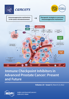
Cover Story (view full-size image): In the last decade, many therapeutic options have been approved, improving survival outcomes in advanced prostate cancer (PCa) patients. The emphasis on immune checkpoint inhibitors (ICIs) has led to many trials in this setting, with generally poor results. We discuss the immunobiology of PCa, the current clinical data, and ongoing trials to understand the potential role of ICIs in this disease and the patients’ subset more likely to benefit from them. Current evidence shows the tumor microenvironment (TME) needs a qualitative evaluation, and the prognostic/predictive role of immunotherapy biomarkers needs further assessment in PCa. ICI monotherapy has shown modest efficacy, while combined strategies with other standard therapies have shown some results. ICIs in biomarker-selected patients and new targets modulating the immune TME should be better investigated. View this paper
  • Issues are regarded as officially published after their release is announced to the table of contents alert mailing list.
  • You may sign up for e-mail alerts to receive table of contents of newly released issues.
  • PDF is the official format for papers published in both, html and pdf forms. To view the papers in pdf format, click on the "PDF Full-text" link, and use the free Adobe Reader to open them.
Order results
Result details
Section
Select all
Export citation of selected articles as:
22 pages, 1926 KiB  
Review
The Hodgkin Lymphoma Immune Microenvironment: Turning Bad News into Good
by Victoria Menéndez, José L. Solórzano, Sara Fernández, Carlos Montalbán and Juan F. García
Cancers 2022, 14(5), 1360; https://doi.org/10.3390/cancers14051360 - 7 Mar 2022
Cited by 18 | Viewed by 4757
Abstract
The classic Hodgkin lymphoma (cHL) tumor microenvironment (TME) is by far the most abundant component of tumors and is responsible for most of their biological and clinical characteristics. Recent advances in our knowledge of these networks in cellular interactions allow us to understand [...] Read more.
The classic Hodgkin lymphoma (cHL) tumor microenvironment (TME) is by far the most abundant component of tumors and is responsible for most of their biological and clinical characteristics. Recent advances in our knowledge of these networks in cellular interactions allow us to understand that the neoplastic Hodgkin and Reed Sternberg (HRS) cells, although they are in the minority, are the main architects of this dysregulated immune milieu. Here, we review the major changes that have happened in recent years: from TME as a helpless bystander, reflecting an ineffective immune response, to a dynamic tumor-promoting and immunosuppressive element. The HRS cells promote survival through interconnected intrinsic and extrinsic alterations, boosting pro-tumoral signaling pathways through genetic aberrations and autocrine growth signals, in parallel with abnormal cytokine secretion for the recruitment and selection of the best cell partners for this immunosuppressive TME. In turn, cHL is already proving to be the perfect model with which to address an immune checkpoint blockade. Preliminary data demonstrate the utility of druggable key signaling pathways in this ensemble, such as JAK-STAT, NF-κB, and others. In addition, myriad biomarkers predicting a response await validation by new in situ multiplex analytical methods, single-cell gene expression, and other techniques. Together, these components will define the functional phenotypes with which we will elucidate the molecular pathogenesis of the disease and improve the survival of patients who are refractory to conventional therapies. Full article
(This article belongs to the Special Issue Immunology of Hodgkin Lymphoma)
Show Figures

Graphical abstract

15 pages, 850 KiB  
Review
Neutrophil Extracellular Traps in Cancer Therapy Resistance
by Muhammad H. Shahzad, Lixuan Feng, Xin Su, Ariane Brassard, Iqraa Dhoparee-Doomah, Lorenzo E. Ferri, Jonathan D. Spicer and Jonathan J. Cools-Lartigue
Cancers 2022, 14(5), 1359; https://doi.org/10.3390/cancers14051359 - 7 Mar 2022
Cited by 34 | Viewed by 6897
Abstract
Neutrophils and their products are increasingly recognized to have a key influence on cancer progression and response to therapy. Their involvement has been shown in nearly every aspect of cancer pathophysiology with growing evidence now supporting their role in resistance to a variety [...] Read more.
Neutrophils and their products are increasingly recognized to have a key influence on cancer progression and response to therapy. Their involvement has been shown in nearly every aspect of cancer pathophysiology with growing evidence now supporting their role in resistance to a variety of cancer therapies. Recently, the role of neutrophils in cancer progression and therapy resistance has been further complicated with the discovery of neutrophil extracellular traps (NETs). NETs are web-like structures of chromatin decorated with a variety of microbicidal proteins. They are released by neutrophils in a process called NETosis. NET-dependent mechanisms of cancer pathology are beginning to be appreciated, particularly with respect to tumor response to chemo-, immuno-, and radiation therapy. Several studies support the functional role of NETs in cancer therapy resistance, involving T-cell exhaustion, drug detoxification, angiogenesis, the epithelial-to-mesenchymal transition, and extracellular matrix remodeling mechanisms, among others. Given this, new and promising data suggests NETs provide a microenvironment conducive to limited therapeutic response across a variety of neoplasms. As such, this paper aims to give a comprehensive overview of evidence on NETs in cancer therapy resistance with a focus on clinical applicability. Full article
(This article belongs to the Special Issue Neutrophils in Cancer: Role and Therapeutic Strategies)
Show Figures

Figure 1

15 pages, 2586 KiB  
Article
Anticancer Activity of (S)-5-Chloro-3-((3,5-dimethylphenyl)sulfonyl)-N-(1-oxo-1-((pyridin-4-ylmethyl)amino)propan-2-yl)-1H-indole-2-carboxamide (RS4690), a New Dishevelled 1 Inhibitor
by Antonio Coluccia, Marianna Bufano, Giuseppe La Regina, Michela Puxeddu, Angelo Toto, Alessio Paone, Amani Bouzidi, Giorgia Musto, Nadia Badolati, Viviana Orlando, Stefano Biagioni, Domiziana Masci, Chiara Cantatore, Roberto Cirilli, Francesca Cutruzzolà, Stefano Gianni, Mariano Stornaiuolo and Romano Silvestri
Cancers 2022, 14(5), 1358; https://doi.org/10.3390/cancers14051358 - 7 Mar 2022
Cited by 4 | Viewed by 3844
Abstract
Wingless/integrase-11 (WNT)/β-catenin pathway is a crucial upstream regulator of a huge array of cellular functions. Its dysregulation is correlated to neoplastic cellular transition and cancer proliferation. Members of the Dishevelled (DVL) family of proteins play an important role in the transduction of WNT [...] Read more.
Wingless/integrase-11 (WNT)/β-catenin pathway is a crucial upstream regulator of a huge array of cellular functions. Its dysregulation is correlated to neoplastic cellular transition and cancer proliferation. Members of the Dishevelled (DVL) family of proteins play an important role in the transduction of WNT signaling by contacting its cognate receptor, Frizzled, via a shared PDZ domain. Thus, negative modulators of DVL1 are able to impair the binding to Frizzled receptors, turning off the aberrant activation of the WNT pathway and leading to anti-cancer activity. Through structure-based virtual screening studies, we identified racemic compound RS4690 (1), which showed a promising selective DVL1 binding inhibition with an EC50 of 0.74 ± 0.08 μM. Molecular dynamic simulations suggested a different binding mode for the enantiomers. In the in vitro assays, enantiomer (S)-1 showed better inhibition of DVL1 with an EC50 of 0.49 ± 0.11 μM compared to the (R)-enantiomer. Compound (S)-1 inhibited the growth of HCT116 cells expressing wild-type APC with an EC50 of 7.1 ± 0.6 μM and caused a high level of ROS production. These results highlight (S)-1 as a lead compound for the development of new therapeutic agents against WNT-dependent colon cancer. Full article
(This article belongs to the Collection Drug Resistance and Novel Therapies in Cancers)
Show Figures

Figure 1

15 pages, 1504 KiB  
Article
Blood Circulating CD133+ Extracellular Vesicles Predict Clinical Outcomes in Patients with Metastatic Colorectal Cancer
by Davide Brocco, Pasquale Simeone, Davide Buca, Pietro Di Marino, Michele De Tursi, Antonino Grassadonia, Laura De Lellis, Maria Teresa Martino, Serena Veschi, Manuela Iezzi, Simone De Fabritiis, Marco Marchisio, Sebastiano Miscia, Alessandro Cama, Paola Lanuti and Nicola Tinari
Cancers 2022, 14(5), 1357; https://doi.org/10.3390/cancers14051357 - 7 Mar 2022
Cited by 16 | Viewed by 3094
Abstract
Colorectal cancer (CRC) is one of the most incident and lethal malignancies worldwide. Recent treatment advances prolonged survival in patients with metastatic colorectal cancer (mCRC). However, there are still few biomarkers to guide clinical management and treatment selection in mCRC. In this study, [...] Read more.
Colorectal cancer (CRC) is one of the most incident and lethal malignancies worldwide. Recent treatment advances prolonged survival in patients with metastatic colorectal cancer (mCRC). However, there are still few biomarkers to guide clinical management and treatment selection in mCRC. In this study, we applied an optimized flow cytometry protocol for EV identification, enumeration, and subtyping in blood samples of 54 patients with mCRC and 48 age and sex-matched healthy controls (HCs). The overall survival (OS) and overall response rate (ORR) were evaluated in mCRC patients enrolled and treated with a first line fluoropyrimidine-based regimen. Our findings show that patients with mCRC presented considerably higher blood concentrations of total EVs, as well as CD133+ and EPCAM+ EVs compared to HCs. Overall survival analysis revealed that increased blood concentrations of total EVs and CD133+ EVs before treatment were significantly associated with shorter OS in mCRC patients (p = 0.001; and p = 0.0001, respectively). In addition, we observed a correlation between high blood levels of CD133+ EVs at baseline and reduced ORR to first-line systemic therapy (p = 0.045). These findings may open exciting perspectives into the application of novel blood-based EV biomarkers for improved risk stratification and optimized treatment strategies in mCRC. Full article
(This article belongs to the Special Issue Liquid Biopsy in Gastrointestinal Cancers)
Show Figures

Figure 1

14 pages, 2692 KiB  
Article
EGFR Mutation-Harboring Lung Cancer Cells Produce CLEC11A with Endothelial Trophic and Tumor-Promoting Activities
by Tzu-Yin Lin, Chi-Hwa Yang, Hsiao-Chin Chou, Chun-Mei Cheng, Ya-Wen Liu, Jiz-Yuh Wang, Li-Rung Huang, Shih-Feng Tsai, Shiu-Feng Huang and Yi-Rong Chen
Cancers 2022, 14(5), 1356; https://doi.org/10.3390/cancers14051356 - 7 Mar 2022
Cited by 8 | Viewed by 3154
Abstract
The formation of new blood vessels in solid tumors is regulated by various endothelial trophic factors. We identified that CLEC11A, an extracellular C-type lectin, was over-expressed in lung cancer cell lines harboring mutated EGFR. CLEC11A expression was also frequently elevated in lung [...] Read more.
The formation of new blood vessels in solid tumors is regulated by various endothelial trophic factors. We identified that CLEC11A, an extracellular C-type lectin, was over-expressed in lung cancer cell lines harboring mutated EGFR. CLEC11A expression was also frequently elevated in lung adenocarcinoma (LAC) tissues with EGFR mutation. CLEC11A-expressing H1299 cells formed larger tumors in nude mice than did the control cells. The CLEC11A-expressing tumors contained more CD31-positive cells, suggesting that they had a higher angiogenic activity. CLEC11A per se did not induce blood vessel formation, but enhanced angiogenesis triggered by VEGF-A or basic FGF in vivo. Additionally, the expression of small hairpin RNA against CLEC11A (shCLEC11A) in HCC827 LAC cells suppressed their tumorigenic ability. Purified CLEC11A exhibited a chemotactic ability, which is dependent on its integrin-binding RGD and LDT motifs, toward endothelial cells. This chemotactic activity was not affected by the presence of a VEGFR inhibitor. Conditioned medium produced by HCC827-shCLEC11A cells had diminished chemotactic ability toward endothelial cells. CLEC11A treatments increased the levels of active integrin β1 that were not associated with activation of focal adhesion kinases in endothelial cells. Our results indicated that CLEC11A was a factor of angiogenic potential and was involved in lung cancer tumorigenesis. Full article
(This article belongs to the Section Tumor Microenvironment)
Show Figures

Figure 1

12 pages, 475 KiB  
Article
Prevalence of Cognitive Impairment before Prostate Cancer Treatment
by Natália Araújo, Adriana Costa, Catarina Lopes, Luisa Lopes-Conceição, Augusto Ferreira, Filipa Carneiro, Jorge Oliveira, Samantha Morais, Luís Pacheco-Figueiredo, Luis Ruano, Vítor Tedim Cruz, Susana Pereira and Nuno Lunet
Cancers 2022, 14(5), 1355; https://doi.org/10.3390/cancers14051355 - 7 Mar 2022
Cited by 5 | Viewed by 2717
Abstract
Cognitive impairment is common among patients with different types of cancer, even before cancer treatment, but no data were reported among patients with prostate cancer (PCa), who may be at high risk due to advanced age. This study aims to estimate the prevalence [...] Read more.
Cognitive impairment is common among patients with different types of cancer, even before cancer treatment, but no data were reported among patients with prostate cancer (PCa), who may be at high risk due to advanced age. This study aims to estimate the prevalence of cognitive impairment before PCa treatment. Between February 2018 and April 2021, the NEON-PC cohort recruited 605 patients with PCa proposed for treatment at the Portuguese Institute of Oncology of Porto. The Montreal Cognitive Assessment (MoCA) was used to assess cognitive performance. Participants with a MoCA < 1.5 standard deviations (SD) of age- and education-specific normative values were considered to have probable cognitive impairment (PCI) and were referred for a comprehensive neuropsychological assessment. Data from the population-based cohort EPIPorto (n = 351 men aged ≥40 years, evaluated in 2013–2015) were used for comparison. The prevalence of PCI was 17.4% in EPIPorto and 14.7% in NEON-PC (age- and education-adjusted odds ratio: 0.82, 95%CI: 0.58,1.18). Neuropsychological assessment was performed in 63 patients with PCa: 54.0% had cognitive impairment. These results suggest that the impact of PCa on cognitive performance could be negligible in the short term, contrary to what other studies have reported regarding other types of cancer. Full article
Show Figures

Figure 1

15 pages, 2162 KiB  
Article
Intraperitoneal Paclitaxel Treatment for Patients with Pancreatic Ductal Adenocarcinoma with Peritoneal Dissemination Provides a Survival Benefit
by Tomohisa Yamamoto, Sohei Satoi, So Yamaki, Daisuke Hashimoto, Mitsuaki Ishida, Tsukasa Ikeura, Satoshi Hirooka, Yuki Matsui, Shogen Boku, Shinji Nakayama, Koh Nakamaru, Nobuhiro Shibata, Utae Katsushima and Mitsugu Sekimoto
Cancers 2022, 14(5), 1354; https://doi.org/10.3390/cancers14051354 - 7 Mar 2022
Cited by 6 | Viewed by 2610
Abstract
Background: Intraperitoneal chemotherapy using paclitaxel (i.p.-PTX) is expected to be a new therapeutic strategy for patients with pancreatic ductal adenocarcinoma (PDAC) and peritoneal dissemination. We evaluated the survival benefit of i.p.-PTX compared with standard systemic chemotherapy. Methods: Clinical data of 101 consecutive PDAC [...] Read more.
Background: Intraperitoneal chemotherapy using paclitaxel (i.p.-PTX) is expected to be a new therapeutic strategy for patients with pancreatic ductal adenocarcinoma (PDAC) and peritoneal dissemination. We evaluated the survival benefit of i.p.-PTX compared with standard systemic chemotherapy. Methods: Clinical data of 101 consecutive PDAC patients with peritoneal dissemination between 2007 and 2018 were analyzed. All patients were determined to have no other sites of distant organ metastasis to the lung, bone, or liver on contrast-enhanced CT imaging. Patients underwent staging laparoscopy or open laparotomy to confirm pathological evidence of peritoneal dissemination, and to exclude occult liver metastasis. Survival curves were estimated using the Kaplan–Meier method, and differences were compared using the log-rank test. Results: Forty-three patients were treated with i.p.-PTX (i.p.-PTX group) and forty-nine patients received standard systemic chemotherapy (Ctrl group). Nine patients did not receive any treatment (BSC group). The median survival time (MST) in the i.p.-PTX group was significantly longer than that in the Ctrl group (17.9 months vs. 10.2 months, p = 0.006). Negative peritoneal washing cytology was observed in 24 out of 43 patients in the i.p.-PTX group. The i.p.-PTX group tended to have a higher proportion of clinical responses than the Ctrl group (30% vs. 18%, p = 0.183). Conversion surgery was performed in 10 patients in the i.p.-PTX group and 2 patients in the Ctrl group after confirming disappearance of peritoneal dissemination with staging laparoscopy or open laparotomy (p = 0.005). The MST in patients who underwent surgical resection was significantly longer than that in patients who did not (27.4 months vs. 11.3 months; p < 0.0001). Conclusion: i.p.-PTX therapy provided improved survival in PDAC patients with peritoneal dissemination, and conversion surgery enhanced it in patients with favorable responses to chemotherapy. i.p.-PTX might become one of the treatment options to PDAC patients with peritoneal dissemination. Full article
(This article belongs to the Special Issue Recent Highly Advanced Surgery for Pancreatic Cancer)
Show Figures

Figure 1

18 pages, 4611 KiB  
Article
Comprehensive Analysis of the Prognosis and Drug Sensitivity of Differentiation-Related lncRNAs in Papillary Thyroid Cancer
by Wenlong Wang, Ning Bai and Xinying Li
Cancers 2022, 14(5), 1353; https://doi.org/10.3390/cancers14051353 - 7 Mar 2022
Cited by 9 | Viewed by 2923
Abstract
Dedifferentiation is the main concern associated with radioactive iodine (RAI) refractoriness in patients with papillary thyroid cancer (PTC), and the underlying mechanisms of PTC dedifferentiation remain unclear. The present work aimed to identify a useful signature to indicate dedifferentiation and further explore its [...] Read more.
Dedifferentiation is the main concern associated with radioactive iodine (RAI) refractoriness in patients with papillary thyroid cancer (PTC), and the underlying mechanisms of PTC dedifferentiation remain unclear. The present work aimed to identify a useful signature to indicate dedifferentiation and further explore its role in prognosis and susceptibility to chemotherapy drugs. A total of five prognostic-related DR-lncRNAs were selected to establish a prognostic-predicting model, and corresponding risk scores were closely associated with the infiltration of immune cells and immune checkpoint blockade. Moreover, we built an integrated nomogram based on DR-lncRNAs and age that showed a strong ability to predict the 3- and 5-year overall survival. Interestingly, drug sensitivity analysis revealed that the low-risk group was more sensitive to Bendamustine and TAS-6417 than the high-risk group. In addition, knockdown of DR-lncRNAs (DPH6-DT) strongly promoted cell proliferation, invasion, and migration via PI3K-AKT signal pathway in vitro. Furthermore, DPH6-DT downregulation also increased the expression of vimentin and N-cadherin during epithelial-mesenchymal transition. This study firstly confirms that DR-lncRNAs play a vital role in the prognosis and immune cells infiltration in patients with PTC, as well as a predictor of the drugs’ chemosensitivity. Based on our results, DR-lncRNAs can serve as a promising prognostic biomarkers and treatment targets. Full article
(This article belongs to the Special Issue Biomarkers of Thyroid Cancer)
Show Figures

Figure 1

7 pages, 1091 KiB  
Article
The Clinical Utility and Impact of Next Generation Sequencing in Gynecologic Cancers
by Vijaya Kadam Maruthi, Mahyar Khazaeli, Devi Jeyachandran and Mohamed Mokhtar Desouki
Cancers 2022, 14(5), 1352; https://doi.org/10.3390/cancers14051352 - 7 Mar 2022
Cited by 4 | Viewed by 2395
Abstract
Next generation sequencing (NGS) has facilitated the identification of molecularly targeted therapies. However, clinical utility is an emerging challenge. Our objective was to identify the clinical utility of NGS testing in gynecologic cancers. A retrospective review of clinico-pathologic data was performed on 299 [...] Read more.
Next generation sequencing (NGS) has facilitated the identification of molecularly targeted therapies. However, clinical utility is an emerging challenge. Our objective was to identify the clinical utility of NGS testing in gynecologic cancers. A retrospective review of clinico-pathologic data was performed on 299 gynecological cancers where NGS testing had been performed to identify (1) recognition of actionable targets for therapy, (2) whether the therapy changed based on the findings, and (3) the impact on survival. High grade serous carcinoma was the most common tumor (52.5%). The number of genetic alterations ranged from 0 to 25 with a mean of 2.8/case. The most altered genes were TP53, PIK3CA, BRCA1 and BRCA2. Among 299 patients, 100 had actionable alterations (79 received a targeted treatment (Group1), 29 did not receive treatment (Group 2), and there were no actionable alterations in 199 (Group3). The death rate in groups 1, 2 and 3 was 54.4%, 42.8% and 50.2%, with an average survival of 18.6, 6.6 and 10.8 months, respectively (p = 0.002). In summary, NGS testing for gynecologic cancers detected 33.4% of actionable alterations with a high clinical action rate. Along with the high clinical utility of NGS, testing also seemed to improve survival for patients who received targeted treatment. Full article
Show Figures

Figure 1

12 pages, 1578 KiB  
Article
Diagnostic Accuracy of Contrast-Enhanced, Spectral Mammography (CESM) and 3T Magnetic Resonance Compared to Full-Field Digital Mammography plus Ultrasound in Breast Lesions: Results of a (Pilot) Open-Label, Single-Centre Prospective Study
by Francesca Romana Ferranti, Federica Vasselli, Maddalena Barba, Francesca Sperati, Irene Terrenato, Franco Graziano, Patrizia Vici, Claudio Botti and Antonello Vidiri
Cancers 2022, 14(5), 1351; https://doi.org/10.3390/cancers14051351 - 7 Mar 2022
Cited by 5 | Viewed by 5244
Abstract
Introduction: To assess the diagnostic accuracy of CESM and 3T MRI compared to full-field digital mammography (FFDM), plus US, in the evaluation of advanced breast lesions. Materials and Methods: Consenting women with suspicious findings underwent FFDM, US, CESM and 3T MRI. Breast lesions [...] Read more.
Introduction: To assess the diagnostic accuracy of CESM and 3T MRI compared to full-field digital mammography (FFDM), plus US, in the evaluation of advanced breast lesions. Materials and Methods: Consenting women with suspicious findings underwent FFDM, US, CESM and 3T MRI. Breast lesions were histologically assessed, with histology being the gold standard. Two experienced breast radiologists, blinded to cancer status, read the images. Diagnostic accuracy of (1) CESM as an adjunct to FFDM and US, and (2) 3T MRI as an adjunct to CESM compared to FFDM and US, was assessed. Measures of accuracy were sensitivity (Se), specificity (Sp), positive predictive value (PPV) and negative predictive value (NPV). Results: There were 118 patients included along with 142 histologically characterized lesions. K agreement values were 0.69, 0.68, 0.63 and 0.56 for concordance between the gold standard and FFDM, FFDM + US, CESM and MRI, respectively (p < 0.001, for all). K concordance for CESM was 0.81 with FFDM + US and 0.73 with MRI (p value < 0.001 for all). Conclusions: CESM may represent a valuable alternative and/or an integrating technique to MRI in the evaluation of breast cancer patients. Full article
(This article belongs to the Special Issue Advanced Breast Cancer: From Biology to Cure)
Show Figures

Figure 1

17 pages, 323 KiB  
Review
Oligometastatic Disease in Non-Small-Cell Lung Cancer: An Update
by Yi-Hsing Chen, Ue-Cheung Ho and Lu-Ting Kuo
Cancers 2022, 14(5), 1350; https://doi.org/10.3390/cancers14051350 - 6 Mar 2022
Cited by 3 | Viewed by 3640
Abstract
Oligometastatic non-small-cell lung cancer (NSCLC) is a distinct entity that is different from localized and disseminated diseases. The definition of oligometastatic NSCLC varies across studies in past decades owing to the use of different imaging modalities; however, a uniform definition of oligometastatic NSCLC [...] Read more.
Oligometastatic non-small-cell lung cancer (NSCLC) is a distinct entity that is different from localized and disseminated diseases. The definition of oligometastatic NSCLC varies across studies in past decades owing to the use of different imaging modalities; however, a uniform definition of oligometastatic NSCLC has been proposed, and this may facilitate trial design and evaluation of certain interventions. Patients with oligometastatic NSCLC are candidates for curative-intent management, in which local ablative treatment, such as surgery or stereotactic radiosurgery, should be instituted to improve clinical outcomes. Although current guidelines recommend that local therapy for thoracic and metastatic lesions should be considered for patients with oligometastatic NSCLC with stable disease after systemic therapy, optimal management strategies for different oligometastatic sites have not been established. Additionally, the development of personalized therapies for individual patients with oligometastatic NSCLC to improve their quality of life and overall survival should also be addressed. Here, we review relevant articles on the management of patients with oligometastatic NSCLC and categorize the disease according to the site of metastases. Ongoing trials are also summarized to determine future directions and expectations for new treatment modalities to improve patient management. Full article
(This article belongs to the Special Issue Oligometastatic Disease)
19 pages, 713 KiB  
Review
Advancements in Oncology with Artificial Intelligence—A Review Article
by Nikitha Vobugari, Vikranth Raja, Udhav Sethi, Kejal Gandhi, Kishore Raja and Salim R. Surani
Cancers 2022, 14(5), 1349; https://doi.org/10.3390/cancers14051349 - 6 Mar 2022
Cited by 26 | Viewed by 7595
Abstract
Well-trained machine learning (ML) and artificial intelligence (AI) systems can provide clinicians with therapeutic assistance, potentially increasing efficiency and improving efficacy. ML has demonstrated high accuracy in oncology-related diagnostic imaging, including screening mammography interpretation, colon polyp detection, glioma classification, and grading. By utilizing [...] Read more.
Well-trained machine learning (ML) and artificial intelligence (AI) systems can provide clinicians with therapeutic assistance, potentially increasing efficiency and improving efficacy. ML has demonstrated high accuracy in oncology-related diagnostic imaging, including screening mammography interpretation, colon polyp detection, glioma classification, and grading. By utilizing ML techniques, the manual steps of detecting and segmenting lesions are greatly reduced. ML-based tumor imaging analysis is independent of the experience level of evaluating physicians, and the results are expected to be more standardized and accurate. One of the biggest challenges is its generalizability worldwide. The current detection and screening methods for colon polyps and breast cancer have a vast amount of data, so they are ideal areas for studying the global standardization of artificial intelligence. Central nervous system cancers are rare and have poor prognoses based on current management standards. ML offers the prospect of unraveling undiscovered features from routinely acquired neuroimaging for improving treatment planning, prognostication, monitoring, and response assessment of CNS tumors such as gliomas. By studying AI in such rare cancer types, standard management methods may be improved by augmenting personalized/precision medicine. This review aims to provide clinicians and medical researchers with a basic understanding of how ML works and its role in oncology, especially in breast cancer, colorectal cancer, and primary and metastatic brain cancer. Understanding AI basics, current achievements, and future challenges are crucial in advancing the use of AI in oncology. Full article
(This article belongs to the Topic Artificial Intelligence in Cancer Diagnosis and Therapy)
Show Figures

Figure 1

11 pages, 1262 KiB  
Article
Testing for Lynch Syndrome in Endometrial Carcinoma: From Universal to Age-Selective MLH1 Methylation Analysis
by Annukka Pasanen, Mikko Loukovaara, Elina Kaikkonen, Alisa Olkinuora, Kirsi Pylvänäinen, Pia Alhopuro, Päivi Peltomäki, Jukka-Pekka Mecklin and Ralf Bützow
Cancers 2022, 14(5), 1348; https://doi.org/10.3390/cancers14051348 - 6 Mar 2022
Cited by 4 | Viewed by 3650
Abstract
International guidelines recommend universal screening of endometrial carcinoma (EC) patients for Lynch syndrome (LS). This screening is generally based on mismatch repair (MMR) protein immunohistochemistry followed by MLH1 methylation analysis of MLH1-negative cases to exclude the likely sporadic cases from germline testing. As [...] Read more.
International guidelines recommend universal screening of endometrial carcinoma (EC) patients for Lynch syndrome (LS). This screening is generally based on mismatch repair (MMR) protein immunohistochemistry followed by MLH1 methylation analysis of MLH1-negative cases to exclude the likely sporadic cases from germline testing. As LS-associated EC is uncommon in the elderly, age-selective methylation testing could improve cost-efficiency. We performed MMR immunohistochemistry on 821 unselected ECs (clinic-based cohort) followed by a MLH1 promoter methylation test of all MLH1/PMS2-negative tumors. Non-methylated MLH1-deficient cases underwent NGS and MLPA-based germline analyses to identify MLH1 mutation carriers. A reduction in the test burden and corresponding false negative rates for LS screening were investigated for various age cut-offs. In addition, the age distribution of 132 MLH1 mutation carriers diagnosed with EC (registry-based cohort) was examined. A germline MLH1 mutation was found in 2/14 patients with non-methylated MLH1-deficient EC. When compared to a universal methylation analysis, selective testing with a cut-off age of 65 years, would have reduced the testing effort by 70.7% with a false negative rate for LS detection of 0% and 3% in the clinic and registry-based cohorts, respectively. The use of age-selective methylation analysis is a feasible way of reducing the costs and laboratory burden in LS screening for EC patients. Full article
(This article belongs to the Special Issue Immunohistochemical Markers in Endometrial Cancer)
Show Figures

Figure 1

16 pages, 906 KiB  
Article
A Target Animal Effectiveness Study on Adjuvant Peptide-Based Vaccination in Dogs with Non-Metastatic Appendicular Osteosarcoma Undergoing Amputation and Chemotherapy
by Laura Marconato, Alessia Melacarne, Marina Aralla, Silvia Sabattini, Luca Tiraboschi, Valentina Ferrari, Offer Zeira, Andrea Balboni, Eugenio Faroni, Dina Guerra, Luciano Pisoni, Erica Ghezzi, Letizia Pettinari and Maria Rescigno
Cancers 2022, 14(5), 1347; https://doi.org/10.3390/cancers14051347 - 6 Mar 2022
Cited by 7 | Viewed by 3520
Abstract
Despite efforts to develop novel treatment strategies, human and canine osteosarcomas continue to have poor prognosis and limited overall survival. The aim of this clinical trial was to test the antitumor effect and safety of multiple dermal administrations of a peptide-based anticancer vaccine [...] Read more.
Despite efforts to develop novel treatment strategies, human and canine osteosarcomas continue to have poor prognosis and limited overall survival. The aim of this clinical trial was to test the antitumor effect and safety of multiple dermal administrations of a peptide-based anticancer vaccine in dogs with non-metastatic appendicular osteosarcoma undergoing standard of care (SOC), consisting of limb amputation and adjuvant chemotherapy. Salmonella-infected canine osteosarcoma cells were induced to release immunogenic peptides in the extracellular space via Cx43 hemichannels opening; the secretome was collected and constituted the vaccine. Dogs with non-metastatic appendicular osteosarcoma were eligible for recruitment. Following limb amputation and adjuvant carboplatin, dogs were vaccinated on a monthly basis for six times and followed up with serial thoracic radiographs. A population of dogs undergoing SOC treatment (amputation and adjuvant carboplatin) before the vaccine was available served as controls. Primary endpoints were time to metastasis (TTM) and tumor-specific survival (TSS). Secondary endpoints were feasibility, toxicity, T-cell and humoral immune responses. A total of 20 dogs were vaccinated along with SOC and 34 received SOC only. Vaccine-specific humoral and T-cell responses were observed; their amplitude correlated with TSS. Vaccine-associated toxicity was not recorded. TTM and TSS were significantly longer in vaccinated versus unvaccinated dogs (TTM: 308 vs. 240 days, respectively; p = 0.010; TSS: 621 vs. 278 days, respectively; p = 0.002). In dogs with non-metastatic osteosarcoma undergoing SOC, the addition of a bacteria-based vaccination strategy increased TTM, thereby prolonging survival, while maintaining a safe profile. Additionally, vaccinated dogs developed a long-term tumor-specific response, as documented by the immunomonitoring of these patients over time. These results hold promise for future management of canine osteosarcoma. Full article
(This article belongs to the Section Cancer Immunology and Immunotherapy)
Show Figures

Figure 1

15 pages, 2402 KiB  
Article
Reproducibility of Gene Expression Signatures in Diffuse Large B-Cell Lymphoma
by Jessica Rodrigues Plaça, Arjan Diepstra, Tjitske Los, Matías Mendeville, Annika Seitz, Pieternella J. Lugtenburg, Josée Zijlstra, King Lam, Wilson Araújo da Silva, Jr., Bauke Ylstra, Daphne de Jong, Anke van den Berg and Marcel Nijland
Cancers 2022, 14(5), 1346; https://doi.org/10.3390/cancers14051346 - 5 Mar 2022
Cited by 2 | Viewed by 3394
Abstract
Multiple gene expression profiles have been identified in diffuse large B-cell lymphoma (DLBCL). Besides the cell of origin (COO) classifier, no signatures have been reproduced in independent studies or evaluated for capturing distinct aspects of DLBCL biology. We reproduced 4 signatures in 175 [...] Read more.
Multiple gene expression profiles have been identified in diffuse large B-cell lymphoma (DLBCL). Besides the cell of origin (COO) classifier, no signatures have been reproduced in independent studies or evaluated for capturing distinct aspects of DLBCL biology. We reproduced 4 signatures in 175 samples of the HOVON-84 trial on a panel of 117 genes using the NanoString platform. The four gene signatures capture the COO, MYC activity, B-cell receptor signaling, oxidative phosphorylation, and immune response. Performance of our classification algorithms were confirmed in the original datasets. We were able to validate three of the four GEP signatures. The COO algorithm resulted in 94 (54%) germinal center B-cell (GCB) type, 58 (33%) activated B-cell (ABC) type, and 23 (13%) unclassified cases. The MYC-classifier revealed 77 cases with a high MYC-activity score (44%) and this MYC-high signature was observed more frequently in ABC as compared to GCB DLBCL (68% vs. 32%, p < 0.00001). The host response (HR) signature of the consensus clustering was present in 55 (31%) patients, while the B-cell receptor signaling, and oxidative phosphorylation clusters could not be reproduced. The overlap of COO, consensus cluster and MYC activity score differentiated six gene expression clusters: GCB/MYC-high (12%), GCB/HR (16%), GCB/non-HR (27%), COO-Unclassified (13%), ABC/MYC-high (25%), and ABC/MYC-low (7%). In conclusion, the three validated signatures identify distinct subgroups based on different aspects of DLBCL biology, emphasizing that each classifier captures distinct molecular profiles. Full article
(This article belongs to the Special Issue Molecular Characterization of Hematological Tumors)
Show Figures

Figure 1

21 pages, 2539 KiB  
Review
CK1 Is a Druggable Regulator of Microtubule Dynamics and Microtubule-Associated Processes
by Aileen Roth, Adrian Gihring, Joachim Bischof, Leiling Pan, Franz Oswald and Uwe Knippschild
Cancers 2022, 14(5), 1345; https://doi.org/10.3390/cancers14051345 - 5 Mar 2022
Cited by 9 | Viewed by 4054
Abstract
Protein kinases of the Casein Kinase 1 family play a vital role in the regulation of numerous cellular processes. Apart from functions associated with regulation of proliferation, differentiation, or apoptosis, localization of several Casein Kinase 1 isoforms to the centrosome and microtubule asters [...] Read more.
Protein kinases of the Casein Kinase 1 family play a vital role in the regulation of numerous cellular processes. Apart from functions associated with regulation of proliferation, differentiation, or apoptosis, localization of several Casein Kinase 1 isoforms to the centrosome and microtubule asters also implicates regulatory functions in microtubule dynamic processes. Being localized to the spindle apparatus during mitosis Casein Kinase 1 directly modulates microtubule dynamics by phosphorylation of tubulin isoforms. Additionally, site-specific phosphorylation of microtubule-associated proteins can be related to the maintenance of genomic stability but also microtubule stabilization/destabilization, e.g., by hyper-phosphorylation of microtubule-associated protein 1A and RITA1. Consequently, approaches interfering with Casein Kinase 1-mediated microtubule-specific functions might be exploited as therapeutic strategies for the treatment of cancer. Currently pursued strategies include the development of Casein Kinase 1 isoform-specific small molecule inhibitors and therapeutically useful peptides specifically inhibiting kinase-substrate interactions. Full article
(This article belongs to the Special Issue Microtubule Dynamics and Cancer)
Show Figures

Figure 1

13 pages, 1148 KiB  
Article
Measuring Walking Speed Failed to Predict Early Death and Toxicity in Elderly Patients with Metastatic Non-Small-Cell Lung Cancer (NSCLC) Selected for Undergoing First-Line Systemic Treatment: An Observational Exploratory Study
by Amélie Aregui, Johan Pluvy, Manuel Sanchez, Theresa Israel, Hélène Esnault, Alice Guyard, Marie Meyer, Antoine Khalil, Gérard Zalcman, Agathe Raynaud Simon and Valérie Gounant
Cancers 2022, 14(5), 1344; https://doi.org/10.3390/cancers14051344 - 5 Mar 2022
Cited by 3 | Viewed by 2291
Abstract
Walking speed (WS) has emerged as a potential predictor of mortality in elderly cancer patients, yet data involving non-small-cell lung cancer (NSCLC) patients are scarce. Our prospective exploratory study sought to determine whether WS would predict early death or toxicity in patients with [...] Read more.
Walking speed (WS) has emerged as a potential predictor of mortality in elderly cancer patients, yet data involving non-small-cell lung cancer (NSCLC) patients are scarce. Our prospective exploratory study sought to determine whether WS would predict early death or toxicity in patients with advanced NSCLC receiving first-line systemic intravenous treatment. Overall, 145 patients of ≥70 years were diagnosed with NSCLC over 19 months, 91 of whom displayed locally-advanced or metastatic cancer. As first-line treatment, 21 (23%) patients received best supportive care, 13 (14%) targeted therapy, and 57 (63%) chemotherapy or immunotherapy. Among the latter, 38 consented to participate in the study (median age: 75 years). Median cumulative illness rating scale for geriatrics (CIRS-G) was 10 (IQR: 8–12), and median WS 1.09 (IQR: 0.9–1.31) m/s. Older age (p = 0.03) and comorbidities (p = 0.02) were associated with Grade 3–4 treatment-related adverse events or death within 6 months of accrual. Overall survival was 14.3 (IQR: 6.1-NR) months for patients with WS < 1 m/s versus 17.3 (IQR: 9.2–26.5) for those with WS ≥ 1 m/s (p = 0.78). This exploratory study revealed WS to be numerically, yet not significantly, associated with early mortality in older metastatic NSCLC patients. Following these hypothesis-generating results, a larger prospective, multicenter study appears to be required to further investigate this outcome. Full article
(This article belongs to the Section Cancer Therapy)
Show Figures

Figure 1

30 pages, 5494 KiB  
Article
Dual Effect of Combined Metformin and 2-Deoxy-D-Glucose Treatment on Mitochondrial Biogenesis and PD-L1 Expression in Triple-Negative Breast Cancer Cells
by Jernej Repas, Mateja Zupin, Maja Vodlan, Peter Veranič, Boris Gole, Uroš Potočnik and Mojca Pavlin
Cancers 2022, 14(5), 1343; https://doi.org/10.3390/cancers14051343 - 5 Mar 2022
Cited by 11 | Viewed by 6004
Abstract
Metformin and 2-deoxy-D-glucose (2DG) exhibit multiple metabolic and immunomodulatory anti-cancer effects, such as suppressed proliferation or PD-L1 expression. Their combination or 2DG alone induce triple-negative breast cancer (TNBC) cell detachment, but their effects on mitochondria, crucial for anchorage-independent growth and metastasis formation, have [...] Read more.
Metformin and 2-deoxy-D-glucose (2DG) exhibit multiple metabolic and immunomodulatory anti-cancer effects, such as suppressed proliferation or PD-L1 expression. Their combination or 2DG alone induce triple-negative breast cancer (TNBC) cell detachment, but their effects on mitochondria, crucial for anchorage-independent growth and metastasis formation, have not yet been evaluated. In the present study, we explored the effects of metformin, 2DG and their combination (metformin + 2DG) on TNBC cell mitochondria in vitro. Metformin + 2DG increased mitochondrial mass in TNBC cells. This was associated with an increased size but not number of morphologically normal mitochondria and driven by the induction of mitochondrial biogenesis rather than suppressed mitophagy. 2DG and metformin + 2DG strongly induced the unfolded protein response by inhibiting protein N-glycosylation. Together with adequate energy stress, this was one of the possible triggers of mitochondrial enlargement. Suppressed N-glycosylation by 2DG or metformin + 2DG also caused PD-L1 deglycosylation and reduced surface expression in MDA-MB-231 cells. PD-L1 was increased in low glucose and normalized by both drugs. 2DG and metformin + 2DG reduced PD-1 expression in Jurkat cells beyond the effects on activation, while cytokine secretion was mostly preserved. Despite increasing mitochondrial mass in TNBC cells, metformin and 2DG could therefore potentially be used as an adjunct therapy to improve anti-tumor immunity in TNBC. Full article
(This article belongs to the Special Issue Metabolic Networks in Cancer)
Show Figures

Graphical abstract

21 pages, 1064 KiB  
Review
Hemodynamic Imaging in Cerebral Diffuse Glioma—Part B: Molecular Correlates, Treatment Effect Monitoring, Prognosis, and Future Directions
by Vittorio Stumpo, Lelio Guida, Jacopo Bellomo, Christiaan Hendrik Bas Van Niftrik, Martina Sebök, Moncef Berhouma, Andrea Bink, Michael Weller, Zsolt Kulcsar, Luca Regli and Jorn Fierstra
Cancers 2022, 14(5), 1342; https://doi.org/10.3390/cancers14051342 - 5 Mar 2022
Cited by 6 | Viewed by 3528
Abstract
Gliomas, and glioblastoma in particular, exhibit an extensive intra- and inter-tumoral molecular heterogeneity which represents complex biological features correlating to the efficacy of treatment response and survival. From a neuroimaging point of view, these specific molecular and histopathological features may be used to [...] Read more.
Gliomas, and glioblastoma in particular, exhibit an extensive intra- and inter-tumoral molecular heterogeneity which represents complex biological features correlating to the efficacy of treatment response and survival. From a neuroimaging point of view, these specific molecular and histopathological features may be used to yield imaging biomarkers as surrogates for distinct tumor genotypes and phenotypes. The development of comprehensive glioma imaging markers has potential for improved glioma characterization that would assist in the clinical work-up of preoperative treatment planning and treatment effect monitoring. In particular, the differentiation of tumor recurrence or true progression from pseudoprogression, pseudoresponse, and radiation-induced necrosis can still not reliably be made through standard neuroimaging only. Given the abundant vascular and hemodynamic alterations present in diffuse glioma, advanced hemodynamic imaging approaches constitute an attractive area of clinical imaging development. In this context, the inclusion of objective measurable glioma imaging features may have the potential to enhance the individualized care of diffuse glioma patients, better informing of standard-of-care treatment efficacy and of novel therapies, such as the immunotherapies that are currently increasingly investigated. In Part B of this two-review series, we assess the available evidence pertaining to hemodynamic imaging for molecular feature prediction, in particular focusing on isocitrate dehydrogenase (IDH) mutation status, MGMT promoter methylation, 1p19q codeletion, and EGFR alterations. The results for the differentiation of tumor progression/recurrence from treatment effects have also been the focus of active research and are presented together with the prognostic correlations identified by advanced hemodynamic imaging studies. Finally, the state-of-the-art concepts and advancements of hemodynamic imaging modalities are reviewed together with the advantages derived from the implementation of radiomics and machine learning analyses pipelines. Full article
(This article belongs to the Special Issue Advanced Neuroimaging Approaches for Malignant Brain Tumors)
Show Figures

Figure 1

21 pages, 16913 KiB  
Article
A Novel and Automated Approach to Classify Radiation Induced Lung Tissue Damage on CT Scans
by Adam Szmul, Edward Chandy, Catarina Veiga, Joseph Jacob, Alkisti Stavropoulou, David Landau, Crispin T. Hiley and Jamie R. McClelland
Cancers 2022, 14(5), 1341; https://doi.org/10.3390/cancers14051341 - 5 Mar 2022
Cited by 3 | Viewed by 2571
Abstract
Radiation-induced lung damage (RILD) is a common side effect of radiotherapy (RT). The ability to automatically segment, classify, and quantify different types of lung parenchymal change is essential to uncover underlying patterns of RILD and their evolution over time. A RILD dedicated tissue [...] Read more.
Radiation-induced lung damage (RILD) is a common side effect of radiotherapy (RT). The ability to automatically segment, classify, and quantify different types of lung parenchymal change is essential to uncover underlying patterns of RILD and their evolution over time. A RILD dedicated tissue classification system was developed to describe lung parenchymal tissue changes on a voxel-wise level. The classification system was automated for segmentation of five lung tissue classes on computed tomography (CT) scans that described incrementally increasing tissue density, ranging from normal lung (Class 1) to consolidation (Class 5). For ground truth data generation, we employed a two-stage data annotation approach, akin to active learning. Manual segmentation was used to train a stage one auto-segmentation method. These results were manually refined and used to train the stage two auto-segmentation algorithm. The stage two auto-segmentation algorithm was an ensemble of six 2D Unets using different loss functions and numbers of input channels. The development dataset used in this study consisted of 40 cases, each with a pre-radiotherapy, 3-, 6-, 12-, and 24-month follow-up CT scans (n = 200 CT scans). The method was assessed on a hold-out test dataset of 6 cases (n = 30 CT scans). The global Dice score coefficients (DSC) achieved for each tissue class were: Class (1) 99% and 98%, Class (2) 71% and 44%, Class (3) 56% and 26%, Class (4) 79% and 47%, and Class (5) 96% and 92%, for development and test subsets, respectively. The lowest values for the test subsets were caused by imaging artefacts or reflected subgroups that occurred infrequently and with smaller overall parenchymal volumes. We performed qualitative evaluation on the test dataset presenting manual and auto-segmentation to a blinded independent radiologist to rate them as ‘acceptable’, ‘minor disagreement’ or ‘major disagreement’. The auto-segmentation ratings were similar to the manual segmentation, both having approximately 90% of cases rated as acceptable. The proposed framework for auto-segmentation of different lung tissue classes produces acceptable results in the majority of cases and has the potential to facilitate future large studies of RILD. Full article
Show Figures

Figure 1

26 pages, 1355 KiB  
Review
Persistent Large Granular Lymphocyte Clonal Expansions: “The Root of Many Evils”—And of Some Goodness
by Carlos Bravo-Pérez, Salvador Carrillo-Tornel, Esmeralda García-Torralba and Andrés Jerez
Cancers 2022, 14(5), 1340; https://doi.org/10.3390/cancers14051340 - 5 Mar 2022
Cited by 7 | Viewed by 4281
Abstract
Large granular lymphocyte leukemia (LGLL) is a chronic disease of either mature phenotype cytotoxic CD3+ T lymphocytes or CD3- NK cells. LGLL diagnosis is hampered by the fact that reactive persistent clonal LGL expansions may fulfill the current criteria for LGLL diagnoses. In [...] Read more.
Large granular lymphocyte leukemia (LGLL) is a chronic disease of either mature phenotype cytotoxic CD3+ T lymphocytes or CD3- NK cells. LGLL diagnosis is hampered by the fact that reactive persistent clonal LGL expansions may fulfill the current criteria for LGLL diagnoses. In addition to the presence of characteristic clinical and hematological signs such as anemia or neutropenia, LGLL/LGL clonal expansions have been associated with an array of conditions/disorders. We review here the presence of these persistent clonal expansions in autoimmune, hematological disorders and solid neoplasms and after hematopoietic stem cell transplantation. These associations are a unique translational research framework to discern whether these persistently expanded LGL clones are causes or consequences of the concomitant clinical settings and, more importantly, when they should be targeted. Full article
(This article belongs to the Special Issue Large Granular Lymphocytic Leukemia: Genomics and Immunome)
Show Figures

Figure 1

13 pages, 1228 KiB  
Article
Fluorescence Polarization Imaging of Methylene Blue Facilitates Quantitative Detection of Thyroid Cancer in Single Cells
by Peter R. Jermain, Andrew H. Fischer, Lija Joseph, Alona Muzikansky and Anna N. Yaroslavsky
Cancers 2022, 14(5), 1339; https://doi.org/10.3390/cancers14051339 - 5 Mar 2022
Cited by 7 | Viewed by 2914
Abstract
Background: Diagnostic accuracy of the standard of care fine-needle aspiration cytology (FNAC) remains a significant problem in thyroid oncology. Therefore, a robust and accurate method for reducing uncertainty of cytopathological evaluation would be invaluable. Methods: In this double-blind study, we employed fluorescence emission [...] Read more.
Background: Diagnostic accuracy of the standard of care fine-needle aspiration cytology (FNAC) remains a significant problem in thyroid oncology. Therefore, a robust and accurate method for reducing uncertainty of cytopathological evaluation would be invaluable. Methods: In this double-blind study, we employed fluorescence emission and quantitative fluorescence polarization (Fpol) confocal imaging for sorting thyroid cells into benign/malignant categories. Samples were collected from malignant tumors, benign nodules, and normal thyroid epithelial tissues. Results: A total of 32 samples, including 12 from cytologically indeterminate categories, were stained using aqueous methylene blue (MB) solution, imaged, and analyzed. Fluorescence emission images yielded diagnostically relevant information on cytomorphology. Significantly higher MB Fpol was measured in thyroid cancer as compared to benign and normal cells. The results obtained from 12 indeterminate samples revealed that MB Fpol accurately differentiated benign and malignant thyroid nodules. Conclusions: The developed imaging approach holds the potential to provide an accurate and objective biomarker for thyroid cancer, improve diagnostic accuracy of cytopathology, and decrease the number of lobectomy and near-total thyroidectomy procedures. Full article
(This article belongs to the Topic Biomedical Photonics)
Show Figures

Figure 1

12 pages, 671 KiB  
Perspective
Stem-Cell Theory of Cancer: Implications for Antiaging and Anticancer Strategies
by Shi-Ming Tu and Louis L. Pisters
Cancers 2022, 14(5), 1338; https://doi.org/10.3390/cancers14051338 - 4 Mar 2022
Cited by 3 | Viewed by 3054
Abstract
A stem-cell theory of cancer predicates that not only does the cell affect the niche, the niche also affects the cell. It implicates that even though genetic makeup may be supreme, cellular context is key. When we attempt to solve the mystery of [...] Read more.
A stem-cell theory of cancer predicates that not only does the cell affect the niche, the niche also affects the cell. It implicates that even though genetic makeup may be supreme, cellular context is key. When we attempt to solve the mystery of a long cancer-free life, perhaps we need to search no further than the genetics and epigenetics of the naked mole-rat. When we try to unlock the secrets in the longevity and quality of life, perhaps we need to look no further than the lifestyle and habits of the super centenarians. We speculate that people with Down’s syndrome and progeria age faster but have fewer cancers, because they are depleted of stem cells, and, as a consequence, have fewer opportunities for stem cell defects that could predispose them to the development of cancer. We contemplate whether these incredible experiments of nature may provide irrefutable evidence that cancer is a stem-cell disease—fewer aberrant stem cells, fewer cancers; no defective stem cells, no cancer. In this perspective, we investigate a stem-cell origin of aging and cancer. We elaborate an intriguing inverse relationship between longevity and malignancy in the naked mole-rat, in Down’s syndrome, and in progeria. We postulate that stem-cell pools and stemness factors may affect aging and dictate cancer. We propose that a healthy microbiome may protect and preserve stem cell reserves and provide meaningful antiaging effects and anticancer benefits. Full article
(This article belongs to the Special Issue Aging and Cancers)
Show Figures

Figure 1

13 pages, 3424 KiB  
Article
Prediction of Early Response to Immunotherapy: DCE-US as a New Biomarker
by Raphael Naccache, Younes Belkouchi, Littisha Lawrance, Baya Benatsou, Joya Hadchiti, Paul-Henry Cournede, Samy Ammari, Hugues Talbot and Nathalie Lassau
Cancers 2022, 14(5), 1337; https://doi.org/10.3390/cancers14051337 - 4 Mar 2022
Viewed by 2573
Abstract
Purpose: The objective of our study is to propose fast, cost-effective, convenient, and effective biomarkers using the perfusion parameters from dynamic contrast-enhanced ultrasound (DCE-US) for the evaluation of immune checkpoint inhibitors (ICI) early response. Methods: The retrospective cohort used in this study included [...] Read more.
Purpose: The objective of our study is to propose fast, cost-effective, convenient, and effective biomarkers using the perfusion parameters from dynamic contrast-enhanced ultrasound (DCE-US) for the evaluation of immune checkpoint inhibitors (ICI) early response. Methods: The retrospective cohort used in this study included 63 patients with metastatic cancer eligible for immunotherapy. DCE-US was performed at baseline, day 8 (D8), and day 21 (D21) after treatment onset. A tumor perfusion curve was modeled on these three dates, and change in the seven perfusion parameters was measured between baseline, D8, and D21. These perfusion parameters were studied to show the impact of their variation on the overall survival (OS). Results: After the removal of missing or suboptimal DCE-US, the Baseline-D8, the Baseline-D21, and the D8-D21 groups included 37, 53, and 33 patients, respectively. A decrease of more than 45% in the area under the perfusion curve (AUC) between baseline and D21 was significantly associated with better OS (p = 0.0114). A decrease of any amount in the AUC between D8 and D21 was also significantly associated with better OS (p = 0.0370). Conclusion: AUC from DCE-US looks to be a promising new biomarker for fast, effective, and convenient immunotherapy response evaluation. Full article
(This article belongs to the Collection Imaging Biomarker in Oncology)
Show Figures

Figure 1

18 pages, 790 KiB  
Systematic Review
Metformin Intervention—A Panacea for Cancer Treatment?
by Angelika Buczyńska, Iwona Sidorkiewicz, Adam Jacek Krętowski, Monika Zbucka-Krętowska and Agnieszka Adamska
Cancers 2022, 14(5), 1336; https://doi.org/10.3390/cancers14051336 - 4 Mar 2022
Cited by 25 | Viewed by 5118
Abstract
The molecular mechanism of action and the individual influence of various metabolic pathways related to metformin intervention are under current investigation. The available data suggest that metformin provides many advantages, exhibiting anti-inflammatory, anti-cancer, hepatoprotective, cardioprotective, otoprotective, radioprotective, and radio-sensitizing properties depending on cellular [...] Read more.
The molecular mechanism of action and the individual influence of various metabolic pathways related to metformin intervention are under current investigation. The available data suggest that metformin provides many advantages, exhibiting anti-inflammatory, anti-cancer, hepatoprotective, cardioprotective, otoprotective, radioprotective, and radio-sensitizing properties depending on cellular context. This literature review was undertaken to provide novel evidence concerning metformin intervention, with a particular emphasis on cancer treatment and prevention. Undoubtedly, the pleiotropic actions associated with metformin include inhibiting inflammatory processes, increasing antioxidant capacity, and improving glycemic and lipid metabolism. Consequently, these characteristics make metformin an attractive medicament to translate to human trials, the promising results of which were also summarized in this review. Full article
(This article belongs to the Special Issue Adjuvant Therapy in Cancer)
Show Figures

Figure 1

20 pages, 1251 KiB  
Review
Genomic and Epigenomic Landscape of Juvenile Myelomonocytic Leukemia
by Claudia Fiñana, Noel Gómez-Molina, Sandra Alonso-Moreno and Laura Belver
Cancers 2022, 14(5), 1335; https://doi.org/10.3390/cancers14051335 - 4 Mar 2022
Cited by 9 | Viewed by 4453
Abstract
Juvenile myelomonocytic leukemia (JMML) is a rare myelodysplastic/myeloproliferative neoplasm of early childhood. Most of JMML patients experience an aggressive clinical course of the disease and require hematopoietic stem cell transplantation, which is currently the only curative treatment. JMML is characterized by RAS signaling [...] Read more.
Juvenile myelomonocytic leukemia (JMML) is a rare myelodysplastic/myeloproliferative neoplasm of early childhood. Most of JMML patients experience an aggressive clinical course of the disease and require hematopoietic stem cell transplantation, which is currently the only curative treatment. JMML is characterized by RAS signaling hyperactivation, which is mainly driven by mutations in one of five genes of the RAS pathway, including PTPN11, KRAS, NRAS, NF1, and CBL. These driving mutations define different disease subtypes with specific clinico-biological features. Secondary mutations affecting other genes inside and outside the RAS pathway contribute to JMML pathogenesis and are associated with a poorer prognosis. In addition to these genetic alterations, JMML commonly presents aberrant epigenetic profiles that strongly correlate with the clinical outcome of the patients. This observation led to the recent publication of an international JMML stratification consensus, which defines three JMML clinical groups based on DNA methylation status. Although the characterization of the genomic and epigenomic landscapes in JMML has significantly contributed to better understand the molecular mechanisms driving the disease, our knowledge on JMML origin, cell identity, and intratumor and interpatient heterogeneity is still scarce. The application of new single-cell sequencing technologies will be critical to address these questions in the future. Full article
(This article belongs to the Special Issue Genomics of Rare Hematologic Cancers)
Show Figures

Figure 1

16 pages, 1053 KiB  
Review
Safety and Feasibility of Lung Cancer Surgery under the COVID-19 Circumstance
by Lawek Berzenji, Leonie Vercauteren, Suresh K. Yogeswaran, Patrick Lauwers, Jeroen M. H. Hendriks and Paul E. Van Schil
Cancers 2022, 14(5), 1334; https://doi.org/10.3390/cancers14051334 - 4 Mar 2022
Cited by 3 | Viewed by 2313
Abstract
The current coronavirus disease 2019 (COVID-19) pandemic has forced healthcare providers worldwide to adapt their practices. Our understanding of the effects of COVID-19 has increased exponentially since the beginning of the pandemic. Data from large-scale, international registries has provided more insight regarding risk [...] Read more.
The current coronavirus disease 2019 (COVID-19) pandemic has forced healthcare providers worldwide to adapt their practices. Our understanding of the effects of COVID-19 has increased exponentially since the beginning of the pandemic. Data from large-scale, international registries has provided more insight regarding risk factors for severe acute respiratory syndrome coronavirus 2 (SARS-CoV-2) infections and has allowed us to delineate specific subgroups of patients that have higher risks for severe complications. One particular subset of patients that have significantly higher risks of SARS-CoV-2 infection with higher morbidity and mortality rates are those that require surgical treatment for lung cancer. Earlier studies have shown that COVID-19 infections in patients that underwent lung cancer surgery is associated with higher rates of respiratory failure and mortality. However, deferral of cancer treatments is associated with increased mortality as well. This creates difficult situations in which healthcare providers are forced to weigh the benefits of surgical treatment against the possibility of SARS-CoV-2 infections. A number of oncological and surgical organizations have proposed treatment guidelines and recommendations for patients planned for lung cancer surgery. In this review, we summarize the latest data and recommendations for patients undergoing lung cancer surgery in the COVID-19 circumstance. Full article
(This article belongs to the Special Issue Surgical Treatment of Lung Cancer)
Show Figures

Figure 1

16 pages, 2699 KiB  
Article
The B-Cell-Specific Ablation of B4GALT1 Reduces Cancer Formation and Reverses the Changes in Serum IgG Glycans during the Induction of Mouse Hepatocellular Carcinoma
by Jichen Sha, Rongrong Zhang, Jiteng Fan, Yong Gu, Yiqing Pan, Jing Han, Xiaoyan Xu, Shifang Ren and Jianxin Gu
Cancers 2022, 14(5), 1333; https://doi.org/10.3390/cancers14051333 - 4 Mar 2022
Cited by 2 | Viewed by 2731
Abstract
Serum immunoglobulin G (IgG) glycosylation, especially galactosylation, has been found to be related to a variety of tumors, including hepatocellular carcinoma (HCC). However, whether IgG glycan changes occur in the early stages of HCC formation remains unclear. We found that the galactosylation level [...] Read more.
Serum immunoglobulin G (IgG) glycosylation, especially galactosylation, has been found to be related to a variety of tumors, including hepatocellular carcinoma (HCC). However, whether IgG glycan changes occur in the early stages of HCC formation remains unclear. We found that the galactosylation level increased and that the related individual glycans showed regular changes over the course of HCC induction. Then, the effect of the B-cell-specific ablation of β1,4galactosyltransferase 1 (CKO B4GALT1) and B4GALT1 defects on the IgG glycans that were modified during the model induction process and HCC formation is investigated in this study. CKO B4GALT1 reduces serum IgG galactosylation levels and reduces cancer formation. Furthermore, insignificant changes in the B-cell B4GALT1 and unchanged serum IgG galactosylation levels were found during cancer induction in female mice, which might contribute to the lower cancer incidence in female mice than in male mice. The gender differences observed during glycan and B4GALT1 modification also add more evidence that the B4GALT1 in B cells and in serum IgG galactosylation may play an important role in HCC. Therefore, the findings of the present research can be used to determine the methods for the early detection of HCC as well as for prevention. Full article
(This article belongs to the Special Issue Glycosylation in Cancer—Biomarkers and Targeted Therapies)
Show Figures

Figure 1

13 pages, 9694 KiB  
Article
Circulating Cell-Free DNA Reflects the Clonal Evolution of Breast Cancer Tumors
by Jouni Kujala, Jaana M. Hartikainen, Maria Tengström, Reijo Sironen, Päivi Auvinen, Veli-Matti Kosma and Arto Mannermaa
Cancers 2022, 14(5), 1332; https://doi.org/10.3390/cancers14051332 - 4 Mar 2022
Cited by 9 | Viewed by 6819
Abstract
Liquid biopsy of cell-free DNA (cfDNA) is proposed as a potential method for the early detection of breast cancer (BC) metastases and following the clonal evolution of BC. Though the use of liquid biopsy is a widely discussed topic in the field, only [...] Read more.
Liquid biopsy of cell-free DNA (cfDNA) is proposed as a potential method for the early detection of breast cancer (BC) metastases and following the clonal evolution of BC. Though the use of liquid biopsy is a widely discussed topic in the field, only a few studies have demonstrated such usage so far. We sequenced the DNA of matched primary tumor and metastatic sites together with the matched cfDNA samples from 18 Eastern Finnish BC patients and investigated how well cfDNA reflected the clonal evolution of BC interpreted from tumor DNA. On average, liquid biopsy detected 56.2 ± 7.2% of the somatic variants that were present either in the matched primary tumor or metastatic sites. Despite the high discordance observed between matched samples, liquid biopsy was found to reflect the clonal evolution of BC and identify novel driver variants and therapeutic targets absent from the tumor DNA. Tumor-specific somatic variants were detected in cfDNA at the time of diagnosis and 8.4 ± 2.4 months prior to detection of locoregional recurrence or distant metastases. Our results demonstrate that the sequencing of cfDNA may be used for the early detection of locoregional and distant BC metastases. Observed discordance between tumor DNA sequencing and liquid biopsy supports the parallel sequencing of cfDNA and tumor DNA to yield the most comprehensive overview for the genetic landscape of BC. Full article
(This article belongs to the Special Issue Non-invasive Monitoring of Cancer Progression)
Show Figures

Figure 1

16 pages, 1820 KiB  
Article
Negative Relationship between Post-Treatment Stromal Tumor-Infiltrating Lymphocyte (TIL) and Survival in Triple-Negative Breast Cancer Patients Treated with Dose-Dense Dose-Intense NeoAdjuvant Chemotherapy
by Sylvie Giacchetti, Lilith Faucheux, Charlotte Gardair, Caroline Cuvier, Anne de Roquancourt, Luca Campedel, David Groheux, Cedric de Bazelaire, Jacqueline Lehmann-Che, Catherine Miquel, Laurence Cahen Doidy, Malika Amellou, Isabelle Madelaine, Fabien Reyal, Laetitia Someil, Hamid Hocini, Christophe Hennequin, Luis Teixeira, Marc Espié, Sylvie Chevret, Vassili Soumelis and Anne-Sophie Hamyadd Show full author list remove Hide full author list
Cancers 2022, 14(5), 1331; https://doi.org/10.3390/cancers14051331 - 4 Mar 2022
Cited by 2 | Viewed by 2822
Abstract
Background: Patients with triple-negative breast cancers (TNBC) have a poor prognosis unless a pathological complete response (pCR) is achieved after neoadjuvant chemotherapy (NAC). Few studies have analyzed changes in TIL levels following dose-dense dose-intense (dd-di) NAC. Patients and methods: From 2009 to [...] Read more.
Background: Patients with triple-negative breast cancers (TNBC) have a poor prognosis unless a pathological complete response (pCR) is achieved after neoadjuvant chemotherapy (NAC). Few studies have analyzed changes in TIL levels following dose-dense dose-intense (dd-di) NAC. Patients and methods: From 2009 to 2018, 117 patients with TNBC received dd-di NAC at our institution. We aimed to identify factors associated with pre- and post-NAC TIL levels, and oncological outcomes relapse-free survival (RFS), and overall survival (OS). Results: Median pre-NAC and post-NAC TIL levels were 15% and 3%, respectively. Change in TIL levels with treatment was significantly correlated with metabolic response (SUV) and pCR. High post-NAC TIL levels were associated with a weak metabolic response after two cycles of NAC, with the presence of residual disease and nodal involvement at NAC completion. In multivariate analyses, high post-NAC TIL levels independently predicted poor RFS and poor OS (HR = 1.4 per 10% increment, 95%CI (1.1; 1.9) p = 0.014 and HR = 1.8 per 10% increment 95%CI (1.3–2.3), p < 0.0001, respectively). Conclusion: Our results suggest that TNBC patients with TIL enrichment after NAC are at higher risk of relapse. These patients are potential candidates for adjuvant treatment, such as immunotherapy, in clinical trials. Full article
Show Figures

Figure 1

Previous Issue
Next Issue
Back to TopTop