Next Issue
Volume 17, October
Previous Issue
Volume 17, August


Mar. Drugs, Volume 17, Issue 9 (September 2019) – 56 articles

Cover Story (view full-size image): 
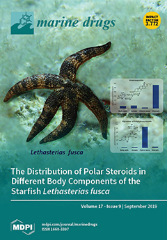
Glycoconjugated and other polar steroids of body walls, gonads, stomach, pyloric caeca, and coelomic fluid of the Far Eastern starfish Lethasterias fusca were studied by nanoflow liquid chromatography/mass spectrometry with captive spray ionization. It has been shown that the levels of polar steroids in the studied body components are qualitatively and quantitatively different. The highest level of polar steroids was found in the stomach. Asterosaponins were found in all body components, the main portion of free polyhydroxysteroids and related glycosides were located in the pyloric caeca. Generally, the obtained data confirmed assumptions about the digestive function of polyhydroxysteroids and protective role of asterosaponins. View this paper.
  • Issues are regarded as officially published after their release is announced to the table of contents alert mailing list.
  • You may sign up for e-mail alerts to receive table of contents of newly released issues.
  • PDF is the official format for papers published in both, html and pdf forms. To view the papers in pdf format, click on the "PDF Full-text" link, and use the free Adobe Reader to open them.
Order results
Result details
Section
Select all
Export citation of selected articles as:
17 pages, 3723 KiB  
Article
Efficiently Anti-Obesity Effects of Unsaturated Alginate Oligosaccharides (UAOS) in High-Fat Diet (HFD)-Fed Mice
by Shangyong Li, Ningning He and Linna Wang
Mar. Drugs 2019, 17(9), 540; https://doi.org/10.3390/md17090540 - 17 Sep 2019
Cited by 56 | Viewed by 4146
Abstract
Obesity and its related complications have become one of the leading problems affecting human health. However, current anti-obesity treatments are limited by high cost and numerous adverse effects. In this study, we investigated the use of a non-toxic green food additive, known as [...] Read more.
Obesity and its related complications have become one of the leading problems affecting human health. However, current anti-obesity treatments are limited by high cost and numerous adverse effects. In this study, we investigated the use of a non-toxic green food additive, known as unsaturated alginate oligosaccharides (UAOS) from the enzymatic degradation of Laminaria japonicais, which showed effective anti-obesity effects in a high-fat diet (HFD) mouse model. Compared with acid hydrolyzed saturated alginate oligosaccharides (SAOS), UAOS significantly reduced body weight, serum lipid, including triacylglycerol (TG), total cholesterol (TC) and free fatty acids (FFA), liver weight, liver TG and TC, serum alanine aminotransferase (ALT), and aspartate aminotransferase (AST) levels, adipose mass, reactive oxygen species (ROS) formation, and accumulation induced in HFD mice. Moreover, the structural differences in β-d-mannuronate (M) and its C5 epimer α-l-guluronate (G) did not cause significant functional differences. Meanwhile, UAOS significantly increased both AMP-activated protein kinase α (AMPKα) and acetyl-CoA carboxylase (ACC) phosphorylation in adipocytes, which indicated that UAOS had an anti-obesity effect mainly through AMPK signaling. Our results indicate that UAOS has the potential for further development as an adjuvant treatment for many metabolic diseases such as fatty liver, hypertriglyceridemia, and possibly diabetes. Full article
(This article belongs to the Collection Marine Polysaccharides)
Show Figures

Figure 1

18 pages, 5106 KiB  
Article
The Inhibitory Effect of Protamine on Platelets is Attenuated by Heparin without Inducing Thrombocytopenia in Rodents
by Joanna Miklosz, Bartlomiej Kalaska, Kamil Kaminski, Malgorzata Rusak, Krzysztof Szczubialka, Maria Nowakowska, Dariusz Pawlak and Andrzej Mogielnicki
Mar. Drugs 2019, 17(9), 539; https://doi.org/10.3390/md17090539 - 17 Sep 2019
Cited by 7 | Viewed by 3723
Abstract
Protamine sulfate (PS) is a polycationic protein drug obtained from the sperm of fish, and is used to reverse the anticoagulant effect of unfractionated heparin (UFH). However, the interactions between PS, UFH, and platelets are still not clear. We measured the platelet numbers [...] Read more.
Protamine sulfate (PS) is a polycationic protein drug obtained from the sperm of fish, and is used to reverse the anticoagulant effect of unfractionated heparin (UFH). However, the interactions between PS, UFH, and platelets are still not clear. We measured the platelet numbers and collagen-induced aggregation, P-selectin, platelet factor 4, β-thromboglobulin, prostacyclin metabolite, D-dimers, activated partial thromboplastin time, prothrombin time, anti-factor Xa, fibrinogen, thrombus weight and megakaryocytopoiesis in blood collected from mice and rats in different time points.. All of the groups were treated intravenously with vehicle, UFH, PS, or UFH with PS. We found a short-term antiplatelet activity of PS in mice and rats, and long-term platelet-independent antithrombotic activity in rats with electrically-induced thrombosis. The antiplatelet and antithrombotic potential of PS may contribute to bleeding risk in PS-overdosed patients. The inhibitory effect of PS on the platelets was attenuated by UFH without inducing thrombocytopenia. Treatment with UFH and PS did not affect the formation, number, or activation of platelets, or the thrombosis development in rodents. Full article
Show Figures

Figure 1

14 pages, 807 KiB  
Article
Effects of Crude Fucus distichus Subspecies evanescens Fucoidan Extract on Retinal Pigment Epithelium Cells―Implications for Use in Age-Related Macular Degeneration
by Kevin Rohwer, Sandesh Neupane, Kaya Saskia Bittkau, Mayra Galarza Pérez, Philipp Dörschmann, Johann Roider, Susanne Alban and Alexa Klettner
Mar. Drugs 2019, 17(9), 538; https://doi.org/10.3390/md17090538 - 16 Sep 2019
Cited by 18 | Viewed by 2864
Abstract
Fucoidan extracts may have beneficial effects in age-related macular degeneration (AMD). Over-the-counter fucoidan preparations are generally undefined, crude extracts. In this study, we investigated the effect of a crude fucoidan extract from Fucus distichus subspecies evanescens (Fe) on the retinal pigment epithelium (RPE). [...] Read more.
Fucoidan extracts may have beneficial effects in age-related macular degeneration (AMD). Over-the-counter fucoidan preparations are generally undefined, crude extracts. In this study, we investigated the effect of a crude fucoidan extract from Fucus distichus subspecies evanescens (Fe) on the retinal pigment epithelium (RPE). Fe extract was investigated for chemical composition and molar mass. It was tested in primary RPE and RPE cell line ARPE19. Oxidative stress was induced with tert-butyl hydroperoxide, cell viability evaluated with MTT assay, VEGF secretion assessed in ELISA. Phagocytosis was evaluated in a fluorescence microscopic assay. Wound healing ability was tested in a scratch assay. Additionally, the inhibition of elastase and complement system by Fe extract was studied. The Fe extract contained about 61.9% fucose and high amounts of uronic acids (26.2%). The sulfate content was not as high as expected (6.9%). It was not toxic and not protective against oxidative stress. However, Fe extract was able to reduce VEGF secretion in ARPE19. Phagocytosis was also reduced. Concerning wound healing, a delay could be observed in higher concentrations. While some beneficial effects could be found, it seems to interfere with RPE function, which may reduce its beneficial effects in AMD treatment. Full article
(This article belongs to the Special Issue Marine Glycobiology, Glycomics and Lectins)
Show Figures

Figure 1

20 pages, 3047 KiB  
Review
Update on Monoterpenes from Red Macroalgae: Isolation, Analysis, and Bioactivity
by Ana-Marija Cikoš, Mladenka Jurin, Rozelindra Čož-Rakovac, Stela Jokić and Igor Jerković
Mar. Drugs 2019, 17(9), 537; https://doi.org/10.3390/md17090537 - 16 Sep 2019
Cited by 12 | Viewed by 4780
Abstract
Macroalgae produce a wide range of monoterpenes as secondary metabolites of mevalonate (MVA) and/or methylerythritol phosphate (MEP) pathway (often including haloperoxidase action). Great biodiversity of macroalgal monoterpenes was reported including acyclic, monocyclic, and bicyclic structures. Halogenated monoterpenes exhibited significant biological activity (e.g., anticancer, [...] Read more.
Macroalgae produce a wide range of monoterpenes as secondary metabolites of mevalonate (MVA) and/or methylerythritol phosphate (MEP) pathway (often including haloperoxidase action). Great biodiversity of macroalgal monoterpenes was reported including acyclic, monocyclic, and bicyclic structures. Halogenated monoterpenes exhibited significant biological activity (e.g., anticancer, antiplasmodial, and insecticidal) that is influenced by the number of present halogens (higher halogen content is preferable, especially bromine) and their position within the monoterpene skeleton. In distinction from the existing reviews, the present review provides novelty with respect to: (a) exclusively monoterpenes from red macroalgae are targeted; (b) biosynthesis, isolation, and analysis, as well as bioactivity of monoterpenes are represented; (c) the methods of their isolation, analysis, and structure elucidation are summarized; (d) the bioactivity of macroalgal monoterpenes is systematically presented with emphasis on anticancer activity; (e) the literature references were updated. Full article
Show Figures

Figure 1

26 pages, 5854 KiB  
Article
Synergistic Cytotoxicity of Renieramycin M and Doxorubicin in MCF-7 Breast Cancer Cells
by Jortan O. Tun, Lilibeth A. Salvador-Reyes, Michael C. Velarde, Naoki Saito, Khanit Suwanborirux and Gisela P. Concepcion
Mar. Drugs 2019, 17(9), 536; https://doi.org/10.3390/md17090536 - 16 Sep 2019
Cited by 29 | Viewed by 6649
Abstract
Renieramycin M (RM) is a KCN-stabilized tetrahydroisoquinoline purified from the blue sponge Xestospongia sp., with nanomolar IC50s against several cancer cell lines. Our goal is to evaluate its combination effects with doxorubicin (DOX) in estrogen receptor positive MCF-7 breast cancer cells. [...] Read more.
Renieramycin M (RM) is a KCN-stabilized tetrahydroisoquinoline purified from the blue sponge Xestospongia sp., with nanomolar IC50s against several cancer cell lines. Our goal is to evaluate its combination effects with doxorubicin (DOX) in estrogen receptor positive MCF-7 breast cancer cells. MCF-7 cells were treated simultaneously or sequentially with various combination ratios of RM and DOX for 72 h. Cell viability was determined using the MTT assay. Synergism or antagonism was determined using curve-shift analysis, combination index method and isobologram analysis. Synergism was observed with pharmacologically achievable concentrations of DOX when administered simultaneously, but not sequentially. The IC95 values of RM and DOX after combination were reduced by up to four-fold and eight-fold, respectively. To gain insights on the mechanism of synergy, real-time profiling, cell cycle analysis, apoptosis assays, and transcriptome analysis were conducted. The combination treatment displayed a similar profile with DNA-damaging agents and induced a greater and faster cell killing. The combination treatment also showed an increase in apoptosis. DOX induced S and G2/M arrest while RM did not induce significant changes in the cell cycle. DNA replication and repair genes were downregulated commonly by RM and DOX. p53 signaling and cell cycle checkpoints were regulated by DOX while ErbB/PI3K-Akt, integrin and focal adhesion signaling were regulated by RM upon combination. Genes involved in cytochrome C release and interferon gamma signaling were regulated specifically in the combination treatment. This study serves as a basis for in vivo studies and provides a rationale for using RM in combination with other anticancer drugs. Full article
Show Figures

Graphical abstract

16 pages, 3760 KiB  
Article
Structure-Function Elucidation of a New α-Conotoxin, MilIA, from Conus milneedwardsi
by Steve Peigneur, Prabha Devi, Andrea Seldeslachts, Samuthirapandian Ravichandran, Loïc Quinton and Jan Tytgat
Mar. Drugs 2019, 17(9), 535; https://doi.org/10.3390/md17090535 - 16 Sep 2019
Cited by 12 | Viewed by 4190
Abstract
The a-Conotoxins are peptide toxins that are found in the venom of marine cone snails and they are potent antagonists of various subtypes of nicotinic acetylcholine receptors (nAChRs). Because nAChRs have an important role in regulating transmitter release, cell excitability, and neuronal integration, [...] Read more.
The a-Conotoxins are peptide toxins that are found in the venom of marine cone snails and they are potent antagonists of various subtypes of nicotinic acetylcholine receptors (nAChRs). Because nAChRs have an important role in regulating transmitter release, cell excitability, and neuronal integration, nAChR dysfunctions have been implicated in a variety of severe pathologies. We describe the isolation and characterization of α-conotoxin MilIA, the first conopeptide from the venom of Conus milneedwardsi. The peptide was characterized by electrophysiological screening against several types of cloned nAChRs that were expressed in Xenopus laevis oocytes. MilIA, which is a member of the α3/5 family, is an antagonist of muscle type nAChRs with a high selectivity for muscle versus neuronal subtype nAChRs. Several analogues were designed and investigated for their activity in order to determine the key epitopes of MilIA. Native MilIA and analogues both showed activity at the fetal muscle type nAChR. Two single mutations (Met9 and Asn10) allowed for MilIA to strongly discriminate between the two types of muscle nAChRs. Moreover, one analogue, MilIA [∆1,M2R, M9G, N10K, H11K], displayed a remarkable enhanced potency when compared to native peptide. The key residues that are responsible for switching between muscle and neuronal nAChRs preference were elucidated. Interestingly, the same analogue showed a preference for α9α10 nAChRs among the neuronal types. Full article
Show Figures

Figure 1

9 pages, 5178 KiB  
Article
New 8-Hydroxybriaranes from the Gorgonian Coral Junceella fragilis (Ellisellidae)
by You-Ying Chen, Lee-Shing Fang, Yu-Hsin Chen, Bo-Rong Peng, Tung-Pin Su, Thanh-Hao Huynh, Feng-Yu Lin, Chiung-Chin Hu, Nai-Cheng Lin, Zhi-Hong Wen, Jih-Jung Chen, Chieh-Yu Lee, Jin-Wei Wang and Ping-Jyun Sung
Mar. Drugs 2019, 17(9), 534; https://doi.org/10.3390/md17090534 - 14 Sep 2019
Cited by 7 | Viewed by 3188
Abstract
Three new 8-hydroxybriaranes—fragilides R–T (13) were obtained from a sea whip gorgonian coral Junceella fragilis. The structures of briaranes 13 were elucidated by using spectroscopic methods, including 1D (1H and 13C NMR), 2D [...] Read more.
Three new 8-hydroxybriaranes—fragilides R–T (13) were obtained from a sea whip gorgonian coral Junceella fragilis. The structures of briaranes 13 were elucidated by using spectroscopic methods, including 1D (1H and 13C NMR), 2D (COSY, HSQC, HMBC, and NOESY experiments) NMR studies, and (+)-HRESIMS. Fragilides S and T (2 and 3) are the only briaranes known to possess 8α-hydroxy and 17β-methyl groups, respectively. Briarane 2 exerted an inhibition effect on iNOS release from RAW264.7; a macrophage cell line that originated from a mouse monocyte macrophage, stimulated with lipopolysaccharides. Full article
(This article belongs to the Special Issue Terpenoids from Marine Organisms II)
Show Figures

Graphical abstract

19 pages, 1198 KiB  
Article
A New Look for the Red Macroalga Palmaria palmata: A Seafood with Polar Lipids Rich in EPA and with Antioxidant Properties
by Diana Lopes, Tânia Melo, Joana Meneses, Maria H. Abreu, Rui Pereira, Pedro Domingues, Ana I. Lillebø, Ricardo Calado and M. Rosário Domingues
Mar. Drugs 2019, 17(9), 533; https://doi.org/10.3390/md17090533 - 13 Sep 2019
Cited by 35 | Viewed by 4938
Abstract
Palmaria palmata is an edible red macroalga widely used for human consumption and valued for its high protein value. Despite its low total lipid content, it is rich in eicosapentaenoic acid (EPA). This seaweed has been scarcely explored with regard to its lipid [...] Read more.
Palmaria palmata is an edible red macroalga widely used for human consumption and valued for its high protein value. Despite its low total lipid content, it is rich in eicosapentaenoic acid (EPA). This seaweed has been scarcely explored with regard to its lipid composition. The polar lipids of seaweeds are nowadays recognized as important phytochemicals contributing to their add value valorization and providing support for claims of potential health benefits. The present study aimed to disclose the polar lipid profile of P. palmata, farmed in an integrated multi-trophic aquaculture (IMTA) through modern lipidomic approaches using high-resolution LC-MS and MS/MS and to screen for the antioxidant properties of this red macroalga. A total of 143 molecular species of lipids were identified, belonging to several classes of polar lipids, such as glycolipids, phospholipids, and betaine lipids. It is noteworthy that the most abundant lipid species in each class were esterified with eicosapentaenoic acid (EPA), accounting for more than 50% of the lipid content. The polar lipid extract rich in EPA showed antioxidant activity with an inhibition concentration (IC) of IC30 = 171 ± 19.8 µg/mL for α,α-diphenyl-β-picrylhydrazyl radical (DPPH) and IC50 = 26.2 ± 0.1 µg/mL for 2,20-azino-bis-3-ethylbenzothiazoline-6-sulfonic acid radical cation (ABTS●+). Overall, this study highlights that P. palmata farmed in an IMTA framework can be a sustainable source of beneficial lipids with antioxidant activity. Moreover, this red macroalga can be exploited for future applications as a source of lipids rich in EPA for food and feed, nutraceuticals, and cosmetics. Full article
Show Figures

Figure 1

13 pages, 2150 KiB  
Article
The Anti-Obesity Effect of Polysaccharide-Rich Red Algae (Gelidium amansii) Hot-Water Extracts in High-Fat Diet-Induced Obese Hamsters
by Tsung-Han Yang, Chen-Yuan Chiu, Ting-Jang Lu, Shing-Hwa Liu and Meng-Tsan Chiang
Mar. Drugs 2019, 17(9), 532; https://doi.org/10.3390/md17090532 - 13 Sep 2019
Cited by 30 | Viewed by 3607
Abstract
This study investigated the anti-obesity effect of a polysaccharide-rich red algae Gelidium amansii hot-water extract (GHE) in high-fat (HF) diet-induced obese hamsters. GHE contained 68.54% water-soluble indigestible carbohydrate polymers. Hamsters were fed with a HF diet for 5 weeks to induce obesity, and [...] Read more.
This study investigated the anti-obesity effect of a polysaccharide-rich red algae Gelidium amansii hot-water extract (GHE) in high-fat (HF) diet-induced obese hamsters. GHE contained 68.54% water-soluble indigestible carbohydrate polymers. Hamsters were fed with a HF diet for 5 weeks to induce obesity, and then randomly divided into: HF group, HF with 3% guar gum diet group, HF with 3% GHE diet group, and HF with orlistat (200 mg/kg diet) group for 9 weeks. The increased weights of body, liver, and adipose in the HF group were significantly reversed by GHE supplementation. Lower plasma leptin, tumor necrosis factor-α, and interleukin-6 levels were observed in the GHE+HF group compared to the HF group. GHE also increased the lipolysis rate and decreased the lipoprotein lipase activity in adipose tissues. GHE induced an increase in the phosphorylation of AMP-activated protein kinase (AMPK) and the protein expressions of peroxisome proliferator-activated receptor alpha (PPARα) and uncoupling protein (UCP)-2 in the livers. The decreased triglyceride and total cholesterol in the plasma and liver were also observed in obese hamsters fed a diet with GHE. These results suggest that GHE exerts a down-regulation effect on hepatic lipid metabolism through AMPK phosphorylation and up-regulation of PPARα and UCP-2 in HF-induced obese hamsters. Full article
Show Figures

Graphical abstract

17 pages, 3279 KiB  
Article
Antioxidant Peptides from the Protein Hydrolysate of Spanish Mackerel (Scomberomorous niphonius) Muscle by in Vitro Gastrointestinal Digestion and Their In Vitro Activities
by Guo-Xu Zhao, Xiu-Rong Yang, Yu-Mei Wang, Yu-Qin Zhao, Chang-Feng Chi and Bin Wang
Mar. Drugs 2019, 17(9), 531; https://doi.org/10.3390/md17090531 - 12 Sep 2019
Cited by 27 | Viewed by 3399
Abstract
For the full use of Spanish mackerel (Scomberomorous niphonius) muscle to produce antioxidant peptides, the proteins of Spanish mackerel muscle were separately hydrolyzed under five kinds of enzymes and in vitro gastrointestinal digestion, and antioxidant peptides were isolated from the protein [...] Read more.
For the full use of Spanish mackerel (Scomberomorous niphonius) muscle to produce antioxidant peptides, the proteins of Spanish mackerel muscle were separately hydrolyzed under five kinds of enzymes and in vitro gastrointestinal digestion, and antioxidant peptides were isolated from the protein hydrolysate using ultrafiltration and multiple chromatography methods. The results showed that the hydrolysate (SMPH) prepared using in vitro GI digestion showed the highest degree of hydrolysis (27.45 ± 1.76%) and DPPH radical scavenging activity (52.58 ± 2.68%) at the concentration of 10 mg protein/mL among the six protein hydrolysates, and 12 peptides (SMP-1 to SMP-12) were prepared from SMPH. Among them, SMP-3, SMP-7, SMP-10, and SMP-11 showed the higher DPPH radical scavenging activities and were identified as Pro-Glu-Leu-Asp-Trp (PELDW), Trp-Pro-Asp-His-Trp (WPDHW), and Phe-Gly-Tyr-Asp-Trp-Trp (FGYDWW), and Tyr-Leu-His-Phe-Trp (YLHFW), respectively. PELDW, WPDHW, FGYDWW, and YLHFW showed high scavenging activities on DPPH radical (EC50 1.53, 0.70, 0.53, and 0.97 mg/mL, respectively), hydroxyl radical (EC50 1.12, 0.38, 0.26, and 0.67 mg/mL, respectively), and superoxide anion radical (EC50 0.85, 0.49, 0.34, and 1.37 mg/mL, respectively). Moreover, PELDW, WPDHW, FGYDWW, and YLHFW could dose-dependently inhibit lipid peroxidation in the linoleic acid model system and protect plasmid DNA (pBR322DNA) against oxidative damage induced by H2O2 in the tested model systems. In addition, PELDW, WPDHW, FGYDWW, and YLHFW could retain their high activities when they were treated under a low temperature (<60 °C) and a moderate pH environment (pH 5–9). These present results indicate that the protein hydrolysate, fractions, and isolated peptides from Spanish mackerel muscle have strong antioxidant activity and might have the potential to be used in health food products. Full article
Show Figures

Figure 1

8 pages, 3127 KiB  
Article
New 1,4-Dienonesteroids from the Octocoral Dendronephthya sp.
by Thanh-Hao Huynh, Pei-Chin Chen, San-Nan Yang, Feng-Yu Lin, Tung-Pin Su, Lo-Yun Chen, Bo-Rong Peng, Chiung-Chin Hu, You-Ying Chen, Zhi-Hong Wen, Tung-Ying Wu and Ping-Jyun Sung
Mar. Drugs 2019, 17(9), 530; https://doi.org/10.3390/md17090530 - 11 Sep 2019
Cited by 6 | Viewed by 2611
Abstract
Two new steroids, dendronesterones D (1) and E (2), featuring with 1,4-dienone moiety, along with three known steroids, methyl 3-oxochola-4,22-diene-24-oate (3), 5α,8α-epidioxy-24(S)- methylcholesta-6,22-dien-3β-ol (4), and 5α,8α-epidioxy-24(S)-methylcholesta-6,9(11),22-trien-3β-ol (5), were isolated [...] Read more.
Two new steroids, dendronesterones D (1) and E (2), featuring with 1,4-dienone moiety, along with three known steroids, methyl 3-oxochola-4,22-diene-24-oate (3), 5α,8α-epidioxy-24(S)- methylcholesta-6,22-dien-3β-ol (4), and 5α,8α-epidioxy-24(S)-methylcholesta-6,9(11),22-trien-3β-ol (5), were isolated from an octocoral Dendronephthya sp. The structures of steroids 1 and 2 were elucidated by using spectroscopic methods and steroid 1 was found to exhibit significant in vitro anti-inflammatory activity in lipopolysaccharides (LPS)-induced RAW264.7 macrophage cells by inhibiting the expression of the iNOS protein. Full article
(This article belongs to the Special Issue Bioactive Compounds from Coral Reef Organisms)
Show Figures

Graphical abstract

11 pages, 1425 KiB  
Article
Antimicrobial Lavandulylated Flavonoids from a Sponge-Derived Streptomyces sp. G248 in East Vietnam Sea
by Duc Danh Cao, Thi Thanh Van Trinh, Huong Doan Thi Mai, Van Nam Vu, Hong Minh Le, Quyen Vu Thi, Mai Anh Nguyen, Thu Trang Duong, Dang Thach Tran, Van Minh Chau, Rui Ma, Gauri Shetye, Sanghyun Cho, Brian T. Murphy and Van Cuong Pham
Mar. Drugs 2019, 17(9), 529; https://doi.org/10.3390/md17090529 - 10 Sep 2019
Cited by 16 | Viewed by 4367
Abstract
Three new lavandulylated flavonoids, (2S,2″S)-6-lavandulyl-7,4′-dimethoxy-5,2′-dihydroxylflavanone (1), (2S,2″S)-6-lavandulyl-5,7,2′,4′-tetrahydroxylflavanone (2), and (2″S)-5′-lavandulyl-2′-methoxy-2,4,4′,6′-tetrahydroxylchalcone (3), along with seven known compounds 4–10 were isolated from culture broth of Streptomyces sp. G248. Their [...] Read more.
Three new lavandulylated flavonoids, (2S,2″S)-6-lavandulyl-7,4′-dimethoxy-5,2′-dihydroxylflavanone (1), (2S,2″S)-6-lavandulyl-5,7,2′,4′-tetrahydroxylflavanone (2), and (2″S)-5′-lavandulyl-2′-methoxy-2,4,4′,6′-tetrahydroxylchalcone (3), along with seven known compounds 4–10 were isolated from culture broth of Streptomyces sp. G248. Their structures were established by spectroscopic data analysis, including 1D and 2D nuclear magnetic resonance (NMR), and high-resolution electrospray ionization mass spectrometry (HR-ESI-MS). The absolute configurations of 1–3 were resolved by comparison of their experimental and calculated electronic circular dichroism spectra. Compounds 1–3 exhibited remarkable antimicrobial activity. Whereas, two known compounds 4 and 5 exhibited inhibitory activity against Mycobacterium tuberculosis H37Rv with minimum inhibitory concentration (MIC) values of 6.0 µg/mL and 11.1 µg/mL, respectively. Full article
Show Figures

Graphical abstract

22 pages, 4535 KiB  
Article
Identification and Characterization of a Novel Protein ASP-3 Purified from Arca subcrenata and Its Antitumor Mechanism
by Zhongyi Guo, Hui Shi, Chunlei Li, Yuanyuan Luo, Sixue Bi, Rongmin Yu, Haoran Wang, Wanying Liu, Jianhua Zhu, Weijuan Huang and Liyan Song
Mar. Drugs 2019, 17(9), 528; https://doi.org/10.3390/md17090528 - 09 Sep 2019
Cited by 6 | Viewed by 3113
Abstract
Diverse bioactive substances derived from marine organisms have been attracting growing attention. Besides small molecules and polypeptides, numerous studies have shown that marine proteins also exhibit antitumor activities. Small anticancer proteins can be expressed in vivo by viral vectors to exert local and [...] Read more.
Diverse bioactive substances derived from marine organisms have been attracting growing attention. Besides small molecules and polypeptides, numerous studies have shown that marine proteins also exhibit antitumor activities. Small anticancer proteins can be expressed in vivo by viral vectors to exert local and long-term anticancer effects. Herein, we purified and characterized a novel protein (ASP-3) with unique antitumor activity from Arca subcrenata Lischke. The ASP-3 contains 179 amino acids with a molecular weight of 20.6 kDa. The spectral characterization of ASP-3 was elucidated using Fourier Transform infrared spectroscopy (FTIR) and Circular Dichroism (CD) spectroscopy. Being identified as a sarcoplasmic calcium-binding protein, ASP-3 exhibited strong inhibitory effects on the proliferation of Human hepatocellular carcinoma (HepG2) cells with an IC50 value of 171.18 ± 18.59 μg/mL, measured by 3-(4,5-dimethylthiazol-2-yl)-2,5-diphenyltetrazolium bromide (MTT) assay. The RNA-seq analysis showed that ASP-3 regulated the vascular endothelial growth factor receptor (VEGFR) signaling pathway in HepG2 cells. Immunofluorescence results indicated that ASP-3 effectively reduced VEGFR2 phosphorylation in HepG2 cells and affected the downstream components of VEGF signaling pathways. The surface plasmon resonance (SPR) analysis further demonstrated that ASP-3 direct interacted with VEGFR2. More importantly, the therapeutic potential of ASP-3 as an anti-angiogenesis agent was further confirmed by an in vitro model using VEGF-induced tube formation assay of human umbilical vein endothelial cells (HUVECs), as well as an in vivo model using transgenic zebrafish model. Taken together, the ASP-3 provides a good framework for the development of even more potent anticancer proteins and provides important weapon for cancer treatment using novel approaches such as gene therapy. Full article
Show Figures

Figure 1

25 pages, 1317 KiB  
Review
Bioactive Aliphatic Sulfates from Marine Invertebrates
by Luis C. Kellner Filho, Bruno W. Picão, Marcio L. A. Silva, Wilson R. Cunha, Patricia M. Pauletti, Gustavo M. Dias, Brent R. Copp, Camila S. Bertanha and Ana H. Januario
Mar. Drugs 2019, 17(9), 527; https://doi.org/10.3390/md17090527 - 09 Sep 2019
Cited by 13 | Viewed by 2721
Abstract
The occurrence of sulfated steroids and phenolics in marine organisms is quite widespread, being typically reported from Echinoderms. In contrast, alkane and alkene aliphatic sulfates are considerably rarer with examples being reported from a diverse array of organisms including echinoderms, sponges and ascidians. [...] Read more.
The occurrence of sulfated steroids and phenolics in marine organisms is quite widespread, being typically reported from Echinoderms. In contrast, alkane and alkene aliphatic sulfates are considerably rarer with examples being reported from a diverse array of organisms including echinoderms, sponges and ascidians. While no ecological roles for these metabolites have been proposed, they do exhibit a diverse array of biological activities including thrombin inhibition; the ability to induce metamorphosis in larvae; antiproliferative, antibacterial and antifungal properties; and metalloproteinase inhibition. Of particular interest and an avenue for future development is the finding of antifouling properties with low or nontoxic effects to the environment. This review focuses on alkyl sulfates and related sulfamates, their structures and biological activities. Spectroscopic and spectrometric techniques that can be used to recognize the presence of sulfate groups are also discussed, data for which will enhance the ability of researchers to recognize this class of chemically- and biologically-interesting marine natural products. Full article
Show Figures

Figure 1

14 pages, 2935 KiB  
Article
Echinochrome A Promotes Ex Vivo Expansion of Peripheral Blood-Derived CD34+ Cells, Potentially through Downregulation of ROS Production and Activation of the Src-Lyn-p110δ Pathway
by Ga-Bin Park, Min-Jung Kim, Elena A. Vasileva, Natalia P. Mishchenko, Sergey A. Fedoreyev, Valentin A. Stonik, Jin Han, Ho Sup Lee, Daejin Kim and Jee-Yeong Jeong
Mar. Drugs 2019, 17(9), 526; https://doi.org/10.3390/md17090526 - 09 Sep 2019
Cited by 14 | Viewed by 3538
Abstract
Intracellular reactive oxygen species (ROS) play an important role in the proliferation and differentiation of hematopoietic stem and progenitor cells (HSPCs). HSPCs are difficult to be expanded ex vivo while maintaining their stemness when they are exposed to oxidative damage after being released [...] Read more.
Intracellular reactive oxygen species (ROS) play an important role in the proliferation and differentiation of hematopoietic stem and progenitor cells (HSPCs). HSPCs are difficult to be expanded ex vivo while maintaining their stemness when they are exposed to oxidative damage after being released from the bone marrow. There have been efforts to overcome this limitation by using various cytokine cocktails and antioxidants. In this study, we investigated the effects of echinochrome A (Ech A)-a well-established and non-toxic antioxidant-on the ex vivo expansion of HSPCs by analyzing a CD34+ cell population and their biological functions. We observed that Ech A-induced suppression of ROS generation and p38-MAPK/JNK phosphorylation causes increased expansion of CD34+ cells. Moreover, p38-MAPK/JNK inhibitors SB203580 and SP600125 promoted ex vivo expansion of CD34+ cells. We also demonstrated that the activation of Lyn kinase and p110δ is a novel mechanism for Ech A to enhance ex vivo expansion of CD34+ cells. Ech A upregulated phospho-Src, phospho-Lyn, and p110δ expression. Furthermore, the Ech A-induced ex vivo expansion of CD34+ cells was inhibited by pretreatment with the Src family inhibitor PP1 and p110δ inhibitor CAL-101; PP1 blocked p110δ upregulation and PI3K/Akt activation, whereas CAL-101 and PI3K/Akt pathway inhibitor LY294002 did not block Src/Lyn activation. These results suggest that Ech A initially induces Src/Lyn activation, upregulates p110δ expression, and finally activates the PI3K/Akt pathway. CD34+ cells expanded in the presence of Ech A produced equal or more hematopoietic colony-forming cells than unexpanded CD34+ cells. In conclusion, Ech A promotes the ex vivo expansion of CD34+ cells through Src/Lyn-mediated p110δ expression, suppression of ROS generation, and p38-MAPK/JNK activation. Hence, Ech A is a potential candidate modality for the ex vivo, and possibly in vivo, expansion of CD34+ cells. Full article
(This article belongs to the Special Issue Selected Papers from the 3rd International Symposium on Life Science)
Show Figures

Figure 1

16 pages, 5018 KiB  
Article
Epigenetic Modification and Differentiation Induction of Malignant Glioma Cells by Oligo-Fucoidan
by Chien-Huang Liao, I-Chun Lai, Hui-Ching Kuo, Shuang-En Chuang, Hsin-Lun Lee, Jacqueline Whang-Peng, Chih-Jung Yao and Gi-Ming Lai
Mar. Drugs 2019, 17(9), 525; https://doi.org/10.3390/md17090525 - 08 Sep 2019
Cited by 15 | Viewed by 4588
Abstract
Malignant glioma (MG) is a poor prognostic brain tumor with inevitable recurrence after multimodality treatment. Searching for more effective treatment is urgently needed. Differentiation induction via epigenetic modification has been proposed as a potential anticancer strategy. Natural products are known as fruitful sources [...] Read more.
Malignant glioma (MG) is a poor prognostic brain tumor with inevitable recurrence after multimodality treatment. Searching for more effective treatment is urgently needed. Differentiation induction via epigenetic modification has been proposed as a potential anticancer strategy. Natural products are known as fruitful sources of epigenetic modifiers with wide safety margins. We thus explored the effects of oligo-fucoidan (OF) from brown seaweed on this notion in MG cells including Grade III U87MG cells and Grade IV glioblastoma multiforme (GBM)8401 cells and compared to the immortalized astrocyte SVGp12 cells. The results showed that OF markedly suppress the proliferation of MG cells and only slightly affected that of SVGp12 cells. OF inhibited the protein expressions of DNA methyltransferases 1, 3A and 3B (DNMT1, 3A and 3B) accompanied with obvious mRNA induction of differentiation markers (MBP, OLIG2, S100β, GFAP, NeuN and MAP2) both in U87MG and GBM8401 cells. Accordingly, the methylation of p21, a DNMT3B target gene, was decreased by OF. In combination with the clinical DNMT inhibitor decitabine, OF could synergize the growth inhibition and MBP induction in U87MG cells. Appropriated clinical trials are warranted to evaluate this potential complementary approach for MG therapy after confirmation of the effects in vivo. Full article
(This article belongs to the Special Issue Marine Glycobiology, Glycomics and Lectins)
Show Figures

Figure 1

13 pages, 1207 KiB  
Review
Haloarchaeal Carotenoids: Healthy Novel Compounds from Extreme Environments
by Micaela Giani, Inés Garbayo, Carlos Vílchez and Rosa María Martínez-Espinosa
Mar. Drugs 2019, 17(9), 524; https://doi.org/10.3390/md17090524 - 06 Sep 2019
Cited by 69 | Viewed by 5154
Abstract
Haloarchaea are halophilic microorganisms belonging to the archaea domain that inhabit salty environments (mainly soils and water) all over the world. Most of the genera included in this group can produce carotenoids at significant concentrations (even wild-type strains). The major carotenoid produced by [...] Read more.
Haloarchaea are halophilic microorganisms belonging to the archaea domain that inhabit salty environments (mainly soils and water) all over the world. Most of the genera included in this group can produce carotenoids at significant concentrations (even wild-type strains). The major carotenoid produced by the cells is bacterioruberin (and its derivatives), which is only produced by this kind of microbes and few bacteria, like Micrococcus roseus. Nevertheless, the understanding of carotenoid metabolism in haloarchaea, its regulation, and the roles of carotenoid derivatives in this group of extreme microorganisms remains mostly unrevealed. Besides, potential biotechnological uses of haloarchaeal pigments are poorly explored. This work summarises what it has been described so far about carotenoids from haloarchaea and their production at mid- and large-scale, paying special attention to the most recent findings on the potential uses of haloarchaeal pigments in biomedicine. Full article
14 pages, 709 KiB  
Article
The Distribution of Asterosaponins, Polyhydroxysteroids and Related Glycosides in Different Body Components of the Far Eastern Starfish Lethasterias fusca
by Roman S. Popov, Natalia V. Ivanchina, Alla A. Kicha, Timofey V. Malyarenko, Boris B. Grebnev, Valentin A. Stonik and Pavel S. Dmitrenok
Mar. Drugs 2019, 17(9), 523; https://doi.org/10.3390/md17090523 - 06 Sep 2019
Cited by 8 | Viewed by 2439
Abstract
Glycoconjugated and other polar steroids of starfish have unique chemical structures and show a broad spectrum of biological activities. However, their biological functions remain not well established. Possible biological roles of these metabolites might be indicated by the studies on their distribution in [...] Read more.
Glycoconjugated and other polar steroids of starfish have unique chemical structures and show a broad spectrum of biological activities. However, their biological functions remain not well established. Possible biological roles of these metabolites might be indicated by the studies on their distribution in the organism–producer. In order to investigate the localization of polar steroids in body components of the Far Eastern starfish Lethasterias fusca, chemical constituents of body walls, gonads, stomach, pyloric caeca, and coelomic fluid were studied by nanoflow liquid chromatography/mass spectrometry with captive spray ionization (nLC/CSI–QTOF–MS). It has been shown that the levels of polar steroids in the studied body components are qualitatively and quantitatively different. Generally, the obtained data confirmed earlier made assumptions about the digestive function of polyhydroxysteroids and protective role of asterosaponins. The highest level of polar steroids was found in the stomach. Asterosaponins were found in all body components, the main portion of free polyhydroxysteroids and related glycosides were located in the pyloric caeca. In addition, a great inter-individual variability was found in the content of most polar steroids, which may be associated with the peculiarities in their individual physiologic status. Full article
(This article belongs to the Special Issue Marine Glycoconjugates: Trends and Perspectives)
Show Figures

Figure 1

10 pages, 1616 KiB  
Article
Penicamide A, A Unique N,N′-Ketal Quinazolinone Alkaloid from Ascidian-Derived Fungus Penicillium sp. 4829
by Senhua Chen, Minghua Jiang, Bin Chen, Jintana Salaenoi, Shah-Iram Niaz, Jianguo He and Lan Liu
Mar. Drugs 2019, 17(9), 522; https://doi.org/10.3390/md17090522 - 05 Sep 2019
Cited by 19 | Viewed by 3123
Abstract
Previously unreported N,N′-ketal quinazolinone enantiomers [(−)-1 and (+)-1] and a new biogenetically related compound (2), along with six known compounds, 2-pyrovoylaminobenzamide (3), N-(2-hydroxypropanoyl)-2 amino benzoic acid amide (4), pseurotin A [...] Read more.
Previously unreported N,N′-ketal quinazolinone enantiomers [(−)-1 and (+)-1] and a new biogenetically related compound (2), along with six known compounds, 2-pyrovoylaminobenzamide (3), N-(2-hydroxypropanoyl)-2 amino benzoic acid amide (4), pseurotin A (5), niacinamide (6), citreohybridonol (7), citreohybridone C (8) were isolated from the ascidian-derived fungus Penicillium sp. 4829 in wheat solid-substrate medium culture. Their structures were elucidated by a combination of spectroscopic analyses (1D and 2D NMR and Electron Circular Dichroism data) and X-ray crystallography. The enantiomeric pair of 1 is the first example of naturally occurring N,N′-ketal quinazolinone possessing a unique tetracyclic system having 4-quinazolinone fused with tetrahydroisoquinoline moiety. The enantiomeric mixtures of 1 displayed an inhibitory effect on NO production in lipopolysaccharide-activated RAW264.7 cells, while the optically pure (–)-1 showed better inhibitory effect than (+)-1. Full article
(This article belongs to the Special Issue Advances in Marine Alkaloids)
Show Figures

Graphical abstract

17 pages, 2889 KiB  
Article
First Results from a Screening of 300 Naturally Occurring Compounds: 4,6-dibromo-2-(2′,4′-dibromophenoxy)phenol, 4,5,6-tribromo-2-(2′,4′-dibromophenoxy)phenol, and 5-epi-nakijinone Q as Substances with the Potential for Anticancer Therapy
by Saskia Mayer, Marie Prechtl, Pia Liebfried, Ron-Patrick Cadeddu, Fabian Stuhldreier, Matthias Kohl, Folker Wenzel, Björn Stork, Sebastian Wesselborg, Peter Proksch, Ulrich Germing, Rainer Haas and Paul Jäger
Mar. Drugs 2019, 17(9), 521; https://doi.org/10.3390/md17090521 - 05 Sep 2019
Cited by 9 | Viewed by 3223
Abstract
There is a variety of antineoplastic drugs that are based on natural compounds from ecological niches with high evolutionary pressure. We used two cell lines (Jurkat J16 and Ramos) in a screening to assess 300 different naturally occurring compounds with regard to their [...] Read more.
There is a variety of antineoplastic drugs that are based on natural compounds from ecological niches with high evolutionary pressure. We used two cell lines (Jurkat J16 and Ramos) in a screening to assess 300 different naturally occurring compounds with regard to their antineoplastic activity. The results of the compounds 4,6-dibromo-2-(2′,4′-dibromophenoxy)phenol (P01F03), 4,5,6-tribromo-2-(2′,4′-dibromophenoxy)phenol (P01F08), and 5-epi-nakijinone Q (P03F03) prompted us to perform further research. Using viability and apoptosis assays on the cell lines of primary human leukemic and normal hematopoietic cells, we found that P01F08 induced apoptosis in the cell lines at IC50 values between 1.61 and 2.95 μM after 72 h. IC50 values of peripheral blood mononuclear cells (PBMNCs) from healthy donors were higher, demonstrating that the cytotoxicity in the cell lines reached 50%, while normal PBMNCs were hardly affected. The colony-forming unit assay showed that the hematopoietic progenitor cells were not significantly affected in their growth by P01F08 at a concentration of 3 μM. P01F08 showed a 3.2-fold lower IC50 value in primary leukemic cells [acute myeloid leukemia (AML)] compared to the PBMNC of healthy donors. We could confirm the antineoplastic effect of 5-epi-nakijinone Q (P03F03) on the cell lines via the induction of apoptosis but noted a similarly strong cytotoxic effect on normal PBMNCs. Full article
Show Figures

Figure 1

22 pages, 3934 KiB  
Article
N-Terminal Acetylation and C-Terminal Amidation of Spirulina platensis-Derived Hexapeptide: Anti-Photoaging Activity and Proteomic Analysis
by Qiaohui Zeng, Jianguo Jiang, Jingjing Wang, Qiuchan Zhou and Xuewu Zhang
Mar. Drugs 2019, 17(9), 520; https://doi.org/10.3390/md17090520 - 04 Sep 2019
Cited by 9 | Viewed by 3377
Abstract
Ultraviolet (UV) irradiation is a potent inducer for skin photoaging. This paper investigated the anti-photoaging effects of the acetylated and amidated hexapeptide (AAH), originally identified from Spirulina platensis, in (Ultraviolet B) UVB-irradiated Human immortalized keratinocytes (Hacats) and mice. The results demonstrated that [...] Read more.
Ultraviolet (UV) irradiation is a potent inducer for skin photoaging. This paper investigated the anti-photoaging effects of the acetylated and amidated hexapeptide (AAH), originally identified from Spirulina platensis, in (Ultraviolet B) UVB-irradiated Human immortalized keratinocytes (Hacats) and mice. The results demonstrated that AAH had much lower toxicity on Hacats than the positive matrixyl (81.52% vs. 5.32%). Moreover, AAH reduced MDA content by 49%; increased SOD, CAT, and GSH-Px activities by 103%, 49%, and 116%, respectively; decreased MMP-1 and MMP-3 expressions by 27% and 29%, respectively, compared to UVB-irradiated mice. Employing isobaric tags for relative and absolute quantitation (iTRAQ)-based proteomics, 60 differential proteins were identified, and major metabolic pathways were determined. Network analysis indicated that these differential proteins were mapped into an interaction network composed of two core sub-networks. Collectively, AAH is protective against UVB-induced skin photoaging and has potential application in skin care cosmetics. Full article
Show Figures

Figure 1

21 pages, 3575 KiB  
Article
Phosphorus-Induced Lipid Class Alteration Revealed by Lipidomic and Transcriptomic Profiling in Oleaginous Microalga Nannochloropsis sp. PJ12
by Jibei Liang, Sunya Iqbal, Fang Wen, Mengmeng Tong and Jianhua Liu
Mar. Drugs 2019, 17(9), 519; https://doi.org/10.3390/md17090519 - 03 Sep 2019
Cited by 12 | Viewed by 3332
Abstract
Phytoplankton are primary producers in the marine ecosystem, where phosphorus is often a limiting factor of their growth. Hence, they have evolved strategies to recycle phosphorus by replacing membrane phospholipids with phosphorus-free lipids. However, mechanisms for replacement of lipid classes remain poorly understood. [...] Read more.
Phytoplankton are primary producers in the marine ecosystem, where phosphorus is often a limiting factor of their growth. Hence, they have evolved strategies to recycle phosphorus by replacing membrane phospholipids with phosphorus-free lipids. However, mechanisms for replacement of lipid classes remain poorly understood. To improve our understanding, we performed the lipidomic and transcriptomic profiling analyses of an oleaginous marine microalga Nannochloropsis sp. PJ12 in response to phosphorus depletion (PD) and replenishing. In this study, by using (liquid chromatography couple with tandem mass spectrometry) LC-MS/MS-based lipidomic analysis, we show that membrane phospholipid levels are significantly reduced upon PD, while phosphorus-free betaine lipid levels are increased. However, levels of phosphorus-free photosynthetic galactolipid and sulfolipid are not increased upon PD, consistent with the reduced photosynthetic activity. RNA-seq-based transcriptomic analysis indicates that enzymes involved in phospholipid recycling and phosphorus-free lipid synthesis are upregulated, supporting the lipidomic analysis. Furthermore, enzymes involved in FASII (type II fatty acid synthesis) elongation cycle upon PD are transcriptionally downregulated. EPA (eicosapentaenoic acid) level decrease upon PD is revealed by both GC-MS (gas chromatography coupled with mass spectrometry) and LC-MS/MS-based lipidomic analyses. PD-induced alteration is reversed after phosphorus replenishing. Taken together, our results suggest that the alteration of lipid classes upon environmental change of phosphorus is a result of remodeling rather than de novo synthesis in Nannochloropsis sp. PJ12. Full article
Show Figures

Figure 1

15 pages, 2740 KiB  
Article
Fucoidan Suppresses Mitochondrial Dysfunction and Cell Death against 1-Methyl-4-Phenylpyridinum-Induced Neuronal Cytotoxicity via Regulation of PGC-1α Expression
by Yong-Seok Han, Jun Hee Lee and Sang Hun Lee
Mar. Drugs 2019, 17(9), 518; https://doi.org/10.3390/md17090518 - 02 Sep 2019
Cited by 14 | Viewed by 3376
Abstract
Mitochondria are considered to be the powerhouses of cells. They are the most commonly damaged organelles within dopaminergic neurons in patients with Parkinson’s disease (PD). Despite the importance of protecting neuronal mitochondria in PD patients, the detailed mechanisms underlying mitochondrial dysfunction during pathogenesis [...] Read more.
Mitochondria are considered to be the powerhouses of cells. They are the most commonly damaged organelles within dopaminergic neurons in patients with Parkinson’s disease (PD). Despite the importance of protecting neuronal mitochondria in PD patients, the detailed mechanisms underlying mitochondrial dysfunction during pathogenesis and pathophysiological progression of PD have not yet been elucidated. We investigated the protective action of fucoidan against the detrimental action of 1-methyl-4-phenyl-pyridinium (MPP+), a neurotoxin used to model PD, in the mitochondria of SH-SY5Y neural cells. Fucoidan increased the expression of peroxisome proliferator-activated receptor gamma coactivator-1 alpha (PGC-1α) and protected the cells from MPP+-induced apoptosis by upregulating the 5′ adenosine monophosphate-activated protein kinase (AMPK)-PGC-1α axis. These effects were blocked by the silencing of the PGC-1α axis. These results indicated that fucoidan protects SH-SY5Y cells from mitochondrial dysfunction and cell death caused by MPP+ treatment, via the AMPK-PGC-1α axis. These findings also suggest that fucoidan could potentially be used as a therapeutic agent for PD. Full article
Show Figures

Figure 1

15 pages, 5183 KiB  
Article
Characterization and Antibiofilm Activity of Mannitol–Chitosan-Blended Paste for Local Antibiotic Delivery System
by Leslie R. Pace, Zoe L. Harrison, Madison N. Brown, Warren O. Haggard and J. Amber Jennings
Mar. Drugs 2019, 17(9), 517; https://doi.org/10.3390/md17090517 - 02 Sep 2019
Cited by 12 | Viewed by 3446
Abstract
Mannitol, a polyalcohol bacterial metabolite, has been shown to activate dormant persister cells within bacterial biofilm. This study sought to evaluate an injectable blend of mannitol, chitosan, and polyethylene glycol for delivery of antibiotics and mannitol for eradication of Staphylococcal biofilm. Mannitol blends [...] Read more.
Mannitol, a polyalcohol bacterial metabolite, has been shown to activate dormant persister cells within bacterial biofilm. This study sought to evaluate an injectable blend of mannitol, chitosan, and polyethylene glycol for delivery of antibiotics and mannitol for eradication of Staphylococcal biofilm. Mannitol blends were injectable and had decreased dissociation and degradation in the enzyme lysozyme compared to blends without mannitol. Vancomycin and amikacin eluted in a burst response, with active concentrations extended to seven days compared to five days for blends without mannitol. Mannitol eluted from the paste in a burst the first day and continued through Day 4. Eluates from the mannitol pastes with and without antibiotics decreased viability of established S. aureus biofilm by up to 95.5% compared to blends without mannitol, which only decreased biofilm when loaded with antibiotics. Cytocompatibility tests indicated no adverse effects on viability of fibroblasts. In vivo evaluation of inflammatory response revealed mannitol blends scored within the 2–4 range at Week 1 (2.6 ± 1.1) and at Week 4 (3.0 ± 0.8), indicative of moderate inflammation and comparable to non-mannitol pastes (p = 0.065). Clinically, this paste could be loaded with clinician-selected antibiotics and used as an adjunctive therapy for musculoskeletal infection prevention and treatment. Full article
(This article belongs to the Special Issue Biofilm Inhibitors of Marine Origin)
Show Figures

Figure 1

10 pages, 2698 KiB  
Article
Verruculosins A–B, New Oligophenalenone Dimers from the Soft Coral-Derived Fungus Talaromyces verruculosus
by Minghui Wang, Longhe Yang, Liubin Feng, Fan Hu, Fang Zhang, Jie Ren, Yan Qiu and Zhaokai Wang
Mar. Drugs 2019, 17(9), 516; https://doi.org/10.3390/md17090516 - 02 Sep 2019
Cited by 18 | Viewed by 3192
Abstract
In an effort to discover new bioactive anti-tumor lead compounds, a specific tyrosine phosphatase CDC25B and an Erb family receptor EGFR were selected as drug screening targets. This work led to the investigation of the soft coral-derived fungus Talaromyces verruculosus and identification of [...] Read more.
In an effort to discover new bioactive anti-tumor lead compounds, a specific tyrosine phosphatase CDC25B and an Erb family receptor EGFR were selected as drug screening targets. This work led to the investigation of the soft coral-derived fungus Talaromyces verruculosus and identification of two new oligophenalenone dimers, verruculosins A–B (12), along with three known analogues, bacillisporin F (3), duclauxin (4), and xenoclauxin (5). Compound 1 was the first structure of the oligophenalenone dimer possessing a unique octacyclic skeleton. The detailed structures and absolute configurations of the new compounds were elucidated on the basis of spectroscopic data, X-ray crystallography, optical rotation, Electronic Circular Dichroism (ECD) analysis, and nuclear magnetic resonance (NMR) calculations. Among which, compounds 1, 3, and 5 exhibited modest inhibitory activity against CDC25B with IC50 values of 0.38 ± 0.03, 0.40 ± 0.02, and 0.26 ± 0.06 µM, respectively. Full article
(This article belongs to the Special Issue Strategies for Enhancing the Metabolome of Marine-Derived Fungi)
Show Figures

Figure 1

16 pages, 7738 KiB  
Article
Chitosan Oleate Coated Poly Lactic-Glycolic Acid (PLGA) Nanoparticles versus Chitosan Oleate Self-Assembled Polymeric Micelles, Loaded with Resveratrol
by Dalila Miele, Laura Catenacci, Milena Sorrenti, Silvia Rossi, Giuseppina Sandri, Lorenzo Malavasi, Giacomo Dacarro, Franca Ferrari and Maria Cristina Bonferoni
Mar. Drugs 2019, 17(9), 515; https://doi.org/10.3390/md17090515 - 01 Sep 2019
Cited by 20 | Viewed by 3232
Abstract
Chitosan oleate (CS-OA), a chitosan salt with amphiphilic properties, has demonstrated the ability to self-assemble in aqueous environment to give polymeric micelles useful to load poorly soluble drugs. More recently, CS-OA was proposed to stabilize nanoemulsions during the preparation by emulsification and solvent [...] Read more.
Chitosan oleate (CS-OA), a chitosan salt with amphiphilic properties, has demonstrated the ability to self-assemble in aqueous environment to give polymeric micelles useful to load poorly soluble drugs. More recently, CS-OA was proposed to stabilize nanoemulsions during the preparation by emulsification and solvent evaporation of poly lactic-glycolic acid (PLGA) nanoparticles (NPs) loaded with curcumin. Positive mucoadhesive behavior and internalization properties were demonstrated for these NPs attributable to the presence of positive charge at the NP surface. In the present paper, two CS-OA-based nanosystems, micelles and PLGA NPs, were compared with the aim of elucidating their physico-chemical characteristics, and especially their interaction with cell substrates. The two systems were loaded with resveratrol (RSV), a hydrophobic polyphenol endowed with anti-cancerogenic, anti-inflammatory, and heart/brain protective effects, but with low bioavailability mainly due to poor aqueous solubility. Calorimetric analysis and X-ray spectra demonstrated amorphization of RSV, confirming its affinity for hydrophobic domains of polymeric micelles and PLGA core of NPs. TGA decomposition patterns suggest higher stability of PLGA-NPs compared with polymeric micelles, that anyway resulted more stable than expected, considering the RSV release profiles, and the cell line interaction results. Full article
Show Figures

Graphical abstract

11 pages, 1968 KiB  
Article
Discovery of Bioactive Indole-Diketopiperazines from the Marine-Derived Fungus Penicillium brasilianum Aided by Genomic Information
by Ya-Hui Zhang, Ce Geng, Xing-Wang Zhang, Hua-Jie Zhu, Chang-Lun Shao, Fei Cao and Chang-Yun Wang
Mar. Drugs 2019, 17(9), 514; https://doi.org/10.3390/md17090514 - 01 Sep 2019
Cited by 20 | Viewed by 3872
Abstract
Identification and analysis of the whole genome of the marine-derived fungus Penicillium brasilianum HBU-136 revealed the presence of an interesting biosynthetic gene cluster (BGC) for non-ribosomal peptide synthetases (NRPS), highly homologous to the BGCs of indole-diketopiperazine derivatives. With the aid of genomic analysis, [...] Read more.
Identification and analysis of the whole genome of the marine-derived fungus Penicillium brasilianum HBU-136 revealed the presence of an interesting biosynthetic gene cluster (BGC) for non-ribosomal peptide synthetases (NRPS), highly homologous to the BGCs of indole-diketopiperazine derivatives. With the aid of genomic analysis, eight indole-diketopiperazines (18), including three new compounds, spirotryprostatin G (1), and cyclotryprostatins F and G (2 and 3), were obtained by large-scale cultivation of the fungal strain HBU-136 using rice medium with 1.0% MgCl2. The absolute configurations of 13 were determined by comparison of their experimental electronic circular dichroism (ECD) with calculated ECD spectra. Selective cytotoxicities were observed for compounds 1 and 4 against HL-60 cell line with the IC50 values of 6.0 and 7.9 μM, respectively, whereas 2, 3, and 5 against MCF-7 cell line with the IC50 values of 7.6, 10.8, and 5.1 μM, respectively. Full article
(This article belongs to the Special Issue Marine Microbial Diversity as a Source of Bioactive Natural Products)
Show Figures

Graphical abstract

13 pages, 2693 KiB  
Article
Bathyptilones: Terpenoids from an Antarctic Sea Pen, Anthoptilum grandiflorum (Verrill, 1879)
by Santana A.L. Thomas, Anthony Sanchez, Younghoon Kee, Nerida G. Wilson and Bill J. Baker
Mar. Drugs 2019, 17(9), 513; https://doi.org/10.3390/md17090513 - 01 Sep 2019
Cited by 10 | Viewed by 4387
Abstract
An Antarctic coral belonging to the order Pennatulacea, collected during the 2013 austral autumn by trawl from 662 to 944 m depth, has yielded three new briarane diterpenes, bathyptilone A-C (13) along with a trinorditerpene, enbepeanone A (4 [...] Read more.
An Antarctic coral belonging to the order Pennatulacea, collected during the 2013 austral autumn by trawl from 662 to 944 m depth, has yielded three new briarane diterpenes, bathyptilone A-C (13) along with a trinorditerpene, enbepeanone A (4), which bears a new carbon skeleton. Structure elucidation was facilitated by one- and two-dimensional NMR spectroscopy, mass spectrometry and confirmed by X-ray crystallography. The three compounds were screened in four cancer cell lines. Bathyptilone A displayed selective nanomolar cytotoxicity against the neurogenic mammalian cell line Ntera-2. Full article
Show Figures

Figure 1

22 pages, 6146 KiB  
Review
Worms’ Antimicrobial Peptides
by Renato Bruno, Marc Maresca, Stéphane Canaan, Jean-François Cavalier, Kamel Mabrouk, Céline Boidin-Wichlacz, Hamza Olleik, Daniela Zeppilli, Priscille Brodin, François Massol, Didier Jollivet, Sascha Jung and Aurélie Tasiemski
Mar. Drugs 2019, 17(9), 512; https://doi.org/10.3390/md17090512 - 29 Aug 2019
Cited by 24 | Viewed by 6007
Abstract
Antimicrobial peptides (AMPs) are natural antibiotics produced by all living organisms. In metazoans, they act as host defense factors by eliminating microbial pathogens. But they also help to select the colonizing bacterial symbionts while coping with specific environmental challenges. Although many AMPs share [...] Read more.
Antimicrobial peptides (AMPs) are natural antibiotics produced by all living organisms. In metazoans, they act as host defense factors by eliminating microbial pathogens. But they also help to select the colonizing bacterial symbionts while coping with specific environmental challenges. Although many AMPs share common structural characteristics, for example having an overall size between 10–100 amino acids, a net positive charge, a γ-core motif, or a high content of cysteines, they greatly differ in coding sequences as a consequence of multiple parallel evolution in the face of pathogens. The majority of AMPs is specific of certain taxa or even typifying species. This is especially the case of annelids (ringed worms). Even in regions with extreme environmental conditions (polar, hydrothermal, abyssal, polluted, etc.), worms have colonized all habitats on Earth and dominated in biomass most of them while co-occurring with a large number and variety of bacteria. This review surveys the different structures and functions of AMPs that have been so far encountered in annelids and nematodes. It highlights the wide diversity of AMP primary structures and their originality that presumably mimics the highly diverse life styles and ecology of worms. From the unique system that represents marine annelids, we have studied the effect of abiotic pressures on the selection of AMPs and demonstrated the promising sources of antibiotics that they could constitute. Full article
Show Figures

Figure 1

19 pages, 4199 KiB  
Article
Characterization, Recombinant Production and Structure-Function Analysis of NvCI, A Picomolar Metallocarboxypeptidase Inhibitor from the Marine Snail Nerita versicolor
by Giovanni Covaleda-Cortés, Martha Hernández, Sebastián Alejandro Trejo, Manuel Mansur, Sergi Rodríguez-Calado, Javier García-Pardo, Julia Lorenzo, Josep Vendrell, María Ángeles Chávez, Maday Alonso-del-Rivero and Francesc Xavier Avilés
Mar. Drugs 2019, 17(9), 511; https://doi.org/10.3390/md17090511 - 29 Aug 2019
Cited by 4 | Viewed by 2936
Abstract
A very powerful proteinaceous inhibitor of metallocarboxypeptidases has been isolated from the marine snail Nerita versicolor and characterized in depth. The most abundant of four, very similar isoforms, NvCla, was taken as reference and N-terminally sequenced to obtain a 372-nucleotide band coding for [...] Read more.
A very powerful proteinaceous inhibitor of metallocarboxypeptidases has been isolated from the marine snail Nerita versicolor and characterized in depth. The most abundant of four, very similar isoforms, NvCla, was taken as reference and N-terminally sequenced to obtain a 372-nucleotide band coding for the protein cDNA. The mature protein contains 53 residues and three disulphide bonds. NvCIa and the other isoforms show an exceptionally high inhibitory capacity of around 1.8 pM for human Carboxypeptidase A1 (hCPA1) and for other A-like members of the M14 CPA subfamily, whereas a twofold decrease in inhibitory potency is observed for carboxypeptidase B-like members as hCPB and hTAFIa. A recombinant form, rNvCI, was produced in high yield and HPLC, mass spectrometry and spectroscopic analyses by CD and NMR indicated its homogeneous, compact and thermally resistant nature. Using antibodies raised with rNvCI and histochemical analyses, a preferential distribution of the inhibitor in the surface regions of the animal body was observed, particularly nearby the open entrance of the shell and gut, suggesting its involvement in biological defense mechanisms. The properties of this strong, small and stable inhibitor of metallocarboxypeptidases envisage potentialities for its direct applicability, as well as leading or minimized forms, in biotechnological/biomedical uses. Full article
(This article belongs to the Collection Bioactive Compounds from Marine Invertebrates)
Show Figures

Figure 1

Previous Issue
Next Issue
Back to TopTop