Next Issue
Volume 14, February
Previous Issue
Volume 13, December


Dent. J., Volume 14, Issue 1 (January 2026) – 70 articles

Cover Story (view full-size image): 
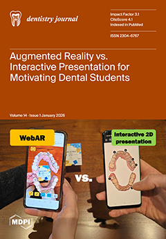
Augmented Reality is a technology that overlays interactive virtual objects onto the real world, with the potential to make learning more engaging. Two resources to train caries detection skills were designed using low-cost authoring tools: a web-based (WebAR) image-tracking experience and, as an active comparator, a 2D interactive presentation. Third-year dental students participated in a randomized controlled experimental study and completed the Reduced Instructional Materials Motivation Survey (RIMMS). As a result, WebAR achieved higher RIMMS motivation scores and higher mean scores in the Attention, Confidence, Satisfaction, and Relevance dimensions. These findings suggest that incorporating 3D spatial interaction may enhance learners’ motivation for caries detection training. View this paper
  • Issues are regarded as officially published after their release is announced to the table of contents alert mailing list.
  • You may sign up for e-mail alerts to receive table of contents of newly released issues.
  • PDF is the official format for papers published in both, html and pdf forms. To view the papers in pdf format, click on the "PDF Full-text" link, and use the free Adobe Reader to open them.
Order results
Result details
Section
Select all
Export citation of selected articles as:
17 pages, 297 KB  
Review
Systemic Bone Loss and Periodontal Disease: An Updated Review of a Bidirectional Association
by Abdulkareem A. Alhumaidan and Ahmed Elakel
Dent. J. 2026, 14(1), 70; https://doi.org/10.3390/dj14010070 - 22 Jan 2026
Viewed by 163
Abstract
Background: Systemic bone loss, particularly osteoporosis, and periodontal disease are highly prevalent chronic conditions that share common risk factors and biological pathways. Increasing evidence suggests a bidirectional relationship between these conditions; however, findings remain heterogeneous and evolving. Objective: This review aims to evaluate [...] Read more.
Background: Systemic bone loss, particularly osteoporosis, and periodontal disease are highly prevalent chronic conditions that share common risk factors and biological pathways. Increasing evidence suggests a bidirectional relationship between these conditions; however, findings remain heterogeneous and evolving. Objective: This review aims to evaluate and update current evidence on the bidirectional association between systemic bone loss and periodontal disease, with emphasis on underlying mechanisms and clinical implications. Methods: A narrative review of the literature was conducted using major electronic databases, focusing on human studies evaluating the relationship between osteoporosis or systemic bone loss and periodontal disease. Relevant experimental, clinical, and epidemiological studies were included. Results: Most studies support an association between reduced bone mineral density and increased severity of periodontal disease, including greater alveolar bone loss and attachment loss. Conversely, periodontal inflammation may contribute to systemic bone remodeling through inflammatory mediators. However, variability in study design, diagnostic criteria, and confounding factors limits definitive conclusions. Conclusions: Current evidence supports a bidirectional association between systemic bone loss and periodontal disease. Greater interdisciplinary awareness is warranted, and future well-designed longitudinal studies are needed to clarify causality and inform preventive and therapeutic strategies. Full article
(This article belongs to the Section Oral Hygiene, Periodontology and Peri-implant Diseases)
16 pages, 461 KB  
Review
Preheated Composite for Prosthetic Cementation to Enamel and Dentin: A Scoping Review
by Anca Labunet, Andreea Kui, Alexandra Vigu, Andrada Voina-Tonea, Alexandru Burde and Sorina Sava
Dent. J. 2026, 14(1), 69; https://doi.org/10.3390/dj14010069 - 21 Jan 2026
Viewed by 123
Abstract
Background and Objectives: Preheated composite resins have been proposed as an alternative to conventional luting agents due to their improved resistance, color stability, and adaptation. This review aims to critically evaluate the current literature on the use of preheated composites as luting agents [...] Read more.
Background and Objectives: Preheated composite resins have been proposed as an alternative to conventional luting agents due to their improved resistance, color stability, and adaptation. This review aims to critically evaluate the current literature on the use of preheated composites as luting agents exclusively on dentin and enamel, focusing on their mechanical behavior, optical properties, and biological effects, in order to determine whether they provide superior clinical outcomes compared with conventional resin cements. Materials and Methods: A comprehensive literature search from 2015 to 2025 was conducted in accordance with PRISMA-ScR guidelines. Eligible studies included in vitro investigations comparing the preheated composite with other luting agents performed on human, bovine, analog dentin or enamel substrates. Studies meeting these criteria were screened, evaluated, and synthesized. Results: Fifteen studies met the inclusion criteria: nine focused on the mechanical performance, and the remaining six studies examined additional properties such as color stability, pulpal temperature changes during preheating, film thickness characteristics, and the influence on marginal discrepancy. Conclusions: Preheated composite resins offer improved mechanical properties, marginal adaptation, and fracture resistance compared with conventional luting agents. However, their performance is highly technique-sensitive, and clinical outcomes depend on operator skill, restoration thickness, and material selection. Preheating generally does not compromise color stability, but it can elevate pulpal temperature, particularly when residual dentin is thin. Overall, preheated composites have potential clinical advantages, provided that careful handling and appropriate application are ensured. Full article
Show Figures

Figure 1

15 pages, 2662 KB  
Case Report
Multidisciplinary Approach for Dental Management of Congenital Insensitivity to Pain with Anhidrosis: Clinical Case Report with 12-Month Follow-Up
by Almoataz B. A. T. Abdel-bari, Mohamed Fawzy, Khaled A. Saad and Hatem A. Alhadainy
Dent. J. 2026, 14(1), 68; https://doi.org/10.3390/dj14010068 - 20 Jan 2026
Viewed by 173
Abstract
Background: Congenital Insensitivity to Pain and Anhidrosis (CIPA) is a rare autosomal recessive disorder characterized by congenital analgesia, anhidrosis, and multisystem involvement affecting the musculoskeletal, cutaneous, oral, and para-oral structures. This case report describes the oral phenotype and multidisciplinary clinical management of a [...] Read more.
Background: Congenital Insensitivity to Pain and Anhidrosis (CIPA) is a rare autosomal recessive disorder characterized by congenital analgesia, anhidrosis, and multisystem involvement affecting the musculoskeletal, cutaneous, oral, and para-oral structures. This case report describes the oral phenotype and multidisciplinary clinical management of a child with CIPA. Case Description: A 9-year-old boy presented with poor oral hygiene, multiple severely damaged teeth, masticatory difficulty, limited mouth opening, impaired bolus control, and para-oral traumatic injuries. Medical and orthopedic history indicated recurrent painless fractures, self-inflicted injuries, cutaneous scarring, and recurrent hyperpyrexia. Oral self-injury associated with CIPA was suspected and supported by the Nociception Assessment Test and Minor’s Iodine–Starch Test. Although the clinical findings were suggestive of CIPA, the diagnosis remained presumptive due to the absence of confirmatory molecular or histopathological testing. Management: A wearable wireless continuous temperature-monitoring device was prescribed to assist in tracking hyperpyrexia associated with CIPA (RHA-CIPA). A conservative, staged, multidisciplinary treatment was planned rather than full-mouth extraction, emphasizing prevention of dental sepsis and mitigation of future self-injury. Dental procedures were performed under local anesthesia to manage discomfort related to tactile hyperesthesia. To reduce nocturnal biting and oral trauma, a hard acrylic occlusal protector was fabricated using an intraoral scanner and a 3D-printed cast. The patient was followed for 12 months. Outcomes: At the 12-month follow-up, clinical improvement was observed, with particularly notable gains in cheek elasticity and soft tissue resilience. Conclusions: This case highlights the considerable challenges involved in the interdisciplinary management of children with CIPA, including oral self-injury prevention, limited mouth opening, and the necessity of close coordination with medical specialties. These findings are descriptive observations of a single case and do not establish efficacy or generalizability of any intervention. Full article
Show Figures

Figure 1

14 pages, 1230 KB  
Article
Guiding Esthetic Crown Lengthening: A CBCT-Based Modified Classification of Altered Passive Eruption
by Kitichai Janaphan and Thanasak Rakmanee
Dent. J. 2026, 14(1), 67; https://doi.org/10.3390/dj14010067 - 20 Jan 2026
Viewed by 158
Abstract
Background: Altered passive eruption (APE) is one of the etiological factors associated with excessive gingival display and is commonly treated with esthetic crown lengthening (ECL). However, existing classification systems provide limited guidance for selecting appropriate treatment approaches. Objectives: The aim of this study [...] Read more.
Background: Altered passive eruption (APE) is one of the etiological factors associated with excessive gingival display and is commonly treated with esthetic crown lengthening (ECL). However, existing classification systems provide limited guidance for selecting appropriate treatment approaches. Objectives: The aim of this study was to evaluate (1) the expected outcome of ECL in eliminating unattractive excessive gingival display (4 mm) based on digital smile assessment and (2) the distribution of teeth and patients according to the modified APE classification. Methods: Forty-two Thai patients with APE underwent clinical examination, digital smile assessment, intraoral scanning, and CBCT. Predicted gingival display (PGD) was calculated to assess the expected outcomes of ECL. The modified APE classification, incorporating CEJ–BC distance and buccal bone thickness, was analyzed at both the tooth and patient levels. Results: A total of 252 maxillary anterior teeth were assessed. Most patients (78.57%) presented with APE and hyperactive upper lip. The mean gingival display (GD) was 6.04 ± 1.76 mm, with GD ≥ 4 mm observed in 92.86% of patients. The mean PGD was 3.56 ± 1.71 mm, and ECL was predicted to reduce GD to < 4 mm in 66.67% of patients. Teeth were classified as Class I (28.97%), II (15.48%), III (41.27%), and IV (14.28%); only Types II (11.9%) and III (88.1%) occurred at the patient level. Conclusions: ECL performed at the CEJ level is predicted to eliminate excessive gingival display in approximately two-thirds of APE patients. The modified APE classification offers guidance for selecting surgical approaches, highlighting the necessity of open-flap procedures and the limited applicability of flapless approaches. Full article
(This article belongs to the Special Issue New Perspectives in Periodontology and Implant Dentistry)
Show Figures

Figure 1

15 pages, 4587 KB  
Article
Bovine Dentin as a Substitute for Human Dentin: Bond Strength Tests on Sound and Eroded Substrate
by Ramona Oltramare, Caroline A. Lutz Guzman, Julia J. Lotz, Thomas Attin and Florian J. Wegehaupt
Dent. J. 2026, 14(1), 66; https://doi.org/10.3390/dj14010066 - 20 Jan 2026
Viewed by 259
Abstract
Objectives: Investigating and comparing the micro-tensile bond strength (µTBS) of etch-and-rinse (ER) or self-etch (SE) adhesives on sound (s) and eroded (e) human (H) and bovine (B) dentin. Methods: Twenty-four human and bovine teeth were divided into eight groups (n = 6) [...] Read more.
Objectives: Investigating and comparing the micro-tensile bond strength (µTBS) of etch-and-rinse (ER) or self-etch (SE) adhesives on sound (s) and eroded (e) human (H) and bovine (B) dentin. Methods: Twenty-four human and bovine teeth were divided into eight groups (n = 6) and coronally ground down, exposing their dentin. Two groups of human (HeER + HeSE) and bovine teeth (BeER + BeSE) were subjected to erosive challenges (citric acid (pH 2.7), 10 × 2 min per day for five days, and stored in artificial saliva). Groups HsER + HeER and BsER + BeER were treated with an etch-and-rinse adhesive (OptiBond FL), and groups HsSE + HeSE and BsSE + BeSE were treated with a self-etch adhesive (OptiBond All-in-One), followed by buildups with a composite restorative material. After seven days of storage in tap water, µTBS was determined and failure type analysis was performed. Data were evaluated using two-way ANOVA and Tukey’s post hoc tests at a level of significance of α = 0.05. Results: Using etch-and-rinse adhesive, sound human dentin (HsER) showed the significantly highest µTBS (p < 0.05) compared to eroded human (HeER) and sound and eroded bovine dentin (BsER + BeER). For sound human and bovine specimens (HsSE + BsSE), there was no significant difference (p ≥ 0.05) in µTBS when self-etch adhesive was applied, as well as in the eroded specimens (HeSE + BeSE). Conclusions: Within the limitations of this study, it can be concluded that for the etch-and-rinse approach, it is not recommended to substitute human dentin with bovine dentin. When using the specific self-etch adhesive used in the present study, bovine dentin can be used to substitute human dentin, as they showed comparable µTBS. Full article
Show Figures

Figure 1

18 pages, 293 KB  
Review
Academic Integrity and Cheating in Dental Education: Prevalence, Drivers, and Career Implications
by Akhilesh Kasula, Gadeer Zahran, Undral Munkhsaikhan, Vivian Diaz, Michelle Walker, Candice Johnson, Kathryn Lefevers, Ammaar H. Abidi and Modar Kassan
Dent. J. 2026, 14(1), 65; https://doi.org/10.3390/dj14010065 - 19 Jan 2026
Viewed by 276
Abstract
Background: Integrity, encompassing honesty, accountability, and ethical conduct, is a cornerstone of the dental profession, essential for patient trust and safety. Despite its importance, academic dishonesty remains a pervasive issue in dental education globally. This review examines the prevalence, causes, and long-term [...] Read more.
Background: Integrity, encompassing honesty, accountability, and ethical conduct, is a cornerstone of the dental profession, essential for patient trust and safety. Despite its importance, academic dishonesty remains a pervasive issue in dental education globally. This review examines the prevalence, causes, and long-term career implications of academic dishonesty in dental education and explores institutional strategies to cultivate a culture of integrity. Method: The study was conducted using PubMed, Scopus, Web of Science, and Google Scholar to identify studies published between 1970 and 2025 on academic dishonesty in dental education. Search terms included dental students, cheating, plagiarism, and clinical falsification. Eligible studies reported prevalence, drivers, or consequences of dishonest behaviors. Data were extracted and thematically synthesized to highlight common patterns and professional implications. Results: Self-reported data indicate alarmingly high rates of cheating among dental students, ranging from 43% to over 90%. Common forms include exam fraud, plagiarism, and the falsification of clinical records. Key drivers include intense academic pressure, competitive environments, and a perception of weak enforcement. Such behaviors are not merely academic violations—they have profound professional consequences. A history of academic dishonesty can damage a student’s reputation, hinder licensure and credentialing processes, and limit postgraduate opportunities. Crucially, studies indicate that unethical behavior in school can normalize dishonesty, predicting a higher likelihood of future professional misconduct, such as insurance fraud or malpractice, thereby jeopardizing patient care and public trust. Conclusions: Academic integrity is a critical predictor of professional ethical conduct. Dental schools must move beyond punitive policies to implement proactive, multi-faceted approaches. This includes integrating comprehensive ethics curricula, fostering reflective practice, promoting faculty role modeling, and empowering student-led initiatives to uphold honor codes. Cultivating an unwavering culture of integrity is essential not only for academic success but for developing trustworthy practitioners committed to lifelong ethical patient care. Full article
Show Figures

Graphical abstract

14 pages, 5665 KB  
Article
Transcrestal Sinus Elevation with Implant Placement Using Autogenous Bone Supporting Multilayer Crosslinked Collagen Xenograft Scaffolding: A Case Series
by David Barack, Chander S. Gupta, Luigi Canullo and Marco Toia
Dent. J. 2026, 14(1), 64; https://doi.org/10.3390/dj14010064 - 19 Jan 2026
Viewed by 229
Abstract
Background/Objectives: Limited residual bone height in the atrophic posterior maxilla complicates implant placement. Transcrestal sinus elevation can be used to correct bone shrinkage after sinus pneumatization or crestal bone loss. This study evaluated a minimally invasive, one-stage transcrestal sinus lift using a [...] Read more.
Background/Objectives: Limited residual bone height in the atrophic posterior maxilla complicates implant placement. Transcrestal sinus elevation can be used to correct bone shrinkage after sinus pneumatization or crestal bone loss. This study evaluated a minimally invasive, one-stage transcrestal sinus lift using a double-layer crosslinked collagen scaffold (MCCS) with autogenous bone from the implant osteotomy site in patients with RBH ≤ 6 mm. Methods: In this prospective series, 11 patients (48–64 years, mean RBH 4.75 mm, SD 0.95 mm) underwent one-stage transcrestal sinus floor elevation with simultaneous implants. After osteotomy, autogenous bone chips collected during drilling were compacted into the site, and two layers of MCCS were placed under the elevated Schneiderian membrane. Buccal and palatal bone heights were measured on CBCT before and after surgery to assess vertical bone gain (ΔRBH). Results: All implants achieved stable osseointegration. Mean ΔRBH was approximately 3.1 ± 0.9 mm (combined buccal–palatal). No postoperative complications occurred. Two small Schneiderian membrane perforations were sealed intraoperatively by MCCS placement, with uneventful healing. Follow-up imaging showed maintenance of the augmented bone around the implants. Conclusions: This double-layer MCCS plus autogenous bone approach is a safe, effective, and minimally invasive transcrestal sinus lift for atrophic maxillae. It yielded crestal bone gains even with minimal initial RBH, leveraging the palatal sinus wall’s osteogenic potential and the implant’s tent-pole effect. The MCCS scaffold maintained space for bone formation and enabled immediate sealing of any membrane perforations. This one-stage protocol is viable for implant placement in low-RBH sites. Full article
Show Figures

Figure 1

15 pages, 245 KB  
Article
The Effect of Age and Use of Enamel Matrix Derivative on Implant Loss
by Stephen K. Harrel, Thomas G. Wilson, Jr., Martha E. Nunn and Charles M. Cobb
Dent. J. 2026, 14(1), 63; https://doi.org/10.3390/dj14010063 - 19 Jan 2026
Viewed by 150
Abstract
Background: Previous reports suggest that patient age at the time of implant placement is not a factor in implant survival. However, analysis of data compiled for a previously published study on the effect of enamel matrix derivative (EMD), a frequently used biomaterial to [...] Read more.
Background: Previous reports suggest that patient age at the time of implant placement is not a factor in implant survival. However, analysis of data compiled for a previously published study on the effect of enamel matrix derivative (EMD), a frequently used biomaterial to aid bone regeneration, on peri-implantitis indicated that age and use of EMD may be a factor in implant survival. The current study further evaluated the existing database to determine the effect of age and EMD use on long term survival of implants. Methods: An existing database from a private periodontal specialty practice was evaluated for the effect of age at the time of implant placement on implant survival. In addition, all available clinical factors were evaluated, including the use of EMD at any point during site preparation or implant placement to determine any effect on implant survival. Results: Patient age at the time of implant placement had a negligible effect on implant survival for younger individuals. However, starting at 58 years of age, an increase in relative risk for implant loss was noted. When the patient age was divided into groups, it was determined that patients ≥ 58 and ≤68 years had a statistically significantly increased relative risk of implant loss (2.75), which was sharply reduced if EMD had been used (1.24). This trend was also noted to a lesser extent in patients older than 68 years. Conclusions: The risk of implant loss was elevated when implants were placed in older patients. This risk was reduced if EMD had been used at any point during implant site preparation or placement. Full article
(This article belongs to the Topic Oral Health Management and Disease Treatment)
11 pages, 919 KB  
Systematic Review
Obstructive Sleep Apnea: The Expanding Role of Dental Sleep Medicine—A Systematic Review of Mandibular Advancement Devices, Treatment Efficacy, and Occlusal Complications
by Jędrzej Szmyt, Tymoteusz Szczapa, Maksymilian Chyła, Adam Bęben and Izabela Maciejewska
Dent. J. 2026, 14(1), 62; https://doi.org/10.3390/dj14010062 - 17 Jan 2026
Viewed by 322
Abstract
Background: Obstructive sleep apnea is characterized by recurrent upper airway obstruction during sleep, leading to intermittent hypoxemia, sleep fragmentation, and excessive daytime sleepiness. Affecting up to 11% of the adult Polish population and more commonly diagnosed in men, OSA poses a major public [...] Read more.
Background: Obstructive sleep apnea is characterized by recurrent upper airway obstruction during sleep, leading to intermittent hypoxemia, sleep fragmentation, and excessive daytime sleepiness. Affecting up to 11% of the adult Polish population and more commonly diagnosed in men, OSA poses a major public health concern due to its association with cardiovascular, metabolic, and neurocognitive complications. This review summarizes the current evidence on diagnostic methods, risk factors, and therapeutic approaches, with particular emphasis on oral appliance therapy using mandibular advancement devices (MADs). Methods: A systematic literature review was conducted using the PubMed and Scopus databases, covering publications from 2020 to 2025, including clinical trials, meta-analyses, and systematic reviews evaluating the efficacy and safety of MAD therapy. Results: Findings demonstrate that MAD effectively reduces apnea–hypopnea index (AHI) values, improves oxygen saturation, and alleviates snoring and daytime fatigue, offering a patient-tolerable alternative for those intolerant to continuous positive airway pressure (CPAP). However, long-term use may cause occlusal or dental changes. Novel techniques, such as Er:YAG laser therapy, show potential in treating mild OSA. Moreover, epidemiological data suggest a correlation between tooth loss and an increased risk of OSA, particularly among men over 65. Conclusions: Dentists play a pivotal role in early detection, screening, and interdisciplinary management of OSA, underscoring the importance of collaboration between dental professionals and sleep medicine specialists for comprehensive care. Full article
(This article belongs to the Topic Oral Health Management and Disease Treatment)
Show Figures

Figure 1

16 pages, 2193 KB  
Article
Perceived Diagnostic Value of Fluorescence-Enhanced 3D Imaging for Detecting Caries Adjacent to Restorations: A Questionnaire-Based Study
by Dimitrios Spagopoulos, Grigoria Gkavela and Christos Rahiotis
Dent. J. 2026, 14(1), 61; https://doi.org/10.3390/dj14010061 - 16 Jan 2026
Viewed by 197
Abstract
Background/Objectives: Caries adjacent to restorations remain a leading cause of restoration failure and replacement. Conventional diagnostic methods are limited by subjectivity and restricted visualization. Fluorescence-enhanced three-dimensional (3D) imaging has been proposed to improve detection accuracy, but evidence on its clinical perception and usability [...] Read more.
Background/Objectives: Caries adjacent to restorations remain a leading cause of restoration failure and replacement. Conventional diagnostic methods are limited by subjectivity and restricted visualization. Fluorescence-enhanced three-dimensional (3D) imaging has been proposed to improve detection accuracy, but evidence on its clinical perception and usability remains scarce. The objective of this study was to evaluate the perceived diagnostic value of fluorescence-enhanced 3D imaging in detecting caries adjacent to direct restorations. Methods: A cross-sectional questionnaire-based survey was distributed to undergraduate dental students and licensed dentists (n = 94). Participants assessed images of extracted teeth with direct restorations presented in three formats: conventional photographs, monochromatic 3D models, and 3D models with fluorescence. Responses were analyzed using descriptive statistics, chi-square tests, and Cohen’s kappa to measure inter-rater agreement. Results: Overall, 64.9% of respondents reported that fluorescence-enhanced images improved their diagnostic decision-making, while 29.8% reported partial benefit. Fluorescence was mainly perceived as helpful in defining cavity margins (53.3%) and assessing lesion volume (42.4%). Most participants preferred 3D models with fluorescence over conventional images for diagnostic value. However, inter-rater agreement was generally poor (κ range: –0.05 to 0.25; median κ = 0.02; only 4 images showed weak but statistically significant agreement), with only a few images demonstrating weak but statistically significant agreement. Notably, 39.3% of participants reported prior experience with 3D imaging, which was associated with greater confidence in interpreting fluorescence-enhanced images. Participants with prior 3D imaging experience reported greater confidence in fluorescence interpretation. Conclusions: While fluorescence-enhanced 3D imaging is perceived as a useful adjunct for visualizing lesion margins and depth, it does not currently yield consistent diagnostic agreement across clinicians. Training, calibration, and integration of artificial intelligence support may enhance the clinical reliability of this technology. Full article
Show Figures

Figure 1

16 pages, 2780 KB  
Article
Multi-Class Malocclusion Detection on Standardized Intraoral Photographs Using YOLOv11
by Ani Nebiaj, Markus Mühling, Bernd Freisleben and Babak Sayahpour
Dent. J. 2026, 14(1), 60; https://doi.org/10.3390/dj14010060 - 16 Jan 2026
Viewed by 219
Abstract
Background/Objectives: Accurate identification of dental malocclusions from routine clinical photographs can be time-consuming and subject to interobserver variability. A YOLOv11-based deep learning approach is presented and evaluated for automatic malocclusion detection on routine intraoral photographs, testing the hypothesis that training on a structured [...] Read more.
Background/Objectives: Accurate identification of dental malocclusions from routine clinical photographs can be time-consuming and subject to interobserver variability. A YOLOv11-based deep learning approach is presented and evaluated for automatic malocclusion detection on routine intraoral photographs, testing the hypothesis that training on a structured annotation protocol enables reliable detection of multiple clinically relevant malocclusions. Methods: An anonymized dataset of 5854 intraoral photographs (frontal occlusion; right/left buccal; maxillary/mandibular occlusal) was labeled according to standardized instructions derived from the Index of Orthodontic Treatment Need (IOTN) A total of 17 clinically relevant classes were annotated with bounding boxes. Due to an insufficient number of examples, two malocclusions (transposition and non-occlusion) were excluded from our quantitative analysis. A YOLOv11 model was trained with augmented data and evaluated on a held-out test set using mean average precision at IoU 0.5 (mAP50), macro precision (macro-P), and macro recall (macro-R). Results: Across 15 analyzed classes, the model achieved 87.8% mAP50, 76.9% macro-P, and 86.1% macro-R. The highest per-class AP50 was observed for Deep bite (98.8%), Diastema (97.9%), Angle Class II canine (97.5%), Anterior open bite (92.8%), Midline shift (91.8%), Angle Class II molar (91.1%), Spacing (91%), and Crowding (90.1%). Moderate performance included Anterior crossbite (88.3%), Angle Class III molar (87.4%), Head bite (82.7%), and Posterior open bite (80.2%). Lower values were seen for Angle Class III canine (76%), Posterior crossbite (75.6%), and Big overjet (75.3%). Precision–recall trends indicate earlier precision drop-off for posterior/transverse classes and comparatively more missed detections in Posterior crossbite, whereas Big overjet exhibited more false positives at the chosen threshold. Conclusion: A YOLOv11-based deep learning system can accurately detect several clinically salient malocclusions on routine intraoral photographs, supporting efficient screening and standardized documentation. Performance gaps align with limited examples and visualization constraints in posterior regions. Larger, multi-center datasets, protocol standardization, quantitative metrics, and multimodal inputs may further improve robustness. Full article
(This article belongs to the Special Issue Artificial Intelligence in Oral Rehabilitation)
Show Figures

Graphical abstract

16 pages, 722 KB  
Review
Intentional Tooth Replantation: Current Evidence and Future Research Directions for Case Selection, Extraction Approaches, and Post-Operative Management
by Rahul Minesh Shah, Thomas Manders and Georgios Romanos
Dent. J. 2026, 14(1), 59; https://doi.org/10.3390/dj14010059 - 15 Jan 2026
Viewed by 296
Abstract
Background: Intentional tooth replantation (ITR) is a promising treatment option for preserving teeth in cases where conventional endodontic therapy is challenging, or when previous endodontic treatment and apicoectomy have been unsuccessful. The procedure involves extracting the compromised tooth, preserving the alveolar socket and [...] Read more.
Background: Intentional tooth replantation (ITR) is a promising treatment option for preserving teeth in cases where conventional endodontic therapy is challenging, or when previous endodontic treatment and apicoectomy have been unsuccessful. The procedure involves extracting the compromised tooth, preserving the alveolar socket and root surface, performing extraoral endodontic therapy, and replanting the tooth in the alveolar socket. Objective: An increase in evidence-based support for ITR has improved the viability of ITR as a treatment option for patients. This review aims to further establish and provide new areas of potential research for ITR with respect to root morphology, extraction, and surgical techniques, maintenance of the tooth socket, and methods for post-op stabilization. Materials and Methods: A literature review was performed across PubMed from 1 January 1980 to 1 July 2025, with a focus on oral surgery techniques, atraumatic extraction techniques, topographical discrepancies in root system anatomy, and ITR procedural outcomes. Conclusions: Although ITR is not a common procedure performed in contemporary clinical practice, gathering sufficient data on the variables influencing the procedure may help patient outcome and improve communication between the endodontist and oral surgeons. Full article
(This article belongs to the Section Dental Education)
Show Figures

Figure 1

20 pages, 720 KB  
Review
Next-Generation S3-Level Clinical Practice Guidelines in Periodontology: Methodology, Current Evidence, and Future Directions
by Nada Tawfig Hashim, Ayman Ahmed, Azza A. Abushama, Salma Musa Adam Abduljalil, Bakri Gobara Gismalla and Muhammed Mustahsen Rahman
Dent. J. 2026, 14(1), 58; https://doi.org/10.3390/dj14010058 - 15 Jan 2026
Viewed by 372
Abstract
Background: S3-level clinical practice guidelines represent the highest standard of evidence-based healthcare, integrating systematic reviews, formal evidence grading, and structured expert consensus. In periodontology, current S3-level guidelines provide robust recommendations for the management of stage I–III periodontitis. However, increasing clinical complexity, emerging [...] Read more.
Background: S3-level clinical practice guidelines represent the highest standard of evidence-based healthcare, integrating systematic reviews, formal evidence grading, and structured expert consensus. In periodontology, current S3-level guidelines provide robust recommendations for the management of stage I–III periodontitis. However, increasing clinical complexity, emerging diagnostic technologies, and the need for patient-centred and implementation-oriented care highlight important gaps that warrant further methodological refinement. Objective: This review aims to critically appraise the conceptual foundations, strengths, and limitations of existing S3-level periodontal guidelines and to propose a structured roadmap for the development of next-generation S3 guidance. Methods: A narrative and methodological review was conducted focusing on key European S3-level guidelines in periodontology and endodontics, with emphasis on guideline methodology, evidence grading, outcome prioritization, and consensus processes. Results: Current S3-level periodontal guidelines demonstrate strong methodological rigor but show limited coverage of stage IV periodontitis, peri-implant diseases, and endo–perio lesions. In addition, emerging domains such as biomarker-based diagnostics, artificial intelligence-assisted decision support, and implementation science are not yet systematically integrated. Conclusions: Future S3-level periodontal guidelines should incorporate clinical complexity, patient-reported outcomes, precision diagnostics, digital technologies, and real-world implementation strategies to enhance personalization, transparency, and clinical impact. Full article
Show Figures

Figure 1

14 pages, 1191 KB  
Article
Cross-Sectional Clinical Evaluation of Subantral Augmentation Using Nano Graft Composite: Implications for Implant Success
by Olexiy Kosinov, Olesya Manukhina, Kristina Volchykhina, Oleg Mishchenko, Andrii Liutyi, Agne Ramanaviciute, Vilma Ratautaite and Arunas Ramanavicius
Dent. J. 2026, 14(1), 57; https://doi.org/10.3390/dj14010057 - 15 Jan 2026
Viewed by 208
Abstract
Objectives: This study aims to evaluate the efficacy of hydroxyapatite-tricalcium phosphate (HAP-TCP) as a bone substitute in subantral augmentation for dental implants. Specifically, it investigates the effects of HAP-TCP on bone quality, density, and integration with implants over time. Methods: A prospective controlled [...] Read more.
Objectives: This study aims to evaluate the efficacy of hydroxyapatite-tricalcium phosphate (HAP-TCP) as a bone substitute in subantral augmentation for dental implants. Specifically, it investigates the effects of HAP-TCP on bone quality, density, and integration with implants over time. Methods: A prospective controlled longitudinal study was conducted on 22 patients (39–75 years of age) undergoing subantral augmentation and dental implantation. A total of 52 sites of augmented bone and 67 sites of native bone were analyzed using computed tomography (CT) to assess bone density in Hounsfield Units (HU), insertion torque measurements, and the Misch classification for bone quality. Augmented and native bone measurements were compared within each patient. Results: The augmented bone exhibited an average density of 1132.6 ± 334.9 HU, which is significantly higher (45.9%) than the average density of native bone at 519.3 ± 395.0 HU. Insertion torque values in the HAP-TCP augmented sites averaged 35 N·cm, showing a 71.4% increase compared to adjacent native bone sites (25 N·cm). The study found notable improvements in bone homogeneity and vascularization within the augmented zones. Conclusion: HAP-TCP demonstrates significant potential as a reliable and effective synthetic bone substitute for subantral augmentation in dental implants. It yields higher radiodensity and insertion torque than adjacent native bone, while mitigating complications associated with autogenous grafts. These observational findings support the potential clinical use of HAP-TCP for sinus augmentation. Full article
(This article belongs to the Topic Advances in Dental Materials)
Show Figures

Figure 1

17 pages, 587 KB  
Review
Prophylactic Antibiotic Therapy in Cleft Surgery—A Scoping Review
by Margareta Budner, Marcelina Podleśna, Aleksandra Domańska, Natalia Pijas, Katarzyna Zyska, Daniel Wiśniewski, Klaudiusz Garbacki, Grzegorz Wilhelm, Kamil Torres, Jerzy Strużyna and Agnieszka Surowiecka
Dent. J. 2026, 14(1), 56; https://doi.org/10.3390/dj14010056 - 15 Jan 2026
Viewed by 250
Abstract
Background/Objectives: Cleft lip and/or palate are common craniofacial anomalies whose surgical repair is classified as clean-contaminated and may be complicated by surgical site infection or palatal fistula. Despite widespread perioperative antibiotic use, there are no standardized, evidence-based recommendations, and rising antimicrobial resistance underlines [...] Read more.
Background/Objectives: Cleft lip and/or palate are common craniofacial anomalies whose surgical repair is classified as clean-contaminated and may be complicated by surgical site infection or palatal fistula. Despite widespread perioperative antibiotic use, there are no standardized, evidence-based recommendations, and rising antimicrobial resistance underlines the need for rational prescribing. This systematic scoping review aimed to map current evidence on prophylactic antibiotic therapy and related perioperative measures in cleft surgery. Methods: A scoping review was conducted using the Arksey and O’Malley framework and reported in line with PRISMA 2020. PubMed, Mendeley and Google Scholar were searched (January 2015–10 February 2025) for English-language retrospective studies, clinical trials, survey studies and systematic reviews concerning prophylactic antibiotics, bone grafting procedures, mouthwash use or oral microbiota in patients undergoing cleft lip and/or palate surgery. Six reviewers independently screened records; two experienced clinicians extracted data on study characteristics, antimicrobial regimens and infectious or microbiological outcomes. Given heterogeneity and the scoping aim, no formal risk-of-bias assessment or meta-analysis was performed. Results: A total of 40 studies met the inclusion criteria, including 21 original research articles. Considerable variation in antibiotic choice, timing and duration was observed, with no clear superiority of any regimen. Single-dose perioperative prophylaxis appeared non-inferior to prolonged courses in several settings. Oral microbiota studies highlighted colonization by resistant and opportunistic pathogens in cleft patients. Conclusions: Current evidence supports individualized, often short-course perioperative antibiotic strategies rather than routine prolonged therapy. High-quality randomized and microbiological studies are required to develop standardized, resistance-conscious guidelines. Full article
(This article belongs to the Special Issue Trends in Orofacial Cleft Research)
Show Figures

Figure 1

17 pages, 1870 KB  
Systematic Review
The Role of Digit- and Pacifier-Sucking Habits on Malocclusion Development in Children: Anterior Open Bite and Posterior Crossbite—A Systematic Review & Meta-Analysis
by Arvin Faryad, Susana Muwaquet Rodriguez and Tawfiq Hijazi Alsadi
Dent. J. 2026, 14(1), 55; https://doi.org/10.3390/dj14010055 - 14 Jan 2026
Viewed by 342
Abstract
Background/Objectives: Malocclusion is one of the most prevalent oral health concerns in paediatric dentistry, with anterior open bite (AOB) and posterior crossbite (PCB) being among the most common forms. Non-nutritive sucking habits (NNSHs), including digit-sucking habits (DSHs) and pacifier-sucking habits (PSHs), have been [...] Read more.
Background/Objectives: Malocclusion is one of the most prevalent oral health concerns in paediatric dentistry, with anterior open bite (AOB) and posterior crossbite (PCB) being among the most common forms. Non-nutritive sucking habits (NNSHs), including digit-sucking habits (DSHs) and pacifier-sucking habits (PSHs), have been linked to malocclusion development. While both habits are known to impact dental and skeletal development, their comparative effects remain unclear. This systematic review and meta-analysis aims to determine the difference in the development and prevalence of anterior open bite and posterior crossbite between patients with digit-sucking and pacifier-sucking habits. Materials and Methods: An exhaustive review of the literature was conducted on the 25 November 2024 across three databases, namely EBSCOhost (including PubMed-Medline), Web of Science and Scopus. The following PICO question was constructed for the systematic review: “In children and teenagers, is there a difference in the development and prevalence of malocclusions (Anterior Open bite & Posterior Crossbite) between patients with a history of digit sucking habits and patients with a history of pacifier sucking habits?”A meta-analysis was also performed with the selected studies, and the software used to carry out the meta-analysis was R 4.3.1 (R Core Team (2023)). Results: From the initial search, 102 articles were found and a further 11 articles were obtained from manual findings. 12 articles were included in the final systematic review and meta-analysis. The meta-analysis indicated that the risk of AOB and PCB was increased by both DSH and PSH. Conclusions: Both DSH and PSH significantly increased the risk of AOB and PCB. PSH posed a significantly higher risk than DSH for PCB development (OR = 2.66, p < 0.001), while no significant difference in AOB prevalence was observed between DSH and PSH (OR = 1.77, p = 0.150). Full article
Show Figures

Figure 1

12 pages, 249 KB  
Article
Genetic Associations with Non-Syndromic Cleft Lip/Palate and Dental Caries in Kuwaiti Patients: A Case–Control Study
by Manal Abu Al-Melh, Fawzi M. Al-Qatami, Maribasappa Karched and Muawia A. Qudeimat
Dent. J. 2026, 14(1), 54; https://doi.org/10.3390/dj14010054 - 13 Jan 2026
Viewed by 215
Abstract
Background: Non-syndromic cleft lip/palate (NCL/P) is a prevalent congenital anomaly. Despite an unclear epidemiological link between orofacial clefts and dental caries, genetic studies suggest that polymorphisms in taste receptor genes may influence caries risk. Objectives: This study had two primary objectives: (1) to [...] Read more.
Background: Non-syndromic cleft lip/palate (NCL/P) is a prevalent congenital anomaly. Despite an unclear epidemiological link between orofacial clefts and dental caries, genetic studies suggest that polymorphisms in taste receptor genes may influence caries risk. Objectives: This study had two primary objectives: (1) to compare SNPs in NCL/P-associated genes (IRF6, FOXE1) between Kuwaiti NCL/P cases and controls, and (2) to explore whether variants in caries-associated (KLK4, DSPP) and taste receptor (TAS1R2, TAS2R38) genes are associated with dental caries susceptibility in individuals with NCL/P, independent of overall caries prevalence. Methods: A case–control design was employed, with 25 NCL/P cases and 25 unaffected controls recruited from a Dental Craniofacial Clinic in Kuwait. Genomic DNA was extracted from buccal swabs, and SNP genotyping was performed using real-time PCR for genes related to NCL/P, dental caries, and taste perception. Caries status was assessed using the dmft/DMFT scoring system. The genotyped genes included NCL/P-related (IRF6, FOXE1), caries-related (KLK4, DSPP), and taste receptor genes (TAS1R2, TAS2R38). Results: At nominal significance, KLK4, DSPP, and TAS1R2 showed associations with NCL/P status, while IRF6 and FOXE1 did not. After applying Benjamini–Hochberg FDR correction across 10 SNPs, no allele- or genotype-level association remained significant (q < 0.05). The strongest signal was KLK4 rs2235091 (allele-level p = 0.016; q = 0.159). An exploratory age- and sex-adjusted logistic model for KLK4 suggested a possible effect (aOR 0.40; 95% CI 0.18–0.87; p = 0.021). Within-group analyses of caries burden revealed no associations that survived FDR control (lowest q = 0.056 for FOXE1 in controls). Conclusions: After controlling for multiple testing, no SNP showed a statistically significant association with NCL/P or caries burden. Nominal signals for KLK4, DSPP, and TAS1R2 did not survive FDR correction; an exploratory adjusted model suggested a possible KLK4 effect, but this requires cautious interpretation. The small sample size is a key limitation, and the findings highlight the need for larger, well-powered studies to clarify genetic contributions to NCL/P and caries risk. Full article
22 pages, 1020 KB  
Article
Prevalence and Socio-Behavioural Determinants of Periodontal Disease Among Adults in the Northern West Bank: A Cross-Sectional Study
by Sura Al-Hassan, Mazen Kazlak and Elham Kateeb
Dent. J. 2026, 14(1), 53; https://doi.org/10.3390/dj14010053 - 13 Jan 2026
Viewed by 392
Abstract
Background & Objectives: Periodontal disease (PD) is a common oral disease that affects the supporting structures of the teeth and is a leading cause of tooth loss worldwide. This study aimed to estimate the prevalence of PD among 9th-grade teachers in the [...] Read more.
Background & Objectives: Periodontal disease (PD) is a common oral disease that affects the supporting structures of the teeth and is a leading cause of tooth loss worldwide. This study aimed to estimate the prevalence of PD among 9th-grade teachers in the northern West Bank and examine its association with key behavioral and socioeconomic factors. Methods: A cross-sectional study was conducted among 920 teachers selected through proportional stratified random sampling from governmental and private schools. Periodontal health was assessed using the WHO Community Periodontal Index for Treatment Needs (CPITN), and oral hygiene status was measured with the Simplified Oral Hygiene Index (S-OHI). A structured questionnaire was administered to collect data on socioeconomic status, oral hygiene practices, dietary habits, and smoking behaviours. Data was analysed using descriptive statistics, bivariate and multivariate logistic regression. Results: Only 11.8% of participants exhibited completely healthy gingiva, with the mean condition ranging between calculus and shallow pockets. Oral hygiene practices were the strongest predictors of periodontal outcomes: frequent tooth brushing (Adjusted Odds Ratio: AOR = 0.015), morning brushing (AOR = 0.015), and regular toothbrush replacement (AOR = 2.514) were protective. Higher red meat intake was negatively associated with periodontal health (AOR = 0.032), while frequent nut consumption was protective (AOR = 0.227). The number of cigarettes smoked per week was positively associated with PD (AOR = 1.085). Conclusions: PD is highly prevalent among Palestinian adults, with significant behavioural and lifestyle-related determinants. Targeted oral health interventions are urgently needed to improve adults’ oral health. Full article
(This article belongs to the Topic Preventive Dentistry and Public Health)
Show Figures

Figure 1

21 pages, 2605 KB  
Article
In Vitro Accuracy Analysis of Intraoral Scanning Strategies: A Comparison of Two Contemporary IOS Systems
by Sabina-Ana Răuță, Vlad Gabriel Vasilescu, Lucian Toma Ciocan, Alexandra Popa, Ana-Maria Cristina Țâncu, Florin Octavian Froimovici, Bogdan Dimitriu, Silviu-Mirel Pițuru and Marina Imre
Dent. J. 2026, 14(1), 52; https://doi.org/10.3390/dj14010052 - 13 Jan 2026
Viewed by 276
Abstract
Background: Digital intraoral scanning has become an essential component of modern restorative dentistry, offering enhanced accuracy, workflow efficiency, and patient comfort compared to conventional impression techniques. Despite these advantages, the accuracy of intraoral scanners (IOS) can be affected by multiple parameters, among [...] Read more.
Background: Digital intraoral scanning has become an essential component of modern restorative dentistry, offering enhanced accuracy, workflow efficiency, and patient comfort compared to conventional impression techniques. Despite these advantages, the accuracy of intraoral scanners (IOS) can be affected by multiple parameters, among which scanning strategy and device design are particularly influential. Purpose: This study aimed to investigate the effect of different scanning strategies on scan accuracy and precision, focusing on two widely used intraoral scanners (Medit i700 and Trios 5) in a controlled in vitro environment. Materials and Methods: A standardized digital test object was created according to ISO 20896-1 specifications to ensure uniformity and comparability. The object was printed using a high-precision 3D printer and scanned multiple times with both IOS systems, employing distinct scanning strategies under identical environmental conditions. Data analysis was performed using descriptive and comparative statistics, including Mean, Median, Mean Absolute Deviation (MAD), Root Mean Square Error (RMSE), Standard Deviation (SD), and Variance, to evaluate trueness and precision. Results: The Medit i700 consistently exhibited lower deviation values and greater precision compared with the Trios 5, reflecting higher trueness and precision. Scanning strategy influenced scan outcomes; structured, systematic scanning paths produced more stable and accurate datasets. The Trios 5 demonstrated higher variability, suggesting increased sensitivity to operator motion and scanning trajectory. Conclusions: Both the scanner type and scanning strategy substantially affect intraoral scan accuracy. The superior performance of the Medit i700 indicates greater robustness and operator-independent stability. Clinically, these results underscore the importance of standardized scanning protocols, as operator consistency may be a key determinant of digital impression accuracy and, consequently, of clinical outcomes. Full article
(This article belongs to the Special Issue Feature Papers in Digital Dentistry)
Show Figures

Graphical abstract

10 pages, 1354 KB  
Article
Palmitic Acid Induces Inflammatory Environment and Is Involved in Pyroptosis in a Human Dental Pulp Cell Line
by Takashi Muramatsu, Akihide Yanagisawa, Keisuke Mitomo, Kana Takada, Masahiro Furusawa, Yoshihiro Abiko and Han-Sung Jung
Dent. J. 2026, 14(1), 51; https://doi.org/10.3390/dj14010051 - 12 Jan 2026
Viewed by 318
Abstract
Background/Objectives: We investigated whether palmitic acid (PA) induced the expression of inflammatory cytokines and was involved in pyroptosis in a human dental pulp cell line. Methods: Human dental pulp cells cultured in Minimum Essential Medium Alpha (αMEM) were treated with 1 [...] Read more.
Background/Objectives: We investigated whether palmitic acid (PA) induced the expression of inflammatory cytokines and was involved in pyroptosis in a human dental pulp cell line. Methods: Human dental pulp cells cultured in Minimum Essential Medium Alpha (αMEM) were treated with 1 µg/mL LPS and/or PA (100, 300 and 500 µM). As a control, αMEM was added in the culture medium. The WST-1 assay was performed to assess cell proliferation, and morphological changes in cells were examined. RNA expression of IL-1β, IL-6, TNF-α, caspase-4 and gasdermin d were detected by quantitative RT-PCR (qPCR). Results: The WST-1 assay showed that cell viability decreased by 36% at 300 µM and 47% at 500 µM PA compared to the control (p < 0.05). Cell morphology revealed slight shrinkage in 100, 300 and 500 µM PA groups. RNA expression of IL-1β and IL-6 in the PA groups was significantly higher than that in the control groups (p < 0.05), while RNA expression of TNF-α in the PA group was the same as that of control group. The mRNA expression of caspase-4 and gasdermin d in PA groups was significantly higher than that in control group (p < 0.05). Likewise, the concentration of IL-1β and IL-6 was significantly higher in both LPS and PA groups than that in the LPS or PA groups (p < 0.05). Conclusions: The results of this study suggest that PA induces the expression of inflammatory cytokines and is involved in pyroptosis in a human dental pulp cell line. Full article
(This article belongs to the Section Restorative Dentistry and Traumatology)
Show Figures

Figure 1

15 pages, 499 KB  
Article
Correlation of Oxidative Stress Markers and Nuclear Abnormalities with Clinical Parameters in Individuals with Periodontitis
by Saulo Oswaldo Sánchez-Rivera, Yveth Marlene Ortiz-Garcia, Blanca Patricia Lazalde-Ramos, Cristina Hermila Martínez-Bugarín, Guillermo Moisés Zúñiga-González, Susana Vanessa Sánchez-De-La-Rosa, Belinda Claudia Gómez-Meda, Vianeth Martínez-Rodríguez, Cristian Gabriel Guerrero-Bernal, Gabriela Morales-Velazquez and Ana Lourdes Zamora-Perez
Dent. J. 2026, 14(1), 50; https://doi.org/10.3390/dj14010050 - 12 Jan 2026
Viewed by 340
Abstract
Background: Chronic periodontitis (CP) is a prevalent inflammatory disease worldwide, characterized by the destruction of periodontal tissue due to an immune response triggered by periodontopathogenic bacteria and the prolonged release of reactive oxygen species (ROS). Excess ROS leads to tissue damage through [...] Read more.
Background: Chronic periodontitis (CP) is a prevalent inflammatory disease worldwide, characterized by the destruction of periodontal tissue due to an immune response triggered by periodontopathogenic bacteria and the prolonged release of reactive oxygen species (ROS). Excess ROS leads to tissue damage through mechanisms such as lipid peroxidation and DNA damage. The aim of this study was to evaluate oxidative and genotoxic damage by quantifying 8-hydroxy-2-deoxiguanosine (8-OHdG), malondialdehyde (MDA), and nuclear abnormalities (NAs) in individuals with CP. Methods: The participants were divided into a CP group (n = 30) and a control group without CP (n = 30). Saliva was collected to quantify 8-OHdG (via ELISA) and MDA (via spectrophotometry). Buccal mucosa samples were collected to assess NAs. Periodontal parameters, probing depth (PD), clinical attachment level (CAL), plaque index (PI), and bleeding on probing (BOP), were recorded. Results: The levels of 8-OHdG and MDA were significantly higher in the CP group. NAs were also significantly increased. Positive correlations were observed between 8-OHdG, MDA levels and NAs with clinical parameters. Conclusions: The elevated levels of 8-OHdG, MDA and NAs reflect oxidative and genotoxic damage correlated with CP severity. These biomarkers could complement diagnosis, monitor progression, and assess treatment efficacy. Their elevation may also indicate increased systemic disease risk. Full article
Show Figures

Graphical abstract

20 pages, 1210 KB  
Systematic Review
Microbiological Effects of Laser-Assisted Non-Surgical Treatment of Peri-Implantitis: A Systematic Review and Meta-Analysis of Randomized Controlled Trials
by Chariklia Neophytou, Elpiniki Vlachodimou, Eleftherios G. Kaklamanos, Dimitra Sakellari and Konstantinos Papadimitriou
Dent. J. 2026, 14(1), 49; https://doi.org/10.3390/dj14010049 - 12 Jan 2026
Viewed by 293
Abstract
Background: Peri-implantitis, a condition characterized by inflammation and progressive bone loss around dental implants, presents a significant challenge in contemporary dentistry. Conventional non-surgical treatments often fail to fully eliminate bacterial biofilms, particularly on complex implant surfaces. Laser therapies have emerged as potential [...] Read more.
Background: Peri-implantitis, a condition characterized by inflammation and progressive bone loss around dental implants, presents a significant challenge in contemporary dentistry. Conventional non-surgical treatments often fail to fully eliminate bacterial biofilms, particularly on complex implant surfaces. Laser therapies have emerged as potential adjuncts due to their antimicrobial and bio-modulatory properties. However, their microbiological effectiveness and suitability for individualized patient treatment planning remain unclear. Objective: Τhis study aims to systematically assess and synthesize the microbiological effects of various laser-assisted non-surgical treatments for peri-implantitis compared to conventional mechanical debridement. Methods: This systematic review and meta-analysis followed PRISMA guidelines and was registered in PROSPERO (CRD420251035354). Randomized controlled trials (RCTs) evaluating microbiological changes following laser-assisted non-surgical treatment of peri-implantitis, with a minimum follow-up of one month, were identified through searches in multiple databases and registries up to February 2025. The ncluded studies used lasers such as diode, Er: YAG, and photodynamic therapy (PDT) either alone or as adjuncts to mechanical debridement. Outcomes of interest included bacterial counts. Risk of bias was assessed using the RoB2 tool, and certainty of evidence was evaluated via GRADE. Quantitative synthesis used random-effects meta-analysis, with standardized mean differences (SMDs) calculated. Results: Eight RCTs involving 266 patients and 335 implants were included in the systematic review. Quantitative synthesis of three pathogens (counts of Fusobacterium nucleatum, P. gingivalis, T. denticola) across three studies displayed no statistically significant differences between laser and control groups at 3 and 6 months (p > 0.05 for all comparisons). When examining individual study findings, PDT, particularly in patients with diabetes or acute abscess, showed short-term reductions in red complex bacteria (e.g., Porphyromonas gingivalis and Treponema denticola). In contrast, diode and Er: YAG lasers demonstrated inconsistent or transient effects. The quality of evidence was rated as very low according to GRADE. Conclusions: Laser-assisted therapies, especially PDT, may provide targeted microbiological benefit in selected patient groups, supporting their adjunctive use within personalized treatment planning rather than as replacements for mechanical debridement, which remains the gold standard. Further high-quality RCTs incorporating well-defined patient risk profiles, such as systemic conditions and behavioral factors, and precision treatment algorithms are needed. Full article
(This article belongs to the Section Dental Implantology)
Show Figures

Figure 1

10 pages, 951 KB  
Case Report
Direct Pulp Capping of Dental Pulp with Two Different Autologous Platelet Concentrates A-PRF+ and H-PRF—A Report on Two Cases
by Janet Kirilova and Dimitar Yovchev
Dent. J. 2026, 14(1), 48; https://doi.org/10.3390/dj14010048 - 12 Jan 2026
Viewed by 266
Abstract
Background: Autologous platelet concentrates, including platelet-rich fibrin (PRF) matrices, have been proposed as biologically active scaffolds for vital pulp therapy. Evidence on the clinical use of different solid PRF matrices for direct pulp capping remains limited. Objective: The aim of this [...] Read more.
Background: Autologous platelet concentrates, including platelet-rich fibrin (PRF) matrices, have been proposed as biologically active scaffolds for vital pulp therapy. Evidence on the clinical use of different solid PRF matrices for direct pulp capping remains limited. Objective: The aim of this study is to describe and monitor two clinical cases of reversible pulpitis treated with direct pulp capping using two PRF membranes prepared by different centrifugation approaches, namely advanced platelet-rich fibrin plus (A-PRF+) and horizontal platelet-rich fibrin plus (H-PRF). Methods: In Case 1, A-PRF+ was prepared using a fixed-angle centrifugation protocol; in Case 2, H-PRF was prepared using a horizontal centrifugation protocol. In both cases, deep carious lesions with small carious pulp exposures (<1.5 mm) were managed by caries removal, ozone-assisted dentin disinfection, and direct pulp capping with the respective PRF membrane, followed by temporary calcium-silicate cement definitive coronal restoration. Clinical and radiographic follow-up, including cone-beam computed tomography, was performed for up to 12 months. Results: In Case 1 (A-PRF+), reparative dentin bridge formation was confirmed at 90 days, with a thickness of 0.2 mm. In Case 2 (H-PRF), reparative dentin was observed within 46 days, with a thickness of 0.28 mm. In both cases, pulp vitality was maintained, and no clinical symptoms or periapical changes were detected during the 12-month follow-up. Conclusions: These two cases suggest that direct pulp capping using PRF membranes (A-PRF+ or H-PRF), combined with ozone-assisted dentin disinfection and adequate coronal sealing, may be associated with maintained pulp vitality and hard-tissue repair after carious pulp exposure diagnosed as reversible pulpitis. Due to the descriptive two-case design and major confounding factors (including age and lesion characteristics), no comparative conclusions can be drawn. Prospective controlled clinical studies with standardized protocols are warranted. Full article
(This article belongs to the Special Issue Regenerative Dentistry: Innovations and Clinical Applications)
Show Figures

Figure 1

24 pages, 547 KB  
Review
Occlusal Dysesthesia (Phantom Bite Syndrome): A Scoping Review
by Ivica Pelivan, Sven Gojsović, Samir Čimić and Nikša Dulčić
Dent. J. 2026, 14(1), 47; https://doi.org/10.3390/dj14010047 - 12 Jan 2026
Viewed by 318
Abstract
Background: Occlusal dysesthesia (OD), also known as phantom bite syndrome, is characterized by the subjective sensation of an uncomfortable or “wrong” bite despite the absence of objective occlusal pathology. This scoping review aimed to synthesize the current evidence on the epidemiology, etiology, [...] Read more.
Background: Occlusal dysesthesia (OD), also known as phantom bite syndrome, is characterized by the subjective sensation of an uncomfortable or “wrong” bite despite the absence of objective occlusal pathology. This scoping review aimed to synthesize the current evidence on the epidemiology, etiology, clinical presentation, diagnosis, and management of OD. Methods: The PubMed, Google Scholar, Scopus, Web of Science, ScienceDirect, and Cochrane Library databases were systematically searched using the terms “phantom bite,” “occlusal dysesthesia,” “occlusal hyperawareness,” “occlusal hypervigilance,” “uncomfortable occlusion,” and “oral cenestopathy.” Studies were screened according to the Preferred Reporting Items for Systematic Reviews and Meta-Analyses criteria (2020), and evidence quality was assessed using the Oxford Center for Evidence-Based Medicine levels of evidence. Results: A total of 20 studies were included. OD predominantly affected middle-aged women, with symptom durations often exceeding several years, and was believed to be caused by disorderly central sensory processing or maladaptive signal processing rather than by a primary occlusal abnormality, with high rates of psychiatric comorbidities reported. Current evidence supports conservative multidisciplinary management, including patient education, cognitive behavioral therapy, and supportive pharmacotherapy, and irreversible dental interventions are contraindicated. Conclusions: OD is a complex biopsychosocial condition requiring multidisciplinary care. The current low-quality evidence is primarily obtained from case reports and case series. Therefore, high-quality controlled trials are urgently required to establish evidence-based diagnostic criteria and treatment protocols. Full article
Show Figures

Graphical abstract

18 pages, 1736 KB  
Article
Impact of Conventional vs. Vertical Tooth Extraction on Three-Dimensional Soft Tissue Remodelling and Aesthetic Parameters of Adjacent Teeth: One-Year Results of a Randomized Clinical Trial
by Jonas Kopp, Ragai Edward Matta, Mayte Buchbender, Werner Adler, Marco Kesting, Manfred Wichmann and Anna Seidel
Dent. J. 2026, 14(1), 46; https://doi.org/10.3390/dj14010046 - 12 Jan 2026
Viewed by 286
Abstract
Objectives: Post-extraction remodelling of hard and soft tissues results in volume reduction, leading to aesthetic challenges in planning prosthetic restorations, particularly in the anterior maxilla. This study assessed whether atraumatic vertical extraction, versus conventional extraction, could reduce postoperative volume loss and aesthetic [...] Read more.
Objectives: Post-extraction remodelling of hard and soft tissues results in volume reduction, leading to aesthetic challenges in planning prosthetic restorations, particularly in the anterior maxilla. This study assessed whether atraumatic vertical extraction, versus conventional extraction, could reduce postoperative volume loss and aesthetic compromises at the extraction site and adjacent teeth. Methods: Following randomized tooth extraction with unassisted healing in the test (Benex® extraction, n = 10) and control group (conventional extraction, n = 10), postoperative scans were conducted at 30 days (t1), 60 days (t2), 90 days (t3) and 12 months (t4). Each scan was aligned with the baseline scan (t0), and surface comparison was performed with five regions of interest (ROIs: central, mesial, distal, papilla mesial and papilla distal). Aesthetic parameters, including recession and Pink Esthetic Score (PES) of adjacent teeth, were clinically evaluated at each follow-up appointment. Statistical analysis used a mixed linear model accounting for confounding factors such as smoking, buccal bone integrity, gingival phenotype, and provisional use. Results: Both groups showed significant volume reduction from baseline to t3 and t4. The largest volume loss occurred in the central ROI in both test (t4: −65.34 ± 36.89 mm3) and control group (t4: −70.85 ± 30.96 mm3), with no significant difference between groups. A decline in PES and recession at the adjacent teeth was noted in both groups at 12 months. Conclusions: Both groups showed significant volume reduction with aesthetic impairment at the adjacent teeth’s soft tissue. Full article
Show Figures

Figure 1

15 pages, 2606 KB  
Article
The Evolution of Extended Platelet-Rich Fibrin Membranes for Socket Grafting: Part Two: A Randomized Clinical Trial Comparing These Membranes with Collagen Membranes
by Nathan E. Estrin, Paras Ahmad, Troy B. Tran, Alan Rene Espinoza, Ryan Holmes, Jean-Claude Imber, Nima Farshidfar and Richard J. Miron
Dent. J. 2026, 14(1), 45; https://doi.org/10.3390/dj14010045 - 12 Jan 2026
Viewed by 360
Abstract
Background: Extended platelet-rich fibrin (e-PRF) membranes are a novel 100% autologous biomaterial with a longer resorption time (4–6 months) than traditional solid-PRF membranes (two weeks). In part 1 of this 2-part publication series, four clinical variations for using these novel e-PRF membranes for [...] Read more.
Background: Extended platelet-rich fibrin (e-PRF) membranes are a novel 100% autologous biomaterial with a longer resorption time (4–6 months) than traditional solid-PRF membranes (two weeks). In part 1 of this 2-part publication series, four clinical variations for using these novel e-PRF membranes for socket preservation were introduced. In this randomized clinical trial (RCT), all four iterations of e-PRF membranes were compared to traditional collagen membranes in alveolar ridge preservation for hard and soft tissue dimensional changes and early wound healing outcomes. Methods: A single-center RCT was conducted, including 55 patients requiring the extraction of a single tooth with planned implant placement. All sockets were grafted with a “sticky bone” (bone allograft mixed with PRF) and secured with either a collagen membrane (control) or e-PRF membranes utilizing the four variations present in Part 1 (both formed extra-orally or intra-orally, each with or without an overlying solid PRF membrane). The time of fabrication and application of each e-PRF iteration was recorded. Cone beam computed tomography was utilized to evaluate horizontal and vertical ridge dimensions at baseline and 3 months post-operatively, and soft tissue thickness was also measured at both time intervals utilizing an endodontic reamer. Early wound healing was recorded at 2 weeks, utilizing the Landry, Turnbull, and Howley Index by three blinded clinicians. Results: The results demonstrated that, at 3 months, the e-PRF membranes fabricated utilizing all 4 treatment variations demonstrated equal improvements in horizontal and vertical ridge dimensions and soft tissue thickness when compared to collagen membranes. Additionally, the membrane (p = 0.029) and membrane w/solid (p = 0.021) groups demonstrated statistically significant superior early wound healing compared to the collagen membrane group. Notably, the Bio-Filler groups demonstrated statistically significant reduction in fabrication/application time compared to the membrane groups. Conclusions: Within the limitations of this RCT, all e-PRF iterations performed comparably to collagen membranes in maintaining both hard and soft tissue ridge dimensions when combined with sticky bone, while also significantly improving soft tissue wound healing. Future RCTs with alternative grafting materials, direct wound-margin assessment, and evaluation of patient-reported outcomes are necessary to clarify the advantages of each membrane type. Full article
Show Figures

Figure 1

11 pages, 511 KB  
Review
The Role of Oxidative Stress and Total Antioxidant Capacity in the Management of Impacted Third Molars: A Narrative Review
by Isis Mateos-Corral, Rogelio González-González, Marcelo Gómez Palacio-Gastelum, Ronell Bologna-Molina, Sandra López-Verdín, Omar Tremillo-Maldonado, Victor H. Toral-Rizo and Nicolás Serafín-Higuera
Dent. J. 2026, 14(1), 44; https://doi.org/10.3390/dj14010044 - 8 Jan 2026
Viewed by 343
Abstract
Oxidative stress (OS) has gained substantial relevance due to its involvement in the pathogenesis of numerous systemic diseases. It is characterized by an imbalance between the production of reactive oxygen species (ROS) and the capacity of endogenous antioxidant systems to neutralize them. Various [...] Read more.
Oxidative stress (OS) has gained substantial relevance due to its involvement in the pathogenesis of numerous systemic diseases. It is characterized by an imbalance between the production of reactive oxygen species (ROS) and the capacity of endogenous antioxidant systems to neutralize them. Various factors, including trauma, immunological alterations, and psychological stress, may contribute to this condition. The aim of this narrative review was to analyze OS markers and total antioxidant capacity (TAC) in asymptomatic and pericoronitis-associated impacted mandibular third molars (ITMs). This review examines the relationship between OS and impacted ITMs, highlighting the importance of timely clinical management to prevent chronic tissue damage. Current evidence indicates that OS biomarkers such as myeloperoxidase (MPO), malondialdehyde (MDA), uric acid (UA), and nitric oxide (NO) are elevated in patients with ITMs, including those classified as asymptomatic, and that a reduction in total antioxidant capacity (TAC) has been observed. The surgical removal of ITMs can effectively reduce OS levels. Following the procedure, oxidative markers typically return to normal within a relatively short period of time, and there is often a significant improvement in TAC. Full article
(This article belongs to the Section Oral and Maxillofacial Surgery)
Show Figures

Figure 1

19 pages, 690 KB  
Review
Methodologies for Assessing the Dimensional Accuracy of Computer-Guided Static Implant Surgery in Clinical Settings: A Scoping Review
by Sorana Nicoleta Rosu, Monica Silvia Tatarciuc, Anca Mihaela Vitalariu, Roxana-Ionela Vasluianu, Irina Gradinaru, Nicoleta Ioanid, Catalina Cioloca Holban, Livia Bobu, Adina Oana Armencia, Alice Murariu, Elena-Odette Luca and Ana Maria Dima
Dent. J. 2026, 14(1), 43; https://doi.org/10.3390/dj14010043 - 8 Jan 2026
Viewed by 373
Abstract
Background: Computer-guided static implant surgery (CGSIS) is widely adopted to enhance the precision of dental implant placement. However, significant heterogeneity in reported accuracy values complicates evidence-based clinical decision-making. This variance is likely attributable to a fundamental lack of standardization in the methodologies [...] Read more.
Background: Computer-guided static implant surgery (CGSIS) is widely adopted to enhance the precision of dental implant placement. However, significant heterogeneity in reported accuracy values complicates evidence-based clinical decision-making. This variance is likely attributable to a fundamental lack of standardization in the methodologies used to assess dimensional accuracy. Objective: This scoping review aimed to systematically map, synthesize, and analyze the clinical methodologies used to quantify the dimensional accuracy of CGSIS. Methods: The review was conducted in accordance with the PRISMA-ScR guidelines. A systematic search of PubMed/MEDLINE, Scopus, and Embase was performed from inception to October 2025. Clinical studies quantitatively comparing planned versus achieved implant positions in human patients were included. Data were charted on study design, guide support type, data acquisition methods, reference systems for superimposition, measurement software, and accuracy metrics. Results: The analysis of 21 included studies revealed extensive methodological heterogeneity. Key findings included the predominant use of two distinct reference systems: post-operative CBCT (n = 12) and intraoral scanning with scan bodies (n = 6). A variety of proprietary and third-party software packages (e.g., coDiagnostiX, Geomagic, Mimics) were employed for superimposition, utilizing different alignment algorithms. Critically, this heterogeneity in measurement approach directly manifests in widely varying reported values for core accuracy metrics. In addition, the definitions and reporting of core accuracy metrics—specifically global coronal deviation (range of reported means: 0.55–1.70 mm), global apical deviation (0.76–2.50 mm), and angular deviation (2.11–7.14°)—were inconsistent. For example, these metrics were also reported using different statistical summaries (e.g., means with standard deviations or medians with interquartile ranges). Conclusions: The comparability and synthesis of evidence on CGSIS accuracy are significantly limited by non-standardized measurement approaches. The reported ranges of deviation values are a direct consequence of this methodological heterogeneity, not a comparison of implant system performance. Our findings highlight an urgent need for a consensus-based minimum reporting standard for future clinical research in this field to ensure reliable and translatable evidence. Full article
(This article belongs to the Special Issue New Trends in Digital Dentistry)
Show Figures

Graphical abstract

13 pages, 264 KB  
Article
External Root Resorption Associated with Orthodontic Treatment—Descriptive Correlations of Biological and Dental Risk Factors
by Maria-Cristina Zlate, Maria-Angelica Bencze, Anca-Oana Dragomirescu, Andreea-Mihaela Bǎluțǎ and Ecaterina Ionescu
Dent. J. 2026, 14(1), 42; https://doi.org/10.3390/dj14010042 - 7 Jan 2026
Viewed by 405
Abstract
Background/Objectives: External root resorption is an undesirable complication of orthodontic treatment, characterized by the loss of dental root structure. The aim of this study was to identify the biological and dental risk factors involved in the development of external root resorption at [...] Read more.
Background/Objectives: External root resorption is an undesirable complication of orthodontic treatment, characterized by the loss of dental root structure. The aim of this study was to identify the biological and dental risk factors involved in the development of external root resorption at the end of orthodontic treatment. Methods: A retrospective observational study was conducted on a sample of 120 patients who underwent orthodontic treatment. External root resorption was assessed using pre- and post-treatment panoramic radiographs. Correlations were established between the severity of external root resorption and various biological and dental risk factors. Results: Out of a total of 2639 teeth analyzed, 52.14% exhibited external root resorption, with most cases being mild to moderate (<3 mm). The maxillary central incisors were the most affected teeth. Age showed a statistically significant correlation with the severity of external root resorption in the lower anterior region (p < 0.01). No significant differences were observed in relation to gender, facial growth pattern, or type of dento-maxillary anomaly. Conclusions: External root resorption is a common consequence of orthodontic treatment, most often presenting with low severity. The type of tooth and the patient’s age influence the severity of root resorption, while factors such as gender, facial growth pattern, and type of dento-maxillary anomaly did not prove to be significant in this context. Full article
13 pages, 3075 KB  
Article
Inhibition of Streptococcus Biofilm Formation by 6′-Sialyllactose and N-Acetylneuraminic Acid
by Yohei Sato, Yuta Watanabe, Tatsuhiro Ayabe and Takeshi Kokubo
Dent. J. 2026, 14(1), 41; https://doi.org/10.3390/dj14010041 - 7 Jan 2026
Viewed by 371
Abstract
Background/Objectives: Oral hygiene is crucial for maintaining overall health, as poor oral care can lead to various systemic diseases. Although xylitol is widely used to inhibit plaque formation, more effective agents are needed to control oral biofilms. Herein, we evaluated the inhibitory [...] Read more.
Background/Objectives: Oral hygiene is crucial for maintaining overall health, as poor oral care can lead to various systemic diseases. Although xylitol is widely used to inhibit plaque formation, more effective agents are needed to control oral biofilms. Herein, we evaluated the inhibitory effects of sialyllactose (SL), a type of human milk oligosaccharide (HMO), and its partial structure N-acetylneuraminic acid (Neu5Ac) against Streptococcus biofilm. Methods: Under a CO2 atmosphere, Streptococcus mutans and mixed Streptococcus species were each cultivated in vitro, and the inhibitory effects of HMOs [2′-fucosyllactose, 3′-sialyllactose (3′-SL) and 6′-sialyllactose (6′-SL)] and Neu5Ac on biofilm formation were evaluated. Bacterial biofilm formation was quantified using the crystal violet assay. Biofilm architecture and viability were visualized using confocal laser-scanning microscopy (CLSM) with SYTO9/propidium iodide staining. Transcriptomic responses of S. mutans biofilms to the test compounds were analyzed by RNA-Seq. Statistical analysis was performed using one-way analysis of variance followed by Tukey’s test. Results: SLs and Neu5Ac at 100 mM significantly inhibited S. mutans biofilm formation, with stronger effects than those of xylitol. The inhibitory effects varied among HMOs, with 6′-SL being more effective than 3′-SL and Neu5Ac being most effective. These effects were consistent in assays targeting biofilms formed by other S. mutans strains and in a mixed biofilm comprising Streptococcus species. Gene expression analysis suggested that the inhibitory mechanism involves the physical inhibition of surface adhesion and stress-induced regulation of gene expression. Conclusions: This study provides insights into the physiological significance of HMOs in the oral cavities of humans. HMOs exhibited potential as functional foods to control oral biofilm formation and reduce the risk of oral and systemic diseases. Full article
Show Figures

Figure 1

Previous Issue
Next Issue
Back to TopTop