Next Issue
Volume 12, July
Previous Issue
Volume 12, May


Bioengineering, Volume 12, Issue 6 (June 2025) – 119 articles

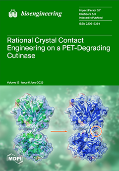
Cover Story (view full-size image): A novel protein engineering approach enhances the crystallization of the leaf branch compost cutinase (LCC) quadruple mutant ICCG, a key enzyme for effective PET recycling under mild conditions. By introducing targeted electrostatic interactions—specifically Arg–Glu pairs—at crystal contact sites, researchers generated the ICCGY T110E mutant with improved crystallizability in terms of faster crystallization at decreased protein concentrations. Controlled mutations at non-interacting sites confirmed the specificity of this strategy, with preservation of activity. This advancement facilitates more efficient downstream processing of biocatalysts, offering a rational method to streamline enzyme purification and support sustainable industrial biotechnology applications. View this paper
  • Issues are regarded as officially published after their release is announced to the table of contents alert mailing list.
  • You may sign up for e-mail alerts to receive table of contents of newly released issues.
  • PDF is the official format for papers published in both, html and pdf forms. To view the papers in pdf format, click on the "PDF Full-text" link, and use the free Adobe Reader to open them.
Order results
Result details
Section
Select all
Export citation of selected articles as:
25 pages, 789 KiB  
Article
A Changepoint Detection-Based General Methodology for Robust Signal Processing: An Application to Understand Preeclampsia’s Mechanisms
by Patricio Cumsille, Felipe Troncoso, Hermes Sandoval, Jesenia Acurio and Carlos Escudero
Bioengineering 2025, 12(6), 675; https://doi.org/10.3390/bioengineering12060675 - 19 Jun 2025
Viewed by 432
Abstract
Motivated by illuminating the underlying mechanisms of preeclampsia, we develop a changepoint detection-based general and versatile methodology that can be applied to any experimental model, effectively addressing the challenges of high uncertainty produced by experimental interventions, intrinsic high variability, and rapidly and abruptly [...] Read more.
Motivated by illuminating the underlying mechanisms of preeclampsia, we develop a changepoint detection-based general and versatile methodology that can be applied to any experimental model, effectively addressing the challenges of high uncertainty produced by experimental interventions, intrinsic high variability, and rapidly and abruptly varying time dynamics in perfusion signals. This methodology provides a systematic and reliable approach for robust perfusion signal analysis. The main innovation of our methodology is a highly efficient automatic data processing system consisting of modular programming components. These components include a signal processing tool for optimal segmentation of perfusion signals by isolating their “genuine” vascular response to experimental interventions, and a novel and suitable normalization to evaluate this response concerning an experimental reference state, typically basal or pre-intervention. In this way, we can identify anomalies in an experimental group compared to a control group by disaggregating noise during the transitions just after experimental interventions. We have successfully applied our general methodology to perfusion signals measured from a preeclampsia-like syndrome model developed by our research group. Our findings revealed impaired brain perfusion in offspring from preeclampsia, particularly dysfunctional brain perfusion signals with inadequate perfusion signal vasoreactivity to thermal physical stimuli. This general methodology represents a significant step towards a systematic, accurate, and reliable approach to robust perfusion signals analysis across various experimental settings with diverse intervention protocols. Full article
(This article belongs to the Special Issue 10th Anniversary of Bioengineering: Biosignal Processing)
Show Figures

Figure 1

13 pages, 1093 KiB  
Article
A Hybrid Deep Learning Framework for Accurate Cell Segmentation in Whole Slide Images Using YOLOv11, StarDist, and SAM2
by Julius Bamwenda, Mehmet Siraç Özerdem, Orhan Ayyıldız and Veysı Akpolat
Bioengineering 2025, 12(6), 674; https://doi.org/10.3390/bioengineering12060674 - 19 Jun 2025
Viewed by 616
Abstract
Accurate segmentation of cellular structures in whole slide images (WSIs) is essential for quantitative analysis in computational pathology. However, the complexity and scale of WSIs present significant challenges for conventional segmentation methods. In this study, we propose a novel hybrid deep learning framework [...] Read more.
Accurate segmentation of cellular structures in whole slide images (WSIs) is essential for quantitative analysis in computational pathology. However, the complexity and scale of WSIs present significant challenges for conventional segmentation methods. In this study, we propose a novel hybrid deep learning framework that integrates three complementary approaches, YOLOv11, StarDist, and Segment Anything Model v2 (SAM2), to achieve robust and precise cell segmentation. The proposed pipeline utilizes YOLOv11 as an object detector to localize regions of interest, generating bounding boxes or preliminary masks that are subsequently used either as prompts to guide SAM2 or to filter segmentation outputs. StarDist is employed to model cell and nuclear boundaries with high geometric precision using star-convex polygon representations, which are particularly effective in densely packed cellular regions. The framework was evaluated on a unique WSI dataset comprising 256 × 256 image tiles annotated with high-resolution cell-level masks. Quantitative evaluations using the Dice coefficient, intersection over union (IoU), F1-score, precision, and recall demonstrated that the proposed method significantly outperformed individual baseline models. The integration of object detection and prompt-based segmentation led to enhanced boundary accuracy, improved localization, and greater robustness across varied tissue types. This work contributes a scalable and modular solution for advancing automated histopathological image analysis. Full article
(This article belongs to the Special Issue Machine Learning and Deep Learning Applications in Healthcare)
Show Figures

Figure 1

13 pages, 2595 KiB  
Article
A Miniaturized Implantable Telemetry Biosensor for the Long-Term Dual-Modality Monitoring of Core Temperature and Locomotor Activity
by Wendi Shi, Hao Huang, Xueting Sun, Qihui Jia, Yu Zhou, Maohua Zhu, Mingqiang Tian, Zhuofan Li, Zepeng Zhang, Tongfei A. Wang and Lei Zhang
Bioengineering 2025, 12(6), 673; https://doi.org/10.3390/bioengineering12060673 - 19 Jun 2025
Viewed by 430
Abstract
Implantable telemetry biosensors have become powerful tools for continuous physiological monitoring with minimal animal perturbation. However, commercially available implants are relatively oversized for small animals such as mice and have limited transmission range, leading to concerns about animal welfare, experiment scenarios, and the [...] Read more.
Implantable telemetry biosensors have become powerful tools for continuous physiological monitoring with minimal animal perturbation. However, commercially available implants are relatively oversized for small animals such as mice and have limited transmission range, leading to concerns about animal welfare, experiment scenarios, and the reliability of the data. In this study, we designed a telemetry system that tracks the animals’ body temperature and locomotor activity in real time. The implant integrates a temperature sensor with a 3-axis accelerometer and is capable of wirelessly transmitting data over a 40 m mesh network. The implant’s temperature performance was evaluated in bench tests, showing a response rate of 0.2 °C/s, drift ≤ 0.03 °C within 31 days, and a standard deviation of 0.035 °C across three identically designed implants. Meanwhile, the in vivo implant’s locomotion recordings showed strong agreement with computer vision analysis with a correlation coefficient of r = 0.95 (p < 0.001), and their body temperature recordings were aligned to differential states of rest, exercise, or post-exercise recovery. The results demonstrate stable and highly accurate performance over the 30-day implantation period. Its ability to minimize behavioral interference while enabling long-term continuous monitoring highlights its value in both biomedical and animal behavior research. Full article
(This article belongs to the Section Biosignal Processing)
Show Figures

Figure 1

21 pages, 1609 KiB  
Article
Resting-State Activity Changes Induced by tDCS in MS Patients and Healthy Controls: A Simultaneous tDCS rs-fMRI Study
by Marco Muccio, Giuseppina Pilloni, Lillian Walton Masters, Peidong He, Lauren Krupp, Abhishek Datta, Marom Bikson, Leigh Charvet and Yulin Ge
Bioengineering 2025, 12(6), 672; https://doi.org/10.3390/bioengineering12060672 - 19 Jun 2025
Viewed by 547
Abstract
Transcranial direct current stimulation (tDCS) is a safe, well-tolerated method of non-invasively eliciting cortical neuromodulation. It has gained recent interest, especially for its positive clinical outcomes in neurodegenerative diseases such as multiple sclerosis (MS). However, its simultaneous (during tDCS) and cumulative effects (following [...] Read more.
Transcranial direct current stimulation (tDCS) is a safe, well-tolerated method of non-invasively eliciting cortical neuromodulation. It has gained recent interest, especially for its positive clinical outcomes in neurodegenerative diseases such as multiple sclerosis (MS). However, its simultaneous (during tDCS) and cumulative effects (following repeated tDCS sessions) on the regional brain activity during rest need further investigation, especially in MS. This study aims to elucidate tDCS’ underpinnings, alongside its therapeutic impact in MS patients, using concurrent tDCS-MRI methods. In total, 20 MS patients (age = 48 ± 12 years; 8 males) and 28 healthy controls (HCs; age = 36 ± 15 years; 12 males) were recruited. They participated in a tDCS-MRI session, during which resting-state functional MRI (rs-fMRI) was used to measure the levels of the fractional amplitude of low-frequency fluctuations (fALFFs), which is an index of regional neuronal activity, before and during left anodal dorsolateral prefrontal cortex (DLPFC) tDCS (2.0 mA for 15 min). MS patients were then asked to return for an identical tDCS-MRI visit (follow-up) after 20 identical at-home tDCS sessions. Simultaneous tDCS-induced changes in fALFF are seen across cortical and subcortical areas in both HC and MS patients, with some regions showing increased and others decreased brain activity. In HCs, fALFF increased in the right pre- and post-central gyrus whilst it decreased in subcortical regions. Conversely, MS patients initially displayed increases in more posterior cortical regions but decreases in the superior and temporal cortical regions. At follow-up, MS patients showed reversed patterns, emphasizing significant cumulative effects of tDCS treatment upon brain excitation. Such long-lasting changes are further supported by greater pre-tDCS fALFFs measured at follow-up compared to baseline, especially around the cuneus. The results were significant after correcting for multiple comparisons (p-FDR < 0.05). Our study shows that tDCS has both simultaneous and cumulative effects on neuronal activity measured with rs-fMRI, especially involving major brain areas distant from the site of stimulation, and it is responsible for fatigue and cognitive and motor skills. Full article
Show Figures

Figure 1

13 pages, 1801 KiB  
Article
Finite Element Analysis of Biomechanical Assessment: Traditional Bilateral Pedicle Screw System vs. Novel Reverse Transdiscal Screw System for Lumbar Degenerative Disc Disease
by Utpal K. Dhar, Kamran Aghayev, Hadi Sultan, Saahas Rajendran, Chi-Tay Tsai and Frank D. Vrionis
Bioengineering 2025, 12(6), 671; https://doi.org/10.3390/bioengineering12060671 - 19 Jun 2025
Viewed by 517
Abstract
The traditional bilateral pedicle screw system has been used for the treatment of various lumbar spine conditions including advanced degenerative disc disease. However, there is an ongoing need to develop more effective and less invasive techniques. The purpose of this study was to [...] Read more.
The traditional bilateral pedicle screw system has been used for the treatment of various lumbar spine conditions including advanced degenerative disc disease. However, there is an ongoing need to develop more effective and less invasive techniques. The purpose of this study was to compare the traditional bilateral pedicle screw system (BPSS) with the novel reverse transdiscal screw system (RTSS) for lumbar disc degenerative disease. A 3D solid lumbar L1–L5 spine model was developed and validated based on a human CT scan. Fusions were simulated at L3–L4. The first scenario comprised a transforaminal lumbar interbody cage in combination with the bilateral pedicle screw-rod system (BPSS-TLIF). In the second scenario, the same TLIF cage was combined with reverse L3–L4 transdiscal screws (RTSS-TLIF). Testing parameters included range of motion (ROM) in three orthogonal axes, hardware (cage and screw) stress, and shear load resistance. The ROM of the surgical model was reduced by approximately 90% compared to the intact model at the fused level. The RTSS model demonstrated less ROM compared to the BPSS model at the fused level for all loading conditions. Overall, the RTSS model exhibited lower stress on both screws and cage compared with the BPSS model in all biomechanical testing conditions. The RTSS model also exhibited higher anterior and posterior shear load resistance than the BPSS model. In conclusion, the RTSS model proved superior to the BPSS model in all respects. These findings indicate that the RTSS could serve as a feasible option for patients undergoing lumbar fusion, especially for adjacent segment disease, potentially enhancing surgical outcomes for disc degeneration. Full article
(This article belongs to the Special Issue Spine Biomechanics)
Show Figures

Figure 1

20 pages, 1771 KiB  
Article
An Innovative Artificial Intelligence Classification Model for Non-Ischemic Cardiomyopathy Utilizing Cardiac Biomechanics Derived from Magnetic Resonance Imaging
by Liqiang Fu, Peifang Zhang, Liuquan Cheng, Peng Zhi, Jiayu Xu, Xiaolei Liu, Yang Zhang, Ziwen Xu and Kunlun He
Bioengineering 2025, 12(6), 670; https://doi.org/10.3390/bioengineering12060670 - 19 Jun 2025
Viewed by 554
Abstract
Significant challenges persist in diagnosing non-ischemic cardiomyopathies (NICMs) owing to early morphological overlap and subtle functional changes. While cardiac magnetic resonance (CMR) offers gold-standard structural assessment, current morphology-based AI models frequently overlook key biomechanical dysfunctions like diastolic/systolic abnormalities. To address this, we propose [...] Read more.
Significant challenges persist in diagnosing non-ischemic cardiomyopathies (NICMs) owing to early morphological overlap and subtle functional changes. While cardiac magnetic resonance (CMR) offers gold-standard structural assessment, current morphology-based AI models frequently overlook key biomechanical dysfunctions like diastolic/systolic abnormalities. To address this, we propose a dual-path hybrid deep learning framework based on CNN-LSTM and MLP, integrating anatomical features from cine CMR with biomechanical markers derived from intraventricular pressure gradients (IVPGs), significantly enhancing NICM subtype classification by capturing subtle biomechanical dysfunctions overlooked by traditional morphological models. Our dual-path architecture combines a CNN-LSTM encoder for cine CMR analysis and an MLP encoder for IVPG time-series data, followed by feature fusion and dense classification layers. Trained on a multicenter dataset of 1196 patients and externally validated on 137 patients from a distinct institution, the model achieved a superior performance (internal AUC: 0.974; external AUC: 0.962), outperforming ResNet50, VGG16, and radiomics-based SVM. Ablation studies confirmed IVPGs’ significant contribution, while gradient saliency and gradient-weighted class activation mapping (Grad-CAM) visualizations proved the model pays attention to physiologically relevant cardiac regions and phases. The framework maintained robust generalizability across imaging protocols and institutions with minimal performance degradation. By synergizing biomechanical insights with deep learning, our approach offers an interpretable, data-efficient solution for early NICM detection and subtype differentiation, holding strong translational potential for clinical practice. Full article
(This article belongs to the Special Issue Bioengineering in a Generative AI World)
Show Figures

Figure 1

34 pages, 720 KiB  
Review
A Comprehensive Review of Unobtrusive Biosensing in Intelligent Vehicles: Sensors, Algorithms, and Integration Challenges
by Shiva Maleki Varnosfaderani, Mohd. Rizwan Shaikh and Mohamad Forouzanfar
Bioengineering 2025, 12(6), 669; https://doi.org/10.3390/bioengineering12060669 - 18 Jun 2025
Viewed by 513
Abstract
Unobtrusive in-vehicle measurement and the monitoring of physiological signals have recently attracted researchers in industry and academia as an innovative approach that can provide valuable information about drivers’ health and status. The main goal is to reduce the number of traffic accidents caused [...] Read more.
Unobtrusive in-vehicle measurement and the monitoring of physiological signals have recently attracted researchers in industry and academia as an innovative approach that can provide valuable information about drivers’ health and status. The main goal is to reduce the number of traffic accidents caused by driver errors by monitoring various physiological parameters and devising appropriate actions to alert the driver or to take control of the vehicle. The research on this topic is in its early stages. While there have been several publications on this topic and industrial prototypes made by car manufacturers, a comprehensive and critical review of the current trends and future directions is missing. This review examines the current research and findings in in-vehicle physiological monitoring and suggests future directions and potential uses. Various physiological sensors, their potential locations, and the results they produce are demonstrated. The main challenges of in-vehicle biosensing, including unobtrusive sensing, vehicle vibration and driver movement cancellation, and privacy management, are discussed, and possible solutions are presented. The paper also reviews the current in-vehicle biosensing prototypes built by car manufacturers and other researchers. The reviewed methods and presented directions provide valuable insights into robust and accurate biosensing within vehicles for researchers in the field. Full article
(This article belongs to the Section Biosignal Processing)
Show Figures

Figure 1

19 pages, 9332 KiB  
Article
Biomechanical Design and Validation of a Novel Elliptical Sleeve Pedicle Screw for Enhanced Spinal Fixation Stability
by Ting-Shuo Hsu, Chang-Jung Chiang, Hsuan-Wen Wang, Yu-San Chen and Chun-Li Lin
Bioengineering 2025, 12(6), 668; https://doi.org/10.3390/bioengineering12060668 - 18 Jun 2025
Viewed by 736
Abstract
This study aimed to develop a novel modular pedicle screw system incorporating an elliptical sleeve to conform the pedicle’s elliptical cross-section and enhance fixation strength with mechanical stability. The biomechanical evaluation was conducted based on fundamental mechanics principles, followed by a finite element [...] Read more.
This study aimed to develop a novel modular pedicle screw system incorporating an elliptical sleeve to conform the pedicle’s elliptical cross-section and enhance fixation strength with mechanical stability. The biomechanical evaluation was conducted based on fundamental mechanics principles, followed by a finite element (FE) analysis to assess stress distribution under compressive and torsional loads. Subsequently, mechanical testing was performed to evaluate static and fatigue bending performance and in vitro biomechanical fatigue in porcine vertebrae by pull-out testing after 5000 and 100,000 cycles to assess fixation stability. The FE analysis demonstrated that the elliptical sleeve design improved bending resistance by 1.21× and torsional resistance by 1.91× compared to conventional cylindrical screws. Mechanical testing revealed greater bending/torsion stiffness and fatigue resistance, with the elliptical sleeve screw withstanding 5 million cycles at 235.4 N, compared to 175.46 N for cylindrical screws. Biomechanical pull-out testing further confirmed significantly higher retention strength after 100,000 cycles (1229.75 N vs. 867.83 N, p = 0.0101), whereas cylindrical screws failed prematurely at 10,663 cycles due to excessive displacement (>2 mm). The elliptical sleeve pedicle screw system demonstrated enhanced fixation strength, reduced micromotion, and superior fatigue resistance, making it a promising alternative to conventional pedicle screws for improving long-term spinal fixation stability. Full article
(This article belongs to the Special Issue Joint Biomechanics and Implant Design)
Show Figures

Figure 1

20 pages, 2303 KiB  
Article
Dynamically Quantifying Vocal Fold Thickness: Effects of Medialization Implant Location on Glottal Shape and Phonation
by Charles Farbos de Luzan, Jacob Michaud-Dorko, Rebecca J. Howell, Ephraim Gutmark and Liran Oren
Bioengineering 2025, 12(6), 667; https://doi.org/10.3390/bioengineering12060667 - 18 Jun 2025
Viewed by 631
Abstract
Unilateral vocal fold paralysis (UVFP) can lead to significant dysphonia. Medialization thyroplasty type 1 (TT1) is a common surgical intervention aiming at improving vocal quality by optimally positioning the paralyzed fold to generate the necessary vibrations for phonation. Implants are generally placed through [...] Read more.
Unilateral vocal fold paralysis (UVFP) can lead to significant dysphonia. Medialization thyroplasty type 1 (TT1) is a common surgical intervention aiming at improving vocal quality by optimally positioning the paralyzed fold to generate the necessary vibrations for phonation. Implants are generally placed through the thyroid cartilage in a sedated patient and positioned either underneath the level of the vocal folds (infraglottal medialization or IM) or at the level of the vocal folds (glottal medialization or GM). Using high-speed three-dimensional digital image correlation (3D-DIC) in an ex vivo canine hemilarynx model, this study explores the impact of implant location, specifically IM versus GM on the pre-phonatory and dynamic vertical thickness, glottal divergence, flow rate (Q), and cepstral peak prominence (CPP) under varying adduction and subglottal pressure conditions. IM consistently increased glottal divergence and dynamic vertical thickness, particularly in under-adducted states (AL1), despite producing lower static thickness than GM. CPP remained unaffected by the implant condition, but Q decreased significantly with IM under AL1, indicating enhanced glottal resistance and closure. These findings suggest that IM may offer superior functional outcomes by restoring divergent glottal shaping and improving vibratory efficiency. This study also introduces a validated method for dynamically quantifying vocal fold thickness and emphasizes the importance of implant depth in medialization thyroplasty strategies. Full article
Show Figures

Figure 1

15 pages, 2466 KiB  
Article
MRI-Based Machine Learning and Radiomics Methods for Assessing Spinal Cord Function in Patients with Mild Cervical Spondylotic Myelopathy
by He Wang, Kai Wang, Yutian Wang, Zhenlei Liu, Lei Zhang, Shanhang Jia, Kun He, Xiangyu Zhang and Hao Wu
Bioengineering 2025, 12(6), 666; https://doi.org/10.3390/bioengineering12060666 - 17 Jun 2025
Viewed by 809
Abstract
(1) Background: Patients with mild cervical spondylotic myelopathy (CSM) who delay surgery risk progression. While PET evaluates spinal cord function, its cost and radiation limit its use. (2) Methods: In this prospective study, patients with mild cervical spondylosis underwent preoperative 18F-FDG PET-MRI. Narrowed [...] Read more.
(1) Background: Patients with mild cervical spondylotic myelopathy (CSM) who delay surgery risk progression. While PET evaluates spinal cord function, its cost and radiation limit its use. (2) Methods: In this prospective study, patients with mild cervical spondylosis underwent preoperative 18F-FDG PET-MRI. Narrowed spinal levels were classified based on whether SUVmax was decreased. Follow-up assessments were conducted. Two machine learning models using MRI T2-based radiomics were developed to identify stenotic levels and decreased SUVmax. (3) Results: Patients with normal SUVmax showed greater symptom improvement. The radiomics models performed well, with AUCs of 0.981/0.962 (training/testing) for stenosis detection and 0.830/0.812 for predicting SUVmax decline. The model outperformed clinicians in predicting SUVmax decline, improving the AUC by 10%. (4) Conclusion: Patients with preserved SUVmax have better outcomes. MRI-based radiomics shows potential for identifying stenosis and predicting spinal cord function changes for preoperative assessment, though larger studies are needed to validate its clinical utility. Full article
Show Figures

Figure 1

35 pages, 8317 KiB  
Article
ResST-SEUNet++: Deep Model for Accurate Segmentation of Left Ventricle and Myocardium in Magnetic Resonance Imaging (MRI) Images
by Abduljabbar S. Ba Mahel, Mehdhar S. A. M. Al-Gaashani, Fahad Mushabbab G. Alotaibi and Reem Ibrahim Alkanhel
Bioengineering 2025, 12(6), 665; https://doi.org/10.3390/bioengineering12060665 - 17 Jun 2025
Viewed by 492
Abstract
The highly precise and trustworthy segmentation of the left ventricle (LV) and myocardium is critical for diagnosing and treating cardiovascular disorders, which includes persistent microvascular obstruction (MVO) as well as myocardial infarction (MI) diseases. This process improves diagnostic accuracy and optimizes the planning [...] Read more.
The highly precise and trustworthy segmentation of the left ventricle (LV) and myocardium is critical for diagnosing and treating cardiovascular disorders, which includes persistent microvascular obstruction (MVO) as well as myocardial infarction (MI) diseases. This process improves diagnostic accuracy and optimizes the planning and implementation of therapeutic interventions, ultimately improving the quality of care and patient prognosis. Limitations of earlier investigations include neglecting the complex image pre-processing required to accurately delineate areas of the LV and myocardium (Myo) in MRI and the absence of a substantial, high-quality dataset. Thus, this paper presents a comprehensive end-to-end framework, which includes contrast-limited adaptive histogram equalization (CLAHE) and bilateral filtering methods for image pre-processing and the development and implementation of a proposed deep model for left ventricular and myocardium segmentation. This study utilizes the EMIDEC database for the training and assessment of the model, allowing for a detailed comparative analysis with six state-of-the-art (SOTA) segmentation models. This approach provides a high accuracy and reliability for the segmentation that is crucial for the diagnosis and treatment of cardiovascular disorders. The achievements of the proposed model are demonstrated by high average values of segmentation rates, such as an Intersection over Union (IoU) of 93.73%, Recall of 96.54%, Dice coefficient of 96.70%, Precision of 96.86%, and F1-score of 96.70%. To verify the generalization capability, we assessed our suggested model on five supplementary databases, which substantiates its exceptional efficiency and adaptability in a diverse environment. The presented findings demonstrate that the proposed deep model surpasses current methods, offering more a precise and resilient segmentation of cardiac structures. Full article
(This article belongs to the Special Issue Medical Artificial Intelligence and Data Analysis)
Show Figures

Figure 1

3 pages, 161 KiB  
Editorial
Recent Advances in Drug Delivery and Oral Health: The Impact of Technology and Digital Advances as a New Frontier
by Cristina Grippaudo, Ludovica Nucci and Marco Farronato
Bioengineering 2025, 12(6), 664; https://doi.org/10.3390/bioengineering12060664 - 17 Jun 2025
Viewed by 433
Abstract
Technological progress is the basis of scientific and clinical advancement in dentistry [...] Full article
18 pages, 1615 KiB  
Article
Effects of Physiological Loading from Patient-Derived Activities of Daily Living on the Wear of Metal-on-Polymer Total Hip Replacements
by Benjamin A. Clegg, Samuel Perry, Enrico De Pieri, Anthony C. Redmond, Stephen J. Ferguson, David E. Lunn, Richard M. Hall, Michael G. Bryant, Nazanin Emami and Andrew R. Beadling
Bioengineering 2025, 12(6), 663; https://doi.org/10.3390/bioengineering12060663 - 16 Jun 2025
Viewed by 600
Abstract
The current pre-clinical testing standards for total hip replacements (THRs), ISO standards, use simplified loading waveforms that do not fully replicate real-world biomechanics. These standards provide a benchmark of data that may not accurately predict in vivo wear, necessitating the evaluation of physiologically [...] Read more.
The current pre-clinical testing standards for total hip replacements (THRs), ISO standards, use simplified loading waveforms that do not fully replicate real-world biomechanics. These standards provide a benchmark of data that may not accurately predict in vivo wear, necessitating the evaluation of physiologically relevant loading conditions. Previous studies have incorporated activities of daily living (ADLs) such as walking, jogging and stair negotiation into wear simulations. However, these studies primarily used simplified adaptations that increased axial forces and applied accelerated sinusoidal waveforms, rather than fully replicating the complex kinematics experienced by THR patients. To address this gap, this study applied patient-derived ADL profiles—jogging and stair negotiation—using a three-station hip simulator, obtained through 3D motion analysis of total hip arthroplasty patients, processed via a musculoskeletal multibody modelling approach to derive realistic hip contact forces (HCFs). The results indicate that jogging significantly increased wear rates compared to the ISO walking gait waveform, with wear increasing from 15.24 ± 0.55 to 28.68 ± 0.87 mm3/Mc. Additionally, wear was highly sensitive to changes in lubricant protein concentration, with an increase from 17 g/L to 30 g/L reducing wear by over 60%. Contrary to predictive models, stair descent resulted in higher volumetric wear (8.62 ± 0.43 mm3/0.5 Mc) compared to stair ascent (4.15 ± 0.31 mm3/0.5 Mc), despite both profiles having similar peak torques. These findings underscore the limitations of current ISO standards in replicating physiologically relevant wear patterns. The application of patient-specific loading profiles highlights the need to integrate ADLs into pre-clinical testing protocols, ensuring a more accurate assessment of implant performance and longevity. Full article
(This article belongs to the Special Issue Medical Devices and Implants, 2nd Edition)
Show Figures

Figure 1

17 pages, 1032 KiB  
Article
Development and Validation of a Virtual Version of the Box and Block Test to Assess Manual Dexterity at Home for Adults with Stroke and Children with Cerebral Palsy
by Zélie Rosselli, Merlin Somville, Edouard Ducoffre, Carlyne Arnould, Geoffroy Saussez and Yannick Bleyenheuft
Bioengineering 2025, 12(6), 662; https://doi.org/10.3390/bioengineering12060662 - 16 Jun 2025
Viewed by 537
Abstract
The REAtouch® Lite device was recently developed to support motor skill learning-based interventions, integrating both games/activities and assessment tools to enable home-based telerehabilitation. Given the importance of hand functions in rehabilitation of patients with brain lesions, this study aimed to validate a [...] Read more.
The REAtouch® Lite device was recently developed to support motor skill learning-based interventions, integrating both games/activities and assessment tools to enable home-based telerehabilitation. Given the importance of hand functions in rehabilitation of patients with brain lesions, this study aimed to validate a virtual version of the Box and Block Test (vBBT) implemented in the REAtouch® device. A total of 205 healthy participants, 37 post-stroke adults, and 37 children with cerebral palsy (CP) performed the standard BBT, various versions of the newly designed vBBT (with/without a separation wall; with 6, 4, and free zones) and the Tower of London test assessing executive function/planning abilities. Friedman’s ANOVA revealed significant differences between the BBT and all versions of the vBBT scores in healthy participants (all p < 0.001). However, the vBBT-4 zones showed the largest intraclass correlation coefficient (ICC) with the BBT in healthy participants (0.58) and even higher correlations in participants with CP and stroke (>0.8). Only the vBBT-6 zones version showed a significant correlation with patients’ planning abilities (p < 0.01; r = −0.28). These findings highlight the vBBT-4 zones as the most relevant version to assess hand dexterity directly with the REAtouch® device, potentially within telerehabilitation modalities. Further normative data must be established. Full article
Show Figures

Figure 1

13 pages, 1678 KiB  
Article
Running and Jumping After Muscle Fatigue in Subjects with a History of Knee Injury: What Are the Acute Effects of Wearing a Knee Brace on Biomechanics?
by Tobias Heß, Thomas L. Milani, Jan Stoll and Christian Mitschke
Bioengineering 2025, 12(6), 661; https://doi.org/10.3390/bioengineering12060661 - 16 Jun 2025
Viewed by 1097
Abstract
The knee is one of the most frequently injured joints, involving various structures. To prevent reinjury after rehabilitation, braces are commonly used. However, most studies on knee supports focus on subjects with anterior cruciate ligament (ACL) injuries and do not account for muscle [...] Read more.
The knee is one of the most frequently injured joints, involving various structures. To prevent reinjury after rehabilitation, braces are commonly used. However, most studies on knee supports focus on subjects with anterior cruciate ligament (ACL) injuries and do not account for muscle fatigue, which typically occurs during prolonged intense training and can significantly increase the risk of injury. Hence, this study investigates the acute effects of wearing a knee brace on biomechanics in subjects with a history of various unilateral knee injuries or pain under muscle fatigue. In total, 50 subjects completed an intense fatigue protocol and then performed counter-movement jumps and running tests on a force plate while tracking kinematics with a marker-based 3D motion analysis system. Additionally, subjects filled out a visual analog scale (VAS) to assess knee pain and stability. Tests were conducted on the injured leg with and without a knee brace (Sports Knee Support, Bauerfeind AG, Zeulenroda-Triebes, Germany) and on the healthy leg. Results indicated that wearing the knee brace stabilized knee movement in the frontal plane, with a significant reduction in maximal medio-lateral knee acceleration and knee abduction moment during running and jumping. The brace also normalized loading on the injured leg. We observed higher maximal knee flexion moments, which were associated with increased vertical ground reaction forces, segment velocities, and knee flexion angles. Subjects reported less pain and greater stability while wearing the knee brace. Therefore, we confirm that wearing a knee brace on the injured leg improves joint biomechanics by enhancing stability and kinematics and reducing pain during running and jumping, even with muscle fatigue. Consequently, wearing a knee brace after a knee joint injury may reduce the risk of reinjury. Full article
(This article belongs to the Special Issue Biomechanics of Orthopaedic Rehabilitation)
Show Figures

Figure 1

11 pages, 1645 KiB  
Brief Report
Assessing the Biocompatibility of Tannic Acid-Based Biomaterials: Addressing Challenges in Standard Cytotoxic Assays
by Silvia Cometta and Dietmar Werner Hutmacher
Bioengineering 2025, 12(6), 660; https://doi.org/10.3390/bioengineering12060660 - 16 Jun 2025
Viewed by 520
Abstract
In this comprehensive study, we delve into the intricate binding properties of tannic acid (TA) and examine their dual role in the realm of biomaterial development. While TA’s properties can enhance the functionality and performance of biomaterials, they also raise concerns regarding potential [...] Read more.
In this comprehensive study, we delve into the intricate binding properties of tannic acid (TA) and examine their dual role in the realm of biomaterial development. While TA’s properties can enhance the functionality and performance of biomaterials, they also raise concerns regarding potential biases in in vitro biocompatibility assessments. We focus on the relevance and constraints of several widely employed cell viability assays, namely the DNA-based PicoGreen assay, the PrestoBlue assay, and the Live/Dead staining technique utilizing fluorescein diacetate (FDA) and propidium iodide (PI). We investigate how these assays perform when applied to TA-coated scaffolds and cell sheets. Through a detailed presentation of our experimental findings, we juxtapose them through a critical review of the existing literature, allowing us to identify and elucidate the limitations these assays face when assessing TA-based biomaterials. In doing so, we aim not only to enhance the understanding of these potential assay biases but also to provide actionable recommendations for accurately evaluating the biocompatibility of TA-modified substances. This dual approach, combining empirical research with literature analysis, offers vital insights for the research community, ensuring that the assessment of TA-coated biomaterials is scientifically sound and reproducible. Full article
(This article belongs to the Section Biomedical Engineering and Biomaterials)
Show Figures

Figure 1

18 pages, 3902 KiB  
Article
XGBoost and SHAP-Based Analysis of Risk Factors for Hypertension Classification in Korean Postmenopausal Women
by Hojeong Kim, Mavlonbek Khomidov and Jong-Ha Lee
Bioengineering 2025, 12(6), 659; https://doi.org/10.3390/bioengineering12060659 - 16 Jun 2025
Viewed by 497
Abstract
In postmenopausal women, the prevalence of hypertension increases sharply, emphasizing the importance of its prevention. This increased risk highlights the critical need for effective prevention strategies specifically designed for this population. To address this issue, the present study aimed to identify easily measurable [...] Read more.
In postmenopausal women, the prevalence of hypertension increases sharply, emphasizing the importance of its prevention. This increased risk highlights the critical need for effective prevention strategies specifically designed for this population. To address this issue, the present study aimed to identify easily measurable risk factors that contribute to hypertension in postmenopausal women using explainable artificial intelligence (XAI) and machine learning (ML) techniques. This study conducted hypertension classification by analyzing health checkup data from 3289 postmenopausal Korean women aged 55–79 years, extracted from the 2022–2023 Korea National Health Insurance Service (KNHIS) database, using XGBoost, SVM and ANN. XGBoost was the most effective model (AUC: 92.12%, MCC: 0.71) in hypertension classification. Shapley Additive exPlanations-based feature importance identified age and waist circumference (WC) as the most important risk factors for hypertension. In this study, blood pressure increased with variations in WC, a modifiable risk factor. These findings suggest that WC should be managed more strictly to prevent hypertension in postmenopausal women. Full article
(This article belongs to the Section Biosignal Processing)
Show Figures

Figure 1

21 pages, 2025 KiB  
Article
BioGAN: Enhancing Transcriptomic Data Generation with Biological Knowledge
by Francesca Pia Panaccione, Sofia Mongardi, Marco Masseroli and Pietro Pinoli
Bioengineering 2025, 12(6), 658; https://doi.org/10.3390/bioengineering12060658 - 16 Jun 2025
Viewed by 512
Abstract
The advancement of computational genomics has significantly enhanced the use of data-driven solutions in disease prediction and precision medicine. Yet, challenges such as data scarcity, privacy constraints, and biases persist. Synthetic data generation offers a promising solution to these issues. However, existing approaches [...] Read more.
The advancement of computational genomics has significantly enhanced the use of data-driven solutions in disease prediction and precision medicine. Yet, challenges such as data scarcity, privacy constraints, and biases persist. Synthetic data generation offers a promising solution to these issues. However, existing approaches based on generative artificial intelligence often fail to incorporate biological knowledge, limiting the realism and utility of generated samples. In this work, we present BioGAN, a novel generative framework that, for the first time, incorporates graph neural networks into a generative adversarial network architecture for transcriptomic data generation. By leveraging gene regulatory and co-expression networks, our model preserves biological properties in the generated transcriptomic profiles. We validate its effectiveness on E. coli and human gene expression datasets through extensive experiments using unsupervised and supervised evaluation metrics. The results demonstrate that incorporating a priori biological knowledge is an effective strategy for enhancing both the quality and utility of synthetic transcriptomic data. On human data, BioGAN achieves a 4.3% improvement in precision and an up to 2.6% higher correlation with real profiles compared to state-of-the-art models. In downstream disease and tissue classification tasks, our synthetic data improves prediction performance by an average of 5.7%. Results on E. coli further confirm BioGAN’s robustness, showing consistently strong recall and predictive utility. Full article
(This article belongs to the Special Issue Computational Genomics for Disease Prediction)
Show Figures

Figure 1

13 pages, 3121 KiB  
Article
Cell-Based Therapies: Ferromagnetic Versus Superparamagnetic Cell Targeting
by Tasneem Halhouli, Lisa Münchhalfen, Sarkawt Hamad, Larissa Schmitz-Ullrich, Frank Nitsche, Felix Gaedke, Astrid Schauss, Linlin Zhang, Quoc-Khanh Pham, Gang Bao and Kurt Paul Pfannkuche
Bioengineering 2025, 12(6), 657; https://doi.org/10.3390/bioengineering12060657 - 16 Jun 2025
Viewed by 974
Abstract
Stem-cell-based therapies rely on the transplantation of stem cells or stem-cell-derived organotypic cells into injured tissues in order to improve or restore tissue function that has been impaired by various diseases. The potential of induced pluripotent stem cells has created many applications in [...] Read more.
Stem-cell-based therapies rely on the transplantation of stem cells or stem-cell-derived organotypic cells into injured tissues in order to improve or restore tissue function that has been impaired by various diseases. The potential of induced pluripotent stem cells has created many applications in the field of cell therapy, for example. Some applications, for example, those in cardiac cell therapy, suffer from low or very low efficiencies of cell engraftment. Therefore, magnetic cell targeting can be discussed as a method for capturing superparamagnetic nanoparticle-labelled cells in the tissue. Here, we employ superparamagnetic iron oxide nanoparticles (SPIONs) for the intracellular magnetic loading of mesenchymal stem cells (MSCs). In addition, we test a novel strategy of labelling MSCs with ferromagnetic particles. The adhesion assays demonstrate a faster adhesion kinetic of SPIONs-loaded MSC spheroids when a magnetic field was applied, resulting in >50% spheroid adhesion after 30 min. Clustering of cells inside the magnetic field is a second potential mechanism of magnetic cell retention and >80% of cells were found to be aggregated in clusters when placed in a magnetic field for 10 min. SPIONs-loaded and ferromagnetic-particle-loaded cells performed equally in the cell clustering assay. In conclusion, the clustering of SPION-labelled cells explains the observation that magnetic targeting reaches maximal efficiency in vivo after only 10 min of magnetic field application. This has significant implications for magnetic-targeting-assisted stem cell and cell replacement therapies. Full article
Show Figures

Graphical abstract

16 pages, 1628 KiB  
Article
Anatomical Characteristics Predict Response to Transcranial Direct Current Stimulation (tDCS): Development of a Computational Pipeline for Optimizing tDCS Protocols
by Giulia Caiani, Emma Chiaramello, Marta Parazzini, Eleonora Arrigoni, Leonor J. Romero Lauro, Alberto Pisoni and Serena Fiocchi
Bioengineering 2025, 12(6), 656; https://doi.org/10.3390/bioengineering12060656 - 15 Jun 2025
Viewed by 547
Abstract
Transcranial direct current stimulation (tDCS) is a non-invasive brain stimulation technique promisingly used to treat neurological and psychological disorders. Nevertheless, the inter-subject heterogeneity in its after-effects frequently limits its efficacy. This can be attributed to fixed-dose methods, which do not consider inter-subject anatomical [...] Read more.
Transcranial direct current stimulation (tDCS) is a non-invasive brain stimulation technique promisingly used to treat neurological and psychological disorders. Nevertheless, the inter-subject heterogeneity in its after-effects frequently limits its efficacy. This can be attributed to fixed-dose methods, which do not consider inter-subject anatomical variations. This work attempts to overcome this constraint by examining the effects of age and anatomical features, including the volume of cerebrospinal fluid (CSF), the thickness of the skull, and the composition of brain tissue, on electric field distribution and cortical excitability. A computational approach was used to map the electric field distribution over the brain tissues of realistic head models reconstructed from MRI images of twenty-three subjects, including adults and children of both genders. Significant negative correlations (p < 0.05) were found in the data between the maximum electric field strength and anatomical variable parameters. Furthermore, this study showed that the percentage of brain tissue exposed to an electric field amplitude above a pre-defined threshold (i.e., 0.227 V/m) was the main factor influencing the responsiveness to tDCS. In the end, the research suggests multiple regression models as useful tool to predict subjects’ responsiveness and to support a personalized approach that tailors the injected current to the morphology of the patient. Full article
Show Figures

Figure 1

16 pages, 5313 KiB  
Article
AI-Powered Spectral Imaging for Virtual Pathology Staining
by Adam Soker, Maya Almagor, Sabine Mai and Yuval Garini
Bioengineering 2025, 12(6), 655; https://doi.org/10.3390/bioengineering12060655 - 15 Jun 2025
Viewed by 734
Abstract
Pathological analysis of tissue biopsies remains the gold standard for diagnosing cancer and other diseases. However, this is a time-intensive process that demands extensive training and expertise. Despite its importance, it is often subjective and not entirely error-free. Over the past decade, pathology [...] Read more.
Pathological analysis of tissue biopsies remains the gold standard for diagnosing cancer and other diseases. However, this is a time-intensive process that demands extensive training and expertise. Despite its importance, it is often subjective and not entirely error-free. Over the past decade, pathology has undergone two major transformations. First, the rise in whole slide imaging has enabled work in front of a computer screen and the integration of image processing tools to enhance diagnostics. Second, the rapid evolution of Artificial Intelligence has revolutionized numerous fields and has had a remarkable impact on humanity. The synergy of these two has paved the way for groundbreaking research aiming for advancements in digital pathology. Despite encouraging research outcomes, AI-based tools have yet to be actively incorporated into therapeutic protocols. This is primary due to the need for high reliability in medical therapy, necessitating a new approach that ensures greater robustness. Another approach for improving pathological diagnosis involves advanced optical methods such as spectral imaging, which reveals information from the tissue that is beyond human vision. We have recently developed a unique rapid spectral imaging system capable of scanning pathological slides, delivering a wealth of critical diagnostic information. Here, we present a novel application of spectral imaging (SI) for virtual Hematoxylin and Eosin (H&E) staining using a custom-built, rapid Fourier-based SI system. Unstained human biopsy samples are scanned, and a Pix2Pix-based neural network generates realistic H&E-equivalent images. Additionally, we applied Principal Component Analysis (PCA) to the spectral information to examine the effect of down sampling the data on the virtual staining process. To assess model performance, we trained and tested models using full spectral data, RGB, and PCA-reduced spectral inputs. The results demonstrate that PCA-reduced data preserved essential image features while enhancing statistical image quality, as indicated by FID and KID scores, and reducing computational complexity. These findings highlight the potential of integrating SI and AI to enable efficient, accurate, and stain-free digital pathology. Full article
Show Figures

Figure 1

21 pages, 5152 KiB  
Article
A Hybrid Soft Sensor Approach Combining Partial Least-Squares Regression and an Unscented Kalman Filter for State Estimation in Bioprocesses
by Lucas Hermann and Andreas Kremling
Bioengineering 2025, 12(6), 654; https://doi.org/10.3390/bioengineering12060654 - 15 Jun 2025
Viewed by 351
Abstract
Real-time information on key state variables during fermentation is crucial for the effective optimization and control of bioprocesses. Specialized sensors for online or at-line monitoring of these variables are often associated with high costs, especially during early-stage process optimization. In this study, fed-batch [...] Read more.
Real-time information on key state variables during fermentation is crucial for the effective optimization and control of bioprocesses. Specialized sensors for online or at-line monitoring of these variables are often associated with high costs, especially during early-stage process optimization. In this study, fed-batch processes of an L-phenylalanine (L-phe) production process were carried out using a recombinant Escherichia coli strain under varying inducer concentrations. The available online process variables from the L-phe production process were used to estimate the state variables biomass, glycerol, L-phe, acetate, and L-tyrosine (L-tyr) via partial least-squares regression (PLSR). These predictions were then incorporated as measurements into an unscented Kalman filter (UKF). The filter uses a coarse-grained model as a state estimator, which, in addition to extracellular variables, also provides information on intracellular states. The results of PLSR showed very good prediction accuracy for L-phe, moderate accuracy for glycerol, biomass, and L-tyr and poor performance for acetate concentrations. In combination with the UKF, the estimation of the L-phe concentrations was greatly improved compared to the CGM, whereas further improvement is still needed for the remaining state variables. Full article
(This article belongs to the Special Issue Strategies for the Efficient Development of Microbial Bioprocesses)
Show Figures

Figure 1

15 pages, 1486 KiB  
Article
Artificial Intelligence Outperforms Physicians in General Medical Knowledge, Except in the Paediatrics Domain: A Cross-Sectional Study
by Joana Miranda, Raquel Pereira-Silva, João Guichard, Jorge Meneses, Andreia Neves Carreira and Daniela Seixas
Bioengineering 2025, 12(6), 653; https://doi.org/10.3390/bioengineering12060653 - 14 Jun 2025
Viewed by 580
Abstract
Generative artificial intelligence (genAI) shows promising results in clinical practice. This study compared a GPT-4-turbo virtual assistant with physicians from Italy, France, Spain, and Portugal on medical knowledge derived from national exams while analysing knowledge retention over time and domain-specific performance. Via a [...] Read more.
Generative artificial intelligence (genAI) shows promising results in clinical practice. This study compared a GPT-4-turbo virtual assistant with physicians from Italy, France, Spain, and Portugal on medical knowledge derived from national exams while analysing knowledge retention over time and domain-specific performance. Via a digital platform, 17,144 physicians provided 221,574 answers to 600 exam questions between December 2022 and February 2024. Physicians were stratified by years since graduation and specialty, and the assistant answered the same questions in each native language. Differences in proportions of correct answers were tested with binomial logistic regression (odds ratios, 95% CI) or Fisher’s exact test (α = 0.05). The assistant outperformed physicians in all countries (72–96% vs. 46–62%; logistic regression, p < 0.001). Physicians also trailed the assistant across most knowledge domains (p < 0.001), except paediatrics (45% vs. 52%; Fisher, p = 0.60). Accuracy declined with seniority, falling 4–10% between the youngest and oldest cohorts (logistic regression, p < 0.001). Overall, genAI exceeds practising doctors on broad medical knowledge and may help counter knowledge attrition, though paediatrics remains a domain requiring targeted refinement. Full article
(This article belongs to the Special Issue Bioengineering in a Generative AI World)
Show Figures

Figure 1

16 pages, 1273 KiB  
Review
The Risks Associated with Inhalation Exposure to Cosmetics and Potential for Assessment Using Lung Organoids
by Yiguang Li, Xin Luo, Rong Hu, Lifeng Tang and Qi Xiang
Bioengineering 2025, 12(6), 652; https://doi.org/10.3390/bioengineering12060652 - 13 Jun 2025
Viewed by 492
Abstract
This review addresses the exposure risks associated with the inhalation of aerosolized cosmetic products and explores the utility of lung organoids in assessing these risks. Aerosolized cosmetics such as sprays pose potential health hazards through inhalation, necessitating a thorough evaluation of exposure levels. [...] Read more.
This review addresses the exposure risks associated with the inhalation of aerosolized cosmetic products and explores the utility of lung organoids in assessing these risks. Aerosolized cosmetics such as sprays pose potential health hazards through inhalation, necessitating a thorough evaluation of exposure levels. Traditional methods for assessing inhalation risks have limitations, prompting the exploration of more sophisticated models. Lung organoids, three-dimensional structures derived from stem cells, offer a biologically relevant model for studying lung responses to inhaled substances. This review discusses the construction of lung organoids, their characteristics, and the advantages that they provide over conventional models. Furthermore, it examines existing studies that have employed lung organoids to evaluate the effects of cosmetic inhalation exposure, highlighting the potential of this approach to enhance the safety assessments of cosmetic products. We aim to establish lung organoids as a reliable tool for future research, ensuring the safety and regulatory compliance of cosmetics. Full article
(This article belongs to the Special Issue 3D Cell Culture Systems: Current Technologies and Applications)
Show Figures

Figure 1

39 pages, 30587 KiB  
Article
Hierarchical Swin Transformer Ensemble with Explainable AI for Robust and Decentralized Breast Cancer Diagnosis
by Md. Redwan Ahmed, Hamdadur Rahman, Zishad Hossain Limon, Md Ismail Hossain Siddiqui, Mahbub Alam Khan, Al Shahriar Uddin Khondakar Pranta, Rezaul Haque, S M Masfequier Rahman Swapno, Young-Im Cho and Mohamed S. Abdallah
Bioengineering 2025, 12(6), 651; https://doi.org/10.3390/bioengineering12060651 - 13 Jun 2025
Cited by 1 | Viewed by 728
Abstract
Early and accurate detection of breast cancer is essential for reducing mortality rates and improving clinical outcomes. However, deep learning (DL) models used in healthcare face significant challenges, including concerns about data privacy, domain-specific overfitting, and limited interpretability. To address these issues, we [...] Read more.
Early and accurate detection of breast cancer is essential for reducing mortality rates and improving clinical outcomes. However, deep learning (DL) models used in healthcare face significant challenges, including concerns about data privacy, domain-specific overfitting, and limited interpretability. To address these issues, we propose BreastSwinFedNetX, a federated learning (FL)-enabled ensemble system that combines four hierarchical variants of the Swin Transformer (Tiny, Small, Base, and Large) with a Random Forest (RF) meta-learner. By utilizing FL, our approach ensures collaborative model training across decentralized and institution-specific datasets while preserving data locality and preventing raw patient data exposure. The model exhibits strong generalization and performs exceptionally well across five benchmark datasets—BreakHis, BUSI, INbreast, CBIS-DDSM, and a Combined dataset—achieving an F1 score of 99.34% on BreakHis, a PR AUC of 98.89% on INbreast, and a Matthews Correlation Coefficient (MCC) of 99.61% on the Combined dataset. To enhance transparency and clinical adoption, we incorporate explainable AI (XAI) through Grad-CAM, which highlights class-discriminative features. Additionally, we deploy the model in a real-time web application that supports uncertainty-aware predictions and clinician interaction and ensures compliance with GDPR and HIPAA through secure federated deployment. Extensive ablation studies and paired statistical analyses further confirm the significance and robustness of each architectural component. By integrating transformer-based architectures, secure collaborative training, and explainable outputs, BreastSwinFedNetX provides a scalable and trustworthy AI solution for real-world breast cancer diagnostics. Full article
(This article belongs to the Special Issue Breast Cancer: From Precision Medicine to Diagnostics)
Show Figures

Figure 1

23 pages, 903 KiB  
Review
OCT in Oncology and Precision Medicine: From Nanoparticles to Advanced Technologies and AI
by Sanam Daneshpour Moghadam, Bogdan Maris, Ali Mokhtari, Claudia Daffara and Paolo Fiorini
Bioengineering 2025, 12(6), 650; https://doi.org/10.3390/bioengineering12060650 - 13 Jun 2025
Viewed by 654
Abstract
Optical Coherence Tomography (OCT) is a relatively new medical imaging device that provides high-resolution and real-time visualization of biological tissues. Initially designed for ophthalmology, OCT is now being applied in other types of pathologies, like cancer diagnosis. This review highlights its impact on [...] Read more.
Optical Coherence Tomography (OCT) is a relatively new medical imaging device that provides high-resolution and real-time visualization of biological tissues. Initially designed for ophthalmology, OCT is now being applied in other types of pathologies, like cancer diagnosis. This review highlights its impact on disease diagnosis, biopsy guidance, and treatment monitoring. Despite its advantages, OCT has limitations, particularly in tissue penetration and differentiating between malignant and benign lesions. To overcome these challenges, the integration of nanoparticles has emerged as a transformative approach, which significantly enhances contrast and tumor vascularization at the molecular level. Gold and superparamagnetic iron oxide nanoparticles, for instance, have demonstrated great potential in increasing OCT’s diagnostic accuracy through enhanced optical scattering and targeted biomarker detection. Beyond these innovations, integrating OCT with multimodal imaging methods, including magnetic resonance imaging (MRI), positron emission tomography (PET), and ultrasound, offers a more comprehensive approach to disease assessment, particularly in oncology. Additionally, advances in artificial intelligence (AI) and biosensors have further expanded OCT’s capabilities, enabling real-time tumor characterization and optimizing surgical precision. However, despite these advancements, clinical adoption still faces several hurdles. Issues related to nanoparticle biocompatibility, regulatory approvals, and standardization need to be addressed. Moving forward, research should focus on refining nanoparticle technology, improving AI-driven image analysis, and ensuring broader accessibility to OCT-guided diagnostics. By tackling these challenges, OCT could become an essential tool in precision medicine, facilitating early disease detection, real-time monitoring, and personalized treatment for improved patient outcomes. Full article
Show Figures

Figure 1

26 pages, 1080 KiB  
Review
Toward Integrative Biomechanical Models of Osteochondral Tissues: A Multilayered Perspective
by Bruna Silva, Marco Domingos, Sandra Amado, Juliana R. Dias, Paula Pascoal-Faria, Ana C. Maurício and Nuno Alves
Bioengineering 2025, 12(6), 649; https://doi.org/10.3390/bioengineering12060649 - 13 Jun 2025
Viewed by 372
Abstract
Understanding the complex mechanical behavior of osteochondral tissues in silico is essential for improving experimental models and advancing research in joint health and degeneration. This review provides a comprehensive analysis of the constitutive models currently used to represent the different layers of the [...] Read more.
Understanding the complex mechanical behavior of osteochondral tissues in silico is essential for improving experimental models and advancing research in joint health and degeneration. This review provides a comprehensive analysis of the constitutive models currently used to represent the different layers of the osteochondral region, from articular cartilage to subchondral bone, including intermediate regions such as the tidemark and the calcified cartilage layer. Each layer exhibits unique structural and mechanical properties, necessitating a layer-specific modeling approach. Through critical comparison of existing mathematical models, the viscoelastic model is suggested as a pragmatic starting point for modeling articular cartilage zones, the tidemark, and the calcified cartilage layer, as it captures essential time-dependent behaviors such as creep and stress relaxation while ensuring computational efficiency for initial coupling studies. On the other hand, a linear elastic model was identified as an optimal starting point for both the subchondral bone plate and the subchondral trabecular bone, reflecting their dense and stiff nature, and providing a coherent framework for early-stage multilayer integration. This layered modeling approach enables the development of physiologically coherent and computationally efficient representations of osteochondral region modeling. Furthermore, by establishing a layer-specific modeling approach, this review paves the way for modular in silico simulations through the coupling of computational models. Such an integrative framework supports scaffold design, in vitro experimentation, preclinical validation, and the mechanobiological exploration of osteochondral degeneration and repair. These efforts are essential for deepening our understanding of tissue responses under both physiological and pathological conditions. Ultimately, this work provides a robust theoretical foundation for future in silico and in vitro studies aimed at advancing osteochondral tissue regeneration strategies. Full article
(This article belongs to the Section Biomechanics and Sports Medicine)
Show Figures

Figure 1

12 pages, 907 KiB  
Article
Development and Evaluation of a 3D Motion Capture Model for Upper Extremity Kinematics During Wheelchair Maneuvering in Individuals with Spinal Cord Injuries: A Pilot Study
by Lina Bunketorp Käll, Gudni Rafn Harðarson, Erik Tullin, Ann-Sofi Lamberg, Roy Tranberg and Johanna Wangdell
Bioengineering 2025, 12(6), 648; https://doi.org/10.3390/bioengineering12060648 - 12 Jun 2025
Viewed by 548
Abstract
Spinal cord injury (SCI) often necessitates the use of a manual wheelchair, which can overload the shoulders and contribute to upper extremity (UE) pain. Currently, no standardized methods exist to assess UE kinematics during wheelchair propulsion. This study aimed to develop and evaluate [...] Read more.
Spinal cord injury (SCI) often necessitates the use of a manual wheelchair, which can overload the shoulders and contribute to upper extremity (UE) pain. Currently, no standardized methods exist to assess UE kinematics during wheelchair propulsion. This study aimed to develop and evaluate a marker-based motion capture model for analyzing UE movement during wheelchair use, with a secondary goal of assessing test–retest reliability. The study was conducted in two phases: (1) development of the motion analysis model and (2) reliability testing. Eleven participants with SCI were included. Reliability was assessed using intraclass correlation coefficients (ICCs) across 15 movement parameters, including total range of motion and minimum and maximum movement values. The model demonstrated good test–retest reliability. For minimum movement, 12 of 15 parameters were significant (ICC = 0.681–0.965). For maximum movement, 13 of 15 were significant (ICC = 0.726–0.981). For total range of motion, 12 of 15 showed significant reliability (ICC = 0.596–0.952). In conclusion, the motion capture model showed promising reliability for assessing UE kinematics during wheelchair maneuvering in individuals with SCI. However, due to the small sample size, further research is needed to validate and refine the model. Full article
(This article belongs to the Special Issue Advances in Physical Therapy and Rehabilitation)
Show Figures

Figure 1

13 pages, 410 KiB  
Review
Steal Syndrome in Free Flap Microvascular Reconstruction of the Lower Extremity: Systematic Review of Incidence, Risk Factors, and Surgical Management
by Georgios Karamitros, Ilias Iliadis, Raymond A. Pensy and Gregory A. Lamaris
Bioengineering 2025, 12(6), 647; https://doi.org/10.3390/bioengineering12060647 - 12 Jun 2025
Viewed by 473
Abstract
Background: Steal syndrome in the setting of microvascular reconstruction refers to a phenomenon whereby blood flow is diverted from the native tissue to the free flap, leading to ischemia and potential limb loss. In the present study, we aim to comprehensively evaluate [...] Read more.
Background: Steal syndrome in the setting of microvascular reconstruction refers to a phenomenon whereby blood flow is diverted from the native tissue to the free flap, leading to ischemia and potential limb loss. In the present study, we aim to comprehensively evaluate the occurrence and management of steal syndrome in free flap reconstruction of the lower extremities. Methods: A thorough literature search was conducted across the MEDLINE, Embase, Cochrane Library, and Scopus databases up to 29 January 2025. Studies were selected based on predefined inclusion criteria focusing on free flap microvascular reconstruction in the lower extremities with a focus on steal syndrome. Two independent reviewers assessed and extracted data. Results: Three studies were included, involving seven patients, with a mean age of 65.66 ± 5.89 years, who developed steal syndrome following free flap microvascular reconstruction. The most common revision involved below-the-knee amputation (BKA) due to ischemic complications. Comorbidities such as peripheral vascular disease (PVD), diabetes, and hypertension were present in all cases. The majority of anastomoses (85.7%) were end-to-side (ETS), with only one case utilizing a flow-through configuration. The majority of cases (n = 5, 71.4%) were reconstructed using latissimus dorsi (LD) flaps, with the remaining two cases using rectus abdominis (n = 1) and gracilis (n = 1) flaps. The recipient vessel was the anterior tibial artery in two patients (28.6%), the dorsalis pedis artery in two patients (28.6%), and the popliteal artery in three patients (42.9%). The most common salvage procedure was below-the-knee amputation (BKA), performed in four patients (57.1%). One patient required revision of the venous anastomosis and flap debridement, followed by a Chopart amputation (n = 1, 14.3%). Conclusions: The occurrence of steal syndrome in free flap microvascular reconstruction of the lower extremities is rare but can lead to significant complications, including amputation. The findings indicate that steal syndrome is more likely in patients with pre-existing vascular conditions such as PVD and diabetes. While surgical technique and flap type may influence its development, further studies are needed to identify specific anatomical and clinical predictors. The absence of a unified treatment guideline underscores the need for further investigation into effective management strategies to prevent amputation and optimize patient outcomes. Full article
(This article belongs to the Special Issue Surgical Wound Infections and Management)
Show Figures

Figure 1

14 pages, 1327 KiB  
Article
Exploration of Cytokines That Impact the Therapeutic Efficacy of Mesenchymal Stem Cells in Alzheimer’s Disease
by Herui Wang, Chonglin Zhong, Yi Mi, Guo Li, Chenliang Zhang, Yaoyao Chen, Xin Li, Yongjun Liu and Guangyang Liu
Bioengineering 2025, 12(6), 646; https://doi.org/10.3390/bioengineering12060646 - 12 Jun 2025
Viewed by 474
Abstract
Current therapies for Alzheimer’s disease (AD) includes acetylcholinesterase inhibitors, NMDA receptor antagonists, and amyloid beta (Aβ)/Tau-targeting drugs. While these drugs improve cognitive decline and target the pathological mechanisms, their outcomes still are still in debate. Mesenchymal stem cells (MSCs) offer a regenerative approach [...] Read more.
Current therapies for Alzheimer’s disease (AD) includes acetylcholinesterase inhibitors, NMDA receptor antagonists, and amyloid beta (Aβ)/Tau-targeting drugs. While these drugs improve cognitive decline and target the pathological mechanisms, their outcomes still are still in debate. Mesenchymal stem cells (MSCs) offer a regenerative approach by modulating neuroinflammation and promoting neuroprotection. Although the paracrine of MSCs is efficient in various AD preclinical studies and the exosomes of MSCs have entered clinical trials, the key cytokines driving the efficacy remain unclear. Here, we evaluated human umbilical cord-derived MSCs (hUC-MSCs) and employed gene-silenced MSCs (siHGF-MSCs, siTNFR1-MSCs, siBDNF-MSCs) in APP/PS1 AD mice to investigate specific mechanisms. hUC-MSCs significantly reduced Aβ/Tau pathology and neuroinflammation, with cytokine-specific contributions: silencing HGF predominantly reduced Aβ/Tau clearance, although silencing TNFR1 or BDNF showed modest effects; silencing TNFR1 or BDNF more prominently weakened anti-neuroinflammation, while silencing HGF exerted a weaker influence. All three cytokines partially contributed to oxidative stress reduction and cognitive improvements. Our study highlights MSC-driven AD alleviation as a multifactorial strategy and reveals specific cytokines alleviating different aspects of AD pathology. Full article
(This article belongs to the Special Issue Nerve Regeneration)
Show Figures

Graphical abstract

Previous Issue
Next Issue
Back to TopTop