Next Issue
Volume 11, February
Previous Issue
Volume 10, December


Diagnostics, Volume 11, Issue 1 (January 2021) – 146 articles

Cover Story (view full-size image): 
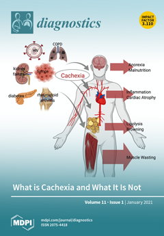
For decades, clinical attention to cachexia, often confused with anorexia, has been neglected. The diagnostic definition of cachexia itself has only recently been approved, which should help to avoid underestimation or mismanagement of this syndrome. Since then, the consensus definition of cachexia has constantly evolved (with growing importance given to functional and biochemical parameters to assess cachexia) and has even been finely tuned to distinguish its stages and degrees of severity. This review aims to pinpoint the major criteria to diagnose cachexia, highlighting the evolving importance of these criteria and distinguishing cachexia from other forms of muscle waste. View this paper.
  • Issues are regarded as officially published after their release is announced to the table of contents alert mailing list.
  • You may sign up for e-mail alerts to receive table of contents of newly released issues.
  • PDF is the official format for papers published in both, html and pdf forms. To view the papers in pdf format, click on the "PDF Full-text" link, and use the free Adobe Reader to open them.
Order results
Result details
Section
Select all
Export citation of selected articles as:
15 pages, 719 KiB  
Article
Pre-Planned and Non-Planned Agility in Patients Ongoing Rehabilitation after Knee Surgery: Design, Reliability and Validity of the Newly Developed Testing Protocols
by Ivan Peric, Miodrag Spasic, Dario Novak, Sergej Ostojic and Damir Sekulic
Diagnostics 2021, 11(1), 146; https://doi.org/10.3390/diagnostics11010146 - 19 Jan 2021
Cited by 2 | Viewed by 2789
Abstract
Background: Due to its association with the risk of falling and consequent injury, the importance of agility is widely recognized, but no study so far has examined the different facets of agility in an untrained/clinical population. The aim of this study was to [...] Read more.
Background: Due to its association with the risk of falling and consequent injury, the importance of agility is widely recognized, but no study so far has examined the different facets of agility in an untrained/clinical population. The aim of this study was to evaluate the reliability, validity, and correlates of newly developed tests of non-planned agility (NPA) and pre-planned agility (PPA) in an untrained/clinical sample. Methods: The sample comprised 38 participants older than 40 years (22 females, age: 56.1 ± 17.3 years, height: 170.4 ± 10.8 cm, mass: 82.54 ± 14.79 kg) who were involved in a rehabilitation program following total knee arthroplasty and knee arthroscopy. Variables included age, gender, type of surgery, history of fall, anthropometrics/body composition, and newly developed tests of NPA and PPA. Results: The results showed the high inter-testing- (ICC > 0.95, CV < 9%), and intra-testing-reliability (ICC > 0.96, CV < 9) of the newly developed tests. PPA and NPA were found to be valid in differentiation between age groups (>50 yrs. vs. <50 yrs.), and genders, with better performance in younger participants and males. Only NPA differentiated participants according to type of surgery, with better performance in those who had arthroscopic surgery, than those who had total knee arthroplasty. No differences in NPA and PPA were established between groups based on fall-history. In females, the body mass (Pearson’s r = 0.58 and 0.59, p < 0.001) and body fatness (Pearson’s r = 0.64 and 0.66, p < 0.001) were negatively correlated, while the lean body mass (Pearson’s r = 0.70 and 0.68, p < 0.001) was positively correlated with PPA and NPA. The NPA and PPA were highly correlated (Pearson’s r = 0.98, p < 0.001). Conclusions: We found that the proposed tests are reliable when evaluating agility characteristics in an untrained/clinical population after knee arthroplasty/arthroscopy. Further evaluation of the specific validity of the proposed tests in other specific subsamples is warranted. Full article
(This article belongs to the Special Issue Skeletal Muscle Diagnostics and Managements)
Show Figures

Figure 1

27 pages, 2121 KiB  
Review
Long Non-Coding RNAs (lncRNAs) in Cardiovascular Disease Complication of Type 2 Diabetes
by Nurruzanna Ismail, Noraidatulakma Abdullah, Nor Azian Abdul Murad, Rahman Jamal and Siti Aishah Sulaiman
Diagnostics 2021, 11(1), 145; https://doi.org/10.3390/diagnostics11010145 - 19 Jan 2021
Cited by 15 | Viewed by 4220
Abstract
The discovery of non-coding RNAs (ncRNAs) has opened a new paradigm to use ncRNAs as biomarkers to detect disease progression. Long non-coding RNAs (lncRNA) have garnered the most attention due to their specific cell-origin and their existence in biological fluids. Type 2 diabetes [...] Read more.
The discovery of non-coding RNAs (ncRNAs) has opened a new paradigm to use ncRNAs as biomarkers to detect disease progression. Long non-coding RNAs (lncRNA) have garnered the most attention due to their specific cell-origin and their existence in biological fluids. Type 2 diabetes patients will develop cardiovascular disease (CVD) complications, and CVD remains the top risk factor for mortality. Understanding the lncRNA roles in T2D and CVD conditions will allow the future use of lncRNAs to detect CVD complications before the symptoms appear. This review aimed to discuss the roles of lncRNAs in T2D and CVD conditions and their diagnostic potential as molecular biomarkers for CVD complications in T2D. Full article
(This article belongs to the Special Issue The Diagnostic and Therapeutic Value of RNAs in Human Diseases)
Show Figures

Figure 1

9 pages, 777 KiB  
Article
The Prognostic Relevance of Poly (ADP-Ribose) Polymerase Expression in Ovarian Cancer Tissue of Wild Type and BRCA-Mutation Carrier Patients
by Szabolcs Molnár, Beáta Vida, Lívia Beke, Gábor Méhes and Róbert Póka
Diagnostics 2021, 11(1), 144; https://doi.org/10.3390/diagnostics11010144 - 19 Jan 2021
Cited by 3 | Viewed by 1925
Abstract
(1) Background: The mechanism of platinum resistance in ovarian cancer is not fully clarified, but the properly functioning DNA repair mechanism can counteract the effect of conventional anticancer treatment. The objective of our study was to evaluate the expression of an important DNA [...] Read more.
(1) Background: The mechanism of platinum resistance in ovarian cancer is not fully clarified, but the properly functioning DNA repair mechanism can counteract the effect of conventional anticancer treatment. The objective of our study was to evaluate the expression of an important DNA repair enzyme, the Poly (ADP-Ribose) Polymerase (PARP) expression in epithelial ovarian cancer (EOC) tissues depending on BRCA status and to assess its relationship with platinum resistance. (2) Methods: Immunostaining to highlight PARP protein expression was performed using a rabbit polyclonal anti-PARP antibody. The intensity and distribution of immunostaining were assessed by light. Somatic BRCA1 or BRCA2 mutation carriers were identified with bidirectional sequencing of DNA from archived tumor tissue, if the test could not be performed due to technical reasons from tumor cells, the sequencing was done from peripheral blood cells to identify germline mutation carriers. The median progression-free survival (PFS) was generated for each semiquantitative group of PARP expression among chemotherapy-naive cases at the time of PARP immunohistochemistry. (3) Results: In the overall population, negative PARP immunohistochemistry predicted significant PFS (20.1 vs. 11.9 months, p = 0.001) and OS (49 vs. 114 months, p = 0.014) benefit. Genotype-stratified subgroup analysis in BRCA-negative cases confirmed the role of PARP positivity indicating an unfavorable prognosis in the entire population (relapsed 73.91% vs. 92%; OR: 4.06; p = 0.04). In the cases of the subgroup carrying the BRCA mutation, the presence of PARP expression was not associated with less favorable relapse rates, but with marginal significance for overall survival predicted a lower chance of survival (OS more than 32 months 72.73% vs. 35%; OR: 0.2; p = 0.05). (4) Conclusion: The BRCA wild type patients with strong expression of PARP enzymes before the first set of chemotherapy have a poor prognosis. Full article
(This article belongs to the Special Issue New Predictive Biomarkers for Ovarian Cancer)
Show Figures

Figure 1

16 pages, 616 KiB  
Review
Viscoelastic Hemostatic Assays and Platelet Function Testing in Patients with Atherosclerotic Vascular Diseases
by Matej Samoš, Ingrid Škorňová, Tomáš Bolek, Lucia Stančiaková, Barbora Korpallová, Peter Galajda, Ján Staško, Peter Kubisz and Marián Mokáň
Diagnostics 2021, 11(1), 143; https://doi.org/10.3390/diagnostics11010143 - 19 Jan 2021
Cited by 6 | Viewed by 2832
Abstract
Platelets play crucial role in acute vascular atherosclerotic diseases, including myocardial infarction and stroke. Additionally, platelet aggregation is a key target of antiplatelet agents, forming the keystone of pharmacotherapy of various atherosclerotic cardiovascular diseases. Thromboelastography and thromboelastometry, representing currently available viscoelastic hemostatic assays [...] Read more.
Platelets play crucial role in acute vascular atherosclerotic diseases, including myocardial infarction and stroke. Additionally, platelet aggregation is a key target of antiplatelet agents, forming the keystone of pharmacotherapy of various atherosclerotic cardiovascular diseases. Thromboelastography and thromboelastometry, representing currently available viscoelastic hemostatic assays (VHA), are designed as whole blood, real-time analyzers of clot formation and clot resolution. These assays could, in theory, overcome some limitations of currently available platelet function testing assays. This article reviews the current experience with the use of VHA for platelet function testing and for monitoring of the response to antiplatelet therapy. Full article
(This article belongs to the Section Pathology and Molecular Diagnostics)
Show Figures

Figure 1

12 pages, 1550 KiB  
Review
A Broader Perspective on the Prenatal Diagnosis of Cornelia de Lange Syndrome: Review of the Literature and Case Presentation
by Anca Maria Panaitescu, Simona Duta, Nicolae Gica, Radu Botezatu, Florina Nedelea, Gheorghe Peltecu and Alina Veduta
Diagnostics 2021, 11(1), 142; https://doi.org/10.3390/diagnostics11010142 - 19 Jan 2021
Cited by 7 | Viewed by 7493
Abstract
Cornelia de Lange syndrome (CDLS) is caused by pathogenic variants in genes which are structural or regulatory components of the cohesin complex. The classical Cornelia de Lange (CDLS) phenotype is characterized by distinctive facial features, growth retardation, upper limb reduction defects, hirsutism, and [...] Read more.
Cornelia de Lange syndrome (CDLS) is caused by pathogenic variants in genes which are structural or regulatory components of the cohesin complex. The classical Cornelia de Lange (CDLS) phenotype is characterized by distinctive facial features, growth retardation, upper limb reduction defects, hirsutism, and developmental delay. Non-classical phenotypes make this condition heterogeneous. Although CDLS is a heterogeneous clinical and genetic condition, clear diagnostic criteria have been described by specialist consensus. Many of these criteria refer to features that can be seen on prenatal ultrasound. The aim of this paper is twofold: to present the ultrasound findings in fetuses affected by CDLS syndrome; to discuss the recent advances and the limitations in the ultrasound and genetic prenatal diagnosis of CDLS. Our review aims to offer, apart from the data needed to understand the genetics and the prenatal presentation of the disease, a joint perspective of the two specialists involved in the prenatal management of this pathology: the fetal medicine specialist and the geneticist. To better illustrate the data presented, we also include a representative clinical case. Full article
(This article belongs to the Section Medical Imaging and Theranostics)
Show Figures

Figure 1

13 pages, 3459 KiB  
Article
Radiological Findings in Multidetector Computed Tomography (MDCT) of Hereditary and Sporadic Pulmonary Veno-Occlusive Disease: Certainties and Uncertainties
by Marta Pérez Núñez, Sergio Alonso Charterina, Carmen Pérez-Olivares, Yolanda Revilla Ostolaza, Rafael Morales Ruiz, Ana Belén Enguita Valls, Jair Antonio Tenorio, Natalia Gallego Zazo, Alicia De Pablo Gafas, Pablo Lapunzina, Adriana Rodríguez Chaverri and Pilar Escribano Subías
Diagnostics 2021, 11(1), 141; https://doi.org/10.3390/diagnostics11010141 - 19 Jan 2021
Cited by 7 | Viewed by 2338
Abstract
Pulmonary veno-occlusive disease (PVOD) is a very infrequent form of pulmonary arterial hypertension with an aggressive clinical course, poor response to specific vasodilator treatment, and low survival. Confirming a definitive diagnosis is essential to guide treatment and assess lung transplantation. However, in the [...] Read more.
Pulmonary veno-occlusive disease (PVOD) is a very infrequent form of pulmonary arterial hypertension with an aggressive clinical course, poor response to specific vasodilator treatment, and low survival. Confirming a definitive diagnosis is essential to guide treatment and assess lung transplantation. However, in the absence of histological or genetic confirmation, the diagnosis is complex, requiring a clinical suspicion. Multidetector computed tomography (MDCT) is an essential part of the non-invasive diagnostic tools of PVOD. We retrospectively reviewed the MDCT findings from a consecutive series of 25 patients diagnosed with PVOD, 9 with the sporadic form and 16 with the hereditary form of the disease. The presence and extent of typical findings of the diagnostic triad were assessed in all patients (ground glass parenchymal involvement, septal lines, and lymphadenopathy). In our series, 92% of patients showed at least two of the radiological findings described as typical of the disease. All patients presented at least one typical radiological characteristic. The incidence of radiological findings considered typical is very high, however was not associated with greater hemodynamic severity nor to the development of acute lung edema. No significant differences were found between the two groups. A poorly expressive MDCT does not exclude the disease. Full article
(This article belongs to the Special Issue Pulmonary Hypertension: Diagnosis and Management)
Show Figures

Figure 1

13 pages, 543 KiB  
Article
Costs of Point-of-Care Viral Load Testing for Adults and Children Living with HIV in Kenya
by Michelle Ann Bulterys, Patrick Oyaro, Evelyn Brown, Nashon Yongo, Enericah Karauki, James Wagude, Leonard Kingwara, Nancy Bowen, Susan Njogo, Anjuli D. Wagner, Irene Mukui, Frederick Oluoch, Lisa Abuogi, Rena Patel and Monisha Sharma
Diagnostics 2021, 11(1), 140; https://doi.org/10.3390/diagnostics11010140 - 19 Jan 2021
Cited by 9 | Viewed by 2461
Abstract
Background: The number of people living with HIV (PLHIV) in need of treatment monitoring in low-and-middle-income countries is rapidly expanding, straining existing laboratory capacity. Point-of-care viral load (POC VL) testing can alleviate the burden on centralized laboratories and enable faster delivery of results, [...] Read more.
Background: The number of people living with HIV (PLHIV) in need of treatment monitoring in low-and-middle-income countries is rapidly expanding, straining existing laboratory capacity. Point-of-care viral load (POC VL) testing can alleviate the burden on centralized laboratories and enable faster delivery of results, improving clinical outcomes. However, implementation costs are uncertain and will depend on clinic testing volume. We sought to estimate the costs of decentralized POC VL testing compared to centralized laboratory testing for adults and children receiving HIV care in Kenya. Methods: We conducted microcosting to estimate the per-patient costs of POC VL testing compared to known costs of centralized laboratory testing. We completed time-and-motion observations and stakeholder interviews to assess personnel structures, staff time, equipment costs, and laboratory processes associated with POC VL administration. Capital costs were estimated using a 5 year lifespan and a 3% annual discount rate. Results: We estimated that POC VL testing cost USD $24.25 per test, assuming a clinic is conducting 100 VL tests per month. Test cartridge and laboratory equipment costs accounted for most of the cost (62% and 28%, respectively). Costs varied by number of VL tests conducted at the clinic, ranging from $54.93 to $18.12 per test assuming 20 to 500 VL tests per month, respectively. A VL test processed at a centralized laboratory was estimated to cost USD $25.65. Conclusion: POC VL testing for HIV treatment monitoring can be feasibly implemented in clinics within Kenya and costs declined with higher testing volumes. Our cost estimates are useful to policymakers in planning resource allocation and can inform cost-effectiveness analyses evaluating POC VL testing. Full article
(This article belongs to the Section Point-of-Care Diagnostics and Devices)
Show Figures

Figure 1

14 pages, 1187 KiB  
Article
White Matter Microstructural Differences between Hallucinating and Non-Hallucinating Schizophrenia Spectrum Patients
by Justyna Beresniewicz, Alexander R. Craven, Kenneth Hugdahl, Else-Marie Løberg, Rune Andreas Kroken, Erik Johnsen and Renate Grüner
Diagnostics 2021, 11(1), 139; https://doi.org/10.3390/diagnostics11010139 - 19 Jan 2021
Cited by 6 | Viewed by 2591
Abstract
The relation between auditory verbal hallucinations (AVH) and white matter has been studied, but results are still inconsistent. This inconsistency may be related to having only a single time-point of AVH assessment in many studies, not capturing that AVH severity fluctuates over time. [...] Read more.
The relation between auditory verbal hallucinations (AVH) and white matter has been studied, but results are still inconsistent. This inconsistency may be related to having only a single time-point of AVH assessment in many studies, not capturing that AVH severity fluctuates over time. In the current study, AVH fluctuations were captured by utilizing a longitudinal design and using repeated (Positive and Negative Symptoms Scale) PANSS questionnaire interviews over a 12 month period. We used a Magnetic Resonance Diffusion Tensor Imaging (MR DTI) sequence and tract-based spatial statistics (TBSS) to explore white matter differences between two subtypes of schizophrenia patients; 44 hallucinating (AVH+) and 13 non-hallucinating (AVH-), compared to 13 AVH- matched controls and 44 AVH+ matched controls. Additionally, we tested for hemispheric fractional anisotropy (FA) asymmetry between the groups. Significant widespread FA-value reduction was found in the AVH+ group in comparison to the AVH- group. Although not significant, the extracted FA-values for the control group were in between the two patient groups, for all clusters. We also found a significant difference in FA-asymmetry between the AVH+ and AVH- groups in two clusters, with significantly higher leftward asymmetry in the AVH- group. The current findings suggest a possible qualitative difference in white matter integrity between AVH+ and AVH- patients. Strengths and limitations of the study are discussed. Full article
(This article belongs to the Special Issue Diagnosis, Treatment, and Management of Psychosis)
Show Figures

Figure 1

10 pages, 476 KiB  
Article
Body Mass Index in Patients Treated with Cabozantinib for Advanced Renal Cell Carcinoma: A New Prognostic Factor?
by Matteo Santoni, Francesco Massari, Sergio Bracarda, Giuseppe Procopio, Michele Milella, Ugo De Giorgi, Umberto Basso, Gaetano Aurilio, Lorena Incorvaia, Angelo Martignetti, Mimma Rizzo, Giacomo Cartenì, Enrique Grande, Marc R. Matrana, Simon J. Crabb, Nuno Vau, Giulia Sorgentoni, Alessia Cimadamore, Rodolfo Montironi and Nicola Battelli
Diagnostics 2021, 11(1), 138; https://doi.org/10.3390/diagnostics11010138 - 18 Jan 2021
Cited by 13 | Viewed by 2258
Abstract
We analyzed the clinical and pathological features of renal cell carcinoma (RCC) patients treated with cabozantinib stratified by body mass index (BMI). We retrospectively collected data from 16 worldwide centers involved in the treatment of RCC. Overall survival (OS) and progression-free survival (PFS) [...] Read more.
We analyzed the clinical and pathological features of renal cell carcinoma (RCC) patients treated with cabozantinib stratified by body mass index (BMI). We retrospectively collected data from 16 worldwide centers involved in the treatment of RCC. Overall survival (OS) and progression-free survival (PFS) were analyzed using Kaplan–Meier curves. Cox proportional models were used at univariate and multivariate analyses. We collected data from 224 patients with advanced RCC receiving cabozantinib as second- (113, 5%) or third-line (111, 5%) therapy. The median PFS was significantly higher in patients with BMI ≥ 25 (9.9 vs. 7.6 months, p < 0.001). The median OS was higher in the BMI ≥ 25 subgroup (30.7 vs. 11.0 months, p = 0.003). As third-line therapy, both median PFS (9.2 months vs. 3.9 months, p = 0.029) and OS (39.4 months vs. 11.5 months, p = 0.039) were longer in patients with BMI ≥ 25. BMI was a significant predictor for both PFS and OS at multivariate analysis. We showed that a BMI ≥ 25 correlates with longer survival in patients receiving cabozantinib. BMI can be easily assessed and should be included in current prognostic criteria for advanced RCC. Full article
(This article belongs to the Special Issue Novel Diagnostic and Predictive Strategies in Renal Cell Tumors)
Show Figures

Figure 1

13 pages, 1638 KiB  
Article
The Added Value of [18F]FDG PET/CT in the Management of Invasive Fungal Infections
by Alfred O. Ankrah, Dina Creemers-Schild, Bart de Keizer, Hans C. Klein, Rudi A. J. O. Dierckx, Thomas C. Kwee, Lambert F. R. Span, Pim A. de Jong, Mike M. Sathekge and Andor W. J. M. Glaudemans
Diagnostics 2021, 11(1), 137; https://doi.org/10.3390/diagnostics11010137 - 17 Jan 2021
Cited by 15 | Viewed by 2471
Abstract
Anatomy-based imaging methods are the usual imaging methods used in assessing invasive fungal infections (IFIs). [18F]FDG PET/CT has also been used in the evaluation of IFIs. We assessed the added value of [18F]FDG PET/CT when added to the most [...] Read more.
Anatomy-based imaging methods are the usual imaging methods used in assessing invasive fungal infections (IFIs). [18F]FDG PET/CT has also been used in the evaluation of IFIs. We assessed the added value of [18F]FDG PET/CT when added to the most frequently used anatomy-based studies in the evaluation of IFIs. The study was conducted in two University Medical Centers in the Netherlands. Reports of [18F]FDG PET/CT and anatomy-based imaging performed within two weeks of the [18F]FDG PET/CT scan were retrieved, and the presence and sites of IFI lesions were documented for each procedure. We included 155 [18F]FDG PET/CT scans performed in 73 patients. A total of 216 anatomy-based studies including 80 chest X-rays, 89 computed tomography studies, 14 magnetic resonance imaging studies, and 33 ultrasound imaging studies were studied. The anatomy-based studies were concordant with the [18F]FDG PET/CT for 94.4% of the scans performed. [18F]FDG PET/CT detected IFI lesions outside of the areas imaged by the anatomy-based studies in 48.6% of the scans. In 74% of the patients, [18F]FDG PET/CT added value in the management of the IFIs. Full article
(This article belongs to the Special Issue Imaging of Infections and Inflammatory Diseases)
Show Figures

Graphical abstract

12 pages, 6737 KiB  
Article
Microperimetry and Optical Coherence Tomography Changes in Type-1 Diabetes Mellitus without Retinopathy
by Elvira Orduna-Hospital, Judit Otero-Rodríguez, Lorena Perdices, Ana Sánchez-Cano, Ana Boned-Murillo, Javier Acha and Isabel Pinilla
Diagnostics 2021, 11(1), 136; https://doi.org/10.3390/diagnostics11010136 - 16 Jan 2021
Cited by 8 | Viewed by 2175
Abstract
Background: We aimed to measure and correlate inner retinal layer (IRL) thickness and macular sensitivity by optical coherence tomography (OCT) and by microperimetry, respectively, in type 1 diabetes mellitus patients (DM1) without diabetic retinopathy (DR). Methods: Fifty-one DM1 patients and 81 age-matched healthy [...] Read more.
Background: We aimed to measure and correlate inner retinal layer (IRL) thickness and macular sensitivity by optical coherence tomography (OCT) and by microperimetry, respectively, in type 1 diabetes mellitus patients (DM1) without diabetic retinopathy (DR). Methods: Fifty-one DM1 patients and 81 age-matched healthy subjects underwent measurement of the axial length (AL), retinal thickness in the macular ETDRS areas by swept source (SS)-OCT and macular sensitivity by microperimeter. Results: The total retinal and IRL thicknesses were thicker in the DM1 group (p < 0.05) in practically all ETDRS areas, and they had a generalized decrease in sensitivity (p < 0.05) in 9 areas between both groups. There was a significant negative correlation between retinal sensitivity and age in all areas and in visual acuity (VA) in 5 out of the 9 areas for DM1 patients. Only a mild negative correlation was observed between retinal sensitivity in the 5° nasal inner (5NI) area and in IRL thickness in the temporal inner (TI) area (−0.309 with p = 0.029) in the DM1 group. Conclusion: Aging and disease evolution in DM1 patients without DR signs generate a decrease in retinal sensitivity. There was a direct relationship between retinal sensitivity and macular thickness in the DM1 group. Full article
(This article belongs to the Section Optical Diagnostics)
Show Figures

Figure 1

15 pages, 789 KiB  
Article
Expression of Transcript Variants of PTGS1 and PTGS2 Genes among Patients with Chronic Rhinosinusitis with Nasal Polyps
by Wioletta Pietruszewska, Wojciech Fendler, Marta Podwysocka, Adam J. Białas, Piotr Kuna, Izabela Kupryś-Lipińska and Maciej Borowiec
Diagnostics 2021, 11(1), 135; https://doi.org/10.3390/diagnostics11010135 - 16 Jan 2021
Cited by 6 | Viewed by 2342
Abstract
To date, there has been no reliable test to identify unfavorable course of Chronic Rhinosinusitis with Nasal Polyps (CRSwNP), especially in aspirin intolerant patients. The research aimed to analyze the expression of transcript variants of PTGS1 and PTGS2 genes in the pathobiology of [...] Read more.
To date, there has been no reliable test to identify unfavorable course of Chronic Rhinosinusitis with Nasal Polyps (CRSwNP), especially in aspirin intolerant patients. The research aimed to analyze the expression of transcript variants of PTGS1 and PTGS2 genes in the pathobiology of the disease. The study was performed on 409 adult patients: 206 CRSwNP patients including 44 (21.36%) aspirin intolerant patients and 203 healthy volunteers in the control group. Transcript variants of the PTGS1 and PTGS2 genes named as follows: COX1.1 for NM_000962, COX1.2 for NM_080591, COX1.3 for NM_001271165.1, COX1.4 for NM_001271368.1, COX1.5 for NM_001271166.1, COX2.1 for NM_000963.3, COX2.2 for AY_151286 and COX2.3 for BQ_722004 were confirmed using direct sequencing and quantified using targeted qPCR. The coexistence of all examined transcript variants in the study and the control group and significant differences between both were found. In aspirin sensitive patients, the levels of COX1.2, COX1.3, COX1.4 and COX1.5 isoforms were higher compared to aspirin-tolerant patients. The severity of symptoms was bigger in patients with higher expressions of variants: COX1.1 (R with dCt = −0.134; p = 0.0490), COX1.3 (R = −0.1429; p = 0.0400) and COX1.5 (Rs = −0.1499; p = 0.032). The expression of COX1.1 (Rs = −0.098; p = 0.049) and COX1.5 (Rs = −0.141; p = 0.043) isoforms increased with polyposis advancement in endoscopy. With the CT extent of sinuses opacification, COX1.1 isoform also significantly increased (Rs = −0.163; p = 0.020). The isoforms COX1.3, COX1.4, COX1.5 and COX2.1 may promote milder CRSwNP course. On the contrary, the variants COX1.1, COX1.2 and COX2.2 may be involved in a more aggressive disease. Full article
(This article belongs to the Section Pathology and Molecular Diagnostics)
Show Figures

Figure 1

16 pages, 7771 KiB  
Review
Endoscopic Ultrasound vs. Computed Tomography for Gastric Cancer Staging: A Network Meta-Analysis
by Bogdan Silviu Ungureanu, Victor Mihai Sacerdotianu, Adina Turcu-Stiolica, Irina Mihaela Cazacu and Adrian Saftoiu
Diagnostics 2021, 11(1), 134; https://doi.org/10.3390/diagnostics11010134 - 16 Jan 2021
Cited by 16 | Viewed by 2912
Abstract
Gastric cancer preoperative staging is of outmost importance to assure proper management of the disease. Providing a relevant clinical stage relies on different imaging methods such as computed tomography (CT) or endoscopic ultrasound (EUS). We aimed to perform a network meta-analysis for gastric [...] Read more.
Gastric cancer preoperative staging is of outmost importance to assure proper management of the disease. Providing a relevant clinical stage relies on different imaging methods such as computed tomography (CT) or endoscopic ultrasound (EUS). We aimed to perform a network meta-analysis for gastric cancer clinical stage diagnostic tests, thus comparing the diagnostic accuracy of EUS vs. multidetector CT (MDCT) and EUS vs. EUS + MDCT. We plotted study estimates of pooled sensitivity and specificity on forest plots and summary receiver operating characteristic space to explore between-study variation in the performance of EUS, MDCT and EUS + MDCT for T1–T4, N0–N3, M0–M1 when data were available. Exploratory analyses were undertaken in RevMan 5. We included twelve studies with 2047 patients. Our results suggest that EUS was superior to MDCT in preoperative T1 and N staging. MDCT is more specific for the M stage but no significant difference in sensitivity was obtained. When comparing EUS vs. EUS + MDCT for T1 both sensitivity and specificity were not relevant. No significant differences were observed in T2–T4 stages. Even though EUS helped differentiate between the presence of invaded nodules, N stages should be carefully assessed by both methods since there is not sufficient data. Full article
(This article belongs to the Special Issue Role of Radiological Imaging in Cancer Histopathology and Staging)
Show Figures

Figure 1

11 pages, 876 KiB  
Article
Soluble ST2 as a Biomarker for Early Complications in Patients with Chronic Thromboembolic Pulmonary Hypertension Treated with Balloon Pulmonary Angioplasty
by Marta Banaszkiewicz, Arkadiusz Pietrasik, Michał Florczyk, Piotr Kędzierski, Michał Piłka, Rafał Mańczak, Janusz Kochman, Grzegorz Opolski, Adam Torbicki, Marcin Kurzyna and Szymon Darocha
Diagnostics 2021, 11(1), 133; https://doi.org/10.3390/diagnostics11010133 - 16 Jan 2021
Cited by 10 | Viewed by 2239
Abstract
Background: The aim of the study was to assess soluble ST2 (sST2) concentration and its dynamic changes in the periprocedural period in patients with chronic thromboembolic pulmonary hypertension (CTEPH) treated with balloon pulmonary angioplasty (BPA). Methods: We prospectively analyzed 57 procedures of BPA [...] Read more.
Background: The aim of the study was to assess soluble ST2 (sST2) concentration and its dynamic changes in the periprocedural period in patients with chronic thromboembolic pulmonary hypertension (CTEPH) treated with balloon pulmonary angioplasty (BPA). Methods: We prospectively analyzed 57 procedures of BPA performed in 37 patients with CTEPH. Biomarkers, such as N-terminal pro B-type natriuretic peptide (NT-proBNP), troponin T (TnT), and sST2 were assessed at four time points: Before the BPA procedure, 24 h and 48 h after the procedure, and at the discharge from hospital. Each postprocedural period was assessed for complications. Results: Before the BPA procedure, median sST2 concentration was 26.56 ng/mL (IQR: 16.66–40.83 ng/mL). sST2 concentration was significantly higher 24 h and 48 h after the BPA compared to the baseline measurements (33.31 ng/mL (IQR: 20.81–62.56), p = 0.000 and 27.45 ng/mL (IQR: 17.66–54.45), p = 0.028, respectively). sST2 level 24 h after the BPA procedure was significantly higher in the group with complications compared to the group without complications in the postprocedural period (97.66 ng/mL (IQR: 53.07–126.18) vs. 26.86 ng/mL (IQR: 19.10–40.12), p = 0.000). Conclusions: sST2 concentration in patients with CTEPH treated with BPA changes significantly in the postprocedural period and is significantly higher in the group with complications in postprocedural period. Full article
(This article belongs to the Special Issue Pulmonary Hypertension: Diagnosis and Management)
Show Figures

Figure 1

14 pages, 1021 KiB  
Article
Prediction of Nonalcoholic Fatty Liver Disease Using Noninvasive and Non-Imaging Procedures in Japanese Health Checkup Examinees
by Kenichiro Murayama, Michiaki Okada, Kenichi Tanaka, Chika Inadomi, Wataru Yoshioka, Yoshihito Kubotsu, Tomomi Yada, Hiroshi Isoda, Takuya Kuwashiro, Satoshi Oeda, Takumi Akiyama, Noriko Oza, Hideyuki Hyogo, Masafumi Ono, Takumi Kawaguchi, Takuji Torimura, Keizo Anzai, Yuichiro Eguchi and Hirokazu Takahashi
Diagnostics 2021, 11(1), 132; https://doi.org/10.3390/diagnostics11010132 - 16 Jan 2021
Cited by 20 | Viewed by 3051
Abstract
Access to imaging is limited for diagnosing nonalcoholic fatty liver disease (NAFLD) in general populations. This study evaluated the diagnostic performance of noninvasive and nonimaging indexes to predict NAFLD in the general Japanese population. Health checkup examinees without hepatitis virus infection or habitual [...] Read more.
Access to imaging is limited for diagnosing nonalcoholic fatty liver disease (NAFLD) in general populations. This study evaluated the diagnostic performance of noninvasive and nonimaging indexes to predict NAFLD in the general Japanese population. Health checkup examinees without hepatitis virus infection or habitual alcohol drinking were included. Fatty liver was diagnosed by ultrasonography. The hepatic steatosis index (HSI), Zhejiang University (ZJU) index, and fatty liver index (FLI) were determined, and risk of advanced liver fibrosis was evaluated by the fibrosis-4 index. NAFLD was diagnosed in 1935 (28.0%) of the 6927 subjects. The area under the receiver operating characteristic (AUROC) curve of the HSI, ZJU index, and FLI was 0.874, 0.886, and 0.884, respectively. The AUROC of the ZJU index (p < 0.001) and FLI (p = 0.002) was significantly greater than that for the HSI. In subjects with a high risk of advanced fibrosis, the sensitivity of the HSI, ZJU index, and FLI were 88.8%, 94.4%, and 83.3% with a low cut-off value and the specificity was 98.5%, 100%, and 100% with a high cut-off value. In conclusion, all indexes were useful to diagnose NAFLD in the general Japanese population and in subjects with potentially advanced liver fibrosis. Full article
(This article belongs to the Special Issue Fatty Liver Disease: Diagnostic, Predictive and Prognostic Markers)
Show Figures

Figure 1

9 pages, 253 KiB  
Article
Feasibility and Safety of Transjugular Liver Biopsy for Japanese Patients with Chronic Liver Diseases
by Makoto Iijima, Takahiro Arisaka, Akira Yamamiya, Keiichi Tominaga, Kazunori Nagashima, Akira Kanamori, Satoshi Masuyama, Yuichi Majima, Kenichi Goda, Kazuyuki Ishida and Atsushi Irisawa
Diagnostics 2021, 11(1), 131; https://doi.org/10.3390/diagnostics11010131 - 16 Jan 2021
Cited by 1 | Viewed by 1656
Abstract
Background and study aim: Transjugular liver biopsy (TJLB) can be used in patients who are ineligible for percutaneous liver biopsy (PLB) with acute and chronic hepatic disease. This study aimed to evaluate the usefulness and safety of TJLB in patients who were not [...] Read more.
Background and study aim: Transjugular liver biopsy (TJLB) can be used in patients who are ineligible for percutaneous liver biopsy (PLB) with acute and chronic hepatic disease. This study aimed to evaluate the usefulness and safety of TJLB in patients who were not indicated for PLB. Methods: Between July 2014 and February 2019, a total of 134 patients underwent liver biopsies at our institution. Among these, PLB was performed in 110 patients and TJLB in 24 patients. A retrospective comparison of clinical results in these patients was then performed. The primary endpoints of this study were the utility and safety of TJLB in patients who were not indicated for PLB. Results: The procedural success rate was 100% in both groups. The clinical response rate and the effective tissue sampling rate were 100% in the TJLB group and 97% in the PLB group (p = 0.55). There was no difference in the number of portal fields examined retrospectively between the two groups. No serious adverse events were observed in either group. Conclusions: It is suggested that TJLB is useful because it can be safely performed in patients with poor general condition who are not indicated for PLB. Full article
(This article belongs to the Section Pathology and Molecular Diagnostics)
12 pages, 2258 KiB  
Review
Cardiac MRI and Myocardial Injury in COVID-19: Diagnosis, Risk Stratification and Prognosis
by Saagar K. Sanghvi, Logan S. Schwarzman and Noreen T. Nazir
Diagnostics 2021, 11(1), 130; https://doi.org/10.3390/diagnostics11010130 - 15 Jan 2021
Cited by 22 | Viewed by 6350
Abstract
Myocardial injury is a common complication of the COVID-19 illness and is associated with a worsened prognosis. Systemic hyperinflammation seen in the advanced stage of COVID-19 likely contributes to myocardial injury. Cardiac magnetic resonance imaging (CMR) is the preferred imaging modality for non-invasive [...] Read more.
Myocardial injury is a common complication of the COVID-19 illness and is associated with a worsened prognosis. Systemic hyperinflammation seen in the advanced stage of COVID-19 likely contributes to myocardial injury. Cardiac magnetic resonance imaging (CMR) is the preferred imaging modality for non-invasive evaluation in acute myocarditis, enabling risk stratification and prognostication. Modified scanning protocols in the pandemic setting reduce risk of exposure while providing critical data regarding cardiac tissue inflammation and fibrosis, chamber remodeling, and contractile function. The growing use of CMR in clinical practice to assess myocardial injury will improve understanding of the acute and chronic sequelae of myocardial inflammation from various pathological etiologies. Full article
Show Figures

Figure 1

2 pages, 453 KiB  
Interesting Images
Incidental Finding of a PSMA-Positive Pancreatic Cancer in a Patient Suffering from a Metastasized PSMA-Positive Prostate Cancer
by Simon Sirtl, Andrei Todica, Harun Ilhan, Michal Zorniak, Peter Bartenstein and Julia Mayerle
Diagnostics 2021, 11(1), 129; https://doi.org/10.3390/diagnostics11010129 - 15 Jan 2021
Cited by 4 | Viewed by 2004
Abstract
An 82-year-old man suffering from prostate cancer that was scheduled for a radioreceptor-ligand therapy (RLT) presented with jaundice to our service. An abdominal ultrasound (US) revealed obstructive extrahepatic cholestasis due to a solid lesion located in the uncinate process of the pancreas. The [...] Read more.
An 82-year-old man suffering from prostate cancer that was scheduled for a radioreceptor-ligand therapy (RLT) presented with jaundice to our service. An abdominal ultrasound (US) revealed obstructive extrahepatic cholestasis due to a solid lesion located in the uncinate process of the pancreas. The Prostate Specific Membrane Antigen (PSMA) PET/CT prior to RLT showed multilocular PSMA positive tumor lesions in the lymph nodes, the lung and the pancreas. On request of the cancer board, an Endoscopic Ultrasound (EUS)-guided Fine-Needle Aspiration (FNA) of the pancreatic mass was performed revealing invasive pancreatic ductal adenocarcinoma incompatible with a prostate cancer metastasis leading to the diagnosis of a PSMA positive pancreatic ductal adenocarcinoma. Full article
Show Figures

Figure 1

9 pages, 211 KiB  
Communication
COVID-19 in Pulmonary Artery Hypertension (PAH) Patients: Observations from a Large PAH Center in New York City
by Roxana Sulica, Frank Cefali, Caroline Motschwiller, Rebecca Fenton, Anabela Barroso and Daniel Sterman
Diagnostics 2021, 11(1), 128; https://doi.org/10.3390/diagnostics11010128 - 15 Jan 2021
Cited by 22 | Viewed by 2988
Abstract
Information on outcomes of COVID-19 in pulmonary arterial hypertension (PAH) patients is limited to a few case series and surveys. Here, we describe our experience at a large Pulmonary Hypertension Center in New York City at the height of the pandemic. We performed [...] Read more.
Information on outcomes of COVID-19 in pulmonary arterial hypertension (PAH) patients is limited to a few case series and surveys. Here, we describe our experience at a large Pulmonary Hypertension Center in New York City at the height of the pandemic. We performed a retrospective chart review of eleven consecutive PAH patients who were diagnosed with SARS-CoV-2 infection. We analyzed demographics, PAH severity, risk factors for COVID-19, and COVID-19 severity and outcomes. We found in our sample that 63.6% of patients required intensive care, and there was a 45.45% overall mortality. Most patients had a known COVID-19 contact and mean duration of symptoms prior to presentation was 12 days. Only 4/11 (36%) patients presented to a center with pulmonary hypertension expertise, all of whom survived. Most patients had at least moderate pulmonary hypertension with an average REVEAL score of 7.81 despite double or triple PAH therapy. Our cases series underscores the gravity of SARS-CoV-2 infection in patients with PAH. It also suggests possible interventions to prevent unfavorable outcomes such as preserving social distancing, PAH management optimization, and early and preferential presentation to a center with specialized expertise in PAH. Full article
(This article belongs to the Special Issue Pulmonary Hypertension: Diagnosis and Management)
17 pages, 3663 KiB  
Review
Alteration of Postural Balance in Patients with Fibromyalgia Syndrome—A Systematic Review and Meta-Analysis
by David Núñez-Fuentes, Esteban Obrero-Gaitán, Noelia Zagalaz-Anula, Alfonso Javier Ibáñez-Vera, Alexander Achalandabaso-Ochoa, María del Carmen López-Ruiz, Daniel Rodríguez-Almagro and Rafael Lomas-Vega
Diagnostics 2021, 11(1), 127; https://doi.org/10.3390/diagnostics11010127 - 15 Jan 2021
Cited by 17 | Viewed by 3290
Abstract
Balance problems are one of the most frequent symptoms in patients with Fibromyalgia Syndrome (FMS). However, the extent and nature of this balance disorder are not known. The objective of this work was to determine the best evidence for the alteration of postural [...] Read more.
Balance problems are one of the most frequent symptoms in patients with Fibromyalgia Syndrome (FMS). However, the extent and nature of this balance disorder are not known. The objective of this work was to determine the best evidence for the alteration of postural balance in patients with FMS and analyze differences with healthy controls. To meet this objective, a systematic review with meta-analysis was performed. A bibliographical search was carried out in PubMed Medline, Scopus, Web of Science, CINAHL and SciELO. Observational studies that assessed postural balance in patients with FMS compared to healthy subjects in baseline conditions, were selected. In a random-effect model, the pooled effect was calculated with the Standardized Mean Difference (SMD) and its 95% confidence interval (CI). Nineteen studies reporting data of 2347 participants (95% female) were included. FMS patients showed poor balance with a large effect on static (SMD = 1.578; 95% CI = 1.164, 1.992), dynamic (SMD = 0.946; 95% CI = 0.598, 1.294), functional balance (SMD = 1.138; 95% CI = 0.689, 1.588) and on balance confidence (SMD = 1.194; 95% CI = 0.914, 1.473). Analysis of the Sensory Organization Test showed large alteration of vestibular (SMD = 1.631; 95% CI = 0.467, 2.795) and visual scores (SMD = 1.317; 95% CI = 0.153, 2.481) compared to healthy controls. Patients with FMS showed worse scores for different measures of postural balance compared to healthy controls. Concretely, FMS patients appear to have poor vestibular and visual scores with a possible somatosensory dependence. Full article
(This article belongs to the Special Issue Rheumatic Diseases: Diagnosis, Treatment and Management)
Show Figures

Figure 1

9 pages, 538 KiB  
Article
Subclavian Vessel Compression Assessed by Duplex Scanning in Patients with Neurogenic Thoracic Outlet Syndrome and No Vascular Signs
by Alban Fouasson-Chailloux, Pierre Menu, Pauline Daley, Giovanni Gautier, Guillaume Gadbled, Pierre Abraham and Marc Dauty
Diagnostics 2021, 11(1), 126; https://doi.org/10.3390/diagnostics11010126 - 15 Jan 2021
Cited by 14 | Viewed by 1996
Abstract
Neurogenic thoracic outlet syndrome (NTOS) is the most frequent form of TOS. It may affect both sides, but specific complementary exams are lacking. We aimed to evaluate duplex scanning results in a group of patients with unilateral or bilateral NTOS and no clinical [...] Read more.
Neurogenic thoracic outlet syndrome (NTOS) is the most frequent form of TOS. It may affect both sides, but specific complementary exams are lacking. We aimed to evaluate duplex scanning results in a group of patients with unilateral or bilateral NTOS and no clinical vascular signs, referred for rehabilitation. We performed a retrospective observational study in patients with unilateral or bilateral NTOS and no vascular symptoms. Subclavian vessels were assessed by duplex scanning. Compressions were considered in case of >50% of increased or decreased blood flow. A total of 101 patients met NTOS criteria; mean age was 40 +/− 10.2; 79.2% women. Seventy patients had a unilateral NTOS and 31 a bilateral form. Duplex scanning showed that 56.4% of the patients had vessels compression, 55.7% in the unilateral group and 58.1% in the bilateral (p = 0.81). In unilateral NTOS, 21 (30%) patients had bilateral vascular compression, 17 (24.3%) had ipsilateral compression and 1 (1.4%) had contralateral compression. In bilateral NTOS, 15 (48.4%) had bilateral compression and 3 (9.7%) compression on only one side. We found a significant difference of the rate of vascular compressions between symptomatic and non-symptomatic upper-limbs, 54.5% vs. 32.9%, respectively, (p = 0.002) and a significant association between symptomatic upper-limbs and vascular compression (OR = 2.45 [95%IC: 1.33–4.49]; p = 0.002). The sensitivity and the specificity of the duplex scanning were 54.5% and 67%, respectively. The ROC curve area was of 0.608 [95%IC: 0.527–0.690]. Despite a highly significant association between symptomatic upper-limbs and vascular compression, duplex scanning did not help make the diagnosis of NTOS. Full article
(This article belongs to the Section Medical Imaging and Theranostics)
Show Figures

Figure 1

9 pages, 1674 KiB  
Article
Sensitive Detection of KRAS Mutations by Clustered Regularly Interspaced Short Palindromic Repeats
by Huifen Zhou, Jen-Hui Tsou, Qixin Leng and Feng Jiang
Diagnostics 2021, 11(1), 125; https://doi.org/10.3390/diagnostics11010125 - 15 Jan 2021
Cited by 6 | Viewed by 2561
Abstract
Kirsten rat sarcoma viral oncogene (KRAS) is the isoform most frequently mutated in human tumors. Testing for activating KRAS mutations has important implications for diagnosis and the personalized medicine of cancers. The current techniques for detecting KRAS mutations have moderate sensitivity. The emerging [...] Read more.
Kirsten rat sarcoma viral oncogene (KRAS) is the isoform most frequently mutated in human tumors. Testing for activating KRAS mutations has important implications for diagnosis and the personalized medicine of cancers. The current techniques for detecting KRAS mutations have moderate sensitivity. The emerging clustered regularly interspaced short palindromic repeats (CRISPR) system shows great promise in the detection of nucleic acids and is revolutionizing medical diagnostics. This study aimed to develop CRISPR–Cas12a as a sensitive test to detect KRAS mutations. Serially diluted DNA samples containing KRAS mutations are subjected to CRISPR–Cas12a and polymerase chain reaction (PCR). CRISPR–Cas12a and PCR can specifically detect 0.01% and 0.1% mutant KRAS DNA in the presence of wild-type KRAS DNA, respectively. Twenty pairs of lung tumor and noncancerous lung tissues are tested by CRISPR–Cas12a, PCR, and direct sequencing. CRISPR–Cas12a could identify the G12C mutation in five of 20 tumor tissues, while both PCR and direct sequencing discovered the KRAS mutation in three of the five tumor tissues. Furthermore, the results of CRISPR–Cas12a for testing the mutation could be directly and immediately visualized by a UV light illuminator. Altogether, CRISPR–Cas12a has a higher sensitivity for the detection of KRAS mutations compared with PCR and sequencing analysis, and thus has diagnostic and therapeutic implications. Nevertheless, the technique needs to be validated for its clinical significance in a large and prospective study. Full article
(This article belongs to the Section Pathology and Molecular Diagnostics)
Show Figures

Figure 1

13 pages, 3539 KiB  
Review
SARS-CoV-2 in Mexico: Beyond Detection Methods, Scope and Limitations
by Cynthia Martinez-Liu, Natalia Martínez-Acuña, Daniel Arellanos-Soto, Kame Galan-Huerta, Sonia Lozano-Sepulveda, María del Carmen Martínez-Guzmán and Ana Maria Rivas-Estilla
Diagnostics 2021, 11(1), 124; https://doi.org/10.3390/diagnostics11010124 - 14 Jan 2021
Cited by 5 | Viewed by 5799
Abstract
The new coronavirus that was first identified in December 2019 in Wuhan China, now called SARS-CoV-2, which causes the disease called COVID-19, has spread from China to the entire world in a few months. Due to its contagious potential (R0: 5.7) and because [...] Read more.
The new coronavirus that was first identified in December 2019 in Wuhan China, now called SARS-CoV-2, which causes the disease called COVID-19, has spread from China to the entire world in a few months. Due to its contagious potential (R0: 5.7) and because there is still no effective treatment to stop the infection, and a vaccine for prevention it is not yet available to the general population, COVID-19 is currently considered a global health problem. The need to implement sensitive methods for the identification of individuals with COVID-19 has led to the development of different molecular and immunological tests. The importance of a timely and accurate diagnosis is essential to determine the course of the pandemic. The interpretation of the results obtained by each test as well as the factors that affect these results have not been fully described. In this review, we describe and analyze the different SARS-CoV-2 detection methods that have been performed in Mexico and are available worldwide, outlining their strengths and weaknesses. Further, a broader perspective of the correct use and interpretation of the results obtained with these diagnostic tools is proposed to improve the containment strategy and identify the true impact of the pandemic. Full article
Show Figures

Figure 1

31 pages, 544 KiB  
Review
Animal Models of Depression: What Can They Teach Us about the Human Disease?
by Maria Becker, Albert Pinhasov and Asher Ornoy
Diagnostics 2021, 11(1), 123; https://doi.org/10.3390/diagnostics11010123 - 14 Jan 2021
Cited by 76 | Viewed by 17334
Abstract
Depression is apparently the most common psychiatric disease among the mood disorders affecting about 10% of the adult population. The etiology and pathogenesis of depression are still poorly understood. Hence, as for most human diseases, animal models can help us understand the pathogenesis [...] Read more.
Depression is apparently the most common psychiatric disease among the mood disorders affecting about 10% of the adult population. The etiology and pathogenesis of depression are still poorly understood. Hence, as for most human diseases, animal models can help us understand the pathogenesis of depression and, more importantly, may facilitate the search for therapy. In this review we first describe the more common tests used for the evaluation of depressive-like symptoms in rodents. Then we describe different models of depression and discuss their strengths and weaknesses. These models can be divided into several categories: genetic models, models induced by mental acute and chronic stressful situations caused by environmental manipulations (i.e., learned helplessness in rats/mice), models induced by changes in brain neuro-transmitters or by specific brain injuries and models induced by pharmacological tools. In spite of the fact that none of the models completely resembles human depression, most animal models are relevant since they mimic many of the features observed in the human situation and may serve as a powerful tool for the study of the etiology, pathogenesis and treatment of depression, especially since only few patients respond to acute treatment. Relevance increases by the fact that human depression also has different facets and many possible etiologies and therapies. Full article
(This article belongs to the Special Issue Animal Models for Studying and Screening Human Diseases)
13 pages, 1497 KiB  
Article
Transarterial Radioembolization of Hepatocellular Carcinoma, Liver-Dominant Hepatic Colorectal Cancer Metastases, and Cholangiocarcinoma Using Yttrium90 Microspheres: Eight-Year Single-Center Real-Life Experience
by Julie Pellegrinelli, Olivier Chevallier, Sylvain Manfredi, Inna Dygai-Cochet, Claire Tabouret-Viaud, Guillaume Nodari, François Ghiringhelli, Jean-Marc Riedinger, Romain Popoff, Jean-Marc Vrigneaud, Alexandre Cochet, Serge Aho, Marianne Latournerie and Romaric Loffroy
Diagnostics 2021, 11(1), 122; https://doi.org/10.3390/diagnostics11010122 - 14 Jan 2021
Cited by 9 | Viewed by 2384
Abstract
Liver tumors are common and may be unamenable to surgery or ablative treatments. Consequently, other treatments have been devised. To assess the safety and efficacy of transarterial radioembolization (TARE) with Yttrium-90 for hepatocellular carcinoma (HCC), liver-dominant hepatic colorectal cancer metastases (mCRC), and cholangiocarcinoma [...] Read more.
Liver tumors are common and may be unamenable to surgery or ablative treatments. Consequently, other treatments have been devised. To assess the safety and efficacy of transarterial radioembolization (TARE) with Yttrium-90 for hepatocellular carcinoma (HCC), liver-dominant hepatic colorectal cancer metastases (mCRC), and cholangiocarcinoma (CCA), performed according to current recommendations, we conducted a single-center retrospective study in 70 patients treated with TARE (HCC, n = 44; mCRC, n = 20; CCA, n = 6). Safety and toxicity were assessed using the National Cancer Institute Common Terminology Criteria. Treatment response was evaluated every 3 months on imaging studies using Response Evaluation Criteria in Solid Tumors (RECIST) or mRECIST criteria. Overall survival and progression-free survival were estimated using the Kaplan-Meier method. The median delivered dose was 1.6 GBq, with SIR-Spheres® or TheraSphere® microspheres. TARE-related grade 3 adverse events affected 17.1% of patients. Median follow-up was 32.1 months. Median progression-free survival was 5.6 months and median overall time from TARE to death was 16.1 months and was significantly shorter in men. Progression-free survival was significantly longer in women (HR, 0.49; 95%CI, 0.26–0.90; p = 0.031). Risk of death or progression increased with the number of systemic chemotherapy lines. TARE can be safe and effective in patients with intermediate- or advanced-stage HCC, CCA, or mCRC refractory or intolerant to appropriate treatments. Full article
(This article belongs to the Special Issue Percutaneous Imaging-Guided Techniques in Interventional Oncology)
Show Figures

Figure 1

9 pages, 1194 KiB  
Article
Application of Innovative TGA/Chemometric Approach for Forensic Purposes: The Estimation of the Time since Death in Contaminated Specimens
by Roberta Risoluti, Giuseppina Gullifa, Vittorio Fineschi, Paola Frati and Stefano Materazzi
Diagnostics 2021, 11(1), 121; https://doi.org/10.3390/diagnostics11010121 - 14 Jan 2021
Cited by 6 | Viewed by 2119
Abstract
Chronothanatology has always been a challenge in forensic sciences. Therefore, the importance of a multidisciplinary approach for the characterization of matrices (organs, tissues, or fluids) that respond linearly to the postmortem interval (PMI) is emerging increasingly. The vitreous humor is particularly suitable for [...] Read more.
Chronothanatology has always been a challenge in forensic sciences. Therefore, the importance of a multidisciplinary approach for the characterization of matrices (organs, tissues, or fluids) that respond linearly to the postmortem interval (PMI) is emerging increasingly. The vitreous humor is particularly suitable for studies aimed at assessing time-related modifications because it is topographically isolated and well-protected. In this work, a novel approach based on thermogravimetry and chemometrics was used to estimate the time since death in the vitreous humor and to collect a databank of samples derived from postmortem examinations after medico–legal evaluation. In this study, contaminated and uncontaminated specimens with tissue fragments were included in order to develop a classification model to predict time of death based on partial least squares discriminant analysis (PLS-DA) that was as robust as possible. Results demonstrate the possibility to correctly predict the PMI even in contaminated samples, with an accuracy not lower than 70%. In addition, the correlation coefficient of the measured versus predicted outcomes was found to be 0.9978, confirming the ability of the model to extend its feasibility even to such situations involving contaminated vitreous humor. Full article
(This article belongs to the Special Issue Progress in the Forensic Diagnosis)
Show Figures

Figure 1

14 pages, 2505 KiB  
Article
Preoperative Evaluation through Dermoscopy and Reflectance Confocal Microscopy of the Lateral Excision Margins for Primary Basal Cell Carcinoma
by Mihai Lupu, Vlad Mihai Voiculescu, Ana Caruntu, Tiberiu Tebeica and Constantin Caruntu
Diagnostics 2021, 11(1), 120; https://doi.org/10.3390/diagnostics11010120 - 14 Jan 2021
Cited by 17 | Viewed by 2417
Abstract
Complete removal of malignant skin lesions with minimal impact on the aesthetic and functional aspects is the ideal of every dermatologic surgeon. Incomplete surgical excisions and tumor recurrences of basal cell carcinomas (BCC) commonly occur due to the subclinical extension of tumor lateral [...] Read more.
Complete removal of malignant skin lesions with minimal impact on the aesthetic and functional aspects is the ideal of every dermatologic surgeon. Incomplete surgical excisions and tumor recurrences of basal cell carcinomas (BCC) commonly occur due to the subclinical extension of tumor lateral margins. Presently, the lateral excision margins for BCC cannot be objectively assessed preoperatively, dermoscopy proving to be relatively inefficient in this respect. The question is whether BCC lateral excision margins can be precisely determined preoperatively through the use of complementary non-invasive imaging techniques such as dermoscopy and reflectance confocal microscopy (RCM), thus permitting the complete removal of the lesion in a single stage, estimation of the post-excisional defect, and planning an appropriate reconstruction, especially in medical centers where Mohs micrographic surgery is not available. We present the results of a prospective, histopathologically controlled study designed to determine the feasibility of preoperative, non-invasive, in vivo evaluation of the lateral excision margins for primary basal cell carcinoma, through dermoscopy and RCM. Full article
(This article belongs to the Section Optical Diagnostics)
Show Figures

Figure 1

11 pages, 1792 KiB  
Article
Diagnostic Performance of F18-FDG PET/CT in Male Breast Cancers Patients
by Andra Piciu, Doina Piciu, Narcis Polocoser, Anita A. Kovendi, Iulia Almasan, Alexandru Mester, Dragos-Stefan Morariu, Calin Cainap and Simona Sorana Cainap
Diagnostics 2021, 11(1), 119; https://doi.org/10.3390/diagnostics11010119 - 13 Jan 2021
Cited by 6 | Viewed by 4828
Abstract
Introduction: F18-FDG PET/CT is the most important hybrid imaging used in the diagnostic, staging, follow-up, and treatment evaluation response in cancer patients. However, it is well-known that in breast cancer the use of F18-FDG is not included in the first line protocol of [...] Read more.
Introduction: F18-FDG PET/CT is the most important hybrid imaging used in the diagnostic, staging, follow-up, and treatment evaluation response in cancer patients. However, it is well-known that in breast cancer the use of F18-FDG is not included in the first line protocol of initial diagnostic, both in female and male breast cancer patients. F18-FDG PET/CT is a valuable tool to provide information on extra-axillary lymph node involvement, distant metastases, and other occult primary cancers. This study assesses F18-FDG PET/CT systemic staging in male patients with diagnosed breast cancer and determines detection rates for unsuspected distant metastases and synchronous malignancies. Methods: We analyzed a number of 170 male patients with breast cancer, seen between 2000–2020, in a tertiary center. From this group, between 2013–2020 a number of 23 patients underwent F18-FDG PET/CT. Rates of upstaging were determined for each case and the detection of other primary malignancies was analyzed. Results: Median age of male breast cancer group was 61.3 y (range, 34–85 y), most had intraductal carcinoma (82.4%) and unsuspected distant metastases, which increased patient stage to IV, observed in 27%. In 4 out 23 patients (17.4%), F18-FDG PET/CT identified synchronous cancers (2 prostate cancers, 1 thyroid and 1 colon cancer). Conclusion: F18-FDG PET/CT is a valuable tool to provide information on extra-axillary lymph node involvement, distant metastases, and other occult primary cancers. Baseline F18-FDG PET/CT has a substantial impact on the initial staging and on clinical management in male breast patients and should be considered for use in newly diagnosed patients. Full article
(This article belongs to the Special Issue Positron Emission Tomography (PET) Imaging for Therapy Monitoring)
Show Figures

Figure 1

13 pages, 3672 KiB  
Article
Simulated Radiation Dose Reduction in Whole-Body CT on a 3rd Generation Dual-Source Scanner: An Intraindividual Comparison
by Andreas S. Brendlin, Moritz T. Winkelmann, Phuong Linh Do, Vincent Schwarze, Felix Peisen, Haidara Almansour, Malte N. Bongers, Christoph P. Artzner, Jakob Weiss, Jong Hyo Kim, Ahmed E. Othman and Saif Afat
Diagnostics 2021, 11(1), 118; https://doi.org/10.3390/diagnostics11010118 - 13 Jan 2021
Cited by 3 | Viewed by 2315
Abstract
To evaluate the effect of radiation dose reduction on image quality and diagnostic confidence in contrast-enhanced whole-body computed tomography (WBCT) staging. We randomly selected March 2016 for retrospective inclusion of 18 consecutive patients (14 female, 60 ± 15 years) with clinically indicated WBCT [...] Read more.
To evaluate the effect of radiation dose reduction on image quality and diagnostic confidence in contrast-enhanced whole-body computed tomography (WBCT) staging. We randomly selected March 2016 for retrospective inclusion of 18 consecutive patients (14 female, 60 ± 15 years) with clinically indicated WBCT staging on the same 3rd generation dual-source CT. Using low-dose simulations, we created data sets with 100, 80, 60, 40, and 20% of the original radiation dose. Each set was reconstructed using filtered back projection (FBP) and Advanced Modeled Iterative Reconstruction (ADMIRE®, Siemens Healthineers, Forchheim, Germany) strength 1–5, resulting in 540 datasets total. ADMIRE 2 was the reference standard for intraindividual comparison. The effective radiation dose was calculated using commercially available software. For comparison of objective image quality, noise assessments of subcutaneous adipose tissue regions were performed automatically using the software. Three radiologists blinded to the study evaluated image quality and diagnostic confidence independently on an equidistant 5-point Likert scale (1 = poor to 5 = excellent). At 100%, the effective radiation dose in our population was 13.3 ± 9.1 mSv. At 20% radiation dose, it was possible to obtain comparably low noise levels when using ADMIRE 5 (p = 1.000, r = 0.29). We identified ADMIRE 3 at 40% radiation dose (5.3 ± 3.6 mSv) as the lowest achievable radiation dose with image quality and diagnostic confidence equal to our reference standard (p = 1.000, r > 0.4). The inter-rater agreement for this result was almost perfect (ICC ≥ 0.958, 95% CI 0.909–0.983). On a 3rd generation scanner, it is feasible to maintain good subjective image quality, diagnostic confidence, and image noise in single-energy WBCT staging at dose levels as low as 40% of the original dose (5.3 ± 3.6 mSv), when using ADMIRE 3. Full article
(This article belongs to the Special Issue Assessment of Radiation Dose in X-ray and CT Exams)
Show Figures

Figure 1

12 pages, 1017 KiB  
Review
Nuclear Imaging for Bone Metastases in Prostate Cancer: The Emergence of Modern Techniques Using Novel Radiotracers
by Wietske I. Luining, Dennie Meijer, Max R. Dahele, André N. Vis and Daniela E. Oprea-Lager
Diagnostics 2021, 11(1), 117; https://doi.org/10.3390/diagnostics11010117 - 13 Jan 2021
Cited by 7 | Viewed by 3090
Abstract
Accurate staging of prostate cancer (PCa) at initial diagnosis and at biochemical recurrence is important to determine prognosis and the optimal treatment strategy. To date, treatment of metastatic PCa has mostly been based on the results of conventional imaging with abdominopelvic computed tomography [...] Read more.
Accurate staging of prostate cancer (PCa) at initial diagnosis and at biochemical recurrence is important to determine prognosis and the optimal treatment strategy. To date, treatment of metastatic PCa has mostly been based on the results of conventional imaging with abdominopelvic computed tomography (CT) and bone scintigraphy. However, these investigations have limited sensitivity and specificity which impairs their ability to accurately identify and quantify the true extent of active disease. Modern imaging modalities, such as those based on the detection of radioactively labeled tracers with combined positron emission tomography/computed tomography (PET/CT) scanning have been developed specifically for the detection of PCa. Novel radiotracers include 18F-sodium fluoride (NaF), 11C-/18F-fluorocholine (FCH), 18F-fluordihydrotestosterone (FDHT), 68Gallium and 18F-radiolabeled prostate-specific membrane antigen (e.g., 68Ga-PSMA-11, 18F-DCFPyL). PET/CT with these tracers outperforms conventional imaging. As a result of this, although their impact on outcome needs to be better defined in appropriate clinical trials, techniques like prostate-specific membrane antigen (PSMA) PET/CT have been rapidly adopted into clinical practice for (re)staging PCa. This review focuses on nuclear imaging for PCa bone metastases, summarizing the literature on conventional imaging (focusing on CT and bone scintigraphy—magnetic resonance imaging is not addressed in this review), highlighting the prognostic importance of high and low volume metastatic disease which serves as a driver for the development of better imaging techniques, and finally discussing modern nuclear imaging with novel radiotracers. Full article
(This article belongs to the Special Issue Bone Imaging in Oncology)
Show Figures

Figure 1

Previous Issue
Next Issue
Back to TopTop