




Next Issue
Volume 18, March-2
Previous Issue
Volume 18, February-2
 
 
cancers-logo

Journal Browser

Journal Browser

Cancers, Volume 18, Issue 5 (March-1 2026) – 169 articles
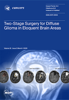
Cover Story (view full-size image): Maximal safe resection is a key prognostic factor in diffuse glioma, yet complete tumour removal is often limited by eloquent brain localisation, multifocal growth, or intraoperative constraints. In this retrospective single-centre study of 36 patients, we evaluated outcomes of a planned two-stage surgical strategy. The second procedure significantly reduced residual contrast-enhancing and non-enhancing tumour volumes and increased rates of supra- and maximal resection without significant deterioration in neurological or functional status. Despite relevant complication rates, staged surgery may offer a favourable risk–benefit profile in carefully selected patients and represents a strategic extension of modern glioma surgery. View this paper
  • Issues are regarded as officially published after their release is announced to the table of contents alert mailing list.
  • You may sign up for e-mail alerts to receive table of contents of newly released issues.
  • PDF is the official format for papers published in both, html and pdf forms. To view the papers in pdf format, click on the "PDF Full-text" link, and use the free Adobe Reader to open them.
Order results
Result details
Section
Select all
Export citation of selected articles as:
19 pages, 1753 KB  
Review
Radiobiological and Clinical Advantages of Proton Therapy in Modern Cancer Treatment
by Spyridon A. Kalospyros, Angeliki Gkikoudi, Athanasios Koutsostathis, Athanasia Adamopoulou, Spyridon N. Vasilopoulos, Vasileios Rangos, Erato Stylianou-Markidou, Ioannis Pantalos, Constantinos Koumenis and Alexandros G. Georgakilas
Cancers 2026, 18(5), 885; https://doi.org/10.3390/cancers18050885 - 9 Mar 2026
Viewed by 1878
Abstract
Background/Objectives: Proton therapy has emerged as an advanced radiotherapy modality due to its unique physical dose distribution and its distinct radiobiological properties. The finite range of protons in tissue enables highly conformal dose delivery with minimal exit dose, significantly reducing irradiation of surrounding [...] Read more.
Background/Objectives: Proton therapy has emerged as an advanced radiotherapy modality due to its unique physical dose distribution and its distinct radiobiological properties. The finite range of protons in tissue enables highly conformal dose delivery with minimal exit dose, significantly reducing irradiation of surrounding normal tissues compared to photon-based radiotherapy. Beyond these physical advantages, proton beams exhibit a spatially varying linear energy transfer that increases toward the distal edge of the spread-out Bragg peak, leading to clustered and complex DNA damage that is more difficult for cancer cells to repair. Methods: This review integrates experimental, computational, and clinical evidence to examine how proton-induced DNA damage, relative biological effectiveness, oxygen effects, and non-targeted responses contribute to tumor control and normal tissue sparing. Results: Comparative analyses with photon intensity-modulated radiotherapy demonstrate consistent reductions in acute and late toxicities across multiple tumor sites, particularly in pediatric patients and in tumors located near critical organs. The review also discusses emerging technologies, including pencil beam scanning, image-guided and adaptive proton therapy, compact accelerator systems, and ultra-high dose rate FLASH proton therapy, which collectively aim to enhance treatment precision, biological effectiveness, and accessibility. Conclusions: Together, these developments support proton therapy as a rapidly evolving modality with significant potential to improve therapeutic outcomes in modern oncology. Full article
(This article belongs to the Special Issue Insights from the Editorial Board Member)
Show Figures

Figure 1

21 pages, 684 KB  
Article
Clinicians’ Experiences of Implementing Clinical Frailty Scale Assessments in Lung Oncology Clinics: A Qualitative Interview Study
by Jessica Pearce, Hayat Hamzeh, Mary Denholm, Alastair Greystoke, Fabio Gomes, Andrew Clegg, Galina Velikova, Suzanne H. Richards and Alexandra Gilbert
Cancers 2026, 18(5), 884; https://doi.org/10.3390/cancers18050884 - 9 Mar 2026
Viewed by 824
Abstract
Background/Objectives: Simple frailty assessments, such as the clinical frailty scale (CFS), are prognostic for worse outcomes in older adults with cancer and could support treatment decision-making. This interview study aims to explore clinicians’ experiences of using simple frailty assessments in oncology, including the [...] Read more.
Background/Objectives: Simple frailty assessments, such as the clinical frailty scale (CFS), are prognostic for worse outcomes in older adults with cancer and could support treatment decision-making. This interview study aims to explore clinicians’ experiences of using simple frailty assessments in oncology, including the impacts on patient care and barriers and facilitators to successful implementation. Methods: Semi-structured individual interviews were conducted with clinicians at three UK sites that had implemented CFS screening in lung cancer clinics as part of a national pilot, to explore how frailty assessments are applied and are impacting care. Purposive sampling targeted a range of professionals involved in assessing frailty and making treatment decisions. Recordings were transcribed verbatim and analysed thematically. Results: Ten clinicians participated, and four main themes were identified. ‘Assessing fitness and frailty’ explores the central role of performance status (PS), as well as its limitations, and what frailty assessments add. ‘Scoring and interpreting CFS’ describes the ease and relative yield of CFS use, particularly for patients with ‘borderline’ PS scores (e.g., PS 1–2 or 2–3), and the importance of contextual interpretation. ‘Role of frailty and impacts of assessment’ highlights how frailty assessments can enhance patient-centered care and support, and clinical and shared decision-making, with potential for streamlined care and system-level benefits. ‘Barriers and facilitators to implementation’ are described, including time, culture, guidance, and training, with recommendations provided. Conclusions: Assessing frailty has wide-ranging potential benefits for patients, oncology teams, and the wider system, but barriers must be overcome. Specific recommendations are provided to support the routine implementation of frailty assessments, which is a key step towards the benefits of frailty-informed care being realised at scale. Full article
(This article belongs to the Section Clinical Research in Cancer)
Show Figures

Figure 1

13 pages, 675 KB  
Article
OSIRIS-Nose: Organ Sparing Using Interventional Radiotherapy (Brachytherapy) for Invasive Squamous Cell Cancer of the Nasal Vestibule
by Tamer Soror, Pierre-Alexander Justenhoven, Warren Bacorro, György Kovács, Dirk Rades, Karl-Ludwig Bruchhage and Anke Leichtle
Cancers 2026, 18(5), 883; https://doi.org/10.3390/cancers18050883 - 9 Mar 2026
Viewed by 728
Abstract
Background/Objectives: Squamous cell carcinoma of the nasal vestibule (SCCNV) represents a rare malignancy traditionally managed by radical surgical resection, frequently at the cost of substantial functional impairment and disfiguring aesthetic consequences. This study investigates an organ-preserving therapeutic strategy integrating high-dose-rate interventional radiotherapy [...] Read more.
Background/Objectives: Squamous cell carcinoma of the nasal vestibule (SCCNV) represents a rare malignancy traditionally managed by radical surgical resection, frequently at the cost of substantial functional impairment and disfiguring aesthetic consequences. This study investigates an organ-preserving therapeutic strategy integrating high-dose-rate interventional radiotherapy (HDR-IRT; brachytherapy) with organ-preserving surgery. Material and Methods: A retrospective analysis of patients with primary SCCNV treated using HDR-IRT between 2008 and 2022, excluding recurrent disease and cutaneous squamous cell carcinomas. Interstitial HDR-IRT catheters were implanted intraoperatively, with radiation delivered twice daily to a target volume encompassing the tumor and a 10–15 mm safety margin. Results: Fifty-one patients were included, with a median age of 71 years. The median total dose was 40 Gy. Gross total resection was performed in 7 patients, and subtotal resection in 44. The median follow-up was 35 months. The 5-year nose preservation rate was 90%, with local control at 84%, regional failure-free survival at 94%, and overall survival at 82%. In total, 49 acute toxicity events were documented, including two grade 3 events, while 35 chronic toxicity events were reported, including one grade 3 event. At 3 years, 84.3% of cosmetic outcomes were rated as satisfactory, 9.8% as acceptable, and 5.9% as unsatisfactory. Conclusions: The OSIRIS approach, combining HDR-IRT with organ-preserving surgery, is an effective treatment for SCCNV, offering high organ preservation and favorable long-term disease control, with manageable toxicity and positive cosmetic outcomes. Full article
(This article belongs to the Special Issue Personalized Radiotherapy in Cancer Care (2nd Edition))
Show Figures

Figure 1

19 pages, 7628 KB  
Article
CF10 Displays Improved Synergy with Oxaliplatin in TP53-Null and Wild-Type CRC Cells from Increased Top1cc and Replication Stress
by Taylor M. Young, Rida Moumouni, Akanksha Behl, Upasana Das and William H. Gmeiner
Cancers 2026, 18(5), 882; https://doi.org/10.3390/cancers18050882 - 9 Mar 2026
Viewed by 910
Abstract
Background/ObjectivesTP53 mutation or deletion status is important for determining cellular responses to DNA-damaging drugs. Oxaliplatin (OXA) is combined with the fluoropyrimidine (FP) drug 5-fluorouracil (5-FU) in the FOLFOX regimen used to treat advanced colorectal cancer (CRC). However, the effects of TP53 [...] Read more.
Background/ObjectivesTP53 mutation or deletion status is important for determining cellular responses to DNA-damaging drugs. Oxaliplatin (OXA) is combined with the fluoropyrimidine (FP) drug 5-fluorouracil (5-FU) in the FOLFOX regimen used to treat advanced colorectal cancer (CRC). However, the effects of TP53 deletion on 5-FU + OXA synergy are not well known. We investigated potential synergy between OXA and 5-FU and compared it with OXA synergy with a novel polymeric FP, CF10, in four cell lines harboring either wild-type (WT) or TP53-null status. Methods: Using CompuSyn and the highest single agent (HSA) models, we compared synergy between CF10 and OXA (COXA) and between 5-FU and OXA (FOXA). Cell cycle analysis was performed, as was Western blot quantification of canonical DNA damage pathway proteins. Likewise, immunofluorescent and confocal analysis allowed us to compare topoisomerase 1 cleavage complex and double-strand DNA break formation. Results: COXA synergy displayed minimal TP53 dependence with greatly improved potency compared to FOXA. COXA synergy resulted from OXA increasing: (i) Topoisomerase 1 (Top1) cleavage complex formation; (ii) DNA double-strand breaks (DSBs), and (iii) Checkpoint Kinase 1 and 2 (p-Chk1/2) phosphorylation, consistent with increased replication stress. Additionally, increased S-phase entry in TP53-null cells enhanced synergy between CF10, 5-FU, and OXA as S-phase drugs. Conclusions: Our results demonstrate that OXA synergizes with CF10 more effectively than with 5-FU through enhanced replication stress in both WT and TP53-null cells by causing greater Top1-mediated DNA double-strand breaks. Our studies provide a foundation for further testing of this combination in an orthotopic liver metastatic setting and eventual clinical development. Full article
(This article belongs to the Special Issue Adjuvant Therapy and the Cytotoxic Effects in Colorectal Cancers)
Show Figures

Figure 1

14 pages, 2190 KB  
Article
Incidence Trends and Geographic Variations of Corpus Uteri and Cervical Cancer in Taiwan, 1995–2022: A Population-Based Study
by Yu Chang, Nari Kay, Liang-Chun Chiu, Chung-I Huang, Hung-Ju Li, Shyh-An Yeh and Yu-Chieh Su
Cancers 2026, 18(5), 881; https://doi.org/10.3390/cancers18050881 - 9 Mar 2026
Viewed by 948
Abstract
Background/Objectives: To examine long-term incidence trends of cervical cancer and corpus uteri cancer in Taiwan from 1995 to 2022, with emphasis on age–period–cohort patterns and regional variation. Methods: Data from the Taiwan cancer registry were analyzed. Age-standardized incidence rates (ASRs) were calculated using [...] Read more.
Background/Objectives: To examine long-term incidence trends of cervical cancer and corpus uteri cancer in Taiwan from 1995 to 2022, with emphasis on age–period–cohort patterns and regional variation. Methods: Data from the Taiwan cancer registry were analyzed. Age-standardized incidence rates (ASRs) were calculated using the 1976 World Standard Population. Temporal trends were evaluated using Joinpoint regression to estimate annual percent changes (APCs) and average annual percent changes (AAPCs). Age–period–cohort modeling was applied to assess net drift, cohort effects, and period effects. Subgroup analyses were conducted by geographic region and urbanization level. Results: Cervical cancer incidence declined markedly, with ASRs decreasing from 20.06 to 6.78 per 100,000 women between 1995 and 2022 (AAPC = −4.43%, 95% CI: −5.39 to −3.45). In contrast, corpus uteri cancer incidence increased substantially, with ASRs rising from 2.91 to 17.42 per 100,000 women (AAPC = 6.32%, 95% CI: 5.86–6.78). Age–period–cohort analysis revealed a negative net drift for cervical cancer (−5.0% per year) and a positive net drift for corpus uteri cancer (6.1% per year). Cohort effects indicated decreasing cervical cancer risk among women born after 1960, whereas corpus uteri cancer risk increased in successive younger cohorts. Period effects showed pronounced declines in cervical cancer incidence after 2000, patterns that are compatible with the implementation of organized screening, while corpus uteri cancer continued to rise. Conclusions: Cervical cancer incidence in Taiwan has declined substantially over the past three decades, a pattern that is compatible with the long-term impact of organized screening programs. In contrast, the increasing burden of corpus uteri cancer may be associated with generational shifts in metabolic and reproductive risk factors. Full article
Show Figures

Figure 1

32 pages, 1391 KB  
Review
Drug Development in Non-Oncogene-Addicted Non-Small Cell Lung Cancer
by Pedro Cruz, Cristina Boixareu, Diogo J. Silva, Joshua Ting, Rayssa Sena, Steph A. Pang, Stephanie Mullings and Anna Minchom
Cancers 2026, 18(5), 880; https://doi.org/10.3390/cancers18050880 - 9 Mar 2026
Cited by 1 | Viewed by 2086 | Correction
Abstract
Non-oncogene-addicted non-small cell lung cancer therapy has seen major advances in recent years. New molecular targets and biomarkers have enabled the development of drugs as diverse as immunotherapies and antibody–drug conjugates, among others. With a pharmacological armamentarium so precise, phase I trials have [...] Read more.
Non-oncogene-addicted non-small cell lung cancer therapy has seen major advances in recent years. New molecular targets and biomarkers have enabled the development of drugs as diverse as immunotherapies and antibody–drug conjugates, among others. With a pharmacological armamentarium so precise, phase I trials have also evolved from exclusively toxicological studies into early efficacy signal-seeking trials. Nonetheless, difficulties remain, with the frequent failure of new drugs when progressing to a phase III setting. Challenges are seen in the setting of later lines therapy (testing against docetaxel), of which there are several examples. These are being tackled with promising new drugs being developed, based on innovative biological rationales. We review the current state of the art of drug development in non-oncogene-addicted non-small cell lung cancer, including advances, new drugs and targets, challenges, and opportunities in drug development. Full article
(This article belongs to the Special Issue Clinical Trials and Outcomes for Non-Small Cell Lung Cancer)
Show Figures

Figure 1

32 pages, 2748 KB  
Review
Pediatric Hepatoblastoma: From Developmental Molecular Mechanisms to Innovative Therapeutic Strategies
by Ana Maria Scurtu, Elena Țarcă, Laura Mihaela Trandafir, Alina Belu, Alina Jehac, Ioana Martu, Valentin Bernic, Rodica Elena Heredea, Viorel Țarcă, Dumitrel Băiceanu and Elena Cojocaru
Cancers 2026, 18(5), 879; https://doi.org/10.3390/cancers18050879 - 9 Mar 2026
Cited by 1 | Viewed by 1686
Abstract
Background/Objectives: Hepatoblastoma, the most common pediatric primary liver cancer, is no longer regarded as a conventional malignancy but rather as a tumor emerging from disrupted hepatic developmental processes. Although improvements in chemotherapy, surgical techniques, and liver transplantation have markedly enhanced survival, therapeutic decision-making [...] Read more.
Background/Objectives: Hepatoblastoma, the most common pediatric primary liver cancer, is no longer regarded as a conventional malignancy but rather as a tumor emerging from disrupted hepatic developmental processes. Although improvements in chemotherapy, surgical techniques, and liver transplantation have markedly enhanced survival, therapeutic decision-making is still primarily guided by anatomical criteria and insufficiently reflects the biological heterogeneity that contributes to variable treatment response and disease recurrence. This narrative review integrates recent advances in molecular biology, tumor stemness, microenvironmental interactions, and translational research models in pediatric hepatoblastoma. We critically examine how developmental signaling pathways, cellular plasticity, and immune–vascular context shape tumor behavior and therapeutic vulnerability, with a focus on emerging targeted, anti-angiogenic, immune, and epigenetic strategies. Results: Hepatoblastoma is characterized by aberrant activation of key developmental pathways, including Wnt/β-catenin, Hippo–YAP, IGF, and mTOR signaling, which cooperate to sustain proliferation, stem-like phenotypes, and treatment resistance. Tumor heterogeneity is further reinforced by cancer stem cell populations and a predominantly immune-cold microenvironment. While innovative therapeutic approaches show promise, their clinical impact has been limited by biological complexity and insufficient integration into current treatment algorithms. Liquid biopsy biomarkers, advanced translational models, and multi-omics approaches offer new opportunities for biologically informed risk stratification and therapy adaptation. Conclusions: Future progress in pediatric hepatoblastoma will require a paradigm shift from purely clinicopathological management toward an integrated molecular and surgical framework. Incorporating biological stratification into therapeutic decision-making may enable personalized treatment, rational therapy de-escalation, and improved outcomes for high-risk disease. This review highlights the foundations and future directions for precision medicine in hepatoblastoma. Full article
(This article belongs to the Section Pediatric Oncology)
Show Figures

Figure 1

17 pages, 11473 KB  
Article
From Black Box to Biological Insight: AttentioFuse Unlocks Multi-Omics Dynamics in Lung Cancer
by Yuhang Huang, Yungang He, Liyan Zeng, Lei Liu and Fan Zhong
Cancers 2026, 18(5), 878; https://doi.org/10.3390/cancers18050878 - 9 Mar 2026
Cited by 1 | Viewed by 814
Abstract
Background: Lung adenocarcinoma (LUAD) and squamous cell carcinoma (LUSC), the major subtypes of non-small cell lung cancer (NSCLC), exhibit distinct molecular landscapes that demand precision in prognosis and therapy. While deep learning models can achieve high predictive accuracy, their black-box nature limits clinical [...] Read more.
Background: Lung adenocarcinoma (LUAD) and squamous cell carcinoma (LUSC), the major subtypes of non-small cell lung cancer (NSCLC), exhibit distinct molecular landscapes that demand precision in prognosis and therapy. While deep learning models can achieve high predictive accuracy, their black-box nature limits clinical translation. Methods: We introduce AttentioFuse, an interpretable deep learning framework employing a Reactome-guided mid-fusion strategy for multi-omics integration. AttentioFuse builds on three pillars: (i) dual-phase learning with omics-specific encoders to preserve modality-unique patterns, (ii) hierarchical attention mechanisms (cross-omics, feature-level, and fusion-layer) to quantify layer contributions dynamically, and (iii) integrated explainability combining DeepSHAP and global attention weights for gene-to-pathway interpretation. Two depth variants are instantiated under identical priors: a three-layer configuration (3F) for main discrimination and a five-layer configuration (AttentioFuse-5X) for deeper hierarchical interpretation; the 5X variant is trained end-to-end and yields comparable accuracy while enhancing pathway-level resolution. Results: Evaluated on The Cancer Genome Atlas (TCGA) LUAD/LUSC cohorts, AttentioFuse matches state-of-the-art performance in TNM staging while uncovering actionable biological insights, including pan-NSCLC AKT/mTOR metabolic control, histology-divergent Notch signaling roles, and additional pathways related to developmental reactivation, microbiota-associated metastasis, and extracellular matrix remodeling. Conclusions: By design, AttentioFuse-5X bridges predictive performance with hierarchical, pathway-resolved explanations, advancing oncology by transforming black-box predictions into biologically grounded decision support. Full article
(This article belongs to the Special Issue AI-Based Applications in Cancers)
Show Figures

Figure 1

5 pages, 213 KB  
Comment
Global Re-Analysis Confirms Absence of the DNAJB1::PRKACA Fusion in Hepatoblastoma. Comment on Fleifil et al. DNAJB1-PKAc Kinase Is Expressed in Young Patients with Pediatric Liver Cancers and Enhances Carcinogenic Pathways. Cancers 2025, 17, 83
by Waqar Arif, Sanford M. Simon, Allison F. O’Neill, Juan Putra, Dolores H. López-Terrada, Mark Yarchoan, Jessica Zucman-Rossi and Theo Z. Hirsch
Cancers 2026, 18(5), 877; https://doi.org/10.3390/cancers18050877 - 9 Mar 2026
Cited by 3 | Viewed by 1094
Abstract
Fleifil et al. recently reported the DNAJB1::PRKACA fusion in 70% of hepatoblastomas (HB), suggesting its diagnostic relevance beyond fibrolamellar carcinoma (FLC). Here, an international consortium re-examined this claim using 225 independent assays across 148 HB samples from five institutions. We found [...] Read more.
Fleifil et al. recently reported the DNAJB1::PRKACA fusion in 70% of hepatoblastomas (HB), suggesting its diagnostic relevance beyond fibrolamellar carcinoma (FLC). Here, an international consortium re-examined this claim using 225 independent assays across 148 HB samples from five institutions. We found no evidence of the DNAJB1::PRKACA fusion in any HB case, in contrast to 286/290 FLC samples. These findings demonstrate that the fusion remains specific to FLC and underscore the need for rigorous validation before clinical interpretation of molecular findings in pediatric liver tumors. Full article
(This article belongs to the Section Molecular Cancer Biology)
14 pages, 771 KB  
Article
Multidisciplinary Treatment of Inguinoscrotal Sarcomas: Analysis of 39 Cases Treated by Surgical Approach
by Roger Homs Samsó, Lorena Cambeiro Cabré, Sandra González Abós, Mireia Solans Solerdelcoll, Katarina Majercakova, Ana Sebio García, Isidre Gracia Alegria, Manuel Fernández Garrido, Antonio Moral Duarte and José Antonio González López
Cancers 2026, 18(5), 876; https://doi.org/10.3390/cancers18050876 - 9 Mar 2026
Viewed by 629
Abstract
Background: Inguinoscrotal sarcomas are a rare sarcoma subtype. The treatment of choice is radical inguinal orchiectomy with wide local resection of the surrounding soft tissues. However, consensus regarding prognostic factors is lacking. We present our experience at a referral sarcoma center concerning the [...] Read more.
Background: Inguinoscrotal sarcomas are a rare sarcoma subtype. The treatment of choice is radical inguinal orchiectomy with wide local resection of the surrounding soft tissues. However, consensus regarding prognostic factors is lacking. We present our experience at a referral sarcoma center concerning the management, oncologic results, and prognostic factors pertaining to this disease. Methods: We conducted a retrospective analysis of patients who underwent surgery for inguinoscrotal sarcomas between 2005 and 2023 at a sarcoma referral hospital. Results: The study included 39 patients. The most frequent histology was liposarcoma. Seven patients required surgical reconstruction with a microvascularized free flap. Four patients presented major postoperative complications. Mean follow-up was 46 months. Overall survival rates were 97.4%, 81.7%, and 64.8% at one, three, and five years. High-grade tumors were correlated with worse overall and disease-free survival. Conclusions: The chance finding of a sarcoma in the inguinal region poses a diagnostic and therapeutic dilemma when considering options for treatment with curative intent. Vascular and muscle resection followed by vascular and/or free flap reconstruction may be necessary to achieve complete surgical resections; therefore, a multidisciplinary approach is needed. A preoperative biopsy should be performed to establish the histological grade, which may be the main prognostic factor. Full article
(This article belongs to the Special Issue Advances in Soft Tissue and Bone Sarcoma (2nd Edition))
Show Figures

Figure 1

16 pages, 2001 KB  
Article
Role of Spatial Heterogeneity in Muscle-Invasive Bladder Cancer on Overall Survival and Immunotherapy Response
by Arjun Venkatesh, Reynier D. Rodriguez Rosales, Jean-Pierre Kanumuambidi, Yudai Ishiyama, Mohammed Al-Toubat, Hunter Sceats, Thomas D. Metzner, Shelby Sparks, Nicole Murray, Mark Bandyk and K. C. Balaji
Cancers 2026, 18(5), 875; https://doi.org/10.3390/cancers18050875 - 9 Mar 2026
Viewed by 922
Abstract
Purpose: Tumor location influences survival in bladder cancer, potentially due to genetic heterogeneity driven by distinct embryological origins and structural compositions. We investigate location-specific somatic gene alterations (GAs) and their potential clinical implications in muscle-invasive bladder cancer (MIBC). Methods: We explored the role [...] Read more.
Purpose: Tumor location influences survival in bladder cancer, potentially due to genetic heterogeneity driven by distinct embryological origins and structural compositions. We investigate location-specific somatic gene alterations (GAs) and their potential clinical implications in muscle-invasive bladder cancer (MIBC). Methods: We explored the role of the intra-bladder tumor location in determining survival and underlying genetic alterations in MIBC patients using multiple large independent databases. We analyzed the tumor location’s impact on survival using the Surveillance, Epidemiology, and End Results (SEER) database and validated these findings using cBioPortal (CBP), which also contains gene sequencing data, enabling a comparison of GA frequency by tumor location. We investigated GA combinations to identify potential synthetic lethal (SL) combinations and co-occurrence signatures for survival prediction. Using the ROC Plotter database, we explored how significantly altered genes affect the response to immune checkpoint inhibitors (ICI). Results: An analysis of 6712 SEER and 570 CBP patients revealed significant (p < 0.001) differences in overall survival stratified by tumor location, with trigone tumors showing the worst survival. Genomic analysis identified 35 genes with location-specific alteration frequencies. Three of these genes, CDKN2A, SPTAN1, and BIRC6, were significantly predictive of ICI response, and three genes were uniquely associated with a specific location: BPTF (anterior wall), RYR1, and OBSCN (dome). Furthermore, we identified 349 SL pairs from the 35 significantly altered genes, and a co-occurrence analysis revealed two novel gene pairs associated with improved survival. Conclusions: Intra-bladder tumor location determines survival and distinct genetic profiles in MIBC. These location-specific alterations predict ICI response and identify novel synthetic lethal targets, guiding precision oncology. Full article
(This article belongs to the Special Issue Advances in Treatment of Bladder Cancer)
Show Figures

Figure 1

6 pages, 198 KB  
Editorial
Editorial: Lung Cancer—From Mechanisms of Action and Risk Factors in Disease Onset to Management
by Irene Giacchetta and Roberto Fabiani
Cancers 2026, 18(5), 874; https://doi.org/10.3390/cancers18050874 - 9 Mar 2026
Viewed by 850
Abstract
Lung cancer is the leading cause of cancer-related mortality worldwide. This editorial accompanies the Special Issue “Lung Cancer: From Mechanisms of Action and Risk Factors in Disease Onset to Management” published in Cancers (MDPI), and introduces the twenty-one research and review articles included [...] Read more.
Lung cancer is the leading cause of cancer-related mortality worldwide. This editorial accompanies the Special Issue “Lung Cancer: From Mechanisms of Action and Risk Factors in Disease Onset to Management” published in Cancers (MDPI), and introduces the twenty-one research and review articles included in the collection. The contributions span a wide spectrum of topics, from risk factors such as allostatic load and telomere biology, to molecular biomarkers including DNA methylation and serum glycopeptides, to advances in low-dose CT screening and the management of incidental findings, to targeted therapy, immunotherapy, surgical techniques, and health economics. Together, the papers highlight the multifactorial and clinically complex nature of lung cancer, and reinforce the importance of integrated, evidence-based strategies to reduce its global burden. Full article
19 pages, 1852 KB  
Review
Nutritional Assessment of Children and Adolescents with Cancer in Various Resource Settings
by Kunanya Suwannaying, Piya Rujkijyanont and Hiroto Inaba
Cancers 2026, 18(5), 873; https://doi.org/10.3390/cancers18050873 - 8 Mar 2026
Cited by 2 | Viewed by 1344
Abstract
Background: Malnutrition has bidirectional effects in childhood cancer, as nutrition affects treatment-related adverse effects and outcomes. In turn, the cancer diagnosis and treatment, along with related psychosocial factors, can affect nutritional status. Nutritional evaluation is challenging because of the heterogeneous nutritional risks associated [...] Read more.
Background: Malnutrition has bidirectional effects in childhood cancer, as nutrition affects treatment-related adverse effects and outcomes. In turn, the cancer diagnosis and treatment, along with related psychosocial factors, can affect nutritional status. Nutritional evaluation is challenging because of the heterogeneous nutritional risks associated with a patient’s cancer diagnosis and socioeconomic status, as well as because of the variation in available resources and capacity in different global settings. Methods: This review summarizes methods for evaluating nutritional status and proposes a structured approach for use across different cancer types and resource settings. Results: Conventional anthropometric measures, including weight, height, and body mass index, along with longitudinal growth curve plotting using World Health Organization or Centers for Disease Control and Prevention growth charts, are widely used but may not adequately detect changes in body composition. In resource-limited (limited-access) countries, where equipment and trained personnel are lacking, history taking, physical examination, and anthropometric measurements should be prioritized, along with basic body composition measures such as mid-upper arm circumference. In partial-access settings, biochemical assessments and bioelectrical impedance analysis may be added to identify micronutrient deficiencies and changes in lean and fat mass, respectively. In full-access settings, advanced body composition imaging techniques (e.g., dual-energy x-ray absorptiometry, computed tomography, and magnetic resonance imaging) can be incorporated. The approaches should also be adjusted based on the cancer diagnosis and treatment. Conclusions: Tailoring nutritional assessment strategies across diverse resource settings and diagnoses would be beneficial for targeted interventions that may improve clinical outcomes. Further research, quality improvement studies, and policy-level initiatives are necessary to develop effective assessments. Full article
(This article belongs to the Section Cancer Causes, Screening and Diagnosis)
Show Figures

Figure 1

23 pages, 2556 KB  
Article
MicroRNA-625-3p Increases Chemosensitivity in Ovarian Cancer Cells Through Decreasing SSX2IP-Mediated Cisplatin Export in Extracellular Vesicles
by Chi-Lam Au-Yeung, Tetsushi Tsuruga, Marina A. Talor, Yadira J. Pacheco, Guangan He, Zahid H. Siddik, Byeong J. Cha, Suet-Ying Kwan, Kwong-Kwok Wong, Kay-Pong Yip and Samuel C. Mok
Cancers 2026, 18(5), 872; https://doi.org/10.3390/cancers18050872 - 8 Mar 2026
Viewed by 918
Abstract
Introduction: Advanced-stage high-grade serous ovarian cancer (HGSC) is a disease that is difficult to manage due to its heterogeneous clinical behavior. No reliable prediction of response to chemotherapy is currently available and the overall survival rate remains poor. Herein, we sought to determine [...] Read more.
Introduction: Advanced-stage high-grade serous ovarian cancer (HGSC) is a disease that is difficult to manage due to its heterogeneous clinical behavior. No reliable prediction of response to chemotherapy is currently available and the overall survival rate remains poor. Herein, we sought to determine the molecular mechanisms by which microRNAs (miRNAs) confer chemoresistance in ovarian cancer and demonstrate the efficacy of targeting miRNAs to sensitize HGSC to cisplatin treatment. Methods: Next-generation miRNA sequencing was performed using microdissected HGSC specimens to identify an miRNA signature for intrinsic chemoresistance, and miR-625-3p was selected for further study. The effects of miR-625-3p on cisplatin sensitivity were evaluated using 3-(4,5-dimethylthiazol-2-yl)-2,5-diphenyltetrazolium bromide assays and cell death enzyme-linked immunosorbent assay. Transcriptome profiling analysis, online prediction algorithms, and reporter assays were used to demonstrate SSX2IP as the direct gene target of miR-625-3p. Cell death enzyme-linked immunosorbent assays, mass spectrometry, and high-speed confocal microscopy were used to determine the roles of SSX2IP in mediating the effects of miR-625-3p in cisplatin sensitivity via the extracellular vesicle (EV) secretion of cisplatin. Results: An miRNA signature for intrinsic chemoresistance was identified. Amongst all the downregulated miRNAs in the chemo-refractory samples, only miR-625-3p was associated with poorer overall survival and progression-free survival rates. Further functional studies showed that the overexpression of miR-625-3p significantly decreased cisplatin resistance in ovarian cancer cells both in vitro and in vivo. SSX2IP (Synovial Sarcoma, X Breakpoint 2 Interacting Protein) was confirmed to be the direct gene target of miR-625-3p and its upregulation abrogated miR-625-3p-mediated cisplatin resistance by enhancing the EV export of cisplatin in ovarian cancer cells. Conclusions: These findings provide a new paradigm for intrinsic cisplatin resistance acquisition by HGSC cells, which will be crucial for developing new treatment strategies for ovarian cancer based on the upregulation of miR-625-3p or downregulation of SSX2IP to enhance cisplatin sensitivity and improve patient survival rates. Full article
(This article belongs to the Section Tumor Microenvironment)
Show Figures

Figure 1

12 pages, 439 KB  
Article
Clinical Utility of PROSTest: A Prospective Study Suggesting Reduction in Unnecessary MRI and Biopsy in Men Evaluated for Prostate Cancer
by Kambiz Rahbar, Martin Bögemann, Philipp Papavasilis, Abdel Halim and Mark Kidd
Cancers 2026, 18(5), 871; https://doi.org/10.3390/cancers18050871 - 8 Mar 2026
Viewed by 835
Abstract
Background/Objectives: Early detection of prostate cancer (PCa) enables timely therapeutic intervention and improved clinical outcomes. Screening strategies are increasingly individualized and now incorporate multiparametric MRI findings, reported using the Prostate Imaging Reporting and Data System (PI-RADS), to refine biopsy decision-making. PROSTest is [...] Read more.
Background/Objectives: Early detection of prostate cancer (PCa) enables timely therapeutic intervention and improved clinical outcomes. Screening strategies are increasingly individualized and now incorporate multiparametric MRI findings, reported using the Prostate Imaging Reporting and Data System (PI-RADS), to refine biopsy decision-making. PROSTest is a novel machine learning (ML)-enhanced, 30-gene mRNA liquid biopsy assay developed to detect PCa from whole blood. In this prospective study (NCT06872619), we evaluated whether PROSTest could function as a pre-biopsy triage tool to inform biopsy decisions while preserving sensitivity for clinically significant prostate cancer (csPCa). Methods: Of 121 men evaluated, 111 (91.7%) completed the full diagnostic work-up—including PSA testing, PROSTest analysis, and PI-RADS assessment—and subsequently underwent image-guided biopsy. Peripheral blood samples for PROSTest were collected prior to biopsy. RNA-stabilized samples underwent RNA isolation followed by reverse transcription and quantitative PCR. Gene expression data were processed using a proprietary machine learning algorithm to generate a continuous range from 0 to 100. A clinically validated cut-off ≥ 50 was applied to produce a binary (positive/negative) result. The diagnostic accuracy of PROSTest was assessed against histology-confirmed prostate cancer. Results: The median age of participants was 69 years (47–83 years) and the median PSA was 7.5 ng/mL (IQR: 5.8–11.4 ng/mL); most patients (104 of 111; 93.7%) had a PI-RADS score of three to five. PCa was diagnosed in 97 men (87.4%) including eight in ISUP Grade Group (GG) 1, 46 in GG2, 33 in GG3, three in GG4 and seven in GG5. PROSTest was positive in 102/111 (91.9%). Among men with biopsy-confirmed PCa, diagnostic accuracy was 99% (93/94). Of the 17 men without histologic evidence of disease, eight (47%) were PROSTest-negative. The overall accuracy was 91% (84.1–95.6%) with an NPV of 89% (51.6–98.4%). Among the nine patients with positive PROSTest but negative biopsy, PI-RADS scores were 4 (n = 6), 3 (n = 1), and 2 (n = 2). Conclusions: PROSTest demonstrated an overall accuracy of 91% (95% CI: 84.1–95.6%) with an NPV of 89%. Among men without a detectable prostate cancer on biopsy, 47% (8/17) were PROSTest-negative. These results suggest that PROSTest may serve as a useful pre-biopsy triage assay. Full article
(This article belongs to the Collection Biomarkers for Detection and Prognosis of Prostate Cancer)
Show Figures

Figure 1

14 pages, 1875 KB  
Article
Pancreatic Head Cancer Masquerading as Distal Cholangiocarcinoma: Diagnostic Challenges, Tumor Characteristics, and Oncologic Outcomes
by Kenta Aso, Ryuji Yoshioka, Atsushi Takahashi, Shoichi Irie, Yoshinori Takeda, Yoshihiro Hirata, Takaaki Kato, Hirofumi Ichida, Yoshihito Kotera, Yoshihiro Mise, Yuki Fukumura and Akio Saiura
Cancers 2026, 18(5), 870; https://doi.org/10.3390/cancers18050870 - 8 Mar 2026
Viewed by 1696
Abstract
Background/Objectives: Differentiating pancreatic head cancer (PHC) from distal cholangiocarcinoma (dCCA) remains clinically challenging and directly influences treatment strategy. This study evaluates the clinicopathologic features and outcomes of patients with PHC who were preoperatively designated as dCCA. Methods: We retrospectively analyzed patients [...] Read more.
Background/Objectives: Differentiating pancreatic head cancer (PHC) from distal cholangiocarcinoma (dCCA) remains clinically challenging and directly influences treatment strategy. This study evaluates the clinicopathologic features and outcomes of patients with PHC who were preoperatively designated as dCCA. Methods: We retrospectively analyzed patients undergoing pancreatoduodenectomy for suspected dCCA or PHC from 2019 to 2023. Patients were stratified by pre- and postoperative diagnoses into three groups: confirmed dCCA (B-B), confirmed PHC (P-P), and dCCA reclassified as PHC (B-P). Clinicopathologic features, perioperative outcomes, and survival were compared. Results: This analysis included 159 patients, B-B = 31, P-P = 115, and B-P = 13. Despite a more advanced stage, a lower R0 rate (p = 0.043), and unplanned portal vein resection confined to B-P (p < 0.001), overall and recurrence-free survival were comparable to P-P (p = 0.363 and 0.183). In multivariable Cox analysis, B-P remained an independent favorable prognostic factor for overall survival (Hazard ratio 0.137, p = 0.020). Conclusions: Approximately one-third of cases initially diagnosed as dCCA were ultimately PHC. These PHC cases mimicking dCCA demonstrated comparable or even superior survival, suggesting a biologically indolent subset. Refinement of diagnostic criteria and integrated clinicopathologic assessment are essential for optimizing preoperative management strategies. Full article
Show Figures

Figure 1

17 pages, 789 KB  
Systematic Review
A Silent Saboteur of Immunotherapy: Antibiotic Use and Its Impact on Immune Checkpoint Inhibitors Efficacy, a Systematic Review and Meta-Analysis of Recent Studies
by Giuliana Ciappina, Enrica Toscano, Giordana Di Mauro, Tindara Franchina, Francesca Basile, Gianluca Vanni, Gaetano Facchini, Guglielmo Nasti, Vincenzo Quagliariello, Nicola Maurea, Mariapia Marafioti, Antonio Bottari, Oreste Claudio Buonomo, Alessandro Ottaiano and Massimiliano Berretta
Cancers 2026, 18(5), 869; https://doi.org/10.3390/cancers18050869 - 8 Mar 2026
Cited by 1 | Viewed by 1144
Abstract
Background: Immune checkpoint inhibitors (ICIs) have transformed the management of solid tumors, yet only a subset of patients achieve durable benefit. The gut microbiota is a key modulator of antitumor immunity, and systemic antibiotic therapy (ABT), frequently prescribed—and sometimes overused—in oncology, can profoundly [...] Read more.
Background: Immune checkpoint inhibitors (ICIs) have transformed the management of solid tumors, yet only a subset of patients achieve durable benefit. The gut microbiota is a key modulator of antitumor immunity, and systemic antibiotic therapy (ABT), frequently prescribed—and sometimes overused—in oncology, can profoundly disrupt microbial homeostasis. Observational studies suggest that ABT may impair ICI efficacy, but results remain heterogeneous, warranting an updated synthesis. Methods: We conducted a systematic review and meta-analysis in accordance with the PRISMA 2020 guidelines. PubMed, Scopus, and EMBASE were searched for studies published between 2018 and 2025 evaluating the association between ABT exposure and time-to-event outcomes in patients with solid tumors treated with ICIs. Studies were required to report explicit definition of the ABT exposure window. Random-effects models were considered primary. A sensitivity analysis was performed in non-small cell lung cancer (NSCLC). Results: Fifteen studies encompassing 52,489 patients were included. ABT exposure was associated with significantly worse OS (random-effects HR 1.16, 95% CI 1.03–1.29) and PFS (random-effects HR 1.11, 95% CI 0.95–1.27), indicating an increased risk of death and disease progression compared with no ABT exposure. In the NSCLC sensitivity analysis, ABT was consistently associated with inferior PFS and, when accounting for heterogeneity, with significantly reduced OS, supporting the robustness of the association. Conclusions: ABT administered in temporal proximity to ICIs is associated with clinically meaningful worsening of survival outcomes across solid tumors, consistent with microbiome-mediated impairment of immunotherapy efficacy. These findings support cautious ABT stewardship in patients receiving ICIs and highlight the need for prospective studies integrating microbiome profiling and standardized ABT exposure assessment. Full article
(This article belongs to the Section Infectious Agents and Cancer)
Show Figures

Figure 1

14 pages, 4609 KB  
Article
Effect of Healthy and Tumor-Associated Breast Adipose Tissue on Breast Cancer Cell Migration and Activation
by Iris L. Holt-Kedde, Hetty Timmer-Bosscha, Frank A. E. Kruyt, Wendy Kelder, Bert van der Vegt, Mieke C. Zwager, Carolien P. Schröder and Marlous Arjaans
Cancers 2026, 18(5), 868; https://doi.org/10.3390/cancers18050868 - 8 Mar 2026
Cited by 1 | Viewed by 764
Abstract
Background: Obesity is a recognized risk factor for developing breast cancer (BC), but factors involved remain unclear. We investigated if breast adipose tissue from healthy women, BRCA1/2 mutation carriers and BC patients, can stimulate BC cell line migration and activation. Methods: adipose tissue [...] Read more.
Background: Obesity is a recognized risk factor for developing breast cancer (BC), but factors involved remain unclear. We investigated if breast adipose tissue from healthy women, BRCA1/2 mutation carriers and BC patients, can stimulate BC cell line migration and activation. Methods: adipose tissue conditioned medium (ATCM), was prepared from breast adipose tissue from healthy subjects (naïve; group 1 (n = 20)), BRCA1/2 mutation carriers (group 2 (n = 22)) and BC patients (group 3 (n = 38)). ATCM effect on migration of BC cell lines MCF-7, SK-BR-3 and MDA-MB-231 was measured with xCELLigence (ACEA Biosciences, San Diego, CA, USA) cell migration assay. Activation of migration was determined by measuring filopodia activation. Migration and filopodia activation were related to body mass index (BMI) and BC subtypes. Luminex multiplex assay was performed to examine the secretory profile of adipose tissue. Results: ATCM from group 1 induced migration and filopodia activation in MCF-7 and MDA-MB-231, but not in SK-BR-3. ATCM from group 2 induced filopodia activation but no migration. ATCM from group 3 induced less migration in MCF-7 than ATCM from group 1. Higher BMI was associated with increased ATCM-induced activation in MCF-7 (group 1) and MDA-MB-231 (group 2). ATCM from group 1 and 2 showed a metabolic secretory profile, whereas group 3 showed higher pro-angiogenic and inflammatory cytokines. Conclusions: This study shows that breast adipose tissue from healthy women, BRCA1/2 mutation carriers and BC patients, can stimulate BC cell line migration and activation. This effect is related to BC subtype and BMI. These data improve insight in adipose tissue as factor in BC development. Full article
(This article belongs to the Special Issue Tumor Microenvironment of Breast Cancer—2nd Edition)
Show Figures

Figure 1

23 pages, 1041 KB  
Review
Beyond Gastric Specificity: V-Set and Immunoglobulin Domain-Containing 1 (VSIG1) in Digestive Tract Tumors
by Catalin-Bogdan Satala, Gabriela Patrichi, Alina-Mihaela Gurau, Andreea Onofrei (Popa) and Daniela Mihalache
Cancers 2026, 18(5), 867; https://doi.org/10.3390/cancers18050867 - 8 Mar 2026
Cited by 5 | Viewed by 1019
Abstract
V-set and immunoglobulin domain-containing 1 (VSIG1) is a member of the immunoglobulin superfamily that has attracted increasing attention as a differentiation-associated protein in gastrointestinal neoplasia. Although initially described as a gastric-specific marker, accumulating evidence indicates that VSIG1 more accurately reflects gastric-enriched epithelial differentiation [...] Read more.
V-set and immunoglobulin domain-containing 1 (VSIG1) is a member of the immunoglobulin superfamily that has attracted increasing attention as a differentiation-associated protein in gastrointestinal neoplasia. Although initially described as a gastric-specific marker, accumulating evidence indicates that VSIG1 more accurately reflects gastric-enriched epithelial differentiation rather than strict anatomical origin. This conceptual shift has implications for phenotype-oriented tumor classification and diagnostic interpretation in the context of lineage plasticity. A structured and transparently reported literature search was conducted in PubMed/MEDLINE, Web of Science, and Scopus, covering studies published between 2000 and 2024. Eligible studies included original research and relevant reviews evaluating VSIG1 expression in normal tissues and digestive tract tumors, with emphasis on immunohistochemical patterns and clinicopathological correlations. In gastric cancer, VSIG1 expression consistently correlates with preserved glandular architecture and epithelial differentiation, whereas reduced or absent expression accompanies dedifferentiation and architectural disorganization. Outside the stomach, VSIG1 positivity is uncommon but reproducible in tumors exhibiting gastric-type or mixed differentiation, including settings of hepato-gastric phenotypic overlap. These patterns support interpretation of VSIG1 as a context-dependent indicator of lineage engagement and differentiation state rather than tumor origin or aggressiveness. Current data on independent prognostic value are limited and partially conflicting, and predictive roles remain unsupported, while functional data remain limited. Full article
(This article belongs to the Section Cancer Immunology and Immunotherapy)
Show Figures

Figure 1

17 pages, 2760 KB  
Article
Integrative In Silico mRNA–miRNA Profiling of mTOR Pathway Dysregulation in High-Grade Serous Ovarian Carcinoma
by Radwa Hablase, Cristina Sisu, Emmanouil Karteris and Jayanta Chatterjee
Cancers 2026, 18(5), 866; https://doi.org/10.3390/cancers18050866 - 7 Mar 2026
Viewed by 1038
Abstract
Introduction and Background: High-grade serous ovarian carcinoma (HGSOC) is notorious for its poor prognosis owing to its inherent biological aggressiveness and development of chemoresistance. The mechanistic target of rapamycin (mTOR) pathway is dysregulated in 55% of epithelial ovarian cancers, representing an appealing [...] Read more.
Introduction and Background: High-grade serous ovarian carcinoma (HGSOC) is notorious for its poor prognosis owing to its inherent biological aggressiveness and development of chemoresistance. The mechanistic target of rapamycin (mTOR) pathway is dysregulated in 55% of epithelial ovarian cancers, representing an appealing therapeutic target. To date, the clinical trials of mTOR inhibitors have shown modest response. In this study, we investigated the mTOR pathway in a clinical cohort of primary, chemo-naive, high-grade ovarian cancer samples, along with its regulatory post-transcriptional miRNA regulation. Methodology: We performed differential gene expression analysis on 100 HGSOC patients from TCGA and 80 healthy controls (i.e., normal ovarian tissue) from GTEx. The differentially expressed genes (DEGs) were overlaid onto the KEGG mTOR signalling pathway, followed by functional enrichment analysis. Next, we conducted differential miRNA expression analysis on the same cohort and identified regulatory miRNA–mTOR gene pairs involved in cancer pathogenesis. Finally, we constructed an interaction network and identified key hub genes and miRNAs with potential prognostic significance. Results: We identified 95 mTOR pathway genes that were significantly differentially expressed, involving upstream regulators, core components, and downstream effectors. Functional pathway analysis revealed a prominent shift toward mTORC1 activation, accompanied by paradoxical activation of autophagy. The let-7 miRNA family was identified as a key regulator of the mTOR pathway, potentially facilitating disease progression. RICTOR downregulation, a key component of the mTORC2 complex, appears to play a critical role in this histotype. In addition, FNIP1, a tumour suppressor gene implicated in mTOR dysregulation, was found to correlate with survival outcomes. Conclusions: We propose a model of dual activation of mTORC1 and autophagy in HGSOC as the metabolic rewiring enabling cancer progression under nutrient and cellular stress. Full article
Show Figures

Graphical abstract

16 pages, 1210 KB  
Article
Comprehensive Comparison of Surgery Followed by Radiotherapy and Radical Radiotherapy for Cervical Cancer: A Multicenter Retrospective Propensity-Score-Matched Analysis
by Junyi Liu, Youwen Zhu, Kun Liu, Dongfeng Deng, Qiuping Yang, Weisong Wang, Xianyu Liu and Hong Zhu
Cancers 2026, 18(5), 865; https://doi.org/10.3390/cancers18050865 - 7 Mar 2026
Viewed by 1032
Abstract
Background: While surgery and radiotherapy are the standard of care for patients with cervical cancer (CC), debate persists regarding the choice of whether treatment should consist of surgery followed by radiotherapy or initial direct radical radiotherapy. The present study was therefore devised to [...] Read more.
Background: While surgery and radiotherapy are the standard of care for patients with cervical cancer (CC), debate persists regarding the choice of whether treatment should consist of surgery followed by radiotherapy or initial direct radical radiotherapy. The present study was therefore devised to compare real-world clinical outcomes and economic assessments associated with these different treatment approaches. Methods: Six tertiary medical centers retrospectively identified patients with International Federation of Gynecology and Obstetrics (FIGO) 2018 stage I-IVA CC who underwent surgery followed by radiotherapy (surgery–radiotherapy group) or radical radiotherapy (radiotherapy group) between 2015 and 2023 in China. The progression-free and overall survival (PFS and OS) of these patients were compared using Kaplan–Meier and propensity-score-weighted proportional risk models. Economic analyses were also conducted based on patient follow-up for up to 8 years from the start of treatment. Results: A total of 980 patients receiving surgery–radiotherapy and radiotherapy were identified for matching. Propensity score weighting revealed no significant statistical differences in PFS (hazard ratio [HR], 0.75; 95% confidence interval [CI], 0.44–1.28; p = 0.29) and OS (HR, 0.49; 95% CI, 0.20–1.21; p = 0.12) when comparing these groups. Subgroup analysis found differences in PFS (HR, 0.17; 95% CI, 0.04–0.77; p = 0.02) among adenocarcinoma. Economic analyses revealed that the incremental cost-effectiveness ratio of the surgery–radiotherapy group versus the radiotherapy group was $40,831/quality-adjusted life-year (QALY), which is higher than the Chinese willingness-to-pay threshold of $35,841/QALY. Conclusions: Survival outcomes were similar for patients with CC who underwent surgery–radiotherapy and radiotherapy. Further, radical radiotherapy may be cost-effective for such patients considering economic factors in China. Full article
Show Figures

Figure 1

16 pages, 800 KB  
Review
Disparities in Lung Cancer Health Outcomes and Access to Lung Cancer Screening Between Rural and Urban Areas in the U.S
by Aishani Gargapati, James Fox and Erminia Massarelli
Cancers 2026, 18(5), 864; https://doi.org/10.3390/cancers18050864 - 7 Mar 2026
Cited by 2 | Viewed by 1239
Abstract
Lung cancer is one of the leading causes of mortality in the United States. Despite overall declines in incidence and mortality nationwide, rural communities continue to experience higher rates of lung cancer incidence and mortality than their urban counterparts, a disparity that has [...] Read more.
Lung cancer is one of the leading causes of mortality in the United States. Despite overall declines in incidence and mortality nationwide, rural communities continue to experience higher rates of lung cancer incidence and mortality than their urban counterparts, a disparity that has persisted over recent decades. This review synthesizes evidence from epidemiologic and clinical studies evaluating rural–urban differences in lung cancer incidence, mortality, diagnostic stage, access to screening, and treatment outcomes. Factors influencing these differences—tobacco use and environmental exposures, socioeconomic inequities, access to healthcare, and psychosocial and spiritual support—are examined as well. The review highlights the importance of increasing access to lung cancer screening and suggests interventions to improve early detection, access to treatment, and enhance psychosocial and spiritual support for patients and caregivers residing in rural areas. In this review, we have followed the urban–rural classification designated by the United States Census Bureau as a rural area consisting of populations, housing, and territory not included within an urban-classified area. Full article
(This article belongs to the Section Cancer Epidemiology and Prevention)
Show Figures

Figure 1

16 pages, 295 KB  
Review
Regional Therapies Utilized in Treating Unresectable Colorectal Adenocarcinoma with Peritoneal Metastases
by Shray Malik, Vanessa Le, Farshid Dayyani, Maheswari Senthil, Oliver S. Eng and Michael P. O’Leary
Cancers 2026, 18(5), 863; https://doi.org/10.3390/cancers18050863 - 7 Mar 2026
Viewed by 735
Abstract
Colorectal peritoneal metastases portend a poor prognosis when compared to other isolated sites of metastatic disease. The advent of regional therapies, including cytoreductive surgery, have improved outcomes for patients with peritoneal carcinomatosis. However, these options are typically only available in patients deemed to [...] Read more.
Colorectal peritoneal metastases portend a poor prognosis when compared to other isolated sites of metastatic disease. The advent of regional therapies, including cytoreductive surgery, have improved outcomes for patients with peritoneal carcinomatosis. However, these options are typically only available in patients deemed to have resectable disease. For patients with unresectable peritoneal disease, non-surgical regional therapy has only been studied in early-phase clinical trials. This represents a gap in therapy in a population with a desperate need. In this review, we highlight the current limited data, as well as postulate on the future direction of regional therapies in patients with unresectable peritoneal metastases from colorectal adenocarcinoma. Full article
15 pages, 4140 KB  
Article
Augmented Prediction of N Parameter in Breast Cancer: Is It Possible with Shear-Wave Elastography Ultrasound Radiomics?
by Martina Caruso, Ludovica Rita La Rocca, Arnaldo Stanzione, Nicola Rocco, Tommaso Pellegrino, Daniela Russo, Maria Salatiello, Andrea de Giorgio, Roberta Pastore, Simone Maurea, Arturo Brunetti, Renato Cuocolo and Valeria Romeo
Cancers 2026, 18(5), 862; https://doi.org/10.3390/cancers18050862 - 7 Mar 2026
Viewed by 877
Abstract
Background/Objectives: The aim was to assess whether a machine learning (ML) algorithm could empower the ability of ultrasound (US) integrated with shear-wave elastography (SWE) to preoperatively define the ALN status in breast cancer (BC). Methods: Patients with at least one histologically proven BC [...] Read more.
Background/Objectives: The aim was to assess whether a machine learning (ML) algorithm could empower the ability of ultrasound (US) integrated with shear-wave elastography (SWE) to preoperatively define the ALN status in breast cancer (BC). Methods: Patients with at least one histologically proven BC lesion, who underwent preoperative breast US and SWE were retrospectively enrolled. BC lesions were segmented on US and SWE images by three different operators and radiomics features were extracted. A multi-step US and SWE feature selection was performed. A Simple Logistic ML classifier was applied to the dataset to predict the ALN status, its performance assessed through the AUC and Matthews Correlation Coefficient (MCC). The performance of the ML classifier was compared to that of an expert radiologist, who evaluated the US B-mode lymph-node features included in the test set. Results: A total of 133 BC lesions were included and divided into a training set, composed of 89 BC lesions (ALN−: 52; ALN+: 37), and a test set, including 44 BC lesions (ALN−: 24; ALN+: 20). Eight features out of the 1098 radiomics features extracted from US and SWE images were selected to build the predictive model. Simple Logistic classifier showed AUC of 0.685 and 0.677, MCC of 0.387 and 0.375 in the training and test set, respectively. The performance of the expert radiologist was higher than that of the ML classifier (AUC = 0.817), but not significantly different (p = 0.481). Conclusions: The inclusion of SWE-derived radiomics features could aid in the preoperative assessment of ALN status in BC using an ML approach. Full article
Show Figures

Figure 1

18 pages, 328 KB  
Review
Chemotherapy-Induced Alopecia in Breast Cancer Patients: Treatment-Specific Incidence and Risk of Persistent Hair Loss
by Simonetta I. Gaumond, Sophie Shrestha, Isabella Kamholtz, Gabriela E. Beraja and Joaquin J. Jimenez
Cancers 2026, 18(5), 861; https://doi.org/10.3390/cancers18050861 - 7 Mar 2026
Cited by 1 | Viewed by 2838
Abstract
Chemotherapy-induced alopecia (CIA) is one of the most common and visible toxicities of breast cancer treatment, yet its true incidence, severity, and long-term outcomes remain inconsistently reported. Although CIA is frequently cited as affecting approximately 65% of patients and persistent alopecia has historically [...] Read more.
Chemotherapy-induced alopecia (CIA) is one of the most common and visible toxicities of breast cancer treatment, yet its true incidence, severity, and long-term outcomes remain inconsistently reported. Although CIA is frequently cited as affecting approximately 65% of patients and persistent alopecia has historically been considered uncommon (1–15%), emerging data suggest a substantially greater burden. We conducted a scoping review of PubMed, EMBASE, SCOPUS, and Cochrane databases to synthesize regimen-specific evidence on the incidence, severity, and persistence of CIA in breast cancer patients. Anthracycline- and taxane-based regimens were associated with the highest risk, with severe alopecia reported in more than 70% of patients and rates approaching 90–100% in combination regimens. Cyclophosphamide further amplified acute CIA when combined with doxorubicin, with reported incidence up to 93%. In contrast, capecitabine and vinorelbine were consistently associated with lower alopecia incidence. Importantly, CIA was not uniformly reversible. Persistent CIA (pCIA) occurred in up to 67% of patients treated with doxorubicin-based regimens and nearly 50% of those receiving docetaxel combinations, substantially higher than historically reported. Despite its high frequency and potential permanence, CIA remains underreported in oncology trials and insufficiently addressed in survivorship care. Recognizing CIA as both an acute toxicity and a potential long-term survivorship concern underscores the need for standardized reporting, longitudinal follow-up, and development of effective preventive strategies in breast cancer care. Full article
Show Figures

Graphical abstract

37 pages, 3264 KB  
Review
Translating Molecular Insights into Effective Targeting of Glioblastoma Stem Cells
by Shilpi Singh, Deepak Singh Kapkoti and Gatikrushna Singh
Cancers 2026, 18(5), 860; https://doi.org/10.3390/cancers18050860 - 7 Mar 2026
Cited by 3 | Viewed by 1272
Abstract
Glioblastoma stem cells (GSCs) function as dynamic regulators of tumor persistence, maintained by interconnected genetic, epigenetic, metabolic, and microenvironment-derived circuits. Rather than fixed entities, GSCs continuously recalibrate their functional state as transcriptional regulators, chromatin architecture, and non-coding RNA networks shift in response to [...] Read more.
Glioblastoma stem cells (GSCs) function as dynamic regulators of tumor persistence, maintained by interconnected genetic, epigenetic, metabolic, and microenvironment-derived circuits. Rather than fixed entities, GSCs continuously recalibrate their functional state as transcriptional regulators, chromatin architecture, and non-coding RNA networks shift in response to microenvironmental cues. Hypoxic, vascular, and immune niches reinforce these adaptive states by stabilizing HIF signaling, modulating cytokine gradients, and sustaining immunosuppression. Metabolic flexibility further supports survival under therapeutic and environmental stress. Standard therapies inadvertently activate these same resilience pathways: TMZ enhances DNA repair and quiescent survival, while radiation promotes mesenchymal transition and immune evasion, thereby enriching GSC-associated circuits that drive recurrence. Understanding how these molecular circuits converge to sustain stemness, plasticity, and microenvironmental crosstalk highlights the need for combinatorial strategies that simultaneously disrupt epigenetic gating, metabolic rewiring, ncRNA-controlled repair, and niche-dependent signaling to achieve durable glioblastoma control. Full article
(This article belongs to the Special Issue Glioblastoma Stem Cells: Molecule Pathways and Cancer Therapy)
Show Figures

Figure 1

17 pages, 591 KB  
Article
Acute Toxicities During Proton Therapy with or Without Simultaneous Chemotherapy in Pediatric CNS Tumors: A Retrospective Cohort Study
by Eicke Schuermann, Sarah Peters, Jonas E. Adolph, Julien Merta, Stefan Rutkowski, Michael C. Frühwald, Philipp Dammann, Hermann L. Müller, Christof M. Kramm, Gudrun Fleischhack, Beate Timmermann and Stephan Tippelt
Cancers 2026, 18(5), 859; https://doi.org/10.3390/cancers18050859 - 7 Mar 2026
Viewed by 803
Abstract
Background: Proton beam therapy (PBT) is a valuable alternative to photon radiotherapy of CNS tumors in children and adolescents. While most recent studies deal with the outcome or long-term side effects of PBT, the aim of this study was to investigate the feasibility [...] Read more.
Background: Proton beam therapy (PBT) is a valuable alternative to photon radiotherapy of CNS tumors in children and adolescents. While most recent studies deal with the outcome or long-term side effects of PBT, the aim of this study was to investigate the feasibility of PBT with a particular focus on the acute toxicity of a simultaneous radiochemotherapy (sPBCT). Patients and methods: We enrolled 199 children [median age 7.4 years (range, 0.9–17.9)], who received altogether 200 courses of PBT/sPBCT at initial diagnosis (n = 121) or at relapse (n = 79) with sPBCT in 52 (26%) courses. Data collection to PBT/sPBCT was based on the medical records and the KiProReg (Registry study of Standard Proton Therapy in Children at West German Proton Therapy Center) with a primarily descriptive-statistical and logistic regression analysis. Results: During PBT/sPBCT a total of n = 704 adverse events (AEs, mean 3.4 per course) were observed. Eighty-seven of them were graded as high-grade adverse events (HGAEs, Common Terminology Criteria for Adverse Eventº ≥3 (CTCAE)) which occurred in 67 (33.5%) PBT/sPBCT courses. HGAEs were in particular hematotoxicity (n = 43; 64.1%) and infections (n = 18; 26.8%). A significantly higher rate of HGAEs was documented in patients treated with sPBCT (n = 33/52; 63.5%) compared to those with PBT only (n = 34/148; 23.0%) (p = 0.001). In children with sPBCT, 15 (28.8%) patients could not receive the recommended dose or schedule of the planned chemotherapy (CTx) due to HGAEs, with the rate of planned CTx courses performed being significantly lower in patients receiving intensive intravenous CTx (p < 0.001). Interruptions of PBT and of simultaneous CTx were both significantly associated with the occurrence of infections [Odds ratios 3.002 (95% CI 1.005–8.971, p = 0.049) and 3.905 (95% CI 1.005–15.174, p = 0.049)]. Total discontinuation of treatment did not occur. Conclusions: Concurrent CTx during proton therapy is associated with a significant increased risk for HGAE occurrence and therapy interruptions requiring individual dose and schedule adjustments dependent on CTx intensity, very experienced interdisciplinary teams as well as intensive care and in-/out-patient oncology facilities on site. Full article
(This article belongs to the Special Issue Proton Therapy of Cancer Treatment)
Show Figures

Figure 1

36 pages, 2413 KB  
Review
Advances in Functional and Metabolic Imaging for Early Tumor Treatment Response and Resistance Evaluation: A Review
by Dengwei Gan, Wenhui Ma, Huan Jie, Cong Huang and Fang Xu
Cancers 2026, 18(5), 858; https://doi.org/10.3390/cancers18050858 - 7 Mar 2026
Cited by 3 | Viewed by 1268
Abstract
Early assessment of tumor treatment response and elucidation of resistance mechanisms are critical for optimizing therapeutic strategies and improving patient outcomes. Functional and metabolic imaging technologies, particularly positron emission tomography (PET) combined with specific tracers, enable dynamic monitoring of tumor cell metabolism and [...] Read more.
Early assessment of tumor treatment response and elucidation of resistance mechanisms are critical for optimizing therapeutic strategies and improving patient outcomes. Functional and metabolic imaging technologies, particularly positron emission tomography (PET) combined with specific tracers, enable dynamic monitoring of tumor cell metabolism and microenvironmental changes during the initial phases of therapy. This capability facilitates early prediction of treatment efficacy and investigation into mechanisms underlying drug resistance. This review synthesizes recent advances in the application of functional and metabolic imaging for early tumor treatment response evaluation and resistance assessment. Emphasis is placed on integrating multimodal imaging techniques with molecular biology approaches to comprehensively analyze the relationships among imaging biomarkers, tumor heterogeneity, immune microenvironment, and molecular pathways. The article further explores the clinical translational potential of these imaging modalities while addressing current challenges and limitations. By providing an updated overview of this rapidly evolving field, this review aims to guide future research and clinical application toward more precise and personalized oncology care. Full article
(This article belongs to the Special Issue Radiomics in Cancer Imaging: Theory and Applications in Solid Tumours)
Show Figures

Figure 1

38 pages, 15512 KB  
Article
Improving Brain Tumor Detection by Cortical Surface and Vessels Segmentation Through RGB-to-HSI Transfer Learning
by Guillermo Vazquez, Alberto Martín-Pérez, Angel Perez-Nuñez, Alfonso Lagares, Eduardo Juarez and Cesar Sanz
Cancers 2026, 18(5), 857; https://doi.org/10.3390/cancers18050857 - 6 Mar 2026
Cited by 1 | Viewed by 999
Abstract
Background: Accurate in vivo brain tumor detection using hyperspectral imaging (HSI), a non-invasive technique that captures spectral information beyond the visible range, is challenging due to the complexity of biological tissues and the difficulty in distinguishing malignant from healthy areas. Conventional neural-network-based [...] Read more.
Background: Accurate in vivo brain tumor detection using hyperspectral imaging (HSI), a non-invasive technique that captures spectral information beyond the visible range, is challenging due to the complexity of biological tissues and the difficulty in distinguishing malignant from healthy areas. Conventional neural-network-based methods often misclassify tumor tissue as blood vessels, largely due to high vascularization and the scarcity of annotated data. Method: To address this issue, this work proposes an underexplored approach that decomposes the problem into two tasks: (1) segmentation of the brain cortical surface and its blood vessels, and (2) segmentation of biological tissues within the segmented craniotomy site. The cortical segmentation task is addressed independently of the segmentation model used in the second stage. To achieve this, a set of pseudo-labels is generated from RGB and HSI captures acquired during in vivo brain surgeries. These pseudo-labels support a multimodal training strategy that leverages both imaging domains, yielding a model capable of segmenting the craniotomy site and the blood vessels contained in it. The model is further refined on HSI using weakly supervised fine-tuning with sparse ground truth annotations. Results: The final segmentation map combines cortical and tissue segmentation outputs, considering only cortex pixels not overlapped by vessels as potential tumor regions. This simplifies the HSI tissue segmentation task, reframing it as a binary segmentation of healthy vs. other tissues, while still enabling a comprehensive multiclass output. Conclusions: The proposed method achieves up to a 15.48% increase in F1 score for the tumor class, while segmenting the brain cortex with a mean Dice similarity coefficient (DSC) of 92.08% and accurately detecting 95.42% of labeled blood vessel samples in the HSI dataset. Full article
Show Figures

Figure 1

30 pages, 358 KB  
Review
Evaluating Tissue-Agnostic Approvals in Thoracic and Head and Neck Malignancies
by Daniel Thomas Jones, Rishi Kumar Nanda, Abbas Ali Hussain, Riccesha Hattin, Yin Mon Myat, Rajat Thawani, Jeremy Cetnar, Mohamed Shanshal, Kyaw Zin Thein and Shivaani Kummar
Cancers 2026, 18(5), 856; https://doi.org/10.3390/cancers18050856 - 6 Mar 2026
Viewed by 1490
Abstract
Background/Objectives: Tissue-agnostic therapy has transformed oncology by enabling treatment selection based on molecular alterations rather than tumor origin. Since 2017, nine U.S. Food and Drug Administration approvals across six biomarker classes have defined this paradigm. Thoracic and head and neck (H&N) cancers have [...] Read more.
Background/Objectives: Tissue-agnostic therapy has transformed oncology by enabling treatment selection based on molecular alterations rather than tumor origin. Since 2017, nine U.S. Food and Drug Administration approvals across six biomarker classes have defined this paradigm. Thoracic and head and neck (H&N) cancers have been underrepresented in the registrational evidence supporting these approvals. This review systematically evaluated biomarker representation, histologic distribution, and clinical applicability of tissue-agnostic therapies in thoracic and H&N malignancies. Methods: A narrative systematic review was conducted using PubMed, ClinicalTrials.gov, and regulatory documents for all tissue-agnostic approvals between January 2017 and October 2025. Data were extracted from pivotal trials, including total enrollment, objective response rate (ORR), histologic distribution, and thoracic/H&N representation. Emerging biomarkers and resistance mechanisms were assessed from phase I–III studies and basket trials. Results: Nine tissue-agnostic approvals encompassing six biomarkers were identified: MSI-H/dMMR, TMB-High, NTRK, RET, BRAF V600E, and HER2 (IHC 3+). Across pivotal datasets (3800 patients), thoracic and H&N cancers accounted for fewer than 8% (n = 290) of enrolled patients. Thoracic representation was dominated by non-small-cell lung cancer (NSCLC) in RET, NTRK, and HER2 programs (150 patients, 4%), while small-cell lung, mesothelioma, and thymic carcinomas contributed <1% combined. H&N cancers comprised 140 patients (3–4%), primarily secretory salivary carcinoma in NTRK trials (n = 12–20), thyroid carcinoma in BRAF (n = 36) and RET (n = 45) programs, and rare HER2-positive salivary duct carcinomas. Conventional HNSCC and sinonasal cancers were limited to 1–2 cases per trial. Only two of nine trials (22%) reported prespecified CNS endpoints, and RNA-based fusion testing was employed in <40%, underscoring diagnostic variability and limited applicability. Conclusions: Although tissue-agnostic therapy has expanded the reach of precision oncology, thoracic and H&N cancers remain underrepresented in registrational evidence. Most approvals rely on single-arm basket studies with small, heterogeneous subsets that preclude histology-specific conclusions. Future research should prioritize histology-enriched trial designs, standardized molecular diagnostics, and real-world validation to establish reliable, equitable standards of care for these underrepresented malignancies. Full article
(This article belongs to the Special Issue Tissue-Agnostic Drug Development in Cancer (2nd Edition))
Previous Issue
Next Issue
Back to TopTop
