Next Issue
Volume 16, November-2
Previous Issue
Volume 16, October-2
 
 
cancers-logo

Journal Browser

Journal Browser

Cancers, Volume 16, Issue 21 (November-1 2024) – 165 articles

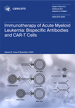
Cover Story (view full-size image): Improving knowledge in AML immunobiology may translate to better clinical developments in immunotherapies for patients with AML. Harnessing the immune system against leukemia cells can be a powerful treatment strategy for AML patients. Immune-based therapies such as BiTE and CAR-T cell therapy have proven to be effective means of targeting chemotherapy resistance in AML, but their therapeutic benefits are not durable and limited only to a fraction of patients. However, despite these limitations, recent studies have shown the peculiar sensitivity of some AML subtypes to immunotherapy, such as multiplex antigen targeting, and have provided the basis for future studies, thus holding the promise of successful clinical developments. View this paper
  • Issues are regarded as officially published after their release is announced to the table of contents alert mailing list.
  • You may sign up for e-mail alerts to receive table of contents of newly released issues.
  • PDF is the official format for papers published in both, html and pdf forms. To view the papers in pdf format, click on the "PDF Full-text" link, and use the free Adobe Reader to open them.
Order results
Result details
Section
Select all
Export citation of selected articles as:
11 pages, 554 KiB  
Article
Whole Exome-Wide Association Identifies Rare Variants in APC Associated with High-Risk Colorectal Cancer in the Middle East
by Abdul Khalid Siraj, Rong Bu, Saud Azam, Zeeshan Qadri, Kaleem Iqbal, Sandeep Kumar Parvathareddy, Fouad Al-Dayel and Khawla S. Al-Kuraya
Cancers 2024, 16(21), 3720; https://doi.org/10.3390/cancers16213720 - 4 Nov 2024
Viewed by 683
Abstract
Background: Colorectal cancer (CRC) displays a complex pattern of inheritance. It is postulated that much of the missing heritability of CRC is enriched in high-impact rare alleles, which might play a crucial role in the etiology and susceptibility of CRC. Methods: [...] Read more.
Background: Colorectal cancer (CRC) displays a complex pattern of inheritance. It is postulated that much of the missing heritability of CRC is enriched in high-impact rare alleles, which might play a crucial role in the etiology and susceptibility of CRC. Methods: In this study, an exome-wide association analysis was performed in 146 patients with high-risk CRC in the Middle East and 1395 healthy controls. The aim was to identify rare germline variants in coding regions and their splicing sites associated with high-risk CRC in the Middle Eastern population. Results: Rare inactivating variants (RIVs) in APC had the strongest association with high-risk CRC (6/146 in cases vs. 1/1395 in controls, OR = 59.7, p = 5.13 × 10⁻¹²), whereas RIVs in RIMS1, an RAS superfamily member, were significantly associated with high-risk CRC (5/146 case vs. 2/1395 controls, OR = 24.7, p = 2.03 × 10⁻⁸). Rare damaging variants in 17 genes were associated with high-risk CRC at the exome-wide threshold (p < 2.5 × 10⁻⁶). Based on the sequence kernel association test, nonsynonymous variants in six genes (TNXB, TAP2, GPSM3, ADGRG4, TMEM229A, and ANKRD33B) had a significant association with high-risk CRC. RIVs in APC—the most common high-penetrance genetic factor—were associated with patients with high-risk CRC in the Middle East. Individuals who inherited APC RIVs had an approximate 60-fold increased risk of developing CRC and were likely to develop the disease earlier. Conclusions: We identified new potential CRC predisposition variants in other genes that could play a role in CRC inheritance. However, large collaborative studies are needed to confirm the association of these variants with high-risk CRC. These results provide information for counseling patients with high-risk CRC and their families in our population. Full article
(This article belongs to the Section Cancer Pathophysiology)
Show Figures

Figure 1

15 pages, 2303 KiB  
Article
The Immunomodulatory Effect of Different FLT3 Inhibitors on Dendritic Cells
by Sebastian Schlaweck, Alea Radcke, Sascha Kampmann, Benjamin V. Becker, Peter Brossart and Annkristin Heine
Cancers 2024, 16(21), 3719; https://doi.org/10.3390/cancers16213719 - 4 Nov 2024
Viewed by 801
Abstract
Background: FMS-like tyrosine kinase 3 (FLT3) mutations or internal tandem duplication occur in 30% of acute myeloid leukemia (AML) cases. In these cases, FLT3 inhibitors (FLT3i) are approved for induction treatment and relapse. Allogeneic hematopoietic stem cell transplantation (alloHSCT) remains the recommended post-induction [...] Read more.
Background: FMS-like tyrosine kinase 3 (FLT3) mutations or internal tandem duplication occur in 30% of acute myeloid leukemia (AML) cases. In these cases, FLT3 inhibitors (FLT3i) are approved for induction treatment and relapse. Allogeneic hematopoietic stem cell transplantation (alloHSCT) remains the recommended post-induction therapy for suitable patients. However, the role of FLT3i therapy after alloHSCT remains unclear. Therefore, we investigated the three currently available FLT3i, gilteritinib, midostaurin, and quizartinib, in terms of their immunosuppressive effect on dendritic cells (DCs). DCs are professional antigen-presenting cells inducing T-cell responses to infectious stimuli. Highly activated DCs can also cause complications after alloHSCT, such as triggering Graft versus Host disease, a serious and potentially life-threatening complication after alloHSCT. Methods: To study the immunomodulatory effects on DCs, we differentiated murine and human DCs in the presence of FLT3i and performed immunophenotyping by flow cytometry and cytokine measurements and investigated gene and protein expression. Results: We detected a dose-dependent immunosuppressive effect of midostaurin, which decreased the expression of costimulatory markers like CD86, and found a reduced secretion of pro-inflammatory cytokines such as IL-12, TNFα, and IL-6. Mechanistically, we show that midostaurin inhibits TLR and TNF signaling and NFκB, PI3K, and MAPK pathways. The immunosuppressive effect of gilteritinib was less pronounced, while quizartinib did not show truncation of relevant signaling pathways. Conclusions: Our results suggest different immunosuppressive effects of these three FLT3i and may, therefore, provide an additional rationale for optimal maintenance therapy after alloHSCT of FLT3-positive AML patients to prevent infectious complications and GvHD mediated by DCs. Full article
(This article belongs to the Special Issue Advancements in Treatment Approaches for AML)
Show Figures

Figure 1

13 pages, 2545 KiB  
Systematic Review
Untreated Vestibular Schwannoma: Analysis of the Determinants of Growth
by Cheng Yang, Daniel Alvarado, Pawan Kishore Ravindran, Max E. Keizer, Koos Hovinga, Martinus P. G. Broen, Henricus (Dirk) P. M. Kunst and Yasin Temel
Cancers 2024, 16(21), 3718; https://doi.org/10.3390/cancers16213718 - 4 Nov 2024
Viewed by 702
Abstract
The growth rate of sporadic VS varies considerably, posing challenges for consistent clinical management. This systematic review examines data on factors associated with VS growth, following a protocol registered in the PROSPERO database. The analysis reveals that key predictors of tumor growth include [...] Read more.
The growth rate of sporadic VS varies considerably, posing challenges for consistent clinical management. This systematic review examines data on factors associated with VS growth, following a protocol registered in the PROSPERO database. The analysis reveals that key predictors of tumor growth include tumor location, initial size, and specific clinical symptoms such as hearing loss and imbalance. Additionally, several studies suggest that growth observed within the first year may serve as an indicator of subsequent progression, enabling the earlier identification of high-risk cases. Emerging factors such as the posture swing test and MRI signal intensity have also been identified as novel predictors that could further refine growth assessments. Our meta-analysis confirms that tumor location, initial size, cystic components, and vestibular symptoms are closely linked to the likelihood of VS growth. This review provides valuable guidance for clinicians in identifying patients who may require closer monitoring or early intervention. By integrating these predictive factors into clinical practice, this review supports more personalized treatment and contributes to the development of more accurate prognostic models for managing untreated sporadic VS. Full article
(This article belongs to the Section Cancer Therapy)
Show Figures

Figure 1

13 pages, 4467 KiB  
Article
ZNF281 Facilitates the Invasion of Cervical Cancer Cell Both In Vivo and In Vitro
by Ye Chong, Kun Zhang, Yuting Zeng, Qian Chen, Qian Feng, Nan Cui, Pengsheng Zheng, Litao Ruan and Wei Hua
Cancers 2024, 16(21), 3717; https://doi.org/10.3390/cancers16213717 - 4 Nov 2024
Viewed by 692
Abstract
Background: Cervical cancer is the fourth most common cancer among women worldwide. The zinc finger transcription factor 281 (ZNF281)/ZBP-99 protein specifically binds to GC-rich DNA sequences and regulates gene expression, and it has been shown to promote tumor progression. In this study, [...] Read more.
Background: Cervical cancer is the fourth most common cancer among women worldwide. The zinc finger transcription factor 281 (ZNF281)/ZBP-99 protein specifically binds to GC-rich DNA sequences and regulates gene expression, and it has been shown to promote tumor progression. In this study, we aim to investigate the function and molecular mechanism of ZNF281 in uterine cervical carcinoma. Methods: We conducted immunohistochemistry and Western blot assays to determine the expression of ZNF281 in eight human cervical cancer tissues. And, xenograft experiments involving the injection of HeLa cells into nude mice was used to determine the function of ZNF281 on proliferation. Transwell assays were used to detect the migration and invasion of HeLa cells after indicated that ZNF281 overexpression. Results: Our results indicated that ZNF281 protein levels were higher in cervical cancer tissues compared to normal cervical tissues. Additionally, ZNF281 was expressed in human cervical carcinoma cell lines, including HeLa, SiHa, C-33 A, CaSki, and HT-3, and is localized in both the cell nucleus and cytoplasm. ZNF281 overexpression did not influence HeLa cell proliferation or tumor size in situ. Moreover, nude mice injected with ZNF281-overexpressing cell lines developed more tumor lesions in the lungs compared to those injected with control cell lines. Conclusions: These findings suggest that ZNF281 is associated with tumor metastasis without affecting cell proliferation, both in vivo and in vitro. Full article
Show Figures

Figure 1

20 pages, 1417 KiB  
Review
Progress in Precision Medicine for Head and Neck Cancer
by Sanaz Vakili, Amir Barzegar Behrooz, Rachel Whichelo, Alexandra Fernandes, Abdul-Hamid Emwas, Mariusz Jaremko, Jarosław Markowski, Marek J. Los, Saeid Ghavami and Rui Vitorino
Cancers 2024, 16(21), 3716; https://doi.org/10.3390/cancers16213716 - 4 Nov 2024
Viewed by 1627
Abstract
This paper presents a comprehensive comparative analysis of biomarkers for head and neck cancer (HNC), a prevalent but molecularly diverse malignancy. We detail the roles of key proteins and genes in tumourigenesis and progression, emphasizing their diagnostic, prognostic, and therapeutic relevance. Our bioinformatic [...] Read more.
This paper presents a comprehensive comparative analysis of biomarkers for head and neck cancer (HNC), a prevalent but molecularly diverse malignancy. We detail the roles of key proteins and genes in tumourigenesis and progression, emphasizing their diagnostic, prognostic, and therapeutic relevance. Our bioinformatic validation reveals crucial genes such as AURKA, HMGA2, MMP1, PLAU, and SERPINE1, along with microRNAs (miRNA), linked to HNC progression. OncomiRs, including hsa-miR-21-5p, hsa-miR-31-5p, hsa-miR-221-3p, hsa-miR-222-3p, hsa-miR-196a-5p, and hsa-miR-200c-3p, drive tumourigenesis, while tumour-suppressive miRNAs like hsa-miR-375 and hsa-miR-145-5p inhibit it. Notably, hsa-miR-155-3p correlates with survival outcomes in addition to the genes RAI14, S1PR5, OSBPL10, and METTL6, highlighting its prognostic potential. Future directions should focus on leveraging precision medicine, novel therapeutics, and AI integration to advance personalized treatment strategies to optimize patient outcomes in HNC care. Full article
(This article belongs to the Collection Advances in Diagnostics and Treatment of Head and Neck Cancer)
Show Figures

Figure 1

15 pages, 2180 KiB  
Review
Time to Rethink Bronchiolitis Obliterans Syndrome Following Lung or Hematopoietic Cell Transplantation in Pediatric Patients
by Tang-Her Jaing, Yi-Lun Wang and Chia-Chi Chiu
Cancers 2024, 16(21), 3715; https://doi.org/10.3390/cancers16213715 - 4 Nov 2024
Viewed by 1044
Abstract
Background: Similar in histological characteristics and clinical manifestations, bronchiolitis obliterans syndrome (BOS) can develop following lung transplantation (LTx) or hematopoietic cell transplantation (HCT). In contrast to lung transplantation, where BOS is restricted to the lung allograft, HCT-related systemic graft-versus-host disease (GVHD) is the [...] Read more.
Background: Similar in histological characteristics and clinical manifestations, bronchiolitis obliterans syndrome (BOS) can develop following lung transplantation (LTx) or hematopoietic cell transplantation (HCT). In contrast to lung transplantation, where BOS is restricted to the lung allograft, HCT-related systemic graft-versus-host disease (GVHD) is the root cause of BOS. Because lung function declines following HCT, diagnosis becomes more difficult. Given the lack of proven effective medicines, treatment is based on empirical evidence. Methods: Cross-disciplinary learning is crucial, and novel therapies are under investigation to improve survival and avoid LTx. Recent advances have focused on updating the understanding of the etiology, clinical features, and pathobiology of BOS. It emphasizes the significance of learning from experts in other transplant modalities, promoting cross-disciplinary knowledge. Results: Our treatment algorithms are derived from extensive research and expert clinical input. It is important to ensure that immunosuppression is optimized and that any other conditions or contributing factors are addressed, if possible. Clear treatment algorithms are provided for each condition, drawing from the published literature and consensus clinical opinion. There are several novel therapies currently being investigated, such as aerosolized liposomal cyclosporine, Janus kinase inhibitors, antifibrotic therapies, and B-cell-directed therapies. Conclusions: We urgently need innovative treatments that can greatly increase survival rates and eliminate the need for LTx or re-transplantation. Full article
Show Figures

Figure 1

15 pages, 1342 KiB  
Systematic Review
The Prognostic Value of the 31-Gene Expression Profile Test in Cutaneous Melanoma: A Systematic Review and Meta-Analysis
by Ryan A. Durgham, Sami I. Nassar, Ramazan Gun, Shaun A. Nguyen, Ameya A. Asarkar and Cherie-Ann O. Nathan
Cancers 2024, 16(21), 3714; https://doi.org/10.3390/cancers16213714 - 4 Nov 2024
Viewed by 1497
Abstract
Background: Cutaneous melanoma is an increasingly common and potentially lethal form of skin cancer. Current staging systems based on clinical and pathological features have limitations in accurately predicting outcomes, particularly for early-stage disease. The 31-gene expression profile (31-GEP) test has emerged as a [...] Read more.
Background: Cutaneous melanoma is an increasingly common and potentially lethal form of skin cancer. Current staging systems based on clinical and pathological features have limitations in accurately predicting outcomes, particularly for early-stage disease. The 31-gene expression profile (31-GEP) test has emerged as a promising tool for improving risk stratification in melanoma patients. Methods: We conducted a systematic review and meta-analysis of studies evaluating the prognostic performance of the 31-GEP test in cutaneous melanoma. A comprehensive literature search was performed in multiple databases. Studies reporting survival outcomes stratified by 31-GEP class were included. Random-effects models were used to determine survival estimates across studies. Results: Thirteen studies comprising 14,760 patients were included in the meta-analysis. The 31-GEP test consistently stratified patients into risk groups with significantly different outcomes. The 5-year melanoma-specific survival rates were 99.8% (95% CI: 98–100%) for Class 1A, 97.6% (95% CI: 92.4–99.3%) for Class 1B/2A, and 83.4% (95% CI: 66.5–92.7%) for Class 2B. Similar trends were observed for recurrence-free and distant metastasis-free survival. Conclusions: This meta-analysis supports the prognostic utility of the 31-GEP test in cutaneous melanoma prognostication. The test consistently stratified patients into clinically meaningful risk groups across multiple survival metrics. These findings support the potential clinical utility of the 31-GEP test in enhancing current staging systems and informing personalized management strategies for melanoma patients. Full article
(This article belongs to the Special Issue Skin Cancers of the Head and Neck)
Show Figures

Figure 1

18 pages, 52984 KiB  
Article
Primitive Resectable Small Bowel Cancer Clinical–Pathological Analysis: A 10-Year Retrospective Study in a General Surgery Unit
by Cosmin Vasile Obleagă, Costin Teodor Streba, Cecil Sorin Mirea, Ionică Daniel Vîlcea, Dan Nicolae Florescu, Mihai Călin Ciorbagiu, Tudor Turcu, Mirela Marinela Florescu, Mircea Sebastian Șerbănescu, Alina-Maria Mehedințeanu and Cristin Constantin Vere
Cancers 2024, 16(21), 3713; https://doi.org/10.3390/cancers16213713 - 4 Nov 2024
Viewed by 797
Abstract
Introduction: Small bowel cancer is very rare; although the incidence of adenocarcinoma and other anatomopathological forms has increased recently, the diagnosis and treatment of this disease are still debatable because of the clinical heterogeneity and the absence of studies including a large number [...] Read more.
Introduction: Small bowel cancer is very rare; although the incidence of adenocarcinoma and other anatomopathological forms has increased recently, the diagnosis and treatment of this disease are still debatable because of the clinical heterogeneity and the absence of studies including a large number of patients. Materials and Methods: We performed a retrospective study over 10 years in which we analyzed the clinical, imaging, and anatomopathological data of 46 patients hospitalized in a surgery clinic and diagnosed with small bowel cancer (duodenum, jejunum, and ileum). Results: After clinical assessment of these patients, including complications (occlusion, bleeding, and perforation), the CT scan established the diagnosis in over 90% of the cases of the complicated form of the disease. Surgery has a curative role in localized cancers; tumor location, local invasion, the presence of locoregional lymph nodes, and the number of multiple tumors influence the type of surgery. The conventional pathological exam was completed via immunohistochemical staining. Adjuvant oncological treatment was performed after surgery (according to the guidelines); in patients with exceptional histopathological forms, the therapy was personalized. Conclusions: Most small bowel cancers were diagnosed with complications (occlusion and bleeding); the tumor type, location, and presence of multiple bowel cancers significantly influenced its management. Independently of the surgical resection (R0/R1 or R2), the prognosis of the disease depends on the tumor aggressivity, location (single/multiple), and locoregional node invasion. Full article
(This article belongs to the Special Issue Histopathology and Diagnosis of Gastrointestinal Tumors)
Show Figures

Figure 1

11 pages, 1927 KiB  
Article
Serum CYFRA 21-1 as a Prognostic Marker in Non-Small-Cell Lung Cancer Patients Treated with Immune Checkpoint Inhibitors
by Keiki Miyadera, Sho Kakuto, Mayu Sugai, Ryosuke Tsugitomi, Yoshiaki Amino, Ken Uchibori, Noriko Yanagitani, Hisatoshi Sugiura, Masahiro Seike, Makoto Nishio and Ryo Ariyasu
Cancers 2024, 16(21), 3712; https://doi.org/10.3390/cancers16213712 - 4 Nov 2024
Viewed by 902
Abstract
Background: A prognostic marker in patients with non-small-cell lung cancer (NSCLC) treated with anti-PD-1/PD-L1 antibodies must be established. This study explored serum cytokeratin fraction 21–1 (CYFRA 21-1), which represents a squamous cell histology, as a prognostic factor in anti-PD-1/PD-L1 antibody treatment, stratifying by [...] Read more.
Background: A prognostic marker in patients with non-small-cell lung cancer (NSCLC) treated with anti-PD-1/PD-L1 antibodies must be established. This study explored serum cytokeratin fraction 21–1 (CYFRA 21-1), which represents a squamous cell histology, as a prognostic factor in anti-PD-1/PD-L1 antibody treatment, stratifying by histology and treatment regimen. Methods: This study retrospectively evaluated patients with advanced NSCLC without driver mutations receiving anti-PD-1/PD-L1 antibodies between November 2015 and March 2023. Cutoff values for CYFRA 21-1 and carcinoembryonic antigen (CEA) were 3.5 and 5.0 ng/mL, respectively. The Kaplan–Meier method and a log-rank test were conducted. The Cox proportional hazards model was utilized for univariate and multivariate analyses. Results: This study included 258 patients. The squamous NSCLC group demonstrated a shorter overall survival (OS) than the non-squamous NSCLC group (median, 17.8 vs. 23.7 months, p = 0.141). Patients with high serum CYFRA 21-1 and CEA levels exhibited a significantly shorter OS than those with normal levels (median, 11.7 vs. 32.7 months, p < 0.005; 15.8 vs. 29.7 months, p < 0.005). The multivariate analysis identified a performance status (PS) of ≥2, a PD-L1 expression of ≥50%, and a serum CYFRA 21-1 of >3.5 ng/mL as independent prognostic factors. Patients with high serum CYFRA 21-1 levels exhibited a significantly shorter OS even focusing on non-squamous NSCLC, anti-PD-1/PD-L1 antibody and chemotherapy combination therapy, or anti-CTLA-4 antibody combination therapy. Conclusion: Serum CYFRA 21-1 is a poor prognostic marker for patients with NSCLC receiving anti-PD-1/PD-L1 antibody treatment even when stratifying by histology or treatment regimen. Full article
(This article belongs to the Section Cancer Immunology and Immunotherapy)
Show Figures

Figure 1

24 pages, 7219 KiB  
Article
Enhancing Anti-PD-1 Immunotherapy by Targeting MDSCs via Hepatic Arterial Infusion in Breast Cancer Liver Metastases
by Minhyung Kim, Colin A. Powers, Daniel T. Fisher, Amy W. Ku, Nickolay Neznanov, Alfiya F. Safina, Jianmin Wang, Avishekh Gautam, Siddharth Balachandran, Anuradha Krishnamurthy, Katerina V. Gurova, Sharon S. Evans, Andrei V. Gudkov and Joseph J. Skitzki
Cancers 2024, 16(21), 3711; https://doi.org/10.3390/cancers16213711 - 3 Nov 2024
Viewed by 1263
Abstract
Background: Surgery, chemotherapy, and radiation often have limited utility for advanced metastatic disease in the liver, and despite its promising activity in select cancers, PD-1 blockade therapy similarly has minimal benefit in this setting. Curaxin, CBL0137, is an experimental anti-cancer drug that disrupts [...] Read more.
Background: Surgery, chemotherapy, and radiation often have limited utility for advanced metastatic disease in the liver, and despite its promising activity in select cancers, PD-1 blockade therapy similarly has minimal benefit in this setting. Curaxin, CBL0137, is an experimental anti-cancer drug that disrupts the binding of DNA to histones, destabilizes chromatin, and induces Z-DNA formation which may stimulate anti-tumor immune responses. Methods: Murine cell lines of colon (CT26) and breast (4T1) cancer were interrogated for survival and CBL0137-associated DNA changes in vitro. Immunocompetent models of liver metastases followed by CBL0137 hepatic arterial infusion (HAI) were used to examine in vivo tumor cell DNA alterations, treatment responses, and the immune contexture associated with CBL0137, both alone and in combination with anti-PD-1 therapy. Results: CBL0137 induced immediate changes to favor tumor cell death in vitro and in vivo with an efficient tumor uptake via the HAI route. Toxicity to CBL0137 was minimal and anti-tumor treatment effects were more efficient with HAI compared to intravenous delivery. Immune effects were pronounced with CBL0137 HAI with concurrent depletion of a specific population of myeloid-derived suppressor cells and maintenance of effector T cell populations. Conclusions: Combination of CBL0137 HAI with PD-1 blockade improved survival in 4T1 tumors but not in CT26 tumors, and therapeutic efficacy relies on the finding of simultaneous and targeted depletion of myeloid-derived suppressor cells and skewing of T cell populations to produce synergy with PD-1 blockade therapy. Full article
(This article belongs to the Special Issue Cancer Immunotherapy in Clinical and Translational Research)
Show Figures

Figure 1

20 pages, 7697 KiB  
Article
MiRNA Profiling of Areca Nut-Induced Carcinogenesis in Head and Neck Cancer
by Hung-Han Huang, Joseph T. Chang, Guo-Rung You, Yu-Fang Fu, Eric Yi-Liang Shen, Yi-Fang Huang, Chia-Rui Shen and Ann-Joy Cheng
Cancers 2024, 16(21), 3710; https://doi.org/10.3390/cancers16213710 - 3 Nov 2024
Viewed by 959
Abstract
Background: While miRNAs are increasingly recognized for their role in tumorigenesis, their involvement in head and neck cancer (HNC) remains insufficiently explored. Additionally, the carcinogenic mechanisms of areca nut, a major habitual carcinogen in Southeast Asia, are not well understood. Methods and results: [...] Read more.
Background: While miRNAs are increasingly recognized for their role in tumorigenesis, their involvement in head and neck cancer (HNC) remains insufficiently explored. Additionally, the carcinogenic mechanisms of areca nut, a major habitual carcinogen in Southeast Asia, are not well understood. Methods and results: This study adopts a systematic approach to identify miRNA profiles associated with areca nut-induced HNC. Using miRNA microarray analysis, we identified 292 miRNAs dysregulated in areca nut-treated HNC cells, with 136 upregulated and 156 downregulated. Bioinformatic analysis of the TCGA-HNSC dataset uncovered a set of 692 miRNAs relevant to HNC development, comprising 449 overexpressed and 243 underexpressed in tumor tissues. Integrating these datasets, we defined a signature of 84 miRNAs, including 39 oncogenic miRNAs (OncomiRs) and 45 tumor-suppressive miRNAs (TsmiRs), highlighting their pivotal role in areca nut-induced carcinogenesis. MultiMiR analysis identified 740 genes cross-regulated by eight hub TsmiRs, significantly impacting key cancer-related pathways (p53, PI3K-AKT, MAPK, and Ras) and critical oncogenic processes. Moreover, we validated miR-499a-5p as a vital regulator, demonstrating its ability to mitigate areca nut-induced cancer progression by reducing cell migration, invasion, and chemoresistance. Conclusions: Thus, this miRNA signature addresses a crucial gap in understanding the molecular underpinnings of areca nut-induced carcinogenesis and offers a promising platform for clinical applications in risk assessment, diagnosis, and prognosis of areca nut-associated malignancies. Full article
(This article belongs to the Special Issue Multi-Omics Analysis in the Study of Carcinogenesis)
Show Figures

Figure 1

20 pages, 1747 KiB  
Article
Predictors and Profile of Severe Infectious Complications in Multiple Myeloma Patients Treated with Daratumumab-Based Regimens: A Machine Learning Model for Pneumonia Risk
by Damian Mikulski, Marcin Kamil Kędzior, Grzegorz Mirocha, Katarzyna Jerzmanowska-Piechota, Żaneta Witas, Łukasz Woźniak, Magdalena Pawlak, Kacper Kościelny, Michał Kośny, Paweł Robak, Aleksandra Gołos, Tadeusz Robak, Wojciech Fendler and Joanna Góra-Tybor
Cancers 2024, 16(21), 3709; https://doi.org/10.3390/cancers16213709 - 3 Nov 2024
Viewed by 1384
Abstract
Background: Daratumumab (Dara) is the first monoclonal antibody introduced into clinical practice to treat multiple myeloma (MM). It currently forms the backbone of therapy regimens in both newly diagnosed (ND) and relapsed/refractory (RR) patients. However, previous reports indicated an increased risk of infectious [...] Read more.
Background: Daratumumab (Dara) is the first monoclonal antibody introduced into clinical practice to treat multiple myeloma (MM). It currently forms the backbone of therapy regimens in both newly diagnosed (ND) and relapsed/refractory (RR) patients. However, previous reports indicated an increased risk of infectious complications (ICs) during Dara-based treatment. In this study, we aimed to determine the profile of ICs in MM patients treated with Dara-based regimens and establish predictors of their occurrence. Methods: This retrospective, real-life study included MM patients treated with Dara-based regimens between July 2019 and March 2024 at our institution. Infectious events were evaluated using the Terminology Criteria for Adverse Events (CTCAE) version 5.0. Results: The study group consisted of a total of 139 patients, including 49 NDMM and 90 RRMM. In the RR setting, the majority (60.0%) of patients received the Dara, bortezomib, and dexamethasone (DVd) regimen, whereas ND patients were predominantly (98%) treated with the Dara, bortezomib, thalidomide, and dexamethasone (DVTd) regimen. Overall, 55 patients (39.6%) experienced ICs. The most common IC was pneumonia (37.5%), followed by upper respiratory tract infections (26.8%). Finally, twenty-five patients had severe ICs (grade ≥ 3) and required hospitalization, and eight patients died due to ICs. In the final multivariable model adjusted for setting (ND/RR) and age, hemoglobin level (OR 0.77, 95% CI: 0.61–0.96, p = 0.0037), and Eastern Cooperative Oncology Group (ECOG) >1 (OR 4.46, 95% CI: 1.63–12.26, p = 0.0037) were significant factors influencing severe IC occurrence. Additionally, we developed predictive models using the J48 decision tree, gradient boosting, and random forest algorithms. After conducting 10-fold cross-validation, these models demonstrated strong performance in predicting the occurrence of pneumonia during treatment with daratumumab-based regimens. Conclusions: Simple clinical and laboratory assessments, including hemoglobin level and ECOG scale, can be valuable in identifying patients vulnerable to infections during Dara-based regimens, facilitating personalized prophylactic strategies. Full article
Show Figures

Figure 1

9 pages, 680 KiB  
Article
The Role of Bladder-Washing Cytology as an Adjunctive Method to Cystoscopy During Follow-Up for Low-Grade TaT1 Non-Muscle-Invasive Bladder Cancer
by Enric Carbonell, Clàudia Mercader, Héctor Alfambra, Paulette Narvaez, Eric Villalba, Rita Pagès, Ignacio Asiain, Meritxell Costa, Agustín Franco, Antonio Alcaraz, María José Ribal and Antoni Vilaseca
Cancers 2024, 16(21), 3708; https://doi.org/10.3390/cancers16213708 - 1 Nov 2024
Viewed by 699
Abstract
Background and Objective: The role of urine cytology during follow-up for low-grade (LG) non-muscle-invasive bladder cancer (NMIBC) is not well established, although cytology has low sensitivity in detecting LG recurrences. Our study aims to evaluate the impact of urine cytology as a complementary [...] Read more.
Background and Objective: The role of urine cytology during follow-up for low-grade (LG) non-muscle-invasive bladder cancer (NMIBC) is not well established, although cytology has low sensitivity in detecting LG recurrences. Our study aims to evaluate the impact of urine cytology as a complementary method to cystoscopy during follow-up for LG NMIBC. Methods: Patients diagnosed with primary LG TaT1 bladder cancer (BC) between 2010 and 2020 were included. Patients were stratified according to the EAU NMIBC scoring model. Urine cytology was performed during follow-up cystoscopy. The outcomes of the study were BC recurrence and upgrading to high-grade (HG). Cytology utility was established by assessing whether its result led to management change. Results: We included 337 patients with LG TaT1 BC. EAU risk group distribution was low in 262 (77.7%), intermediate in 57 (16.9%), and high-risk in 18 (5.3%) cases. With a median follow-up of 5 years, 166 (49.3%) patients experienced recurrence. Cystoscopy was positive in 154 (92.8%) and suspicious in 12 (7.2%) cases. Urine cytology was positive in 33 (19.9%) cases but only changed management in 3 (0.89%), all with suspicious cystoscopy. Positive cytology at first recurrence was associated with higher risk of upgrading during follow-up (HR 2.781, p = 0.006) and lower upgrading-free survival (p = 0.001). Conclusions: The role of urine cytology to detect first recurrences during follow-up for primary LG TaT1 NMIBC might be limited to patients with non-conclusive lesions in the cystoscopy. A positive cytology at first recurrence is associated with a higher risk of upgrading to HG BC during follow-up. Full article
(This article belongs to the Special Issue Urogenital Neoplasms Pathology)
Show Figures

Figure 1

16 pages, 495 KiB  
Article
The Influence of the Microbiome on the Complications of Radiotherapy and Its Effectiveness in Patients with Laryngeal Cancer
by Karolina Dorobisz, Tadeusz Dorobisz, Katarzyna Pazdro-Zastawny, Katarzyna Czyż and Marzena Janczak
Cancers 2024, 16(21), 3707; https://doi.org/10.3390/cancers16213707 - 1 Nov 2024
Viewed by 778
Abstract
Introduction: Radiotherapy is an effective method of treating cancer and affects 50% of patients. Intensity-modulated radiotherapy (IMRT) is a modernized method of classical radiation used in the treatment of laryngeal cancer. Treatment with intent to preserve the larynx is not always safe or [...] Read more.
Introduction: Radiotherapy is an effective method of treating cancer and affects 50% of patients. Intensity-modulated radiotherapy (IMRT) is a modernized method of classical radiation used in the treatment of laryngeal cancer. Treatment with intent to preserve the larynx is not always safe or complication-free. The microbiome may significantly influence the effectiveness of oncological treatment, especially radiotherapy, and may also be modified by the toxic response to radiation. Objective: The aim of the study was to prospectively assess the microbiome and its influence on radiotherapy toxicity in patients with laryngeal cancer. Results: Statistically significant risk factors for complications after radiotherapy were the percentage of Porphyromonas of at least 6.7%, the percentage of Fusobacterium of at least 2.6% and the percentage of Catonella of at least 2.6%. Conclusions: The importance of the microbiome in oncology has been confirmed in many studies. Effective radiotherapy treatment and the prevention of radiation-induced oral mucositis is a challenge in oncology. The microbiome may be an important part of personalized cancer treatment. The assessment of the microbiome of patients diagnosed with cancer may provide the opportunity to predict the response to treatment and its effectiveness. The influence of the microbiome may be important in predicting the risk group for radiotherapy treatment failure. The possibility of modifying the microbiome may become a goal to improve the prognosis of patients with laryngeal cancer. Fusobacterium, Porphyromonas and Catonella are important risk factors for radiation-induced oral mucositis in patients with laryngeal cancer. Full article
(This article belongs to the Section Tumor Microenvironment)
Show Figures

Figure 1

31 pages, 7387 KiB  
Review
Impact of Modern Lifestyle on Circadian Health and Its Contribution to Adipogenesis and Cancer Risk
by Oxana Dobrovinskaya, Javier Alamilla and Miguel Olivas-Aguirre
Cancers 2024, 16(21), 3706; https://doi.org/10.3390/cancers16213706 - 1 Nov 2024
Viewed by 1264
Abstract
Background: Recent research underscores a crucial connection between circadian rhythm disruption and cancer promotion, highlighting an urgent need for attention. Objectives: Explore the molecular mechanisms by which modern lifestyle factors—such as artificial light exposure, shift work, and dietary patterns—affect cortisol/melatonin regulation and cancer [...] Read more.
Background: Recent research underscores a crucial connection between circadian rhythm disruption and cancer promotion, highlighting an urgent need for attention. Objectives: Explore the molecular mechanisms by which modern lifestyle factors—such as artificial light exposure, shift work, and dietary patterns—affect cortisol/melatonin regulation and cancer risk. Methods: Employing a narrative review approach, we synthesized findings from Scopus, Google Scholar, and PubMed to analyze lifestyle impacts on circadian health, focusing on cortisol and melatonin chronobiology as molecular markers. We included studies that documented quantitative changes in these markers due to modern lifestyle habits, excluding those lacking quantitative data or presenting inconclusive results. Subsequent sections focused solely on articles that quantified the effects of circadian disruption on adipogenesis and tumor microenvironment modifications. Results: This review shows how modern habits lead to molecular changes in cortisol and melatonin, creating adipose microenvironments that support cancer development. These disruptions facilitate immune evasion, chemotherapy resistance, and tumor growth, highlighting the critical roles of cortisol dysregulation and melatonin imbalance. Conclusions: Through the presented findings, we establish a causal link between circadian rhythm dysregulation and the promotion of certain cancer types. By elucidating this relationship, the study emphasizes the importance of addressing lifestyle factors that contribute to circadian misalignment, suggesting that targeted interventions could play a crucial role in mitigating cancer risk and improving overall health outcomes. Full article
(This article belongs to the Special Issue Circadian Rhythms, Cancers and Chronotherapy)
Show Figures

Graphical abstract

17 pages, 2450 KiB  
Article
TGF-β Signaling Loop in Pancreatic Ductal Adenocarcinoma Activates Fibroblasts and Increases Tumor Cell Aggressiveness
by Noemi di Miceli, Chiara Baioni, Linda Barbieri, Davide Danielli, Emiliano Sala, Lucia Salvioni, Stefania Garbujo, Miriam Colombo, Davide Prosperi, Metello Innocenti and Luisa Fiandra
Cancers 2024, 16(21), 3705; https://doi.org/10.3390/cancers16213705 - 1 Nov 2024
Viewed by 1073
Abstract
Background: The interaction between cancer cells and cancer-associated fibroblasts (CAFs) is a key determinant of the rapid progression, high invasiveness, and chemoresistance of aggressive desmoplastic cancers such as pancreatic ductal adenocarcinoma (PDAC). Tumor cells are known to reprogram fibroblasts into CAFs by secreting [...] Read more.
Background: The interaction between cancer cells and cancer-associated fibroblasts (CAFs) is a key determinant of the rapid progression, high invasiveness, and chemoresistance of aggressive desmoplastic cancers such as pancreatic ductal adenocarcinoma (PDAC). Tumor cells are known to reprogram fibroblasts into CAFs by secreting transforming growth factor beta (TGF-β), amongst other cytokines. In turn, CAFs produce soluble factors that promote tumor-cell invasiveness and chemoresistance, including TGF-β itself, which has a major role in myofibroblastic CAFs. Such a high level of complexity has hampered progress toward a clear view of the TGFβ signaling loop between stromal fibroblasts and PDAC cells. Methods: Here, we tackled this issue by using co-culture settings that allow paracrine signaling alone (transwell systems) or paracrine and contact-mediated signaling (3D spheroids). Results: We found that TGF-β is critically involved in the activation of normal human fibroblasts into alpha-smooth muscle actin (α-SMA)-positive CAFs. The TGF-β released by CAFs accounted for the enhanced proliferation and resistance to gemcitabine of PDAC cells. This was accompanied by a partial epithelial-to-mesenchymal transition in PDAC cells, with no increase in their migratory abilities. Nevertheless, 3D heterospheroids comprising PDAC cells and fibroblasts allowed monitoring the pro-invasive effects of CAFs on cancer cells, possibly due to combined paracrine and physical contact-mediated signals. Conclusions: We conclude that TGF-β is only one of the players that mediates the communication between PDAC cells and fibroblasts and controls the acquisition of aggressive phenotypes. Hence, these advanced in vitro models may be exploited to further investigate these events and to design innovative anti-PDAC therapies. Full article
(This article belongs to the Special Issue Targeting the Tumor Microenvironment (Volume II))
Show Figures

Figure 1

2 pages, 770 KiB  
Correction
Correction: Lepore Signorile et al. c-MYC Protein Stability Is Sustained by MAPKs in Colorectal Cancer. Cancers 2022, 14, 4840
by Martina Lepore Signorile, Valentina Grossi, Candida Fasano, Giovanna Forte, Vittoria Disciglio, Paola Sanese, Katia De Marco, Francesca La Rocca, Raffaele Armentano, Anna Maria Valentini, Gianluigi Giannelli and Cristiano Simone
Cancers 2024, 16(21), 3704; https://doi.org/10.3390/cancers16213704 - 1 Nov 2024
Viewed by 538
Abstract
In the original publication [...] Full article
Show Figures

Figure 4

55 pages, 1491 KiB  
Review
Microplastics in the Human Body: Exposure, Detection, and Risk of Carcinogenesis: A State-of-the-Art Review
by Eliasz Dzierżyński, Piotr J. Gawlik, Damian Puźniak, Wojciech Flieger, Katarzyna Jóźwik, Grzegorz Teresiński, Alicja Forma, Paulina Wdowiak, Jacek Baj and Jolanta Flieger
Cancers 2024, 16(21), 3703; https://doi.org/10.3390/cancers16213703 - 1 Nov 2024
Viewed by 3394
Abstract
Background: Humans cannot avoid plastic exposure due to its ubiquitous presence in the natural environment. The waste generated is poorly biodegradable and exists in the form of MPs, which can enter the human body primarily through the digestive tract, respiratory tract, or damaged [...] Read more.
Background: Humans cannot avoid plastic exposure due to its ubiquitous presence in the natural environment. The waste generated is poorly biodegradable and exists in the form of MPs, which can enter the human body primarily through the digestive tract, respiratory tract, or damaged skin and accumulate in various tissues by crossing biological membrane barriers. There is an increasing amount of research on the health effects of MPs. Most literature reports focus on the impact of plastics on the respiratory, digestive, reproductive, hormonal, nervous, and immune systems, as well as the metabolic effects of MPs accumulation leading to epidemics of obesity, diabetes, hypertension, and non-alcoholic fatty liver disease. MPs, as xenobiotics, undergo ADMET processes in the body, i.e., absorption, distribution, metabolism, and excretion, which are not fully understood. Of particular concern are the carcinogenic chemicals added to plastics during manufacturing or adsorbed from the environment, such as chlorinated paraffins, phthalates, phenols, and bisphenols, which can be released when absorbed by the body. The continuous increase in NMP exposure has accelerated during the SARS-CoV-2 pandemic when there was a need to use single-use plastic products in daily life. Therefore, there is an urgent need to diagnose problems related to the health effects of MP exposure and detection. Methods: We collected eligible publications mainly from PubMed published between 2017 and 2024. Results: In this review, we summarize the current knowledge on potential sources and routes of exposure, translocation pathways, identification methods, and carcinogenic potential confirmed by in vitro and in vivo studies. Additionally, we discuss the limitations of studies such as contamination during sample preparation and instrumental limitations constraints affecting imaging quality and MPs detection sensitivity. Conclusions: The assessment of MP content in samples should be performed according to the appropriate procedure and analytical technique to ensure Quality and Control (QA/QC). It was confirmed that MPs can be absorbed and accumulated in distant tissues, leading to an inflammatory response and initiation of signaling pathways responsible for malignant transformation. Full article
(This article belongs to the Section Cancer Epidemiology and Prevention)
Show Figures

Figure 1

23 pages, 4829 KiB  
Review
The Evolution of Artificial Intelligence in Medical Imaging: From Computer Science to Machine and Deep Learning
by Michele Avanzo, Joseph Stancanello, Giovanni Pirrone, Annalisa Drigo and Alessandra Retico
Cancers 2024, 16(21), 3702; https://doi.org/10.3390/cancers16213702 - 1 Nov 2024
Viewed by 2789
Abstract
Artificial intelligence (AI), the wide spectrum of technologies aiming to give machines or computers the ability to perform human-like cognitive functions, began in the 1940s with the first abstract models of intelligent machines. Soon after, in the 1950s and 1960s, machine learning algorithms [...] Read more.
Artificial intelligence (AI), the wide spectrum of technologies aiming to give machines or computers the ability to perform human-like cognitive functions, began in the 1940s with the first abstract models of intelligent machines. Soon after, in the 1950s and 1960s, machine learning algorithms such as neural networks and decision trees ignited significant enthusiasm. More recent advancements include the refinement of learning algorithms, the development of convolutional neural networks to efficiently analyze images, and methods to synthesize new images. This renewed enthusiasm was also due to the increase in computational power with graphical processing units and the availability of large digital databases to be mined by neural networks. AI soon began to be applied in medicine, first through expert systems designed to support the clinician’s decision and later with neural networks for the detection, classification, or segmentation of malignant lesions in medical images. A recent prospective clinical trial demonstrated the non-inferiority of AI alone compared with a double reading by two radiologists on screening mammography. Natural language processing, recurrent neural networks, transformers, and generative models have both improved the capabilities of making an automated reading of medical images and moved AI to new domains, including the text analysis of electronic health records, image self-labeling, and self-reporting. The availability of open-source and free libraries, as well as powerful computing resources, has greatly facilitated the adoption of deep learning by researchers and clinicians. Key concerns surrounding AI in healthcare include the need for clinical trials to demonstrate efficacy, the perception of AI tools as ‘black boxes’ that require greater interpretability and explainability, and ethical issues related to ensuring fairness and trustworthiness in AI systems. Thanks to its versatility and impressive results, AI is one of the most promising resources for frontier research and applications in medicine, in particular for oncological applications. Full article
(This article belongs to the Section Cancer Informatics and Big Data)
Show Figures

Figure 1

25 pages, 15136 KiB  
Article
TRBP2, a Major Component of the RNAi Machinery, Is Subjected to Cell Cycle-Dependent Regulation in Human Cancer Cells of Diverse Tissue Origin
by Eleni I. Theotoki, Panos Kakoulidis, Athanassios D. Velentzas, Konstantinos-Stylianos Nikolakopoulos, Nikolaos V. Angelis, Ourania E. Tsitsilonis, Ema Anastasiadou and Dimitrios J. Stravopodis
Cancers 2024, 16(21), 3701; https://doi.org/10.3390/cancers16213701 - 1 Nov 2024
Viewed by 986
Abstract
Background: Transactivation Response Element RNA-binding Protein (TRBP2) is a double-stranded RNA-binding protein widely known for its critical contribution to RNA interference (RNAi), a conserved mechanism of gene-expression regulation mediated through small non-coding RNA moieties (ncRNAs). Nevertheless, TRBP2 has also proved to be involved [...] Read more.
Background: Transactivation Response Element RNA-binding Protein (TRBP2) is a double-stranded RNA-binding protein widely known for its critical contribution to RNA interference (RNAi), a conserved mechanism of gene-expression regulation mediated through small non-coding RNA moieties (ncRNAs). Nevertheless, TRBP2 has also proved to be involved in other molecular pathways and biological processes, such as cell growth, organism development, spermatogenesis, and stress response. Mutations or aberrant expression of TRBP2 have been previously associated with diverse human pathologies, including Alzheimer’s disease, cardiomyopathy, and cancer, with TRBP2 playing an essential role(s) in proliferation, invasion, and metastasis of tumor cells. Methods: Hence, the present study aims to investigate, via employment of advanced flow cytometry, immunofluorescence, cell transgenesis and bioinformatics technologies, new, still elusive, functions and properties of TRBP2, particularly regarding its cell cycle-specific control during cancer cell division. Results: We have identified a novel, mitosis-dependent regulation of TRBP2 protein expression, as clearly evidenced by the lack of its immunofluorescence-facilitated detection during mitotic phases, in several human cancer cell lines of different tissue origin. Notably, the obtained TRBP2-downregulation patterns seem to derive from molecular mechanisms that act independently of oncogenic activities (e.g., malignancy grade), metastatic capacities (e.g., low versus high), and mutational signatures (e.g., p53−/− or p53ΔΥ126) of cancer cells. Conclusions: Taken together, we herein propose that TRBP2 serves as a novel cell cycle-dependent regulator, likely exerting mitosis-suppression functions, and, thus, its mitosis-specific downregulation can hold strong promise to be exploited for the efficient and successful prognosis, diagnosis, and (radio-/chemo-)therapy of diverse human malignancies, in the clinic. Full article
(This article belongs to the Section Tumor Microenvironment)
Show Figures

Figure 1

12 pages, 4944 KiB  
Article
Nanopore Sequencing for T-Cell Receptor Rearrangement Analysis in Cutaneous T-Cell Lymphoma
by Cassandra Cieslak, Carsten Hain, Christian Rückert-Reed, Tobias Busche, Levin Joe Klages, Katrin Schaper-Gerhardt, Ralf Gutzmer, Jörn Kalinowski and Rudolf Stadler
Cancers 2024, 16(21), 3700; https://doi.org/10.3390/cancers16213700 - 1 Nov 2024
Viewed by 1032
Abstract
Background: Analysis of T-cell receptor (TCR) clonality is a major diagnostic tool for lymphomas, particularly for cutaneous T-cell lymphomas (CTCL) like Mycosis fungoides and Sézary syndrome. However, a fast and cost-effective workflow is needed to enable widespread use of this method. Methods: [...] Read more.
Background: Analysis of T-cell receptor (TCR) clonality is a major diagnostic tool for lymphomas, particularly for cutaneous T-cell lymphomas (CTCL) like Mycosis fungoides and Sézary syndrome. However, a fast and cost-effective workflow is needed to enable widespread use of this method. Methods: We established a procedure for TCR rearrangement analysis via Oxford Nanopore Technology (ONT) sequencing. TCR receptor rearrangements (TCR-gamma and TCR-beta chains) were analyzed in samples from 45 patients with various diagnoses: Mycosis fungoides (37/45), Sézary Syndrome (2/45), folliculotropic CTCL (1/45), and non-CTCL diagnoses as polyclonal controls (5/45). Sample types included formalin-fixed paraffin-embedded (FFPE) samples (27/45), fresh frozen samples (9/45), and CD3-isolated cells (9/45). In addition, DNA of a Jurkat cell line was used as a monoclonal control. TCR amplicons were generated employing an optimized version of the protocol from the Euro Clonality consortium. Sequencing was conducted on the ONT GridION and Illumina MiSeq platforms, followed by similar bioinformatic analysis protocols. The tumor clone frequency (TCF), a crucial prognostic factor for CTCL patients, was used for method comparison. Results: The use of an optimized amplicon protocol and adapted bioinformatic tools demonstrated a strong correlation in TCF values between both sequencing methods across all sample types (range R: 0.992–0.996; range r2: 0.984–0.991). Conclusions: In summary, ONT sequencing was able to detect TCR clonality comparable to NGS, indicating its potential as a faster and more cost-effective option for routine diagnostic use. Full article
(This article belongs to the Special Issue Targets and Biomarkers in Cutaneous Lymphoma)
Show Figures

Graphical abstract

17 pages, 6848 KiB  
Article
Deciphering CD59: Unveiling Its Role in Immune Microenvironment and Prognostic Significance
by Bhaumik Patel, Ashok Silwal, Mohamed Ashraf Eltokhy, Shreyas Gaikwad, Marina Curcic, Jalpa Patel and Sahdeo Prasad
Cancers 2024, 16(21), 3699; https://doi.org/10.3390/cancers16213699 - 1 Nov 2024
Viewed by 995
Abstract
Background: CD59, a GPI-anchored membrane protein, protects cancer cells from complement-dependent cytotoxicity (CDC) by inhibiting the formation of the membrane attack complex (MAC). It has been demonstrated to be overexpressed in most solid tumors, where it facilitates tumor cell escape from complement surveillance. [...] Read more.
Background: CD59, a GPI-anchored membrane protein, protects cancer cells from complement-dependent cytotoxicity (CDC) by inhibiting the formation of the membrane attack complex (MAC). It has been demonstrated to be overexpressed in most solid tumors, where it facilitates tumor cell escape from complement surveillance. The role of CD59 in cancer growth and interactions between CD59 and immune cells that modulate immune evasion has not been well explored. Methods: Using cancer patient database from The Cancer Genome Atlas (TCGA) and other public databases, we analyzed CD59 expression, its prognostic significance, and its association with immune cell infiltration in the tumor microenvironment, identifying associated genomic and functional networks and validating findings with invitro cell-line experimental data. Results: This article describes the abundant expression of CD59 in multiple tumors such as cervical squamous cell carcinoma (CESC), kidney renal cell carcinoma (KIRC), glioblastoma multiforme (GBM), head and neck squamous cell carcinoma (HNSC), and stomach adenocarcinoma (STAD), as well as in pan-cancer, using The Cancer Genome Atlas (TCGA) database and confirmed using multiple cancer cell lines. The expression of CD59 significantly alters the overall survival (OS) of patients with multiple malignancies such as CESC, GBM, HNSC, and STAD. Further, the correlation between CD59 and Treg and/or MDSC in the tumor microenvironment (TME) has shown to be strongly associated with poor outcomes in CESC, GBM, HNSC, and STAD as these tumors express high FOXP3 compared to KIRC. Moreover, unfavorable outcomes were strongly associated with the expression of CD59 and M2 tumor-associated macrophage infiltration in the TME via the IL10/pSTAT3 pathway in CESC and GBM but not in KIRC. In addition, TGFβ1-dominant cancers such as CESC, GBM, and HNSC showed a high correlation between CD59 and TGFβ1, leading to suppression of cytotoxic T cell activity. Conclusion: Overall, the correlation between CD59 and immune cells predicts its prognosis as unfavorable in CESC, GBM, HNSC, and STAD while being favorable in KIRC. Full article
Show Figures

Figure 1

13 pages, 1524 KiB  
Article
Peri-Interventional Hemodynamic Management Strategies for Percutaneous Chemosaturation of the Liver in Metastatic Cancer
by Patrick Rehn, Benjamin Tan, Jan Turra, Patrick Adler, Philipp Mayer, Dania Fischer, Mascha O. Fiedler-Kalenka, Felix C. F. Schmitt, De-Hua Chang, Christoph Lichtenstern, Mark O. Wielpütz, Hans-Ulrich Kauczor, Markus A. Weigand and Maximilian Dietrich
Cancers 2024, 16(21), 3698; https://doi.org/10.3390/cancers16213698 - 1 Nov 2024
Viewed by 875
Abstract
Background: Hepatic chemosaturation for inoperable liver tumors is a palliative treatment option with a beneficial effect on survival. However, the procedure regularly leads to circulatory failure during the filtration phase, and hemodynamic management is challenging. Our study aimed to compare two different strategies [...] Read more.
Background: Hepatic chemosaturation for inoperable liver tumors is a palliative treatment option with a beneficial effect on survival. However, the procedure regularly leads to circulatory failure during the filtration phase, and hemodynamic management is challenging. Our study aimed to compare two different strategies for hemodynamic management during chemosaturation to develop hypotheses for improving patient care and reducing peri-interventional morbidity. Methods: We conducted a single-center retrospective cohort study including 66 procedures of chemosaturation between May 2016 and March 2024. Procedures were divided into two groups: group 1 was managed with norepinephrine as the only vasopressor and liberal use of hydroxyethyl starch (HES). Group 2 was managed with norepinephrine and vasopressin and the preferred use of balanced crystalloids. We compared these two groups with respect to hemodynamic parameters, laboratory values, and post-interventional complications. Results: The heart rate was highest and the mean arterial pressure (MAP) was lowest during the filtration phase in both groups (p = 0.868, p = 0.270). The vasoactive inotropic score (VIS) was significantly higher in group 2 during the filtration phase (31.5 vs. 89, p < 0.001). Group 1 received significantly more HES overall (1000 mL vs. 0 mL, p < 0.001). Lactate levels at admission to the ICU were higher in group 1 (22.9 vs. 14.45 mg/dL, p = 0.041). Platelet counts were lower in group 2 from directly after chemosaturation through day 2 (p = 0.022, p = 0.001, p = 0.032). The INR differed significantly directly after chemosaturation (1.13 vs. 1.26, p = 0.015). Overall, group 1 received significantly more blood products peri-interventionally. There were two bleedings and one ischemic stroke in the overall cohort. There was no peri-interventional mortality. Conclusions: Advanced hemodynamic management ensures low peri-interventional mortality and morbidity. High-dose vasopressors, including vasopressin and the preferred use of balanced crystalloids, are sufficient to stabilize circulatory function during chemosaturation. Full article
(This article belongs to the Section Clinical Research of Cancer)
Show Figures

Figure 1

15 pages, 1367 KiB  
Article
Real-World Outcomes for Localised Gastro-Oesophageal Adenocarcinoma Cancer Treated with Perioperative FLOT and Prophylactic GCSF Support in a Single Asian Centre
by Wanyi Kee, Kennedy Yao Yi Ng, Shun Zi Liong, Siqin Zhou, Sharon Keman Chee, Chiew Woon Lim, Justina Yick Ching Lam, Jeremy Tian Hui Tan, Hock Soo Ong, Weng Hoong Chan, Eugene Kee Wee Lim, Chin Hong Lim, Alvin Kim Hock Eng, Christabel Jing Zhi Lee and Matthew Chau Hsien Ng
Cancers 2024, 16(21), 3697; https://doi.org/10.3390/cancers16213697 - 1 Nov 2024
Viewed by 785
Abstract
Background: Perioperative FLOT (5-fluorouracil, oxaliplatin and docetaxel) is a standard of care for patients with locally advanced gastro-oesophageal adenocarcinoma (GEA) in Western guidelines, but its use is limited in Asian patients. We report outcomes from a single Asian centre of perioperative FLOT with [...] Read more.
Background: Perioperative FLOT (5-fluorouracil, oxaliplatin and docetaxel) is a standard of care for patients with locally advanced gastro-oesophageal adenocarcinoma (GEA) in Western guidelines, but its use is limited in Asian patients. We report outcomes from a single Asian centre of perioperative FLOT with concomitant granulocyte colony-stimulating factor (GCSF) prophylaxis. Methods: A retrospective analysis of all 56 stage II to III GEA patients treated with perioperative FLOT at the National Cancer Centre Singapore between June 2017 and February 2024 was performed. All patients were discussed at a multidisciplinary tumour board, underwent preoperative laparoscopic staging, and received prophylactic GCSF with perioperative FLOT. Surgery was performed across four partner institutions. The primary endpoints were the tolerability of FLOT and pathological complete response (pCR). A univariate analysis of factors associated with survival and adverse events was also performed. Results: Overall, 33 patients (58.9%) completed eight cycles of pre- and postoperative FLOT, and 92.9% underwent resection. The commonest grade 3 to 4 adverse events (AEs) were diarrhoea (10.7%) and neutropenia (5.6%). The 30- and 90-day postoperative mortality rates were 0% and 1.9%, respectively. In resected tumours, the pCR was 15.4%. The median DFS was 27.5 months, but the median OS was not reached. The values for 1-, 2-, and 3-year DFS were 74.6%, 61.0%, and 46.5%, respectively. The values for 1-, 2-, and 3-year OS were 85.0%, 67.4%, and 61.0%, respectively. In the univariate analysis of patients who underwent resection, an ECOG status of 0 was associated with better DFS, while ypN0, R0 resection, and pathological stages 0-II were associated with better DFS and OS. Patients ≥ 65 years benefited from FLOT similarly to those <65 years in terms of DFS (HR 1.03; p = 0.940) and OS (HR 1.08; p = 0.869), with similar rates of grade 3 to 4 AEs. Patients with a higher housing index (HI) were less likely to experience ≥grade 3 AEs compared to those with a lower HI (OR 0.16, p = 0.029). Conclusions: This study presents a unique real-world Asian experience of perioperative FLOT with prophylactic GCSF use, with low rates of G3 to 4 neutropenia. The tolerability of FLOT was similar to that reported in Western populations. Furthermore, similar survival and rates of grade 3 to 4 AEs were observed in elderly patients. Patients of lower socioeconomic status were more likely to experience severe AEs, highlighting the need to proactively support vulnerable groups during treatment. Full article
(This article belongs to the Special Issue Gastrointestinal Cancer Surgery)
Show Figures

Figure 1

10 pages, 932 KiB  
Article
Efficacy of Atezolizumab in Subsequent Lines of Therapy for NSCLC Patients: Insights from Real-World Data
by Milica Kontić, Filip Marković, Nikola Nikolić, Natalija Samardžić, Goran Stojanović, Petar Simurdić, Svetlana Petkov, Daliborka Bursać, Bojan Zarić and Mihailo Stjepanović
Cancers 2024, 16(21), 3696; https://doi.org/10.3390/cancers16213696 - 1 Nov 2024
Viewed by 759
Abstract
Immune checkpoint inhibitors (ICIs) like atezolizumab have improved outcomes in advanced non-small cell lung cancer (NSCLC) patients, especially in the second-line setting after progression on platinum-based chemotherapy. However, access to ICIs remains limited in many developing nations. This study evaluated the efficacy of [...] Read more.
Immune checkpoint inhibitors (ICIs) like atezolizumab have improved outcomes in advanced non-small cell lung cancer (NSCLC) patients, especially in the second-line setting after progression on platinum-based chemotherapy. However, access to ICIs remains limited in many developing nations. This study evaluated the efficacy of atezolizumab as a second-line versus later-line treatment for advanced NSCLC patients in Serbia. Methods: This retrospective study involved 147 advanced NSCLC patients treated with atezolizumab following progression on prior platinum-based chemotherapy at two academic centers in Serbia. Data on demographics and clinical, pathological, and molecular characteristics were collected. Median progression-free survival (PFS) and overall survival (OS) were estimated using the Kaplan–Meier method, and multivariable Cox proportional hazards regression identified outcome predictors. Results: The median PFS was 7.13 months, and median OS was 38.6 months. The overall response rate (ORR) was 15%, with a disease control rate (DCR) of 57.9%. No significant PFS differences were observed between patients treated with atezolizumab in the second line versus later lines. Patients with good performance status (ECOG 0–1) had significantly better PFS compared to those with poorer status (12.03 vs. 1.63 months, p < 0.0001). Conclusions: Atezolizumab is effective in both second-line and later-line settings for advanced NSCLC, particularly in patients with good performance status. This highlights the importance of patient selection based on performance status, as well as the need for wider access to ICIs in resource-limited regions. Full article
(This article belongs to the Section Cancer Therapy)
Show Figures

Figure 1

18 pages, 939 KiB  
Review
Perineural Invasion in Head and Neck Cutaneous Squamous Cell Carcinoma
by Michelle Pei, Matthew Wiefels, Danielle Harris, Jaylou M. Velez Torres, Carmen Gomez-Fernandez, Jennifer C. Tang, Leonel Hernandez Aya, Stuart E. Samuels, Zoukaa Sargi, Donald Weed, Christine Dinh and Erin R. Kaye
Cancers 2024, 16(21), 3695; https://doi.org/10.3390/cancers16213695 - 1 Nov 2024
Viewed by 2950
Abstract
Background/Objectives: Cutaneous squamous cell carcinoma (cSCC) is the second most common skin cancer, with a lifetime risk of 14–20% that is rising every year. Although prognosis for cSCC is generally good, certain high-risk features of cSCC portend increased rates of nodal and distant [...] Read more.
Background/Objectives: Cutaneous squamous cell carcinoma (cSCC) is the second most common skin cancer, with a lifetime risk of 14–20% that is rising every year. Although prognosis for cSCC is generally good, certain high-risk features of cSCC portend increased rates of nodal and distant metastasis, recurrence, and disease-specific mortality. One such high-risk factor is perineural invasion (PNI), which is broadly defined as the invasion of cancer into and around nerves. Compared to other high-risk factors, PNI presence is associated with the highest risk for locoregional and distant metastasis. Still, the mechanisms underlying the pathogenesis of PNI remain poorly understood. Recent studies suggest the migration and invasion of tumors into nerves is a result of complex molecular crosstalk within the tumor-nerve microenvironment, wherein the milieu of signaling molecules simultaneously promote neuronal growth and tumor cell invasion. Methods: Understanding the molecular and cellular mechanisms that promote PNI will lead to future developments of targeted therapies that may improve locoregional control and survival. Results/Conclusions: In our article, we aim to provide a comprehensive review of recent findings about the pathogenesis of PNI, clinical implications of PNI-positive disease in cSCC, available treatment modalities, and potential future therapeutic targets. Full article
(This article belongs to the Special Issue Cell Biology of Cancer Invasion)
Show Figures

Figure 1

12 pages, 1380 KiB  
Article
Tumor-Infiltrating Lymphocytes in Resected Esophageal and Gastric Adenocarcinomas Do Not Correlate with Tumor Regression Score After Neoadjuvant Chemotherapy: Results of a Case-Series Study
by Fotios Seretis, Chrysoula Glava, Spyridon Smparounis, Dimitra Riga, Georgios Karantzikos, Maria Theochari, Dimitrios Theodorou and Tania Triantafyllou
Cancers 2024, 16(21), 3694; https://doi.org/10.3390/cancers16213694 - 1 Nov 2024
Viewed by 742
Abstract
Background/Objectives: Adenocarcinomas of the esophagogastric junction and stomach present clinical entities with significant cancer-related morbidity and mortality, often requiring multimodal treatments. Preoperative chemotherapy, mainly the FLOT regimen, is increasingly being utilized in the neoadjuvant setting for the treatment of these malignancies, with [...] Read more.
Background/Objectives: Adenocarcinomas of the esophagogastric junction and stomach present clinical entities with significant cancer-related morbidity and mortality, often requiring multimodal treatments. Preoperative chemotherapy, mainly the FLOT regimen, is increasingly being utilized in the neoadjuvant setting for the treatment of these malignancies, with varying degrees of tumor response. Methods: We conducted a retrospective, single-institution review on 75 patients operated on for adenocarcinoma of the esophagogastric junction and stomach after neoadjuvant FLOT. We investigated whether tumor response correlates with disease response in lymph nodes examined on surgical specimens. We also investigated the role of tumor-infiltrating lymphocytes (TILs) in correlation with primary tumor response and disease response in lymph nodes on pathological specimens. Results: Our results suggest that TILs correlate in a differential manner with regards to primary tumors versus lymph nodes, thus suggesting that there are different biologic processes in place. Conclusions: Our results provide unique evidence on tumor-infiltrating lymphocytes in the adenocarcinoma histology of the esophagogastric junction and stomach and might be important for further studies. Full article
(This article belongs to the Special Issue New Insights in the Management of Resectable Esophageal Malignancy)
Show Figures

Figure 1

19 pages, 2151 KiB  
Review
Molecular Mechanisms of Kaposi Sarcoma-Associated Herpesvirus (HHV8)-Related Lymphomagenesis
by Caroline J. Yu and Blossom Damania
Cancers 2024, 16(21), 3693; https://doi.org/10.3390/cancers16213693 - 31 Oct 2024
Viewed by 1008
Abstract
Approximately 15–20% of cancers are caused by viruses. Kaposi sarcoma-associated herpesvirus (KSHV), also known as human herpesvirus 8 (HHV8), is an oncogenic virus that is the etiologic agent of not only Kaposi sarcoma but also the lymphoproliferative disorders, primary effusion lymphoma (PEL) and [...] Read more.
Approximately 15–20% of cancers are caused by viruses. Kaposi sarcoma-associated herpesvirus (KSHV), also known as human herpesvirus 8 (HHV8), is an oncogenic virus that is the etiologic agent of not only Kaposi sarcoma but also the lymphoproliferative disorders, primary effusion lymphoma (PEL) and multicentric Castleman disease (MCD). KSHV can infect a broad tropism of cells, including B lymphocytes, wherein KSHV encodes specific viral proteins that can transform the cell. KSHV infection precedes the progression of PEL and MCD. KSHV establishes lifelong infection and has two phases of its lifecycle: latent and lytic. During the latent phase, viral genomes are maintained episomally with limited gene expression. Upon sporadic reactivation, the virus enters its replicative lytic phase to produce infectious virions. KSHV relies on its viral products to modulate host factors to evade immune detection or to co-opt their function for KSHV persistence. These manipulations dysregulate normal cell pathways to ensure cell survival and inhibit antiviral immune responses, which in turn, contribute to KSHV-associated malignancies. Here, we highlight the known molecular mechanisms of KSHV that promote lymphomagenesis and how these findings identify potential therapeutic targets for KSHV-associated lymphomas. Full article
(This article belongs to the Special Issue Oncogenesis of Lymphoma)
Show Figures

Figure 1

24 pages, 2138 KiB  
Review
The Role of CT and MR Imaging in Stereotactic Body Radiotherapy of the Spine: From Patient Selection and Treatment Planning to Post-Treatment Monitoring
by Javid Azadbakht, Amy Condos, David Haynor, Wende N. Gibbs, Pejman Jabehdar Maralani, Arjun Sahgal, Samuel T. Chao, Matthew C. Foote, John Suh, Eric L. Chang, Matthias Guckenberger, Mahmud Mossa-Basha and Simon S. Lo
Cancers 2024, 16(21), 3692; https://doi.org/10.3390/cancers16213692 - 31 Oct 2024
Viewed by 1173
Abstract
Spine metastases (SMs) are common, arising in 70% of the cases of the most prevalent malignancies in males (prostate cancer) and females (breast cancer). Stereotactic body radiotherapy, or SBRT, has been incorporated into clinical treatment algorithms over the past decade. SBRT has shown [...] Read more.
Spine metastases (SMs) are common, arising in 70% of the cases of the most prevalent malignancies in males (prostate cancer) and females (breast cancer). Stereotactic body radiotherapy, or SBRT, has been incorporated into clinical treatment algorithms over the past decade. SBRT has shown promising rates of local control for oligometastatic spinal lesions with low radiation dose to adjacent critical tissues, particularly the spinal cord. Imaging is critically important in SBRT planning, guidance, and response monitoring. This paper reviews the roles of imaging in spine SBRT, including conventional and advanced imaging approaches for SM detection, treatment planning, and post-SBRT follow-up. Full article
(This article belongs to the Section Methods and Technologies Development)
Show Figures

Graphical abstract

15 pages, 646 KiB  
Review
Copper and Colorectal Cancer
by Maciej Małyszko and Adam Przybyłkowski
Cancers 2024, 16(21), 3691; https://doi.org/10.3390/cancers16213691 - 31 Oct 2024
Viewed by 1021
Abstract
Minerals constitute only 5% of the typical human diet but are vital for health and functionality. Copper, a trace element, is absorbed by the human gut at 30–40% from diets typical of industrialized countries. The liver produces metallothioneins, which store copper. Copper is [...] Read more.
Minerals constitute only 5% of the typical human diet but are vital for health and functionality. Copper, a trace element, is absorbed by the human gut at 30–40% from diets typical of industrialized countries. The liver produces metallothioneins, which store copper. Copper is crucial for mitochondrial respiration, pigmentation, iron transport, antioxidant defense, hormone production, and extracellular matrix biosynthesis. Copper deficiency, often caused by mutations in the ATP7A gene, results in Menkes disease, an X-linked recessive disorder. On the contrary, Wilson disease is characterized by toxic copper accumulation. Cuproptosis, a unique form of cell death regulated by copper, is a subtype of necrosis induced by enhanced mitochondrial metabolism and intracellular copper accumulation. This process can reduce the malignant potential of tumor cells by inhibiting glucose metabolism. Therapeutically, copper and its complexes have shown efficacy in malignancy treatments. The disruption of copper homeostasis and excessive cuproplasia are significant in colorectal cancer development and metastasis. Therefore, manipulating copper status presents a potential therapeutic target for colorectal cancer, using copper chelators to inhibit copper formation or copper ion carriers to promote cuproptosis. This review highlights the role of copper in human physiology and pathology, emphasizing its impact on colorectal cancer and potential therapeutic strategies. Future AI-based approaches are anticipated to accelerate the development of new compounds targeting cuproptosis and copper disruption in colorectal cancer. Full article
Show Figures

Figure 1

Previous Issue
Back to TopTop