Next Issue
Volume 16, December-1
Previous Issue
Volume 16, November-1


Cancers, Volume 16, Issue 22 (November-2 2024) – 169 articles

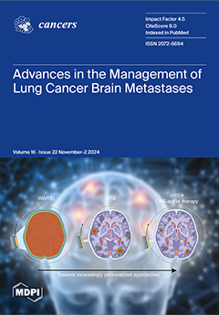
Cover Story (view full-size image): Brain metastases are common in lung cancer, posing distinct clinical challenges. Advances in radiation and systemic therapies underscore the need for an improved integration of multidisciplinary approaches to optimize both survival and symptomatic burden. Radiotherapy prioritizing durable control with minimal neurocognitive toxicity, as well as systemic agents with central nervous system activity, can be leveraged to deliver increasingly personalized regimens. Clinical trials are ongoing to elucidate how to integrate clinical features, tumor biology, and predictive biomarkers to effectively downstage intracranial disease and consolidate persistent metastases. Further work is needed to design risk-adapted strategies to balance intracranial efficacy with patient values while preserving neurocognitive. View this paper
  • Issues are regarded as officially published after their release is announced to the table of contents alert mailing list.
  • You may sign up for e-mail alerts to receive table of contents of newly released issues.
  • PDF is the official format for papers published in both, html and pdf forms. To view the papers in pdf format, click on the "PDF Full-text" link, and use the free Adobe Reader to open them.
Order results
Result details
Section
Select all
Export citation of selected articles as:
20 pages, 3568 KiB  
Article
Prealbumin Prognostic Score: A Novel Prognostic Indicator After Radical Gastrectomy in Patients with Gastric Cancer
by Ryota Matsui, Souya Nunobe, Motonari Ri, Rie Makuuchi, Tomoyuki Irino, Masaru Hayami, Manabu Ohashi and Takeshi Sano
Cancers 2024, 16(22), 3889; https://doi.org/10.3390/cancers16223889 - 20 Nov 2024
Viewed by 484
Abstract
Background: This study aimed to determine whether the prealbumin prognostic score (PPS), a novel indicator using prealbumin instead of albumin in the modified Glasgow Prognostic Score (mGPS), is a better predictive marker postoperatively in patients with gastric cancer. Methods: This retrospective [...] Read more.
Background: This study aimed to determine whether the prealbumin prognostic score (PPS), a novel indicator using prealbumin instead of albumin in the modified Glasgow Prognostic Score (mGPS), is a better predictive marker postoperatively in patients with gastric cancer. Methods: This retrospective study included consecutive patients who underwent radical gastrectomy for primary pStages I–III gastric cancer between 2006 and 2017. The cutoff values for preoperative prealbumin and C-reactive protein (CRP) were 22 mg/dL and 0.5 mg/dL, respectively. According to the prealbumin and CRP levels, a PPS of zero was defined as both being above the cutoff value, of one as either being below the cutoff value, and of two as both being below the cutoff value. Results: Of the 4663 patients, 3421 (73.4%) had a score of zero, 984 (21.1%) had a score of one, and 258 (5.5%) had a score of two. The higher the PPS, the poorer the overall survival [OS] (p < 0.001). When comparing OS by the PPS in patients with an mGPS of zero, a PPS of one indicated poorer OS than a PPS of zero (p < 0.001). In the multivariate analysis, PPSs of one (hazard ratio [HR]: 1.603; 95% confidence interval [CI]: 1.378–1.866; p < 0.001) and two (HR: 1.322; 95% CI: 1.055–1.656; p = 0.015) were independent poor prognostic factors for OS. Conclusions: The PPS, which is based on a combination of prealbumin and CRP levels, can identify a wider range of patients with poor OS than mGPS in patients with gastric cancer after gastrectomy. Full article
Show Figures

Figure 1

17 pages, 2237 KiB  
Article
GPX4 and FSP1 Expression in Lung Adenocarcinoma: Prognostic Implications and Ferroptosis-Based Therapeutic Strategies
by Hirotomo Takahara, Takumi Kanazawa, Haruna Oshita, Yoshinobu Tomita, Yuri Hananoi, Sachiko Ishibashi, Masumi Ikeda, Asuka Furukawa, Mayumi Kinoshita, Kurara Yamamoto, Yuki Kato, Hironori Ishibashi, Kenichi Okubo, Morito Kurata, Masanobu Kitagawa, Kenichi Ohashi and Kouhei Yamamoto
Cancers 2024, 16(22), 3888; https://doi.org/10.3390/cancers16223888 - 20 Nov 2024
Viewed by 492
Abstract
Background: Primary lung cancer is among the cancers with the poorest prognosis, having the highest mortality rate among men and the second highest among women in Japan. While surgery is the primary treatment, advanced stages often require pharmacotherapy. Recently, ferroptosis, an iron-dependent form [...] Read more.
Background: Primary lung cancer is among the cancers with the poorest prognosis, having the highest mortality rate among men and the second highest among women in Japan. While surgery is the primary treatment, advanced stages often require pharmacotherapy. Recently, ferroptosis, an iron-dependent form of cell death caused by lipid peroxidation, has gained attention as a potential therapeutic strategy. This study investigated the prognostic impact of lipid peroxidation marker and regulators involved in ferroptosis in lung adenocarcinoma. Methods: We analyzed 207 patients who underwent resection surgery for lung adenocarcinoma at Tokyo Medical and Dental University Hospital. Immunohistochemistry was used to evaluate the expression levels of glutathione peroxidase 4 (GPX4), ferroptosis suppressor protein 1 (FSP1), and 4-hydroxy-2-nonenal (4-HNE). The association between these markers and clinicopathological factors was assessed, and in vitro experiments were conducted to examine the effects of these markers on cell death. Results: Low cytoplasmic accumulation of 4-HNE and low expression of GPX4 were associated with a worse prognosis, and low FSP1 expression was associated with unfavorable relapse-free survival. In vitro experiments demonstrated that 4-HNE inhibited cell proliferation, and combined inhibition of GPX4 and FSP1 induced ferroptosis. Conclusions: These findings suggest that lipid peroxidation markers and regulators can serve as prognostic biomarkers and therapeutic targets in lung adenocarcinoma. Full article
(This article belongs to the Special Issue The Role of Ferroptosis in Cancer)
Show Figures

Figure 1

24 pages, 2814 KiB  
Systematic Review
Head and Neck Squamous Cell Carcinoma with Distant Metastasis: A Systematic Review and Meta-Analysis
by Antonio Daloiso, Leonardo Franz, Tiziana Mondello, Matteo Tisato, Michael Negrisolo, Paolo Zanatta, Cosimo de Filippis, Laura Astolfi and Gino Marioni
Cancers 2024, 16(22), 3887; https://doi.org/10.3390/cancers16223887 - 20 Nov 2024
Viewed by 439
Abstract
Background/Objectives: Distant metastasis (DM), though uncommon at initial presentation, significantly worsens the prognosis of head and neck squamous cell carcinomas (HNSCCs). This review aimed to investigate the occurrence rates, patterns, and implications of HNSCC DM. Methods: A systematic search was performed in Scopus, [...] Read more.
Background/Objectives: Distant metastasis (DM), though uncommon at initial presentation, significantly worsens the prognosis of head and neck squamous cell carcinomas (HNSCCs). This review aimed to investigate the occurrence rates, patterns, and implications of HNSCC DM. Methods: A systematic search was performed in Scopus, PubMed and Web of Science. Results: Out of 7576 identified titles, 35 studies were included, encompassing 28,193 patients. The pooled rate of DM was 10.01%, with significant heterogeneity existing among the studies (I2: 94.13%). The most common metastatic sites were the lungs, bones, and brain (58%, 15%, 4%, respectively). Treatment modalities varied: overall, 20.4% of patients received radiotherapy alone, 7% underwent chemotherapy, and 4.5% received surgical metastasectomies. Combined treatments accounted for 18.3% of patients. However, 41.3% of patients received no treatment for DM. The median overall survival (OS) after DM diagnosis was 10.1 months. Studies highlighted a 36.3% two-year survival rate for patients with oligo-metastases, compared to the 7.4% rate for those with multiple metastases. At the time of DM diagnosis, half of the studied population presented with locoregional failure. Conclusions: Advanced imaging techniques and emerging systemic therapies offer hope for improved DM detection and treatment. However, continuous research is essential to develop therapeutic strategies that can enhance survival and improve the quality of life for patients with DM. Full article
(This article belongs to the Section Systematic Review or Meta-Analysis in Cancer Research)
Show Figures

Figure 1

11 pages, 421 KiB  
Article
Cladribine-Based Therapy for Acute Myeloid Leukemia in Child, Adolescent, and Early Young Adult Patients: The MD Anderson Cancer Center Experience
by David McCall, Shaikha Alqahtani, Moriah Budak, Irtiza Sheikh, Aaron E. Fan, Ramya Ramakrishnan, Cesar Nunez, Michael Roth, Miriam B. Garcia, Amber Gibson, Naval Daver, Sofia Garces, Nicholas J. Short, Ghayas C. Issa, Farhad Ravandi, Courtney D. DiNardo, Guillermo Montalban Bravo, Guillermo Garcia-Manero, Branko Cuglievan and Tapan Kadia
Cancers 2024, 16(22), 3886; https://doi.org/10.3390/cancers16223886 - 20 Nov 2024
Viewed by 494
Abstract
Background: Cladribine-based combination chemotherapy has demonstrated promising efficacy in patients with relapsed/refractory adult acute myeloid leukemia (AML), prompting its increased utilization in the frontline; in pediatrics, it has been typically reserved for relapsed or refractory cases. While fludarabine has been used more commonly [...] Read more.
Background: Cladribine-based combination chemotherapy has demonstrated promising efficacy in patients with relapsed/refractory adult acute myeloid leukemia (AML), prompting its increased utilization in the frontline; in pediatrics, it has been typically reserved for relapsed or refractory cases. While fludarabine has been used more commonly as a purine analog in intensive regimens, cladribine may be an important alternative. Methods: We performed a retrospective study at MD Anderson Cancer Center from January 2015 to July 2023, which included patients aged 1–21 years with refractory or relapsed AML who received cladribine outside of a transplant conditioning. Results: A total of 30 patients were included, with a median age of 20 years (range, 2–21), and 55% being male. Similar to adults, cladribine exhibited good tolerability in pediatric and adolescent patients, with the most common adverse events being febrile neutropenia and myelosuppression. The most common grade 3 or 4 adverse events included febrile neutropenia (55%) and sepsis (26%), and there were no treatment discontinuations due to adverse events. Among patients with a median number of 2 (0–7) prior treatments, the overall response rate (CR/CRi) was 45%, and median event-free and overall survival were 6 and 12 months, respectively. Disease progression resulted in 4 deaths within 30 days of treatment. Conclusions: Cladribine was tolerated in pediatrics. No new safety signals were seen with cladribine regimens in this cohort. Response assessment is limited due to the heavily pretreated cohort. Further prospective studies are warranted on the safety and efficacy of cladribine and establish its role in pediatric, adolescent, and early young adult patients with AML. Full article
Show Figures

Figure 1

12 pages, 662 KiB  
Review
Clinical Advances and Challenges in Targeting KRAS Mutations in Non-Small Cell Lung Cancer
by Simone E. Dekker and Lei Deng
Cancers 2024, 16(22), 3885; https://doi.org/10.3390/cancers16223885 - 20 Nov 2024
Viewed by 573
Abstract
KRAS mutation is one of the most common oncogenic drivers in non-small cell lung cancer. Since its discovery about four decades ago, drug development targeting KRAS has been met with countless failures. Recently, KRAS G12C, a subvariant of KRAS, became the first druggable [...] Read more.
KRAS mutation is one of the most common oncogenic drivers in non-small cell lung cancer. Since its discovery about four decades ago, drug development targeting KRAS has been met with countless failures. Recently, KRAS G12C, a subvariant of KRAS, became the first druggable KRAS mutation. The efficacy of the first-generation KRAS inhibitor is modest, but with scientific advancement, KRAS G12C inhibitors with higher potency are on the horizon. Additionally, novel therapeutic approaches targeting other KRAS subvariants are also being explored in clinical trials with encouraging early data. We will review the clinical advances and challenges for patients with KRAS-mutated non-small cell lung cancer, with a focus on small molecule inhibitors. Full article
Show Figures

Figure 1

21 pages, 1245 KiB  
Perspective
Artificial Intelligence–Driven Computational Approaches in the Development of Anticancer Drugs
by Pankaj Garg, Gargi Singhal, Prakash Kulkarni, David Horne, Ravi Salgia and Sharad S. Singhal
Cancers 2024, 16(22), 3884; https://doi.org/10.3390/cancers16223884 - 20 Nov 2024
Viewed by 635
Abstract
The integration of AI has revolutionized cancer drug development, transforming the landscape of drug discovery through sophisticated computational techniques. AI-powered models and algorithms have enhanced computer-aided drug design (CADD), offering unprecedented precision in identifying potential anticancer compounds. Traditionally, cancer drug design has been [...] Read more.
The integration of AI has revolutionized cancer drug development, transforming the landscape of drug discovery through sophisticated computational techniques. AI-powered models and algorithms have enhanced computer-aided drug design (CADD), offering unprecedented precision in identifying potential anticancer compounds. Traditionally, cancer drug design has been a complex, resource-intensive process, but AI introduces new opportunities to accelerate discovery, reduce costs, and optimize efficiency. This manuscript delves into the transformative applications of AI-driven methodologies in predicting and developing anticancer drugs, critically evaluating their potential to reshape the future of cancer therapeutics while addressing their challenges and limitations. Full article
Show Figures

Figure 1

19 pages, 2768 KiB  
Systematic Review
Anthracycline-Induced Subclinical Right Ventricular Dysfunction in Breast Cancer Patients: A Systematic Review and Meta-Analysis
by Andrea Faggiano, Elisa Gherbesi, Chiara Giordano, Giacomo Gamberini, Marco Vicenzi, Cesare Cuspidi, Stefano Carugo, Carlo M. Cipolla and Daniela M. Cardinale
Cancers 2024, 16(22), 3883; https://doi.org/10.3390/cancers16223883 - 20 Nov 2024
Viewed by 489
Abstract
Aim: This meta-analysis aims to evaluate the impact of anthracycline chemotherapy on subclinical right ventricular (RV) dysfunction in breast cancer patients, using traditional echocardiographic parameters and strain-based measures, such as the RV global longitudinal strain (RV GLS) and the RV free-wall longitudinal strain [...] Read more.
Aim: This meta-analysis aims to evaluate the impact of anthracycline chemotherapy on subclinical right ventricular (RV) dysfunction in breast cancer patients, using traditional echocardiographic parameters and strain-based measures, such as the RV global longitudinal strain (RV GLS) and the RV free-wall longitudinal strain (RV FWLS). Methods and Results: A systematic search was conducted according to PRISMA guidelines, including 15 studies with a total of 1148 breast cancer patients undergoing anthracycline chemotherapy. The primary outcome was the evaluation of changes in RV GLS and RV FWLS pre- and post-chemotherapy. Secondary outcomes included changes in traditional echocardiographic parameters: TAPSE, FAC, and TDI S’. Meta-analysis revealed significant declines in RV function post-chemotherapy across all parameters. RV GLS decreased from 23.99% to 20.35% (SMD: −0.259, p < 0.0001), and RV FWLS from 24.92% to 21.56% (SMD: −0.269, p < 0.0001). Traditional parameters like TAPSE, FAC, and TDI S’ also showed reductions, but these were less consistent across studies. A meta-regression analysis showed no significant relationship between post-chemotherapy left ventricular ejection fraction (LVEF) and the changes in RV GLS and RV FWLS, suggesting that RV dysfunction may not be solely a consequence of LV impairment. Conclusions: Anthracycline chemotherapy induces subclinical RV dysfunction in breast cancer patients. RV strain analysis, especially 3D strain, shows greater sensitivity in detecting early dysfunction. However, further research is needed to clarify the clinical significance and prognostic value of these findings, as well as the role of routine RV strain analysis in guiding early interventions. Full article
(This article belongs to the Special Issue Clinical Research and Progress in the Treatment of Breast Cancer)
Show Figures

Figure 1

8 pages, 1216 KiB  
Communication
Adverse Events as a Function of Biological Sex in a Multicenter Clinical Trial of Melanoma Vaccines
by Catherine E. Lyons, Ruyun Jin, Aaron D. Smith, Hong Zhu and Craig L. Slingluff, Jr.
Cancers 2024, 16(22), 3882; https://doi.org/10.3390/cancers16223882 - 20 Nov 2024
Viewed by 477
Abstract
Background/Objective: Biological females experience more autoimmune disease than males and more treatment-related adverse events (TRAEs) after immune checkpoint blockade therapy. However, little is known about sex-related differences in TRAEs after cancer vaccines. Methods: The Mel44 clinical trial (NCT00118274) enrolled 167 eligible patients [...] Read more.
Background/Objective: Biological females experience more autoimmune disease than males and more treatment-related adverse events (TRAEs) after immune checkpoint blockade therapy. However, little is known about sex-related differences in TRAEs after cancer vaccines. Methods: The Mel44 clinical trial (NCT00118274) enrolled 167 eligible patients with high-risk melanoma to treatment with either of two melanoma multipeptide vaccines. We hypothesized that females would experience higher rates and grades of TRAEs. TRAE rates and grades were compared between sexes, with adjustment for multiple comparisons, and with mixed-effects models. Results: Multiple sex-related differences in TRAE rate and grade were observed in unadjusted comparisons, but only hyperglycemia and hypopigmentation were significantly higher-grade by sex after correcting for multiple comparisons: they were increased in males. In mixed-effect models, vaccination strategy, but not patient sex, was independently associated with TRAE rates and grades. Conclusions: These data do not support our hypothesis that TRAEs would be increased in females. Vaccine safety was supported for both males and females. Full article
(This article belongs to the Collection Oncology: State-of-the-Art Research in the USA)
Show Figures

Figure 1

11 pages, 820 KiB  
Review
Kill Two Birds with One Stone? The Effect of Helicobacter pylori Eradication in Decreased Prevalence of Gastric Cancer and Colorectal Cancer
by Yang-Che Kuo, Hung-Ju Ko, Lo-Yip Yu, Shou-Chuan Shih, Horng-Yuan Wang, Ying-Chun Lin and Kuang-Chun Hu
Cancers 2024, 16(22), 3881; https://doi.org/10.3390/cancers16223881 - 20 Nov 2024
Viewed by 578
Abstract
The connection between microbial infections and tumor formation is notably exemplified by Helicobacter pylori (H. pylori) and its association with gastric cancer (GC) and colorectal cancer (CRC). While early studies hinted at a link between H. pylori and colorectal neoplasms, comprehensive retrospective cohort [...] Read more.
The connection between microbial infections and tumor formation is notably exemplified by Helicobacter pylori (H. pylori) and its association with gastric cancer (GC) and colorectal cancer (CRC). While early studies hinted at a link between H. pylori and colorectal neoplasms, comprehensive retrospective cohort studies were lacking. Recent research indicates that individuals treated for H. pylori infection experience a significant reduction in both CRC incidence and mortality, suggesting a potential role of this infection in malignancy development. Globally, H. pylori prevalence varies, with higher rates in developing countries (80–90%) compared to developed nations (20–50%). This infection is linked to chronic gastritis, peptic ulcers, and GC, highlighting the importance of understanding its epidemiology for public health interventions. H. pylori significantly increases the risk of non-cardia GC. Some meta-analyses have shown a 1.49-fold increased risk for colorectal adenomas and a 1.70-fold increase for CRC in infected individuals. Additionally, H. pylori eradication may lower the CRC risk, although the relationship is still being debated. Although eradication therapy shows promise in reducing GC incidence, concerns about antibiotic resistance pose treatment challenges. The role of H. pylori in colorectal tumors remains contentious, with some studies indicating an increased risk of colorectal adenoma, while others find minimal association. Future research should investigate the causal mechanisms between H. pylori infection and colorectal neoplasia, including factors like diabetes, to better understand its role in tumor formation and support widespread eradication efforts to prevent both gastric and colorectal cancers. Full article
Show Figures

Figure 1

12 pages, 654 KiB  
Article
Sequential Use of Sorafenib and Regorafenib in Hepatocellular Cancer Recurrence After Liver Transplantation: Treatment Strategies and Outcomes
by Mehmet Fatih Ozbay, Hakan Harputluoglu, Mustafa Karaca, Omer Tekin, Mehmet Ali Nahit Şendur, Muhammed Ali Kaplan, Berksoy Sahin, Caglayan Geredeli, Fatih Teker, Deniz Tural, Sezer Saglam, Timuçin Çil, Ahmet Bilici, Cihan Erol, Ziya Kalkan, Ertugrul Bayram, Oguzhan Selvi, İlkay Gültürk, Sema Sezgin Göksu and Ali Murat Tatlı
Cancers 2024, 16(22), 3880; https://doi.org/10.3390/cancers16223880 - 20 Nov 2024
Viewed by 562
Abstract
Background and Aims: During liver transplantation, hepatocellular carcinoma (HCC) recurrence remains a critical challenge for patient survival. Targeted therapies, such as sorafenib and regorafenib, have been utilized to manage relapsed HCC in this unique setting. This study aimed to assess the efficacy of [...] Read more.
Background and Aims: During liver transplantation, hepatocellular carcinoma (HCC) recurrence remains a critical challenge for patient survival. Targeted therapies, such as sorafenib and regorafenib, have been utilized to manage relapsed HCC in this unique setting. This study aimed to assess the efficacy of Sorafenib and Regorafenib in patients with HCC who experienced recurrence after liver transplantation. We focused on survival outcomes, treatment responses, and the management of side effects in this patient group. Methods: We conducted a retrospective analysis of 73 patients who experienced HCC recurrence post-liver transplantation between 2012 and 2022 across 11 oncology centers in Turkey. Patients were categorized according to Child–Pugh classification and treated with sorafenib as first-line therapy and Regorafenib in case of progression. Survival rates were analyzed using the Kaplan–Meier method, and risk factors were evaluated using Cox regression analysis. Results: Of the 73 patients included in the study, 62 were male (84.9%), and 11 were female (15.1%), with a mean age of 61.5 ± 10.9 years. All patients received sorafenib as first-line treatment. Among patients who experienced progression with sorafenib or discontinued treatment due to toxicity, 45.2% (n = 33) continued treatment with regorafenib. The median progression-free survival (PFS1) time with sorafenib was 5.6 months, and the one-year survival rate was 24.3%. The median progression-free survival (PFS2) time with regorafenib, which was administered as second-line treatment, was also calculated as 5.9 months. Overall survival (OS) duration was determined as 35.9 months. The most common side effects associated with both drugs included fatigue, hand and foot syndrome, and hypertension. Significantly better survival outcomes were shown in the Child–Pugh A group compared to other patients. Conclusions: These results suggest that Sorafenib and Regorafenib treatments offer a survival advantage in patients with relapsed HCC post-transplantation. However, individualized treatment strategies and close follow-up are crucial for optimizing outcomes. Further studies are needed to refine therapeutic protocols and enhance the care of this specific patient group. Full article
Show Figures

Figure 1

28 pages, 1152 KiB  
Article
Lung and Colon Cancer Detection Using a Deep AI Model
by Nazmul Shahadat, Ritika Lama and Anna Nguyen
Cancers 2024, 16(22), 3879; https://doi.org/10.3390/cancers16223879 - 20 Nov 2024
Viewed by 488
Abstract
Lung and colon cancers are among the leading causes of cancer-related mortality worldwide. Early and accurate detection of these cancers is crucial for effective treatment and improved patient outcomes. False or incorrect detection is harmful. Accurately detecting cancer in a patient’s tissue is [...] Read more.
Lung and colon cancers are among the leading causes of cancer-related mortality worldwide. Early and accurate detection of these cancers is crucial for effective treatment and improved patient outcomes. False or incorrect detection is harmful. Accurately detecting cancer in a patient’s tissue is crucial to their effective treatment. While analyzing tissue samples is complicated and time-consuming, deep learning techniques have made it possible to complete this process more efficiently and accurately. As a result, researchers can study more patients in a shorter amount of time and at a lower cost. Much research has been conducted to investigate deep learning models that require great computational ability and resources. However, none of these have had a 100% accurate detection rate for these life-threatening malignancies. Misclassified or falsely detecting cancer can have very harmful consequences. This research proposes a new lightweight, parameter-efficient, and mobile-embedded deep learning model based on a 1D convolutional neural network with squeeze-and-excitation layers for efficient lung and colon cancer detection. This proposed model diagnoses and classifies lung squamous cell carcinomas and adenocarcinoma of the lung and colon from digital pathology images. Extensive experiment demonstrates that our proposed model achieves 100% accuracy for detecting lung, colon, and lung and colon cancers from the histopathological (LC25000) lung and colon datasets, which is considered the best accuracy for around 0.35 million trainable parameters and around 6.4 million flops. Compared with the existing results, our proposed architecture shows state-of-the-art performance in lung, colon, and lung and colon cancer detection. Full article
(This article belongs to the Collection Oncology: State-of-the-Art Research in the USA)
Show Figures

Figure 1

13 pages, 845 KiB  
Article
Patient Outcomes in Resected Combined Hepatocellular Cholangiocarcinoma (cHCC-ICC) and Intrahepatic Cholangiocarcinoma: A Single Center Study
by Rick Y. Lin, Doga Kahramangil, Muhammet Ozer, Thomas J. George, Ibrahim Nassour, Steven J. Hughes, Ali Zarrinpar and Ilyas Sahin
Cancers 2024, 16(22), 3878; https://doi.org/10.3390/cancers16223878 - 20 Nov 2024
Viewed by 456
Abstract
Background/Objectives: Combined hepatocellular cholangiocarcinoma (cHCC-ICC) is a rare malignancy that involves a combination of features of hepatocellular carcinoma and intrahepatic cholangiocarcinoma (ICC) and exhibits a more aggressive clinical course; however, its risk factors and outcomes remain largely undefined. Methods: This study is a [...] Read more.
Background/Objectives: Combined hepatocellular cholangiocarcinoma (cHCC-ICC) is a rare malignancy that involves a combination of features of hepatocellular carcinoma and intrahepatic cholangiocarcinoma (ICC) and exhibits a more aggressive clinical course; however, its risk factors and outcomes remain largely undefined. Methods: This study is a single-center retrospective study of 82 patients diagnosed with ICC or cHCC-ICC who underwent surgical resection from June 2011 to January 2023. Our analysis included 70 patients with resected ICC and 12 with resected cHCC-ICC. Results: The overall survival (OS) for the entire cohort was 21.6 months, with a recurrence-free survival (RFS) of 11.8 months. The cHCC-ICC group had significantly higher levels of AST and ALT (AST median 206 U/L vs. 46 U/L; ALT median 165.5 U/L vs. 48 U/L; p = 0.012 and p = 0.013, respectively), whereas the ICC group had higher alkaline phosphatase (median 66 U/L vs. 104 U/L; p = 0.03). CA 19-9 values (76 U/mL vs. 22 U/mL; p = 0.02) were higher in the ICC group, while AFP values were higher in the cHCC-ICC group (7.3 ng/mL vs. 3.2 ng/mL; p = 0.0004). The cHCC-ICC group had a significantly higher rate of recurrence (83% vs. 47%, p = 0.028) with a significantly decreased RFS (4.7 months vs. 12.4 months; log-rank p = 0.007). In multivariate analysis, patients with resected ICC had a significantly reduced risk of recurrence by 73% compared to their counterparts (HR 0.27 [0.10–0.73], p = 0.01). Conclusions: cHCC-ICC is a rare entity that needs to be further studied to improve patient outcomes. Further studies are warranted and may suggest the need for more aggressive initial treatment strategies in patients diagnosed with cHCC-ICC. Full article
Show Figures

Figure 1

17 pages, 897 KiB  
Article
Association of HLA-G Expression, Its Genetic Variants and Related Neuro-Immunomodulation with Characteristics of Bladder Carcinoma
by Vladimira Durmanova, Iveta Mikolaskova, Eszter Zsemlye, Agata Ocenasova, Helena Bandzuchova, Magda Suchankova, Boris Kollarik, Patrik Palacka, Milan Zvarik, Maria Bucova and Luba Hunakova
Cancers 2024, 16(22), 3877; https://doi.org/10.3390/cancers16223877 - 20 Nov 2024
Viewed by 647
Abstract
Background: Human leukocyte antigen G (HLA-G) is an immune checkpoint molecule with immunosuppressive and anti-inflammatory activities. It belongs to class I non-classical major histocompatibility complex molecules and has been upregulated in various cancer types. In bladder cancer (BC) tumors, the association of HLA-G [...] Read more.
Background: Human leukocyte antigen G (HLA-G) is an immune checkpoint molecule with immunosuppressive and anti-inflammatory activities. It belongs to class I non-classical major histocompatibility complex molecules and has been upregulated in various cancer types. In bladder cancer (BC) tumors, the association of HLA-G with cancer progression has to be explained. Methods: A total of 89 BC patients and 74 control subjects were genotyped for the HLA-G 14 bp ins/del polymorphism. In urine cell samples, HLA-G mRNA expression was analyzed using real-time PCR. Soluble HLA-G (sHLA-G) serum levels were measured by ELISA. The associations between the HLA-G 14 bp ins/del polymorphism, HLA-G mRNA expression, and/or sHLA-G levels and selected variables including tumor grade, disease stage, body mass index, and heart rate variability (HRV) parameters were evaluated. Results: The protective HLA-G 14 bp ins/ins genotype under the recessive genetic model was associated with lower HLA-G mRNA expression in the BC group (p = 0.049). Significantly higher HLA-G mRNA expression was detected in patients with pT2 + pT3 as compared to those with pTa + pT1 stages (p = 0.0436). Furthermore, higher HLA-G mRNA expression was observed in high-grade muscle-infiltrating BC (MIBC) than in the low-grade non-MIBC group (p = 0.0365). Patients with a level of sHLA-G above 29 U/mL had shorter disease-free survival than patients with lower sHLA-G levels. Furthermore, the opposite HRV correlations with sHLA-G levels in BC patients as compared to controls probably reflect the different roles of HLA-G in health and cancer. Conclusions: Our results suggest the impact of the HLA-G 14 bp ins/del variant, HLA-G expression, and autonomic nervous system imbalance on advanced stages of BC. Full article
(This article belongs to the Topic Anti-Tumor Immune Responses 2.0)
Show Figures

Graphical abstract

17 pages, 1791 KiB  
Article
Molecular and In Silico Analysis of the CHEK2 Gene in Individuals with High Risk of Cancer Predisposition from Türkiye
by Ozkan Ozdemir, Brittany L. Bychkovsky, Busra Unal, Gizem Onder, Ufuk Amanvermez, Eylul Aydin, Berk Ergun, Ilayda Sahin, Merve Gokbayrak, Cansu Ugurtas, Merve Nur Koroglu, Berfin Cakir, Irem Kalay, Naci Cine, Ugur Ozbek, Huma Q. Rana, Ozden Hatirnaz Ng and Nihat Bugra Agaoglu
Cancers 2024, 16(22), 3876; https://doi.org/10.3390/cancers16223876 - 20 Nov 2024
Viewed by 649
Abstract
Background and Objectives: Checkpoint kinase 2 (CHEK2) is a tumor suppressor gene involved in DNA repair and cell cycle regulation. Pathogenic or likely pathogenic (P/LP) variants in CHEK2 are associated with increased cancer risk. Conversely, recent large cohort studies have [...] Read more.
Background and Objectives: Checkpoint kinase 2 (CHEK2) is a tumor suppressor gene involved in DNA repair and cell cycle regulation. Pathogenic or likely pathogenic (P/LP) variants in CHEK2 are associated with increased cancer risk. Conversely, recent large cohort studies have identified certain variants that, despite being classified as P/LP by in silico analysis, are considered low risk. Thus, the genotype–phenotype correlations of CHEK2 require a better understanding. In this study, we aimed to characterize germline CHEK2 variants from a group of individuals who applied to cancer genetic clinics in the Marmara Region of Türkiye. We also aimed to assess the phenotypic impacts of these variants by using a new score of statistically significant in silico predictors (SSIPs). Methods: We analyzed 1707 individuals with high risk cancer predisposition, focusing on germline CHEK2 variants, using SSIP scores and population-specific data. Results:CHEK2 variants appeared in approximately 8% of cases. The SSIP scores indicated that the missense mutation, p.Arg117Gly, significantly impairs DNA repair. Almost half of the variants had higher allele frequencies than the variants listed in the Genome Aggregation Database (gnomAD), and three variants had significantly higher frequencies compared to the variants listed on the Turkish Variome database (p.Thr476Met, p.Arg137Gln, c.592+3A>T), emphasizing the importance of population-specific data. Conclusions: This comprehensive analysis of CHEK2 variants in the Turkish population provides crucial insights for cancer geneticists and oncologists. Our findings will help to enhance the evaluation and management of cancer predisposition associated with CHEK2 in Türkiye and other regions that have significant Turkish populations. Full article
(This article belongs to the Section Molecular Cancer Biology)
Show Figures

Figure 1

18 pages, 2168 KiB  
Article
Low-Dose Eribulin Promotes NK Cell-Mediated Therapeutic Efficacy in Bladder Cancer
by Zaineb Hassouneh, Onika D. V. Noel, Niannian Ji, Michelle E. Kim, Jordan Svatek, Robert S. Svatek, April L. Risinger and Neelam Mukherjee
Cancers 2024, 16(22), 3875; https://doi.org/10.3390/cancers16223875 - 19 Nov 2024
Viewed by 564
Abstract
Despite its immunogenic nature, bladder cancer (BCa) responds sub-optimally to FDA-approved immunotherapy. Background/Objectives: We have previously shown that natural killer (NK) cells are major contributors to overall patient survival in BCa. In our efforts to identify clinically approved agents that enhance NK cell [...] Read more.
Despite its immunogenic nature, bladder cancer (BCa) responds sub-optimally to FDA-approved immunotherapy. Background/Objectives: We have previously shown that natural killer (NK) cells are major contributors to overall patient survival in BCa. In our efforts to identify clinically approved agents that enhance NK cell activation, we identified eribulin, a microtubule destabilizer primarily used in breast cancer. Ongoing clinical trials are investigating the potential integration of eribulin into the standard of care in BCa; however, the mechanistic rationale for these trials remains unclear. Methods: Here, we explore the effects of low-dose eribulin on direct NK cell activation in vitro, including on primary patient samples, and in vivo utilizing multiple murine models. Flow cytometry and RNA sequencing were employed to identify the mechanism of NK cell activation by eribulin, which was associated with increased migration and cytotoxicity of NK cells against BCa cells. Results: We found that localized eribulin instillation significantly reduces bladder tumor burden and improves survival in primary BCa in an NK cell-dependent manner. Importantly, eribulin promoted the shift of patient-derived intratumoral NK cells towards an anti-tumor CD49a+ CD103+ NK subset (ieILC1-like) while diminishing the dysfunctional NR4A2-expressing CD49a NK subset. Moreover, it decreased the overall expression of exhaustion markers on NK cells, a pattern replicated in our murine models. Conclusions: These findings are paradigm-shifting given that chemotherapy is traditionally considered immunosuppressive. Our study reveals the novel effect of low-dose eribulin chemotherapy in inhibiting bladder tumor growth by enhancing anti-tumor NK cell immunity, challenging previous assumptions and opening new therapeutic approaches to improve antitumor immunity. Full article
Show Figures

Graphical abstract

9 pages, 485 KiB  
Review
Adjuvant Radiation Therapy in Desmoplastic Melanoma: A Scoping Review
by Christina Setareh Sharafi, B. Ashleigh Guadagnolo, Kelly C. Nelson and Devarati Mitra
Cancers 2024, 16(22), 3874; https://doi.org/10.3390/cancers16223874 - 19 Nov 2024
Viewed by 377
Abstract
Desmoplastic melanoma (DM) is an uncommon subtype of cutaneous melanoma that presents distinct diagnostic and treatment challenges. This review aims to explore the role of adjuvant radiation therapy (RT) in managing DM. To evaluate this question, we reviewed relevant published reports on DM [...] Read more.
Desmoplastic melanoma (DM) is an uncommon subtype of cutaneous melanoma that presents distinct diagnostic and treatment challenges. This review aims to explore the role of adjuvant radiation therapy (RT) in managing DM. To evaluate this question, we reviewed relevant published reports on DM and its treatment and synthesized these findings. It was found that the clinical behavior of DM varies significantly based on its classification as either “pure” DM (pDM, ≥90% desmoplastic features) or mixed DM (mDM, ≤90% desmoplastic features). Patients with pDM have a uniquely high risk of local recurrence but a relatively lower likelihood of nodal disease. Recent studies question the necessity of sentinel lymph node biopsy for pDM patients while illustrating impressive response rates to immune checkpoint inhibition. Most data supporting adjuvant RT predate these changes in surgical management and systemic therapy, yet consistently demonstrate that adjuvant RT reduces the absolute risk of local recurrence by >50%, without significant long-term toxicity. Thus, the existing literature continues to support the conclusion that adjuvant RT effectively reduces the likelihood of local recurrence in pDM patients. Although evolving surgical and systemic therapies are reshaping treatment approaches, adjuvant RT should remain a standard of care. Full article
(This article belongs to the Special Issue Radiotherapy in Melanoma)
Show Figures

Figure 1

17 pages, 1015 KiB  
Review
The Role of eHsp90 in Extracellular Matrix Remodeling, Tumor Invasiveness, and Metastasis
by Pragya Singh and Daniel G. Jay
Cancers 2024, 16(22), 3873; https://doi.org/10.3390/cancers16223873 - 19 Nov 2024
Viewed by 450
Abstract
Identifying proteins that act in tumor invasiveness and metastasis remains a critical unmet need in our search for effective cancer therapy. Hsp90, an abundant intracellular chaperone protein, plays a key role in maintaining cell homeostasis, and its elevated activity is pivotal in cancer [...] Read more.
Identifying proteins that act in tumor invasiveness and metastasis remains a critical unmet need in our search for effective cancer therapy. Hsp90, an abundant intracellular chaperone protein, plays a key role in maintaining cell homeostasis, and its elevated activity is pivotal in cancer progression. Due to the reliance of cancer cells on Hsp90’s chaperone function to sustain tumor growth and spread, Hsp90 inhibitors have been the subject of numerous clinical trials over the past two decades. However, these efforts have largely been unsuccessful, primarily due to the cellular toxicity caused by pan-Hsp90 inhibitors at doses required for anticancer efficacy. Therefore, novel approaches to target Hsp90 are necessary. An identified subpopulation of Hsp90 located outside cells (eHsp90) may offer a promising alternative as a therapeutic target against cancer. Studies including our own have shown that eHsp90 is released specifically by cancer cells, and eHsp90 has unique interactors and functions extracellularly to promote tumor invasiveness, the initial step in metastasis. Inhibition of eHsp90 has been shown to suppress metastasis in animal models, indicating its therapeutic potential, although the underlying mechanisms remain incompletely understood. Cancer cells modulate the tumor microenvironment (TME) during the invasion, especially the ECM proteins and the state of the ECM is a strong predictor of invasive and metastatic cancer. Given that most of the known eHsp90 clients are ECM proteins or are proteins involved in ECM modulation, ECM remodelling could be the key mechanism through which eHsp90 enhances invasiveness. This review will focus on ECM modulation by eHsp90 as a driver of cancer invasion and metastasis. We will also discuss the potency of inhibiting eHsp90 in inhibiting invasion and metastatic spread in preclinical models and the using circulating Hsp90 patient samples as a biomarker of cancer invasion and metastasis. Full article
(This article belongs to the Special Issue Extracellular Matrix Proteins in Cancer)
Show Figures

Graphical abstract

14 pages, 2426 KiB  
Article
Dose-Reduced FLA-IDA in Combination with Venetoclax Is an Effective and Safe Salvage Therapy in Relapsed and Refractory Acute Myeloid Leukemia (R/R AML)
by Martin Schönrock, Piet Sonnemann, Nina Michalowski, Michael Heuser, Felicitas Thol, Francis Ayuketang Ayuk, Christine Wolschke, Evgeny Klyuchnikov, Carsten Bokemeyer, Walter Fiedler and Sophia Cichutek
Cancers 2024, 16(22), 3872; https://doi.org/10.3390/cancers16223872 - 19 Nov 2024
Viewed by 564
Abstract
Background: Despite the development of targeted therapies in first-line AML, complete remissions (CR) cannot be achieved in 30–40%, and relapse rates remain high. In R/R AML the intensive treatment regimen of fludarabine, cytarabine, idarubicin combined with venetoclax (FLA-VIDA) showed improved remission rates compared [...] Read more.
Background: Despite the development of targeted therapies in first-line AML, complete remissions (CR) cannot be achieved in 30–40%, and relapse rates remain high. In R/R AML the intensive treatment regimen of fludarabine, cytarabine, idarubicin combined with venetoclax (FLA-VIDA) showed improved remission rates compared to FLA-IDA. In this retrospective single-center analysis, we investigated the efficacy and safety of dose-reduced FLA-IDA with and without venetoclax to minimize the risk of infectious complications and excessive myelosuppression; Methods: Between 2011 and 2023, 89 R/R AML patients were treated with dose-reduced FLA-IDA (fludarabine 30 mg/m2 day 1–4, cytarabine 2000 mg/m2 day 1–4, idarubicin 10 mg/m2 day 1 + 4). From 2019 onwards, venetoclax was added (day 1 100 mg, day 2 200 mg, day 3–14 400 mg); Results: Significantly improved response rates were observed with 60.0% vs. 38.8% CR/CRi (p = 0.0297) and 74.5% vs. 47.3% (p = 0.032) CR/CRi/MLFS for FLA-VIDA vs. FLA-IDA. Further, with FLA-VIDA significantly improved event-free survival (EFS) was observed (p = 0.026). Overall survival (OS) was similar in FLA-VIDA and FLA-IDA treated patients. The most common treatment-related toxicities were hematological adverse events, but they were comparable between groups. The time to neutrophil and platelet recovery were similar in responding patients treated with FLA-VIDA vs. FLA-IDA; Conclusions: Dose-reduced FLA-VIDA significantly improved response rates without increases in toxicity, showing promise for an improved R/R AML treatment. Full article
(This article belongs to the Special Issue The Clinical Trials and Management of Acute Myeloid Leukemia)
Show Figures

Figure 1

13 pages, 2077 KiB  
Article
Prognostic Role of Inflammatory and Nutritional Biomarkers in Non-Small-Cell Lung Cancer Patients Treated with Immune Checkpoint Inhibitors Alone or in Combination with Chemotherapy as First-Line
by Antonello Veccia, Mariachiara Dipasquale, Stefania Kinspergher and Orazio Caffo
Cancers 2024, 16(22), 3871; https://doi.org/10.3390/cancers16223871 - 19 Nov 2024
Viewed by 478
Abstract
Introduction: In recent years, several inflammation-related factors and nutritional parameters have been evaluated to develop prognostic scores as potential biomarkers in non-small-cell lung cancer (NSCLC) patients receiving immune checkpoint inhibitors (ICIs). The aim of this study was to retrospectively investigate the prognostic role [...] Read more.
Introduction: In recent years, several inflammation-related factors and nutritional parameters have been evaluated to develop prognostic scores as potential biomarkers in non-small-cell lung cancer (NSCLC) patients receiving immune checkpoint inhibitors (ICIs). The aim of this study was to retrospectively investigate the prognostic role of the advanced lung cancer inflammation (ALI) index, lung immune prognostic index (LIPI), prognostic nutritional index (PNI) and systemic inflammation score (SIS) in metastatic NSCLC patients receiving ICI alone or in combination with chemotherapy. Methods and patients: We retrospectively included 191 patients with advanced NSCLC who received first-line ICI with or without chemotherapy from 2017 to 2024. The association between pretreatment ALI, LIPI, PNI, and SIS and overall survival (OS) was evaluated using the Kaplan–Meier method and Cox regression models. Results: After a median follow-up of 27.7 months, significantly longer OS was associated with an ALI score > 18 vs. ≤18 (18.0 vs. 7.3 months; p = 0.00111), LIPI score 0 vs. 1 and 2 [18.9 vs. 8.2 and 4.2 months; (p = 0.001)], PNI ≥ 45 vs. <45 (22.7 vs. 9.6 months; p = 0.002), and SIS score 0 vs. 1 and 2 (27.4 vs. 7.1 and 8.6 months, respectively; p < 0.001). The OS benefit was independent of treatment (ICI vs. ICI + chemotherapy). At multivariate analysis, pretreatment albumin was positively associated with OS, while ECOG PS 1 and liver metastases were negatively associated with OS. Conclusions: Inflammatory and nutritional biomarkers such as the ALI, LIPI, PNI, and SIS represent useful tools to prognosticate survival in metastatic lung cancer patients treated with ICI alone or in combination with chemotherapy as first-line. Full article
(This article belongs to the Special Issue Feature Papers in Section "Cancer Biomarkers" in 2023–2024)
Show Figures

Figure 1

17 pages, 1976 KiB  
Review
Precision Medicine for Metastatic Colorectal Cancer: Where Do We Stand?
by Patrick W. Underwood and Timothy M. Pawlik
Cancers 2024, 16(22), 3870; https://doi.org/10.3390/cancers16223870 - 19 Nov 2024
Viewed by 585
Abstract
Metastatic colorectal cancer is a leading cause of cancer-related death across the world. The treatment paradigm has shifted away from systemic chemotherapy alone to include targeted therapy and immunotherapy. The past two decades have been characterized by increased investigation into molecular profiling of [...] Read more.
Metastatic colorectal cancer is a leading cause of cancer-related death across the world. The treatment paradigm has shifted away from systemic chemotherapy alone to include targeted therapy and immunotherapy. The past two decades have been characterized by increased investigation into molecular profiling of colorectal cancer. These molecular profiles help physicians to better understand colorectal cancer biology among patients with metastatic disease. Additionally, improved data on genetic pathways allow for specific therapies to be targeted at the underlying molecular profile. Investigation of the EGFR, VEGF, HER2, and other pathways, as well as deficient mismatch repair, has led to the development of multiple targeted therapies that are now utilized in the National Comprehensive Cancer Network guidelines for colon and rectal cancer. While these new therapies have contributed to improved survival for metastatic colorectal cancer, long-term survival remains poor. Additional investigation to understand resistance to targeted therapy and development of new targeted therapy is necessary. New therapies are under development and are being tested in the preclinical and clinical settings. The aim of this review is to provide a comprehensive evaluation of molecular profiling, currently available therapies, and ongoing obstacles in the field of colorectal cancer. Full article
Show Figures

Figure 1

20 pages, 7201 KiB  
Review
A Pathological Assessment of the Microvasculature of Biliary Tract Neoplasms Referring to Pre-Existing Blood Vessels and Vessel Co-Option
by Yasuni Nakanuma, Zihan Li, Yasunori Sato, Motoko Sasaki, Kenichi Harada, Yuko Kakuda and Takashi Sugino
Cancers 2024, 16(22), 3869; https://doi.org/10.3390/cancers16223869 - 19 Nov 2024
Viewed by 398
Abstract
There are several types of microvasculature supplying neoplasms: “newly formed blood vessels” (neoangiogenesis), which are a component of the tumor microenvironment (TME) of invasive carcinoma with wound healing-like reaction; and “pre-existing blood vessels”, which are used as tumor-supplying vessels by neoplasms (co-option vessels) [...] Read more.
There are several types of microvasculature supplying neoplasms: “newly formed blood vessels” (neoangiogenesis), which are a component of the tumor microenvironment (TME) of invasive carcinoma with wound healing-like reaction; and “pre-existing blood vessels”, which are used as tumor-supplying vessels by neoplasms (co-option vessels) and are likely to develop in hypervascularized organs. We herein review the microvasculature of neoplasms of biliary tract with reference to pre-existing vessels and vessel co-options. In the hepatobiliary system, intrahepatic large and extrahepatic bile ducts (large bile ducts) and the gallbladder as well as hepatic lobules are highly vascularized regions. In large bile ducts, the biliary lining epithelia and underlining capillaries (peribiliary capillary plexus [PCP]) form the biliary epithelia–PCP alignment, whereas the hepatocyte–sinusoid alignment composes hepatic lobules. Cholangiocarcinoma (CCA) and gallbladder carcinoma (GBC) are the main biliary tract carcinomas. CCA is subdivided into distal (d/CCA), perihilar (pCCA), and intrahepatic (iCCA), and iCCA is subdivided into small duct type (SD-iCCA) and large duct type (LD-iCCA). High-grade biliary intraepithelial neoplasm (BilIN), intraductal papillary neoplasm of the bile duct (IPNB), pyloric gland adenoma (PGA), and intracholecystic papillary neoplasm (ICPN) have recently been proposed as the precursors of LD-iCCA, p/dCCA, and GBC. In the large bile ducts and gallbladder, all cases of high-grade BilIN and PGA, about half of IPNB, and one-third of ICPN with less-complicated structure were found to have hijacked the PCP as their supporting vessels (vessel co-option), while p/dCCA, LD-iCCA, and GBC were supplied by neo-angiogenetic vessels associated with fibrous stroma. The intraluminal components of the remaining cases of ICPN and IPNB with complicated structure presented sparse capillaries without fibrous stroma, a unique microvasculature different from that of co-option or neoangiogenesis. Regarding iCCA showing invasion into the hepatic lobules, some SD-iCCAs replaced hepatocytic cords and used pre-existing sinusoids as co-opted vessels. Visualization of pre-existing vessels could be a new pathological tool for the evaluation of malignant progression and of vascular supply in CCAs and its precursors. Full article
(This article belongs to the Collection Treatment of Hepatocellular Carcinoma and Cholangiocarcinoma)
Show Figures

Figure 1

17 pages, 31067 KiB  
Article
Feasibility of Non-Invasive Sentinel Lymph Node Identification in Early-Stage NSCLC Through Ultrasound Guided Intra-Tumoral Injection of 99mTc-Nanocolloid and Iodinated Contrast Agent During Navigation Bronchoscopy
by Desi K. M. ter Woerds, Roel L. J. Verhoeven, Erik H. J. G. Aarntzen and Erik H. F. M. van der Heijden
Cancers 2024, 16(22), 3868; https://doi.org/10.3390/cancers16223868 - 19 Nov 2024
Viewed by 629
Abstract
Background: As the first sentinel lymph nodes (SLN) in lung cancer are most likely to harbor metastasis, their non-invasive identification could have a significant role in future treatments. We investigated the feasibility of adding an SLN procedure to a diagnostic navigation bronchoscopy. [...] Read more.
Background: As the first sentinel lymph nodes (SLN) in lung cancer are most likely to harbor metastasis, their non-invasive identification could have a significant role in future treatments. We investigated the feasibility of adding an SLN procedure to a diagnostic navigation bronchoscopy. Methods: Thirty-one patients were included for injection of 99mTc-nanocolloid and an iodinated contrast agent intra-/peritumorally and assessment of tracer dissipation via SPECT and CBCT imaging. Injections were performed endobronchially using a multi-modal catheter (Pioneer Plus), combining radial ultrasound and an angulated retractable needle to place injections under fluoroscopy and real-time ultrasound. Results: The injection of an imaging tracer was feasible in all cases using the catheter. Ultrasound visualized 29/30 tumors, and tracer injection was performed in 100% of patients. An SLN was subsequently identified in 10 out of 31 cases (32.3%) via SPECT/CT imaging. Iodinated contrast agent injection under CBCT imaging prior to 99mTc nanocolloid injection visualized dissipation pathways and enabled needle relocation for subsequent 99mTc-nanocolloid injection. Conclusions: Performing imaging tracer injections with a multi-modal catheter provided safe and local depot placement immediately following diagnostic navigation bronchoscopy. SPECT/CT imaging using 99mTc-nanocolloid showed inconsistent results for SLN identification. Full article
(This article belongs to the Special Issue Clinical Applications of Ultrasound in Cancer Imaging and Treatment)
Show Figures

Figure 1

24 pages, 853 KiB  
Review
Cellular Therapies for Multiple Myeloma: Engineering Hope
by Sarah Vera-Cruz, Maria Jornet Culubret, Verena Konetzki, Miriam Alb, Sabrina R. Friedel, Michael Hudecek, Hermann Einsele, Sophia Danhof and Lukas Scheller
Cancers 2024, 16(22), 3867; https://doi.org/10.3390/cancers16223867 - 19 Nov 2024
Viewed by 755
Abstract
Multiple myeloma (MM) treatment remains challenging due to its relapsed/refractory disease course as well as intra- and inter-patient heterogeneity. Cellular immunotherapies, especially chimeric antigen receptor (CAR)-T cells targeting B cell maturation antigen (BCMA), mark a major breakthrough, achieving long-lasting remissions and instilling hope [...] Read more.
Multiple myeloma (MM) treatment remains challenging due to its relapsed/refractory disease course as well as intra- and inter-patient heterogeneity. Cellular immunotherapies, especially chimeric antigen receptor (CAR)-T cells targeting B cell maturation antigen (BCMA), mark a major breakthrough, achieving long-lasting remissions and instilling hope for a potential cure. While ongoing clinical trials are increasingly driving approved cellular products towards earlier lines of therapy, novel targets as well as advanced approaches employing natural killer (NK) cells or dendritic cell (DC) vaccines are currently under investigation. Treatment resistance, driven by tumor-intrinsic factors such as antigen escape and the intricate dynamics of the tumor microenvironment (TME), along with emerging side effects such as movement and neurocognitive treatment-emergent adverse events (MNTs), are the major limitations of approved cellular therapies. To improve efficacy and overcome resistance, cutting-edge research is exploring strategies to target the microenvironment as well as synergistic combinatorial approaches. Recent advances in CAR-T cell production involve shortened manufacturing protocols and “off-the-shelf” CAR-T cells, aiming at decreasing socioeconomic barriers and thereby increasing patient access to this potential lifesaving therapy. In this review, we provide an extensive overview of the evolving field of cellular therapies for MM, underlining the potential to achieve long-lasting responses. Full article
(This article belongs to the Special Issue Stem Cell Transplant and Cellular Therapies on Multiple Myeloma)
Show Figures

Figure 1

11 pages, 1756 KiB  
Article
Cervical CT Angiography: The Advantage of Ultra-High-Resolution CT Versus Conventional HRCT
by Junji Ito, Tsuneo Yamashiro, Hayato Tomita, Joichi Heianna, Sadayuki Murayama and Akihiro Nishie
Cancers 2024, 16(22), 3866; https://doi.org/10.3390/cancers16223866 - 19 Nov 2024
Viewed by 378
Abstract
Background/Objectives: Pre-treatment depiction of the cervical arteries is important for better intra-arterial infusion therapy of malignant head and neck tumors. There have not been any studies on the image quality of ultra-high-resolution computed tomography (U-HRCT) for cervical CT angiography (CTA). The aim of [...] Read more.
Background/Objectives: Pre-treatment depiction of the cervical arteries is important for better intra-arterial infusion therapy of malignant head and neck tumors. There have not been any studies on the image quality of ultra-high-resolution computed tomography (U-HRCT) for cervical CT angiography (CTA). The aim of this study is to evaluate the advantages of U-HRCT over conventional HRCT for cervical CTA; Methods: Forty-one patients underwent cervical CTA prior to selective intra-arterial infusion chemotherapy for malignant head and neck tumors. Twenty-two patients were scanned on conventional HRCT, while the remaining nineteen on U-HRCT. U-HRCT super-high-resolution (SHR) mode was used in 8 patients, while high-resolution (HR) mode was used in 11 patients. On CTA, the visibility of 18 branches from bilateral external carotid arteries was evaluated using a 5-point scale by three radiologists in consensus. Prior to the patient study, a head–neck CT phantom study regarding mock arterial density and its visibility was performed; Results: Regarding the patient study, the mean score of the SHR mode for visibility was significantly higher than that of conventional HRCT in 17 of 18 arteries (p < 0.05). The mean score of the HR mode for visibility was significantly higher than that of conventional HRCT in all arteries (p < 0.05). Regarding the phantom study, the maximum density of the SHR mode was significantly higher than that of conventional HRCT for mock proximal and peripheral arteries (p < 0.01). In addition, the visual score of the SHR mode for mock arteries was significantly higher than that of conventional HRCT (p < 0.05); Conclusions: U-HRCT provides higher image quality in terms of visualization of the arteries than conventional HRCT in cervical CTA. Full article
(This article belongs to the Section Methods and Technologies Development)
Show Figures

Figure 1

14 pages, 2192 KiB  
Systematic Review
Prostate-Specific Membrane Antigen (PSMA) PET/CT in the Detection and Diagnosis of Hepatocellular Carcinoma (HCC): A Systematic Review and Meta-Analysis
by Nicholas Hannah, Catherine Yu, Leya Nedumannil, James Haridy, Grace Kong, Alex Boussioutas and Siddharth Sood
Cancers 2024, 16(22), 3865; https://doi.org/10.3390/cancers16223865 - 19 Nov 2024
Viewed by 500
Abstract
Prostate-specific membrane antigen (PSMA) positron emission tomography (PET) is widely used in prostate cancer. Recent studies indicate hepatocellular carcinoma (HCC) demonstrates PSMA PET uptake. The diagnostic accuracy of PSMA PET for HCC is not known. We conducted a systematic review and meta-analysis of [...] Read more.
Prostate-specific membrane antigen (PSMA) positron emission tomography (PET) is widely used in prostate cancer. Recent studies indicate hepatocellular carcinoma (HCC) demonstrates PSMA PET uptake. The diagnostic accuracy of PSMA PET for HCC is not known. We conducted a systematic review and meta-analysis of studies assessing 68Ga-PSMA-11 in HCC. Nine studies were included, with 196 patients and a total of 491 HCC lesions. Per-patient analysis yielded a pooled sensitivity of 89.8% (95% CI 78.5–95.5). Specificity was poorly reported, with insufficient data. When per-lesion level analysis was performed on seven studies, the pooled sensitivity was 94.5% (95% CI 82.9–98.4), and specificity was again poorly reported with insufficient data. Among the three studies with adequate data for full per-lesion meta-analysis, 115 lesions in 41 patients demonstrated sensitivity of 97.1% (95% CI 87.8–99.4), while specificity was 42.2% (95% CI 0.3–99.4). Two studies provided sufficient data for meta-analysis on a per-patient level (n = 50 patients), demonstrating a sensitivity of 92.5% (95% CI 64.0–98.9) and specificity of 72.4% (95% CI 1.3–99.8). PSMA PET demonstrates a high sensitivity for HCC and shows promise as an imaging modality for diagnosis and staging of HCC. However, the existing literature does not provide enough data to confidently evaluate its specificity and, therefore, accuracy. Further prospective studies are necessary, with a focus on the accurate reporting of benign lesions and inclusion of patients with an intermediate probability of HCC. Full article
(This article belongs to the Special Issue New Approaches in Radiotherapy for Cancer)
Show Figures

Figure 1

2 pages, 157 KiB  
Editorial
Editorial for Special Issue “Brain Tumor Microenvironment”
by Gianluca Trevisi and Annunziato Mangiola
Cancers 2024, 16(22), 3864; https://doi.org/10.3390/cancers16223864 - 18 Nov 2024
Viewed by 354
Abstract
The tumor microenvironment (TME) is a complex interplay of cells, extracellular matrix, and signaling molecules that significantly influences tumor growth, invasion, and resistance to therapy [...] Full article
(This article belongs to the Special Issue Brain Tumor Microenvironment)
19 pages, 4763 KiB  
Article
Altered Mechanobiology of PDAC Cells with Acquired Chemoresistance to Gemcitabine and Paclitaxel
by Alessandro Gregori, Cecilia Bergonzini, Mjriam Capula, Rick Rodrigues de Mercado, Erik H. J. Danen, Elisa Giovannetti and Thomas Schmidt
Cancers 2024, 16(22), 3863; https://doi.org/10.3390/cancers16223863 - 18 Nov 2024
Viewed by 616
Abstract
Background: Pancreatic ductal adenocarcinoma acquired resistance to chemotherapy poses a major limitation to patient survival. Despite understanding some biological mechanisms of chemoresistance, much about those mechanisms remains to be uncovered. Mechanobiology, which studies the physical properties of cells, holds promise as a [...] Read more.
Background: Pancreatic ductal adenocarcinoma acquired resistance to chemotherapy poses a major limitation to patient survival. Despite understanding some biological mechanisms of chemoresistance, much about those mechanisms remains to be uncovered. Mechanobiology, which studies the physical properties of cells, holds promise as a potential target for addressing the challenges of chemoresistance in PDAC. Therefore, we, here in an initial step, assessed the altered mechanobiology of PDAC cells with acquired chemoresistance to gemcitabine and paclitaxel. Methods: Five PDAC cell lines and six stably resistant subclones were assessed for force generation on elastic micropillar arrays. Those measurements of mechanical phenotype were complemented by single-cell motility and invasion in 3D collagen-based matrix assays. Further, the nuclear translocation of Yes-associated protein (YAP), as a measure of active mechanical status, was compared, and biomarkers of the epithelial-to-mesenchymal transition (EMT) were evaluated using RT-qPCR. Results: The PDAC cells with acquired chemoresistance exert higher traction forces than their parental/wild-type (WT) cells. In 2D, single-cell motility was altered for all the chemoresistant cells, with a cell-type specific pattern. In 3D, the spheroids of the chemoresistant PDAC cells were able to invade the matrix and remodel collagen more than their WT clones. However, YAP nuclear translocation and EMT were not significantly altered in relation to changes in other physical parameters. Conclusions: This is the first study to investigate and report on the altered mechanobiological features of PDAC cells that have acquired chemoresistance. A better understanding of mechanical features could help in identifying future targets to overcome chemoresistance in PDAC. Full article
(This article belongs to the Section Cancer Metastasis)
Show Figures

Figure 1

13 pages, 908 KiB  
Systematic Review
Response to Bridging Therapy as a Prognostic Indicator of Post-Transplantation Hepatocellular Carcinoma Recurrence and Survival: A Systematic Review
by Paweł Topolewski, Dariusz Łaski, Martyna Łukasiewicz, Piotr Domagała, Roeland F. de Wilde and Wojciech G. Polak
Cancers 2024, 16(22), 3862; https://doi.org/10.3390/cancers16223862 - 18 Nov 2024
Viewed by 510
Abstract
Liver transplantation (LT) is one of the most effective treatments for hepatocellular carcinoma (HCC) in cirrhotic livers. Neoadjuvant bridging treatment in patients qualifying and listed for LT is advised but is still debatable owing to the low level of evidence. The aim of [...] Read more.
Liver transplantation (LT) is one of the most effective treatments for hepatocellular carcinoma (HCC) in cirrhotic livers. Neoadjuvant bridging treatment in patients qualifying and listed for LT is advised but is still debatable owing to the low level of evidence. The aim of this study was to perform a systematic review to assess the prognostic value of bridging therapy, in terms of radiological and histopathological examination outcomes, for survival after LT. The systematic review was performed according to the PRISMA 2020 guidelines. The MEDLINE and Web of Science databases were searched. In total, five studies were included. An evaluation with the ROBINS-I resulted in studies classified as the following: moderate risk of bias (n = 1) and serious risk of bias (n = 4). The results of the analysis indicated that favorable LT outcomes were most common with complete response or partial radiological response. Poor radiological response or progressive disease during bridging treatment was generally associated with worse overall LT survival. There were not enough data to support the use of this approach to achieve a complete pathologic response. Radiological, pathological, histological, cellular, and molecular tumor features should be included in future LT qualification models. Full article
(This article belongs to the Collection Primary Liver Cancer)
Show Figures

Figure 1

15 pages, 2622 KiB  
Article
Small Leucine Zipper Protein Regulates Glucose Metabolism of Prostate Cancer Cells via Induction of Phosphoglycerate Kinase 1
by Sila Han, Sungyeon Park, Suhyun Kim, Sujin Kwon and Jesang Ko
Cancers 2024, 16(22), 3861; https://doi.org/10.3390/cancers16223861 - 18 Nov 2024
Viewed by 588
Abstract
Background: Cancer cells exhibit altered metabolism whereby glucose is preferentially utilized to produce lactate through aerobic glycolysis. The increase in lactate production creates an acidic microenvironment that supports tumor progression and metastasis. Human small leucine zipper protein (sLZIP) is involved in the transcriptional [...] Read more.
Background: Cancer cells exhibit altered metabolism whereby glucose is preferentially utilized to produce lactate through aerobic glycolysis. The increase in lactate production creates an acidic microenvironment that supports tumor progression and metastasis. Human small leucine zipper protein (sLZIP) is involved in the transcriptional regulation of genes related to migration and invasion of prostate cancer. However, the role of sLZIP in modulating glucose metabolism in prostate cancer remains unknown. This study investigates whether sLZIP regulates the transcription of glycolysis-related genes to promote metabolic reprogramming in prostate cancer. Methods: Depletion of sLZIP resulted in the downregulation of several glycolytic genes, including glucose transporter 1, phosphofructokinase liver type, phosphoglycerate kinase 1 (PGK1), and lactate dehydrogenase. Among these, only PGK1 showed a prominent dose-dependent decrease in mRNA and protein expression after sLZIP silencing. Results: Mechanistically, increasing or decreasing sLZIP affected the promoter activity of PGK1 in a similar manner. Moreover, the absence of sLZIP attenuated the maximum glycolytic rate in prostate cancer cells. These results were further supported by a reduction in lactate secretion, glucose uptake, and ATP production in sLZIP-knockout prostate cancer cells. sLZIP deficiency hindered cancer growth, as demonstrated by proliferation assays. However, overexpression of PGK1 in sLZIP knockout cells resulted in recovery of aerobic glycolysis. Results of the xenograft experiment revealed that mice injected with sLZIP knockout cells exhibited a decrease in tumor mass compared to those injected with control cells. Conclusion: These findings suggest that sLZIP contributes to the metabolic reprogramming of prostate cancer cells via the transcriptional regulation of PGK1. Full article
(This article belongs to the Section Molecular Cancer Biology)
Show Figures

Figure 1

23 pages, 4959 KiB  
Article
Brief Magnetic Field Exposure Stimulates Doxorubicin Uptake into Breast Cancer Cells in Association with TRPC1 Expression: A Precision Oncology Methodology to Enhance Chemotherapeutic Outcome
by Viresh Krishnan Sukumar, Yee Kit Tai, Ching Wan Chan, Jan Nikolas Iversen, Kwan Yu Wu, Charlene Hui Hua Fong, Joline Si Jing Lim and Alfredo Franco-Obregón
Cancers 2024, 16(22), 3860; https://doi.org/10.3390/cancers16223860 - 18 Nov 2024
Viewed by 1021
Abstract
Background/Objectives: Doxorubicin (DOX) is commonly used as a chemotherapeutic agent for the treatment of breast cancer. Nonetheless, its systemic delivery via intravenous injection and toxicity towards healthy tissues commonly result in a broad range of detrimental side effects. Breast cancer severity was [...] Read more.
Background/Objectives: Doxorubicin (DOX) is commonly used as a chemotherapeutic agent for the treatment of breast cancer. Nonetheless, its systemic delivery via intravenous injection and toxicity towards healthy tissues commonly result in a broad range of detrimental side effects. Breast cancer severity was previously shown to be correlated with TRPC1 channel expression that conferred upon it enhanced vulnerability to pulsed electromagnetic field (PEMF) therapy. PEMF therapy was also previously shown to enhance breast cancer cell vulnerability to DOX in vitro and in vivo that correlated with TRPC1 expression and mitochondrial respiratory rates. Methods: DOX uptake was assessed by measuring its innate autofluorescence within murine 4T1 or human MCF7 breast cancer cells following magnetic exposure. Cellular vulnerability to doxorubicin uptake was assessed by monitoring mitochondrial activity and cellular DNA content. Results: Here, we demonstrate that 10 min of PEMF exposure could augment DOX uptake into 4T1 and MCF7 breast cancer cells. DOX uptake could be increased by TRPC1 overexpression, whereas inhibiting the activity of TRPC1 channels with SKF-96356 or genetic knockdown, precluded DOX uptake. PEMF exposure enhances DOX-mediated killing of breast cancer cells, reducing the IC50 value of DOX by half, whereas muscle cells, representative of collateral tissues, were less sensitive to PEMF-enhanced DOX-mediated cytotoxicity. Vesicular loading of DOX correlated with TRPC1 expression. Conclusions: This study presents a novel TRPC1-mediated mechanism through which PEMF therapy may enhance DOX cytotoxicity in breast cancer cells, paving the way for the development of localized non-invasive PEMF platforms to improve cancer outcomes with lower systemic levels of DOX. Full article
(This article belongs to the Special Issue Advances and Novel Multidisciplinary Strategies for Breast Cancer)
Show Figures

Graphical abstract

Previous Issue
Back to TopTop