


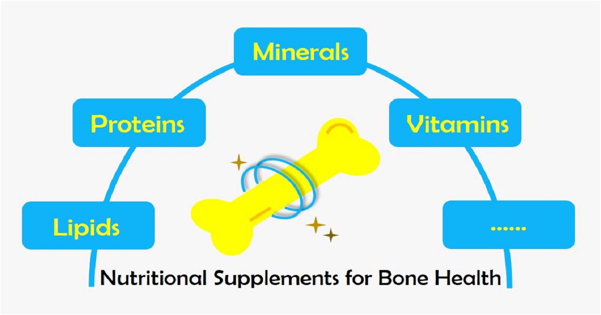
Nutritional Supplements for Bone Health
A special issue of Nutrients (ISSN 2072-6643). This special issue belongs to the section "Nutrition and Public Health".
Deadline for manuscript submissions: closed (5 October 2024) | Viewed by 45941
Special Issue Editor

Interests: bone and cartilage integrity; osteoarthropathy; Kashin–Beck disease; mycotoxin
Special Issue Information
Dear Colleagues,
Bone health is of great importance to our daily activities and is maintained using multiple nutrients under a complicated regulatory system. Nutritional supplements are developed and utilized to keep and improve our bone health under pathological status or during specific life stages. In this context, there are many issues to address. Numerous studies have focused on this topic to help improve the supplement usage strategy. We organized this Special Issue aiming to collect and spread the most current and valuable data on this topic, which we believe will eventually boost the general bone health of the public.
We are enthusiastic about your original research, no matter whether it is population-based or animal-based, in vivo or in vitro, and clinical or basic. Meanwhile, we also welcome meta-analyses and reviews in which up-to-date summaries and informative results are provided. Any other opinions from experts on this topic are more than welcome.
Dr. Yan Wen
Guest Editor
Manuscript Submission Information
Manuscripts should be submitted online at www.mdpi.com by registering and logging in to this website. Once you are registered, click here to go to the submission form. Manuscripts can be submitted until the deadline. All submissions that pass pre-check are peer-reviewed. Accepted papers will be published continuously in the journal (as soon as accepted) and will be listed together on the special issue website. Research articles, review articles as well as short communications are invited. For planned papers, a title and short abstract (about 250 words) can be sent to the Editorial Office for assessment.
Submitted manuscripts should not have been published previously, nor be under consideration for publication elsewhere (except conference proceedings papers). All manuscripts are thoroughly refereed through a single-blind peer-review process. A guide for authors and other relevant information for submission of manuscripts is available on the Instructions for Authors page. Nutrients is an international peer-reviewed open access semimonthly journal published by MDPI.
Please visit the Instructions for Authors page before submitting a manuscript. The Article Processing Charge (APC) for publication in this open access journal is 2900 CHF (Swiss Francs). Submitted papers should be well formatted and use good English. Authors may use MDPI's English editing service prior to publication or during author revisions.
Keywords
- bone health
- nutrients
- nutritional supplements
- bone development
- osteochondropathy
Benefits of Publishing in a Special Issue
- Ease of navigation: Grouping papers by topic helps scholars navigate broad scope journals more efficiently.
- Greater discoverability: Special Issues support the reach and impact of scientific research. Articles in Special Issues are more discoverable and cited more frequently.
- Expansion of research network: Special Issues facilitate connections among authors, fostering scientific collaborations.
- External promotion: Articles in Special Issues are often promoted through the journal's social media, increasing their visibility.
- Reprint: MDPI Books provides the opportunity to republish successful Special Issues in book format, both online and in print.
Further information on MDPI's Special Issue policies can be found here.