

Neuroglia 2026, 7(2), 15; https://doi.org/10.3390/neuroglia7020015 - 17 May 2026
Abstract
Introduction: Rutin is a heterocyclic flavonol glycoside found in plants like apples, citrus fruits and buckwheat, with demonstrated anti-inflammatory properties. However, the molecular mechanisms underlying rutin’s direct effects on microglia, the main immune effector cells in the central nervous system, are not fully
[...] Read more.
Introduction: Rutin is a heterocyclic flavonol glycoside found in plants like apples, citrus fruits and buckwheat, with demonstrated anti-inflammatory properties. However, the molecular mechanisms underlying rutin’s direct effects on microglia, the main immune effector cells in the central nervous system, are not fully understood. The SHH/GLI-1 pathway is a neuronal repair pathway that modulates microglial activity and cell proliferation. Objective: For better compression of the rutin anti-inflammatory effects, this work evaluated the action of rutin on SHH/GLI-1 regulation. Methodology: For this, primary cultures of microglia from postnatal P0–2 days Wistar rats were stimulated with LPS (1 µg/mL) and/or treated with rutin (0.5–1 µM). Microglia morphology was characterized by immunofluorescence for Iba1. Gene expression of cytokines, inflammasome, glial-derived neurotrophic factors (GDNFs), and Sonic Hedgehog and family zinc finger-1 (SHH/GLI) were evaluated by real-time qPCR. Result: The results demonstrated that rutin inhibited the LPS-induced inflammatory response in microglia regulating negatively TNF-alpha, IL-6, and NLR family pyrin domain-containing 3 (NLRP3) mRNA expression. In addition, rutin increased GDNF and SHH-GLI-1 mRNA expression. Furthermore, conditioned medium from rutin-treated microglia showed a protective effect on PC-12 cells against LPS-induced cytotoxicity, reducing cell death as measured by the propidium iodide test and preserving cell morphology. Conclusions: This is the first evidence of the effect of rutin in SHH-GLI-1 signaling, contributing to the understanding of its pharmacological mechanisms and potentially revealing new molecular targets for treatment of neuroinflammatory diseases.
Full article
(This article belongs to the Special Issue The Multifaceted Roles of Glia: From Cellular Functions to Neurological Implications, 2nd Edition)
►
Show Figures
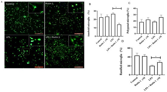
Figure 1


