Review

The Interplay Between Suicidal Behavior and Mental Disorders: Focusing on the Role of Glial Cells

by
Maya N. Abou Chahla
1,2
1
Department of Biological and Chemical Sciences, School of Arts and Sciences, Lebanese International University, Khiyara-West Bekaa, Beirut 11072809, Lebanon
2
Department of Biological Sciences, Faculty of Science, Beirut Arab University, Beirut 11072809, Lebanon
Neuroglia 2025, 6(3), 24; https://doi.org/10.3390/neuroglia6030024
Submission received: 2 March 2025 / Revised: 14 June 2025 / Accepted: 17 June 2025 / Published: 20 June 2025

Abstract

:
Glial cells exhibit multifaceted functions and represent essential contributors to various physiological processes in the brain, rather than just being silent supportive cells to neurons. Different glial populations of the central nervous system within involved brain regions play various functions, express different proteins, and result in fluctuating effects when altered. Glial cell pathologies were detected in most mental disorders including suicidal behavior. Suicidal behavior represents a health problem of high importance worldwide, where protective measures are required to be taken at many levels. Studies on patients with mental disorders that represent risk factors for suicidal behavior revealed multiple changes in the glia at diverse levels, including variations regarding the expressed glial markers. This review summarizes the role of glia in some psychiatric disorders and highlights the crosslink between changes at the level of glial cells and development of suicidal behavior in patients with an underlying psychiatric condition; in addition, the interplay and interconnection between suicidal behavior and other mental diseases will shed light on the routes of personalized therapy involving the development of glia-related drugs.

1. Introduction

Glial cells (glia) or “glue” cells are numerous cells in the brain comprising about 33–66% of brain mass; they were described in 1858 as the connective tissue involved in binding nervous elements together before the cellular nature was fully explored by Camillo Golgi (1873–1903) [1]. Glial cells are found in the central nervous system (CNS) and the peripheral nervous system (PNS); those in the CNS are represented by three major lineages as follows: astroglia, microglia, and oligodendroglia (including the OL-progenitors or NG2 cells) [2]. Glia of the PNS include satellite glial cells (SGCs), enteric glial cells, and myelinating and non-myelinating Schwann cells [3,4]. Glial cells have a wide array of functions with variations between CNS and PNS [3]; they represent half of the total CNS cells, and they usually outnumber neurons with region-specific variations. The proper expression of cell/stage specific molecular markers (Table 1) that usually have variable sensitivity and specificity determines differentiation, functional diversification, and the fate specification of glia [5,6]. Mainly, glia play regulatory roles [7], crucial roles at the level of synapses, secrete specific factors and gliotransmitters, and participate in the control of the neural processes in the CNS and PNS [8,9,10]. The neurophysiological mechanisms performed by glial cells include synaptic remodeling, neuronal development, neuropathic pain, neuronal health maintenance, regulation of brain function/development, and activity by mutually interacting with neurons [11]. They thereby represent a vital metabolic and structural component of the nervous system [12]. Initially, glial cell populations were meant to only support and sustain neuronal cells [13]; however, they are now being reconsidered for playing critical roles underlying a healthy or diseased brain. They contribute to neuro-inflammation, CNS damage, and neurodegeneration [13,14].
Astroglia, oligodendroglia, and microglia are major cells in the CNS (Figure 1) involved in defense against invasion, maintenance of brain homeostasis, and response to stress [15]; in addition to the regulation of blood flow and metabolism, glia are involved in the production of myelin and the modulation of synapse formation/functionality/elimination [6]. They support and interact with neurons and shape neuronal circuits as well. Some features in glial cells facilitate their interaction with neurons in early stages of brain development. Glial cells, especially oligodendrocytes and astrocytes, play a crucial role in synaptic patterning. Synaptic patterning refers to particular organization and arrangement of synapses involved in regulating learning, memory, and neural functions; shaped by genetic, epigenetic, and environmental factors, this process is essential for the strength and plasticity of synapses, as well as the specificity of neural connections [16].
Out of the three CNS glial populations, astroglia is the most abundant group of epithelial cells derived from glioblasts during development and subclassified into protoplasmic (in the gray matter) and fibrous astrocytes (in the white matter) [1]; they are located among synapses and blood vessels, and they are implicated in the maintenance of homeostatic and metabolic functions; this glia group is also involved in synapse formation and elimination (via the multiple epidermal growth factor (EGF)-like domain 10 (MEGF10) and myeloid–epithelial–reproductive tyrosine kinase (MERTK) phagocytic pathways [9]), as well as in plasticity and transmission. Notably, astrocyte-conditioned media contain variable excitatory and inhibitory synaptogenic features, depending on the brain region involved. Astroglia also maintain an ideal milieu for proper neuronal function and the maintenance of the blood–brain barrier. They also conserve water balance in the CNS, due to the presence of aquaporin AQP4. In addition, they regulate and represent the main controllers of K+ homeostasis, which plays a critical role in neuronal activity [17,18]. Moreover, astrocytes are involved in the maintenance of the glutamate/GABA balance in the CNS [19].
On the other hand, the highly active microglia enter the CNS during embryogenesis; they are yolk-sac-derived cells of mesodermal origin that possess a repopulation capacity [2,20]. During brain injury, for instance, microglia activate a mechanism of self-defense as the brain’s resident innate immune cells [21]. In addition, they are involved in phagocytosis (contributing to the clearance of disease-specific protein aggregates such as alfa-synuclein, beta-amyloid, and huntingtin [9]), impact synaptic functions and elimination, and are vital for proper neuronal activity [17].
The cells that control the neuronal axons’ diameter, affecting and sculpting their electrical and structural properties, are the oligodendroglia, and they have the same origin as astroglia. They play a myelinating role in the white matter of the CNS to ensure saltatory conduction or action potential propagation against the axons and provide axons with trophic support [22,23], express the glutamine synthetase (GS) enzyme, and are involved in the excitatory glutamatergic transmission in the CNS as well. They connect with astrocytes via connexin gap junctions (GJs) [24] in the panglial syncytium, which serves a necessary function especially regarding long-distance siphoning of K+ ions from the paranodal peri-axonal space to the cerebrospinal fluid, or the blood vessels. Like astrocytes, they maintain K+ homeostasis [17,22]. On the other hand, oligodendrocyte progenitor cells (OPCs), identified as NG2-glia, contribute to CNS function and dysfunction too. They play a crucial role in brain plasticity, by modulating neurotransmission and interacting with neurons and other glia. It has been reported through clinical studies that NG2-glia are involved in responding to stress, and their role in stress-related psychiatric diseases is still disputable. Studies also showed that early-life adversity causes long-lasting changes in NG2-glia [25].
Taking into consideration the indispensable roles of glial cells in the brain, their dysfunction is inevitably linked to the progression of mental, psychiatric, and neurodegenerative diseases as well as suicidal behavior. In this review, we aim to investigate the role of glial cell dysfunction in the progression of suicidal behavior, by focusing on the possible relative interplay between suicidal behavior and other mental conditions or disorders (such as major depressive disorder (MDD), schizophrenia, bipolar disorder (BD), borderline personality disorder (BPD), attention deficit hyperactivity disorder (ADHD), post-traumatic stress disorder PTSD, and others) after this dysfunction. Suicidal behavior (SB) is a very complex behavior and a multifactorial phenomenon with multiple aspects [26,27]. SB exhibits several phenotypes including suicidal ideation (SI), suicide attempts (SAs), and suicide. These phenotypes are complex and involve many risk factors. Suicide is considered the 17th leading cause of death globally by WHO. Recent advancements in metabolic, transcriptomic, and proteomic studies on postmortem brains and blood samples of individuals with SB, in addition to data from systems biology in silico, have provided novel insights into the key mechanisms and markers underlying this behavior. Research indicated the involution of alterations in neuroplasticity, immune responses, energy homeostasis, neurotransmission (glutamatergic and GABAergic systems), and pathways associated with glial cells (mainly microglia and astrocytes). In other words, the neurobiology of suicide is related to noradrenergic and HPA axis hyperactivity; dysfunction of glutamatergic, dopaminergic and GABAergic, and serotonergic systems; microgliosis (depending on the activation state and brain context, microglia polarization can shift between three different phenotypes: the quiescent M0, which is a resting-state microglia involved in maintaining brain homeostasis; the classically activated M1 in response to tissue damage or pathogens, which produces pro-inflammatory cytokines; and the alternatively activated M2 microglia in response to brain tumors and other stimuli, which produces anti-inflammatory and neuroprotective cytokines to promote tissue repair); signaling failures; and glial cell abnormalities [28]. Moreover, conducting comparative studies and concentrating on the balance between excitatory and inhibitory synapses and pathways associated with psychiatric disorders is key to understanding SB. Further mechanisms—such as specific neuronal–glial interactions, cortical connection formation, glio-genesis, glia-mediated epigenetic changes, and mRNA splicing-associated transcriptional factors—need to be further investigated to fill the missing gaps leading to SB [29].

2. Glio-Pathologies

A hallmark of various psychiatric diseases is synaptic dysfunction; modern tools have helped in investigating the role of glial cells in healthy and diseased brains [17]. In the brain, regulation of neuronal activity occurs by tripartite synapses (constant bidirectional communication between ramified and polarized astrocytes and neurons), suggesting a mutual interaction between the cells involved [30,31]; the latter include the presynaptic neuron (the transmitter), the postsynaptic neuron (the receiver), and the perisynaptic astrocyte fine protrusions or processes (these processes are involved in glutamate synthesis from glucose, glutamate buffering and recycling, synapse formation and pruning, maturation of synaptic activity and strength, provision of metabolites to neurons, and gliotransmitter release such as ATP, glutamate, and D-serine) [32,33,34]. Through the tripartite synapse, the astrocyte can impact the firing behavior of the neurons by sequentially activating/deactivating various pathways within this synapse. In other words, the ability of the astrocyte to introduce new configurations and routes of relevant parameters leads to the modulation and shaping of neuronal behavior, thus introducing irregularities in the firing pattern of both transmitter and receiver neurons [35,36]. Communication at the tripartite synapse involves neurotransmitters (ATP, norepinephrine, GABA, glutamate, etc.) receptors, transporters, ion channels (Ca2+), and gliotransmitters (involved in synaptic plasticity and strength) [33,37]. The majority of synaptic connections in the CNS represent the excitatory glutamatergic synapses [32]. Moreover, one of the crucial players in orchestrating the molecular interactions among the constituents of the tripartite synapse is purinergic signaling (ATP and its chief metabolite adenosine released by the astrocytes mediate an astrocyte–neuron crosstalk through purinergic receptors) [38]. Another more recent concept underlying the control of synaptic transmission is known as the “active milieu”; this latest concept relies on the fact that there is an interaction and dynamic interposition among neuronal compartments, CNS glial cells, extracellular matrix and space, and blood vessels as well. It is also important to note that the maturation of neural development necessitates synapse formation and synapse pruning (a key mechanism in refining neural circuits where connections between neurons are strengthened or weakened based on the level of usage of a neuron), referred to as the consolidation of neural wiring [37,39].
Recently, knowledge about glial cells in health and disease has increased incredibly. According to “gliocentric theory”, alterations and abnormalities in glial cells are responsible for the pathophysiology of mental disorders. But the question to be resolved is how these abnormalities develop and what impact they do have on cognitive and emotional disturbances [40]. Brain pathologies, especially neurodegenerative diseases (NDs), revealed progressive dysfunction in glial cells and damaged neurons; spatial and temporal heterogeneity of glial cells leads to glia-mediated neurodegeneration, as reported in NDs. Some genes expressed in glial cells were found mutated in patients with NDs. These genes include the following: apolipoprotein E (APOE) and the cluster of differentiation 33 (CD33), which represent major genetic risk determinants for late-onset Alzheimer’s disease [41]; glucosylceramidase Beta 1 (GBA1), whose mutation leads to an increased risk for Parkinson’s disease development; granulin precursor (GRN), whose mutation may cause frontotemporal lobar degeneration, cerebral ataxia, and epilepsy [42,43]; and triggering receptor of myeloid cells 2 (TREM2), which is also involved in the pathogenesis of Alzheimer’s disease. Pathways involved in the pathology also described the roles of α-synuclein (α-syn) (Parkinson’s disease) [44], G-protein-coupled receptors (GPCRs), pattern recognition receptors (PRRs) (Parkinson’s disease, schizophrenia, and major depressive disorder) [45,46], Toll-like receptors (TLRs) (major depressive disorder, schizophrenia, and bipolar disorder) [47], NOD-like receptors, and glial extracellular vesicles [48,49,50]. Three main brain regions depict alterations in glial cell functions; the limbic areas (hippocampus (HC) included), the prefrontal cortex, and the amygdala. Progressive plasticity in the amygdala and regressive plasticity in both the prefrontal cortex and HC are the results of failure in adapting to the varying neurophysiology as a response to stress [19]. The unique protein composition of glial cells implies their diverse roles, and these compositions vary according to activity or disease condition [6].
As mentioned earlier, glial cells play essential roles in the brain’s reaction to disease or injury [20]. For instance, the astrocyte response to disease (also known as astrogliosis) is common in several brain disorders. Astrogliosis is heterogeneous, and, depending on different stimuli, astrocytes can shift between two phenotypes or reactive states: the A1 astrocytes are pro-inflammatory and neurotoxic and are usually found in neurodegenerative diseases; they promote inflammation by activating the NF-kB pathway, thus contributing to neuronal loss and damage by impairing the synaptic function, and exhibit the ability to kill oligodendrocytes and neurons. In contrast, A2 astrocytes are anti-inflammatory and gain neuroprotective properties, usually induced by microglia after brain damage. A2 astrocytes suppress inflammation and express protective mediators such as chitin-like3 and prokineticin-2 (PK-2); they also promote synaptic repair and neuronal survival. There is also a correlation between the impairment of astrogliosis and defects in the blood–brain barrier [51]. Astrogliosis, combined with the extracellular matrix, forms what is known as the “glial scar” after injury to fill spaces formed as a result of neural tissue cavitation [51], which, in some contexts, has a prejudicial effect on neural repair and regeneration. Moreover, microglia, fibroblasts, and endothelial cells play a role in the formation of glial scars. Additionally, in the CNS, one oligodendrocyte is able to contact and myelinate many axons at a time. This intimate interaction between axons and oligodendrocytes can be disrupted by injury or disease, a condition known as dysmyelination (or demyelination). Demyelination is a result of autoimmune, metabolic, genetic hypoxic–ischemic or mechanical insults. This could lead to the loss of axonal support and thus result in permanent loss, disability, and degeneration of axons. Moreover, after injury, the microglia undergo dramatic changes in protein expression and shape to protect the brain, and they also release cytokines and phagocytose dead cells and debris. They also strip dysfunctional synapses. The loss of microglial main role and behavior can result in behavioral deficits and impaired learning-dependent synaptic plasticity [6]. In brief, dysfunctional astrocytes affect glutamate clearance and ion homeostasis and impact synaptic communication. Dysfunctional oligodendrocytes alter the connectivity of neural networks; and hyperactivation of microglia is an indication of a neuro-inflammatory process [40]. Furthermore, a study has likewise indicated the significant role of N6-methyladenosine (m6A), an epi-transcriptomic modification in the glial cells of mammalian brains that affects the cells’ functions by regulating gene expression patterns, thus resulting in the development of neurological diseases [52]. The understanding of which abnormality leads to which, as well as the sequence of events behind the main cause of abnormalities, will help identify the unique and/or the intersecting pathways between mental disorders.

3. Glial Cells and SB

The precise role of glial-associated mechanisms and processes in the pathophysiology of disease is still poorly understood and requires further investigations by applying and implementing new methods and diverse study groups [53]. The association of glial cells to SB was based on morphological changes, postmortem brain gene expression, cytokines, and neurotrophic factors [15]. Suicide represents a major health issue gradually rising worldwide and is considered an independent disorder with a unique genetic/molecular background [54,55,56]. In humans, a subcortical brain region called the hippocampus—part of the limbic–cortical–hypothalamic circuit—is rich in glucocorticoid receptors, is very sensitive to stress, and seems to be implicated in the pathophysiology of suicide. Suicidal samples show a smaller HC accompanied with histopathological changes by neuroimaging studies, with anatomical and functional differences in the hippocampal subregions [54].
Table 1. The impact of specific glial marker expression in mental illnesses.
Mental DisordersGlial MarkersImpactReferences
MDD
  • GFAP
  • Its decrease causes decreased hippocampal neurogenesis
  • Hyper-inflammation of astrocytes
[57,58,59]
  • S100B
  • Elevated in the serum of MDD patients
  • Related to the pathogenesis and plasticity changes in MDD
[59,60]
  • GS
  • Its alteration causes depressive behaviors with hypoactive glutamatergic neurotransmission
[61]
  • AQP4
  • Leads to poor water balance when altered
[59]
  • QUIN
  • Its elevated levels lead to reducing the size and density of neurons and glial cells
  • Involved in memory problems, mood swings, behavioral changes, and cognitive difficulties
[62]
  • Cytokines
  • Increased pro-inflammatory cytokines levels lead to hyperactivity of the hypothalamic–pituitary–adrenal (HPA) axis/dysfunction of the glutamatergic system/impairment of neuroplasticity/alterations in tryptophan (TRP) metabolism
[63]
  • Connexins
  • Their disruption leads to alterations in calcium wave propagation and communication between astrocytes
  • Involved in the weakened blood–brain barrier when altered
[59]
Schizophrenia
  • AQP4
  • It is upregulated in the PFC of schizophrenic patients
  • Related to the seriousness of negative symptoms and poor control of neuro-inflammation
  • Plays a role in neurovascular dysfunction and BBB hyperpermeability when altered
[64]
  • MAG
  • Altered regulation in the cortical gray matter in the parietal and temporal regions
  • Its downregulation causes variations in brain morphometry in schizophrenic patients
[65,66]
  • D-Amino-acid-oxidase
  • Involved in reduced D-serine and thus in impaired NMDAR functioning
[67]
  • Glutaminase
  • Leads to an increased glutamatergic synaptic release
[68]
  • DISC1
  • Leads to dopaminergic dysregulation when disrupted
[69]
  • GS
  • Its alteration disturbs glutamate metabolism
[70]
  • GFAP
  • Its expression level varies among brain regions
  • Elevated levels indicate reactive astrogliosis
[71]
  • S100B
  • Oligodendrocyte and astrocyte markers
  • Increased expression is not uniform among brain regions. Its increase leads to metabolic disturbances in neurons and astrocytes
  • Its release can be provoked by an excess of serotonin
  • Its serum level correlates with the development of insulin resistance in schizophrenic patients
[71,72]
  • ADAM12
  • Its reduced expression causes deviant metabolism of some of its substrates that are parts of chemical components of myelin (neurofascin-ankyrin) or important signaling cascades (EGF, betacellulin, TGF-beta)
[73]
  • OLIG2
  • Alterations in its expression cause alterations in the development of oligodendrocytes and motor neurons and in the fate of subtypes of those cells
[74]
  • CNPase
  • Its reduced expression indicates disrupted oligodendrocyte function in schizophrenic brains
  • Alteration in the regulation of the microtubule distribution in the cytoplasm and thus involved in cellular morphology
[75,76]
BD
  • PLP1
  • Dysregulation in the production of myelin (fatty insulation around nerve cell axons that enables efficient communication in the central nervous system)
  • Its alteration causes changes in the assembly and functioning of the nodes of Ranvier and other axonal functional rearrangements
[77,78]
  • GFAP
  • Elevated levels in the dorsolateral prefrontal cortex but downregulated in the orbitofrontal cortex of BD patients, indicating an astrocytic dysfunction and correlating with the disease mechanisms of psychosis
[79,80]
  • Transferrin
  • Elevated levels of transferrin receptors in the plasma of BD patients during acute mania
[81]
  • MOG
  • An oligodendrocyte marker found to be downregulated in BD patients, leading to alterations in myelination
[77,82]
  • MBP
  • Involved in myelin damage and inflammation when downregulated in BD patients
[83]
SUDs
  • Connexin43
  • Its increased hippocampal astrocytic expression increases drug-induced behavioral responses
[84]
  • GLT-1
  • Its region-specific downregulation is involved in addiction-related glutamate homeostasis
[85]
Suicidal Behavior
  • QUIN
  • Elevated level is neurotoxic and could contribute to structural deficits and functional changes
[86,87,88]
  • IDO
  • Activated in response to pro-inflammatory cytokines
  • IDO2, in particular, plays a role in the metabolic changes observed in the KYN pathway in individuals with SB
[89]
  • IBA1
  • Increased microglial marker expression in the anterior midcingulate cortex of individuals who died by suicide, indicating an increase in microglial cell density
[15]
  • MCP-1
  • Lower expression in suicide attempters and individuals with suicide ideation and MDD, but shows upregulation in depressed individuals who died by suicide
[90,91]
  • HLA-DR
  • Elevated levels of some genotypes in suicide attempters
[92]
  • CRP
  • Elevated level in SB patients
  • Associated with various inflammatory conditions
[93]
  • IL-6
  • Increased level plays a significant role in neuro-inflammation
[94,95]
  • IL-2
  • A pleiotropic cytokine showing lower levels in suicidal samples. Altered function as a key cytokine with pro-inflammatory potential, promoting the expansion of NK cells and T-cells
[96]
  • TNF-α
  • Increased levels in suicidal samples
  • Its polymorphisms affect the age at which suicide attempts begin
[96,97]
The pathophysiology of suicide is mainly caused by the disruption of serotonergic functions—serotonin deficiency [98]—in the dorsal raphe nucleus (DRN); the DRN itself is composed of several important subregions, and it is implicated in sending serotonergic projections to other locations such as the HC, thalamus, frontal cortex, nucleus accumbens, striatum, habenular complex, and lateral septal nuclei [99]. The evidence for the role of glial cells, especially microglia, in suicide was revealed through the analysis of the cerebrospinal fluid and serum/plasma of patients, in addition to positron emission tomography (PET) imaging and postmortem studies. Most studies involving SB demonstrated its association with alteration in cytokines levels—mainly interleukin (IL)-6—released by microglia (also in IL-2, IL-8, vascular endothelial growth factor (VEGF), and tumor necrosis factor (TNF)- α) [99]. Collective studies also revealed that astrocyte-related suicides (suicides related to alterations in astrocytes) are associated with abnormalities and changes in methylation, transcription, and protein content. For instance, the major marker for astrocytes is the glial fibrillary acidic protein (GFAP), which has shown reduced transcription in the caudate nucleus, mediodorsal thalamic nucleus (MD), locus ceruleus (LC), and dorsolateral prefrontal cortex (DLPFC) by real-time polymerase chain reaction (PCR), unlike in the cerebral cortex (Cb), primary visual cortex (PVC), and primary motor cortex (PMC) [15]. Only in individuals who died by suicide was the prefrontal cortical GFAP (sp4005) protein identified, indicating that its higher phosphorylation state might be implicated in the pathophysiology of SB. Other astrocytic markers with decreased transcriptional levels in the DLPFC of suicidal samples include S100 calcium-binding proteins B (S100B), aldehyde dehydrogenase 1 family member L1 (ALDH1L1), glutamate–ammonia ligase (GLUL), SRY-Box Transcription Factor 9 (SOX9), and solute-carrier family 1 member 3 (SLC1A3) [15]. In addition, about eight postmortem studies on the brains of individuals who died by suicide have revealed morphological changes (increased cell density) in microglia, supporting the relationship between SB and the dysregulation of microglial; in most cases, this is independent of any psychiatric illness [27].

4. Suicidal Behavior and Mental Disorders

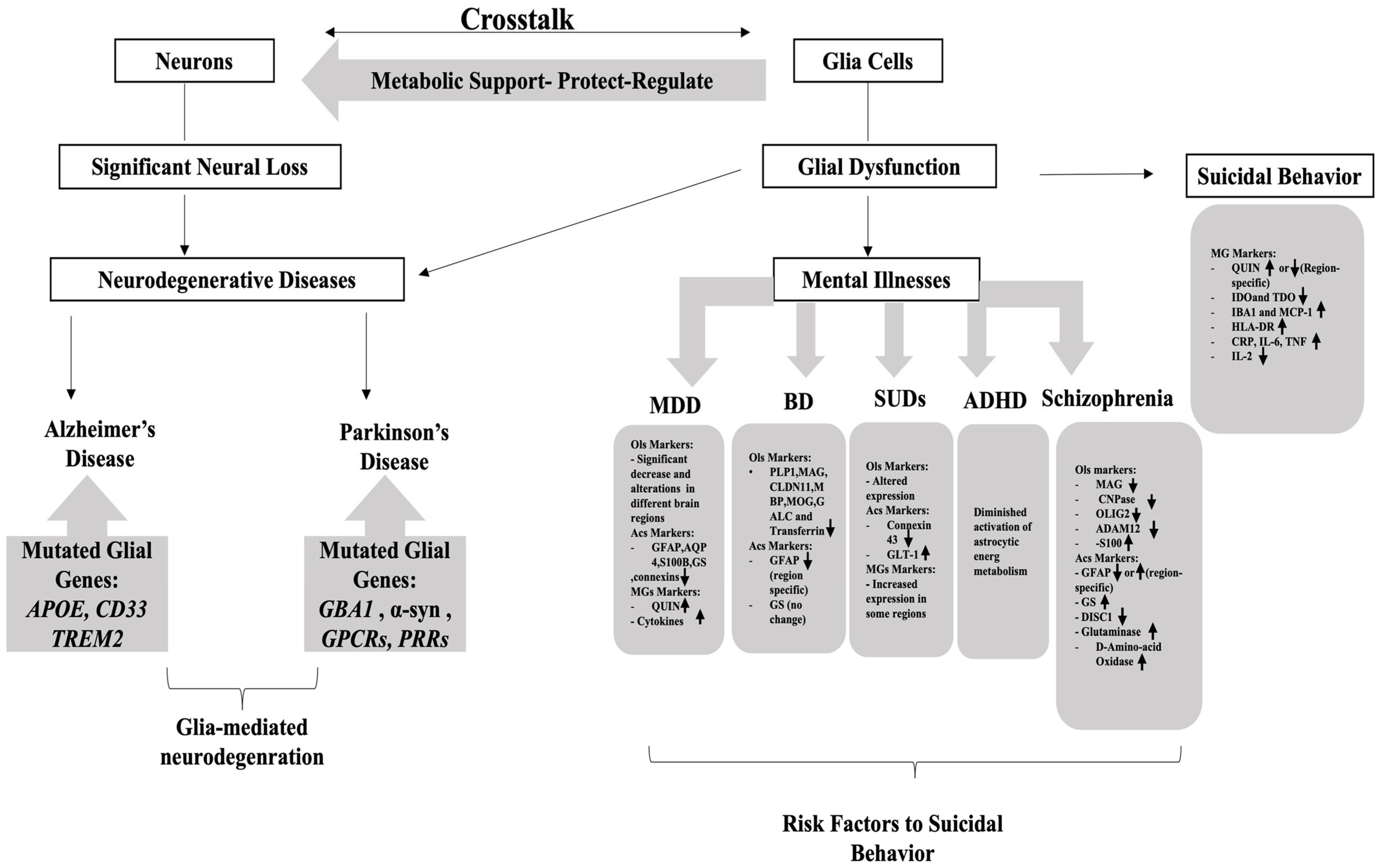
Studies about mental diseases and their interconnected basis have increased in the last decade. Unlike NDs, mental disorders are marked by remarkable glial pathology, rather than loss of neurons [100]. Different groups with different variables showed multiple and complex variations, and thus, repetition in studies is required to confirm these results. It is also interesting here to understand the behavior of glial cells and their expression profiles under different circumstances or triggers. Mental disorders represent a high-risk factor for the predisposition of SB (Figure 2), which is reported to be the most catastrophic outcome for psychiatric disorders. SB phenotypes and severity vary from one patient to another based on the underlying psychiatric state or illness [101].
The brain gene expression profiling of individuals who died by suicide but with a dual diagnosis (co-occurrence of one or more mental disorders or one substance use disorder in an individual) was distinct from that of those who died by suicide with a single disorder, highlighting the presence of common disrupted pathways [102]. Most mental disorders or diseases have a tight connection with inflammation/neuro-inflammation. Yet, the mechanisms underlying the full image of inflammatory dysfunction are not fully elucidated. A possible pivotal player is microglial dysfunction. When microglia are overactivated in response to neuro-inflammation (or injury or infection), they become primed microglia with heightened responsiveness, which means they can respond to subsequent stimuli. This microglial priming process involves alterations in gene expression, changes in cell morphology, and secretion of pro-inflammatory cytokines, excessively modifying the glutamate signaling and the kynurenine pathway (a key metabolic pathway that degrades the amino-acid tryptophan into various metabolites). This overactivation of microglia leads to consequent glutamate release as a result of increased astrocytic activity; this is toxic to the CNS. Moreover, the change in the products of the kynurenine pathway (as a result of excessive microglial activation) will in turn impact the serotonergic, glutamatergic, and dopaminergic signaling pathways [103].

4.1. SB and Major Depressive Disorder (MDD)

MDD is a common mental disease (reported in about 350 million individuals worldwide) in which patients suffer from permanent low mood and related changes in biological functions, thoughts, and behavior. MDD patients reveal dysregulation in the HPA axis, cardiac autonomic regulation, and the immune system; still, MDD pathogenesis and precise etiology are only partially understood [104,105].
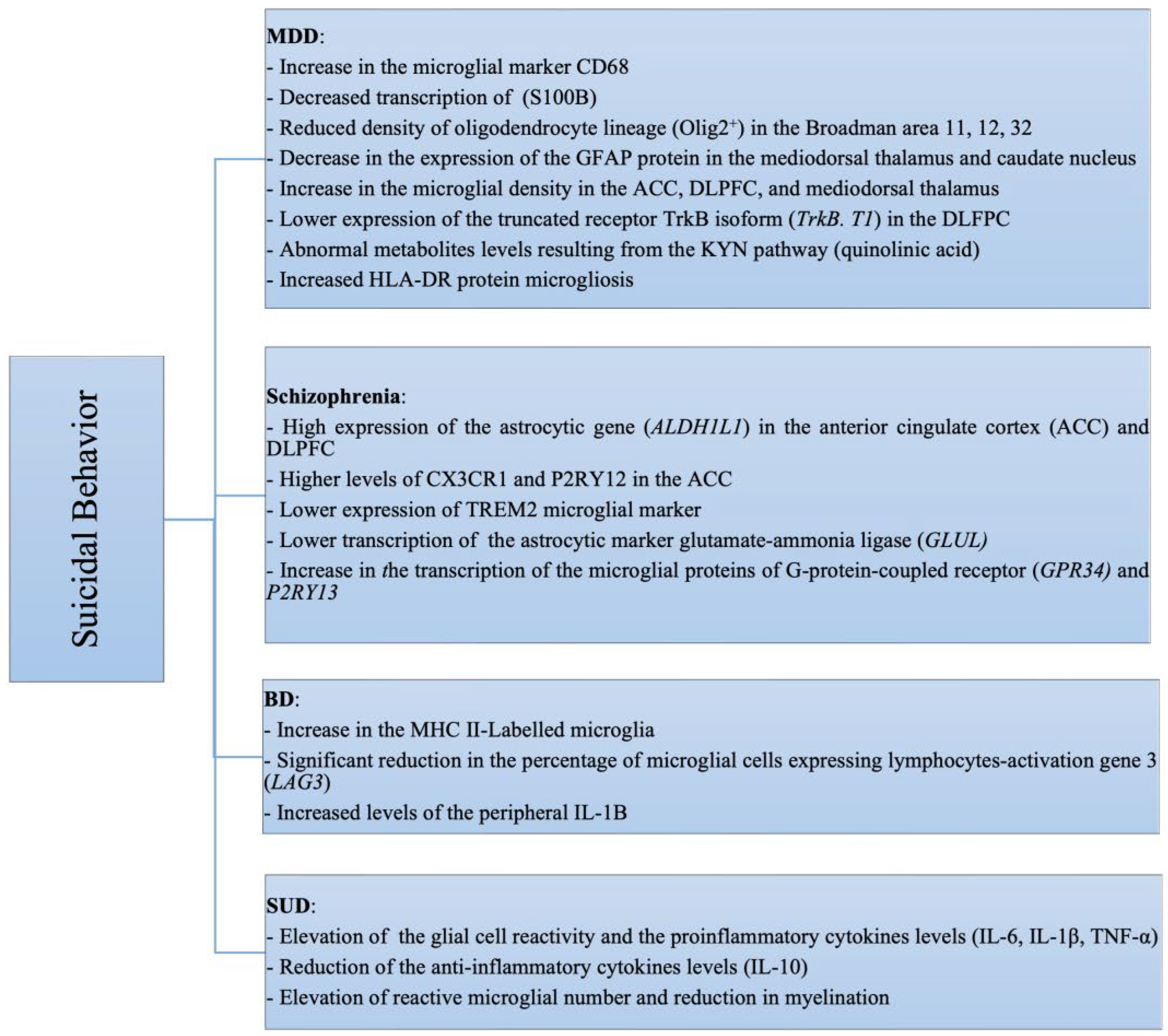
Studies have confirmed the relationship between different types of mental disorders, including MDD, affected by alterations in glial gene expression with variations across different brain regions [106]. In brain studies of MDD patients, reports showed significant reductions in glial numbers in the ventromedial PFC and amygdala [40]. Another study involving 17 glial-related genes in the DLPFC showed an increase in the microglial marker CD68 in patients with MDD who died by suicide, compared to controls. Other gene transcripts, such as the myelin basic protein (MBP) mRNA in the DLPFC, were increased in MDD patients experiencing psychotic features, but not in those who died by suicide [107]. Postmortem investigations of individuals with MDD who died by suicide showed a decreased transcription of S100 calcium-binding protein B (S100B) in their prefrontal cortex. This marker is specifically expressed by protoplasmic astrocytes located in the gray matter (and found in myelinating oligodendrocytes in the white matter), which exhibit special morphological patterns. Another study of depressed suicide samples with a history of child abuse reported a reduced density of oligodendrocyte lineage (Olig2+) in Broadman area (BA) 11, BA12, and BA32 in comparison to controls, which is associated with an increased density of mature oligodendrocytes (Adenomatous Polyposis Coli protein (APC+) and Reticulon-4 (Nogo+)), indicating a shift towards a more mature phenotype of OL lineage.
In the PFC of individuals with depression who died by suicide, mRNA expression for some astrocyte-related genes (including the astrocytic GFAP) was significantly decreased, and the methylation associated with those genes was also decreased. Another study revealed a significant decrease in the expression of GFAP and mRNA in the mediodorsal thalamus and caudate nucleus of individuals with depression who died by suicide, unlike in the primary motor, cerebral, or visual cortex [108]. In addition, compared to controls, suicidal patients with depression showed an increase in microglial density in the ACC, DLPFC, and mediodorsal thalamus. This was also confirmed by another study that reported an increase in microglial density in the white matter of the PFC for the same type of patients. Samples from depressed suicidal patients also showed an increase in the primed microglial cells in the white matter of the dorsal ACC [99]. Repeated stress exposure of patients with MDD can cause microglial inflammation or even SB. In comparison to controls, depressed suicides showed a lower expression of the truncated receptor TrkB isoform (TrkB. T1) in the DLFPC and frontal eye field (FEF) [15,109]. Several studies also focused on the role of monoamine oxidase (MAO) on the severity of MDD and other neuro- and mental pathologies. MAO-B is expressed by fibrous white matter astrocytes and cortical astrocytes in the frontal, temporal, and occipital lobes. This gene is closely related to disorders such as MDD, and its knockdown can result in mild to aggressive behavioral changes that might lead to suicide; but further investigation of this gene’s expression profiles is required, particularly for MAO-B, to better understand its role in brain disorders [110,111,112,113,114,115].
Glial alterations (Figure 3) can lead to defective information processing in the PFC in addition to emotional and cognitive disturbances as seen in MDD patients. This causes loss of interest, impaired decision making, and an increased tendency towards SB; similar results are reported in patients with reductions in hippocampal neurogenesis [19]. Glial cells can also affect the kynurenine (KYN) pathway in the CNS. Abnormal metabolite levels resulting from the KYN pathway (such as quinolinic acid) were recorded in the HC of suicidal depressed patients, which showed a decrease when compared to healthy control subjects [99]. In addition, the hippocampal subregions of individuals with MDD who died by suicide showed that more neurons but fewer astrocytes exist in the Cornu Ammonis (CA) 2/3 subareas of HC, and fewer granular neurons but more glia with larger nuclei were found in the dentate gyrus (DG), indicating a functionally distinct role of the basic circuits of HC in suicidal development [54]. Furthermore, studies showed an increased HLA-DR protein microgliosis in suicide patients with MDD. A few other studies involving the DLPFC and ACC of suicide patients with MDD reported no variation in the transcription of the microglial markers (major histocompatibility complex, class II, DR-α (HLA-DRA), CD68, Integrin alpha M (ITGAM), chemokine receptor (CX3CR1), Allograft inflammatory factor 1 (AIF1)), and oligodendrocyte proteins (myelin oligodendrocyte glycoprotein (MOG), proteolipid protein (PLP), oligodendrocyte transcription factor (OLIG2), and the astrocytic marker (ALDH1L1) [15].

4.2. SB and Schizophrenia

Schizophrenia affects 1 in 300 people worldwide, and it is considered a serious brain disorder in which glial cell loss (especially oligodendrocytes and astrocytes) is a key feature in this disorder’s histopathology [39]. Samples from schizophrenic patients showed suicidal-related differences and diverse glial gene alterations through postmortem RNA-sequencing of transcriptional profiles in the DLPFC examination. The results of studies revealed a high expression of the astrocytic gene (ALDH1L1), an astrocytic marker in the anterior cingulate cortex (ACC), and DLPFC in schizophrenic patients who died by suicide in comparison to controls. On the other hand, mRNA expression of the following microglial markers—CX3CR1 and purinergic receptor (P2RY12)—in the ACC was higher in schizophrenic suicidal patients compared to schizophrenic non-suicidal patients, unlike the following microglial marker the triggering receptor expressed on myeloid cells 2 (TREM2) whose expression was lower [53]. However, clinical findings on schizophrenic brains showed no change (normal) in glial cell numbers unlike in other disorders [40].
Other research involving postmortem studies and PET scans showed discrepancies in the findings concerning microglial activation in schizophrenia due to the effect of comorbid factors such as suicidal tendencies [99]. Surprisingly, other studies reported that in comparison to non-suicides, suicide patients with schizophrenia indicated an elevation in microglial markers (TREM2, P2RY12, and CX3CR1) in the ACC and a lower transcription of astrocytic markers (glutamate–ammonia ligase (GLUL) and ALDH1L1) in the DLPFC. Moreover, MBP was decreased in the anterior PFC. Furthermore, the microglial density of the HLA-DR protein was increased for the same category of patients in the following brain regions: DLPFC, MD, ACC, and HC. The transcription of the microglial proteins of G-protein-coupled receptor (GPR34), P2RY12, and P2RY13 was also increased [15]. This indicates that there is a confirmed heterogeneity in glia gene alterations among schizophrenic patients with SB, and it emphasizes the complexity of this interconnected system between glia and diseases, which may also suggest the possible role of a third player, such as other involved regulators or the relative combination of concentrations of specific glial markers, which might underlie the determination of the route of the onset of a particular illness.
The pathogenesis of schizophrenia is also related to alterations in the glial-cell-derived neurotrophic factor (GDNF) and GFAP; their serum levels in schizophrenic patients were lower than in healthy controls [79].

4.3. SB and Bipolar Disorder (BD)

BD is a psychiatric and behavioral condition characterized by functional disability, cognitive impairment, and fluctuating mood states [116]. BD progression and pathophysiology involve several important brain mechanisms such as oxidative stress, abnormalities in both glutamatergic and monoaminergic neurotransmission, reductions in neurotrophins, mitochondrial dysfunctions, and impairments in neuroplasticity. Marked reductions in glial cell density have been reported in BD patients in the ventromedial and dorsolateral PFC, as well as in ACC [40]. Many studies have revealed a link between SB and bipolar disorder [116,117]. It is estimated that in patients with BD, the risk of SB increases 20–30 times in comparison to healthy controls; and BD represents the psychiatric condition with the highest frequency of SB. It is also indicated that in patients with these psychiatric disorders, the testosterone hormone (high or low) might contribute to suicidality, as it seems to affect behavior and mood and regulate the proactive and reactive aspects of aggression. Patients with BD have a reduced quality of life and experience manic and hypomanic episodes at least once. It is estimated that for men and women with BD, respectively, suicidal attempts are about 19% and 34% [118,119]. Suicidal risk in BD patients increases with rapid cycling, the early onset of BD, and the presence of familial and genetic risk factors that differ between countries and regions [119].
At another level, a study revealed a notable increase in MHC II-labeled microglia, as well as elevated hippocampal microglial density (labeled by the microglial-specific marker P2RY12) compared to controls. Furthermore, there was a significant reduction in the percentage of microglial cells expressing lymphocyte activation gene 3 (LAG3) in suicidal BD patients. This study also confirms the absence of correlations between the density of activated microglia and LAG3 expression levels [117]. In addition, peripheral IL-1β levels are increased in suicide BD patients compared with non-suicide BD patients. In addition, the deficiency of serotonin is one of the biological bases underlying BD. In addition, there is a role played by the activated glycogen synthase kinase-3 β (GSK-3β) in the Wnt-signaling pathway and the progression of BD. Moreover, microglia during neuro-inflammation provide a link between those two pathways [116].

4.4. SB and Borderline Personality Disorder (BPD)

BPD represents a severe personality disorder with abnormal patterns of behavior and inner experience. BPD patients usually suffer from other coexisting mood or mental illnesses including MDD, BD, or substance use disorders (SUDs). Individuals with BPD are characterized by a wide range of behaviors including SB [120]. A study has detected that suicidal risk increases more in the case of BPD than in patients with affective disorders. Among personality disorders, BPD is considered the most suicidal. The suicidal tendencies in patients with BPD are due to the high reactivity of emotions with anxiety, tense relationships, and episodic depression, in addition to impulsiveness in decision making. In 87% of the cases, suicidal attempts occurred suddenly without prior planning [121]; and 10% of BPD patients die by suicide after a mean of three lifetime suicide attempts and a long course of unsuccessful treatment [122]. Further investigations are required at the molecular level, including alterations in glial markers, to assess the precise molecular link and variations between BPD suicidal and non-suicidal patients.

4.5. SB and Attention Deficit Hyperactivity Disorder (ADHD)

Inattention, hyperactivity, and impulsivity are the main characteristics of ADHD, which is a common neurodevelopmental disorder that disrupts brain functioning. ADHD is diagnosed in males more than in females and is estimated to have occurred at a stable rate of 7.2% over the past 40 years. Suicide and ADHD are significantly associated with each other in different age groups and in children [123]. The risk of SB increased in groups diagnosed with ADHD, as some studies confirmed [124,125,126,127,128]. Studies have revealed that glial markers other than S100B have differences in different groups of study. For instance, ADHD patients showed lower levels of the toxic 3-hydroxykynurenine (3HK) [129], whereas patients with ADHD who are sensitive to allergy, especially children, also show higher levels of IL-10 and IL-6 [124].

4.6. SB and Post-Traumatic Stress Disorder (PTSD)
PTSD patients are characterized by poor brain health [130]; it is a mental illness that results after a trauma caused by an incident leading to anxiety or physical damage (mental shock). In adults, PTSD represents a risk factor of SB [131,132]. Glial—mainly astrocytic—dysfunction in PTSD represents an emerging area of investigation [133]. According to studies, changes in glial activation were detected in PTSD patients, and PTSD is linked to neuro-inflammation. GFAP is released by glial cells after reacting to stress and is considered a putative biomarker of proglial activation. Moreover, it is postulated that GFAP distributions are associated with PTSD severity [130]. PTSD is associated with unnatural causes of death, including suicide [134,135]. Based on reports, diseases in glia (astrocytes and microglia) can cause structural, functional, and behavioral changes related to PTSD. Furthermore, increased levels of inflammatory cytokine biomarkers (C-reactive protein, TNF-α, NF-kb, IL-1, and IL-6) were reported in PTSD patients in comparison to controls [136].
Figure 3. Interconnected roles of glial cell dysfunctions and the resulting diseases (arrows indicate the increase/decrease in the expression of a specific marker at the transcriptional level; OL markers = oligodendrocyte markers; Acs = astrocyte markers; MG markers = microglial markers) [39,41,44,99,100,137].
Neuroglia 06 00024 g003

4.7. SB and Anxiety Disorders

Growing evidence exists to confirm the association between suicide and anxiety, but the precise role of anxiety in SB is not yet understood. And since SB is multifactorial, patients must be evaluated at several levels including anxiety, personality, depression, and others to assess their SB. Anxiety is a neuropsychiatric impairment that usually follows blast-related traumatic brain injury (bTBI). Two brain regions, the motor cortex and the HC, play a critical role in the manifestation of anxiety. Glial pathological changes such as astrocyte pathology (GFAP) and dendritic alteration (microtubule-associated protein, MAP2) can contribute to anxiety [138].

4.8. SB and Substance Use Disorders (SUDs)

According to statistics, almost 40 million people are affected by SUDs. Research reported that SUDs or drug addiction usually affect functional changes in the pathways involved in self-control and stress response, as well as brain circuits that control pleasure or rewards. Opioids (morphine) and psychostimulants (methamphetamine or cocaine) seem to elevate glial cell reactivity and pro-inflammatory cytokine levels as well (IL-6, IL-1β, TNF-α), whereas the levels of anti-inflammatory cytokines were reduced (IL-10). It has also been reported that SUDs alter glutamate neurotransmission and metabolism by GLT-1 (specific astrocytic glutamate transporter) and reduce the number of astrocyte cells, but they elevate the number of reactive microglia and decrease myelination by impairing oligodendrocytes as well. These events represented by glial dysfunction affect the permeability of the blood–brain barrier (BBB), increase neurotoxicity, and thus promote further addictive behavior [139].
According to various studies, SB and SUDs are substantially associated, and SUDs are comorbid with various other psychiatric disorders and can result in suicide [139,140,141,142]. Recent data confirms that 13.3% of patients suffering from SUDs seem to be associated with unplanned suicide. SUDs refer to the use of substances in an unhealthy pattern, leading to significant impairment in daily life. Substances such as cannabis [143], heroin, alcohol, cocaine, methylenedioxymethamphetamine (MDMA), lysergic acid diethylamide (LSD), nicotine, and methamphetamine have deleterious effects on suicide as well [144].

5. Pharmacology

The combination of pharmacological treatment, psychological support, and cognitive behavioral therapy (CBT) is considered a promising anti-suicidal approach for preventing suicidal behavior [145,146,147,148]. Ketamine, Lithium, Clozapine are also approved drugs that show high efficacy in reducing suicidality and suicidal behavior phenotypes such as SI and SA [149,150,151]. For suicidal behavior caused by dysfunction in glia, neuroglia can also be targeted for therapeutic approaches. Understanding the precise mechanisms by which glial cells are involved in suicide is critical to uncovering suicide-related biomarkers and developing glia-targeted therapeutics [15,152].

6. Conclusions and Future Perspectives

Suicidal behavior is a multifactorial independent mental disorder, as defined by the fifth edition of the Diagnostic and Statistical Manual of Mental Disorders, which demands substantial attention and efforts worldwide to reduce its impact on society. Understanding the role of glial cells and their interconnected mechanisms in diverse psychiatric illnesses will have an impact on personalized methods of therapy and will pave and clarify the routes leading to SB. Astroglia, microglia, and oligodendroglia, with their diverse roles, are separately considered wide topics with huge impact on mental disorders and pathological conditions in the brain. Glio-pathologies vary across different psychiatric disorders. Their differences have diverse effects on neurons. These discrepancies vary from the density of glial cells to secreted markers, expressed proteins, and affected brain regions. Different age groups diagnosed with SB and other psychiatric disorders have to be monitored to assess the initial trigger/s causing this disruption in natural pathways and the baseline communication between glia and neurons in a “healthy” brain. This complexity that surrounds the multiple combinations of events requires extensive research of wide and diverse groups of patients to better address the interplay between SB and other psychiatric disorders, at the level of alterations in glial cells. It is also essential to navigate and map the interlocking and separate/unique pathways and fill the gaps leading to each mental disorder. Understanding what leads to what—and under which conditions the involved mechanisms are well-orchestrated or disrupted—is a matter of great interest and intricacy and still remains a subject of ongoing debate. This represents a key area that requires a huge amount of research efforts to unlock the “missing link”. Advancements in artificial intelligence and technologies, combined with social and medicinal interventions, are promising in defining glial-related drug targets; developing efficient and case-specific anti-inflammatory agents, inhibitors, or natural drugs; establishing personalized therapeutic procedures based on personalized diagnosis; identifying glial stem cell research gaps; and predicting the possibility of occurrence of a specific mental disorder and/or SB. These efforts will also help to map the unique/intersecting pathways among psychiatric disorders and SB, paving the road towards possible effective/preventive measures at younger ages.

Funding

This research received no external funding.

Data Availability Statement

The data presented in this review are available within the article text and figures.

Acknowledgments

I would like to express my deepest gratitude to the peer reviewers who dedicated their time and expertise to providing valuable feedback and constructive criticism on this manuscript.

Conflicts of Interest

The author declares no conflicts of interest.

References

  1. Parpura, V.; Heneka, M.T.; Montana, V.; Oliet, S.H.R.; Schousboe, A.; Haydon, P.G.; Stout, R.F.; Spray, D.C.; Reichenbach, A.; Pannicke, T.; et al. Glial cells in (patho)physiology. J. Neurochem. 2012, 121, 4–27. [Google Scholar] [CrossRef]
  2. Jäkel, S.; Dimou, L. Glial Cells and Their Function in the Adult Brain: A Journey through the History of Their Ablation. Front. Cell. Neurosci. 2017, 11, 24. [Google Scholar] [CrossRef] [PubMed]
  3. Andreeva, D.; Murashova, L.; Burzak, N.; Dyachuk, V. Satellite Glial Cells: Morphology, functional heterogeneity, and role in pain. Front. Cell. Neurosci. 2022, 16, 1019449. [Google Scholar] [CrossRef]
  4. Gazerani, P. Satellite Glial Cells in Pain Research: A Targeted Viewpoint of Potential and Future Directions. Front. Pain. Res. 2021, 2, 646068. [Google Scholar] [CrossRef] [PubMed]
  5. Huang, H.; He, W.; Tang, T.; Qiu, M. Immunological Markers for Central Nervous System Glia. Neurosci. Bull. 2022, 39, 379–392. [Google Scholar] [CrossRef] [PubMed]
  6. Rasband, M.N. Glial Contributions to Neural Function and Disease. Mol. Cell. Proteom. 2016, 15, 355–361. [Google Scholar] [CrossRef]
  7. Kato, D.; Eto, K.; Nabekura, J.; Wake, H. Activity-dependent functions of non-electrical glial cells. J. Biochem. 2018, 163, 457–464. [Google Scholar] [CrossRef]
  8. Um, J.W. Roles of Glial Cells in Sculpting Inhibitory Synapses and Neural Circuits. Front. Mol. Neurosci. 2017, 10, 381. [Google Scholar] [CrossRef]
  9. Jung, Y.-J.; Chung, W.-S. Phagocytic Roles of Glial Cells in Healthy and Diseased Brains. Biomol. Ther. 2018, 26, 350–357. [Google Scholar] [CrossRef]
  10. Lee, Y.J.; Kim, Y.-K. Neuron-to-microglia Crosstalk in Psychiatric Disorders. Curr. Neuropharmacol. 2020, 18, 84. [Google Scholar] [CrossRef]
  11. Kong, E.; Li, Y.; Deng, M.; Hua, T.; Yang, M.; Li, J.; Feng, X.; Yuan, H. Glycometabolism Reprogramming of Glial Cells in Central Nervous System: Novel Target for Neuropathic Pain. Front. Immunol. 2022, 13, 861290. [Google Scholar] [CrossRef] [PubMed]
  12. Hyung, S.; Park, J.-H.; Jung, K. Application of optogenetic glial cells to neuron-glial communication. Front. Cell. Neurosci. 2023, 17, 1249043. [Google Scholar] [CrossRef] [PubMed]
  13. Magni, G.; Riboldi, B.; Ceruti, S. Human Glial Cells as Innovative Targets for the Therapy of Central Nervous System Pathologies. Cells 2024, 13, 606. [Google Scholar] [CrossRef]
  14. Rajkowska, G.; Miguel-Hidalgo, J.J. Gliogenesis and Glial Pathology in Depression. CNS Neurol. Disord. Drug Targets 2007, 6, 219. [Google Scholar] [CrossRef]
  15. Yamamoto, M.; Sakai, M.; Yu, Z.; Nakanishi, M.; Yoshii, H. Glial Markers of Suicidal Behavior in the Human Brain-A Systematic Review of Postmortem Studies. Int. J. Mol. Sci. 2024, 25, 5750. [Google Scholar] [CrossRef] [PubMed]
  16. Kim, Y.S.; Choi, J.; Yoon, B.-E. Neuron-Glia Interactions in Neurodevelopmental Disorders. Cells 2020, 9, 2176. [Google Scholar] [CrossRef]
  17. Shen, W.; Pristov, J.B.; Nobili, P.; Nikolić, L. Can glial cells save neurons in epilepsy? Neural Regen. Res. 2023, 18, 1417–1422. [Google Scholar] [CrossRef]
  18. Ramsey, J.; Martin, E.C.; Purcell, O.M.; Lee, K.M.; MacLean, A.G. Self-injurious behaviours in rhesus macaques: Potential glial mechanisms. J. Intellect. Disabil. Res. 2018, 62, 1008–1017. [Google Scholar] [CrossRef]
  19. Mayegowda, S.B.; Thomas, C. Glial pathology in neuropsychiatric disorders: A brief review. J. Basic Clin. Physiol. Pharmacol. 2019, 30, 20180120. [Google Scholar] [CrossRef]
  20. Garden, G.A.; Campbell, B.M. Glial Biomarkers in Human Central Nervous System Disease. Glia 2016, 64, 1755–1771. [Google Scholar] [CrossRef]
  21. Ochocka, N.; Kaminska, B. Microglia Diversity in Healthy and Diseased Brain: Insights from Single-Cell Omics. Int. J. Mol. Sci. 2021, 22, 3027. [Google Scholar] [CrossRef] [PubMed]
  22. Zlomuzica, A.; Plank, L.; Kodzaga, I.; Dere, E. A fatal alliance: Glial connexins, myelin pathology and mental disorders. J. Psychiatr. Res. 2023, 159, 97–115. [Google Scholar] [CrossRef] [PubMed]
  23. Wang, J.; Lu, Q.R. Convergent epigenetic regulation of glial plasticity in myelin repair and brain tumorigenesis: A focus on histone modifying enzymes. Neurobiol. Dis. 2020, 144, 105040. [Google Scholar] [CrossRef]
  24. He, C.; Duan, S. Novel Insight into Glial Biology and Diseases. Neurosci. Bull. 2023, 39, 365–367. [Google Scholar] [CrossRef]
  25. Poggi, G.; Wennström, M.; Müller, M.B.; Treccani, G. NG2-glia: Rising stars in stress-related mental disorders? Mol. Psychiatry 2023, 28, 518–520. [Google Scholar] [CrossRef] [PubMed]
  26. Abou Chahla, M.N.; Khalil, M.I.; Comai, S.; Brundin, L.; Erhardt, S.; Guillemin, G.J. Biological Factors Underpinning Suicidal Behaviour: An Update. Brain Sci. 2023, 13, 505. [Google Scholar] [CrossRef]
  27. Baharikhoob, P.; Kolla, N.J. Microglial Dysregulation and Suicidality: A Stress-Diathesis Perspective. Front. Psychiatry 2020, 11, 781. [Google Scholar] [CrossRef]
  28. Aydin, M.; Hacimusalar, Y.; Hocaoğlu, Ç. İntihar Davranışının Nörobiyolojisi. Psikiyatr. Güncel Yaklaşımlar 2019, 11, 1–23. [Google Scholar] [CrossRef]
  29. Pereira, C.A.; Reis-de-Oliveira, G.; Pierone, B.C.; Martins-de-Souza, D.; Kaster, M.P. Depicting the molecular features of suicidal behavior: A review from an “omics” perspective. Psychiatry Res. 2024, 332, 115682. [Google Scholar] [CrossRef]
  30. Supakul, S.; Murakami, R.; Oyama, C.; Shindo, T.; Hatakeyama, Y.; Itsuno, M.; Bannai, H.; Shibata, S.; Maeda, S.; Okano, H. Mutual interaction of neurons and astrocytes derived from iPSCs with APP V717L mutation developed the astrocytic phenotypes of Alzheimer’s disease. Inflamm. Regen. 2024, 44, 8. [Google Scholar] [CrossRef]
  31. Sočan, V.; Dolinar, K.; Kržan, M. Transporters involved in adult rat cortical astrocyte dopamine uptake: Kinetics, expression and pharmacological modulation. Eur. J. Neurosci. 2024, 59, 1296–1310. [Google Scholar] [CrossRef] [PubMed]
  32. Thomas, C.I.; Ryan, M.A.; McNabb, M.C.; Kamasawa, N.; Scholl, B. Astrocyte coverage of excitatory synapses correlates to measures of synapse structure and function in ferret primary visual cortex. Glia 2024, 72, 1785–1800. [Google Scholar] [CrossRef]
  33. Doliwa, M.; Kuzniewska, B.; Nader, K.; Reniewicz, P.; Kaczmarek, L.; Michaluk, P.; Kalita, K. Astrocyte-Secreted Lcn2 Modulates Dendritic Spine Morphology. Cells 2025, 14, 159. [Google Scholar] [CrossRef]
  34. Nozawa, O.; Miyata, M.; Shiotani, H.; Kameyama, T.; Komaki, R.; Shimizu, T.; Kuriu, T.; Kashiwagi, Y.; Sato, Y.; Koebisu, M.; et al. Necl2/3-mediated mechanism for tripartite synapse formation. Development 2023, 150, dev200931. [Google Scholar] [CrossRef]
  35. Garcia, D.W.; Jacquir, S. Astrocyte-mediated neuronal irregularities and dynamics: The complexity of the tripartite synapse. Biol. Cybern. 2024, 118, 249–266. [Google Scholar] [CrossRef]
  36. Chahinian, H.; Yahi, N.; Fantini, J. Glutamate, Gangliosides, and the Synapse: Electrostatics at Work in the Brain. Int. J. Mol. Sci. 2024, 25, 8583. [Google Scholar] [CrossRef] [PubMed]
  37. Bhoi, R.; Mitra, T.; Tejaswi, K.; Manoj, V.; Ghatak, S. Role of Ion Channels in Alzheimer’s Disease Pathophysiology. J. Membr. Biol. 2025, 258, 187–212. [Google Scholar] [CrossRef] [PubMed]
  38. Vitureira, N.; Rafael, A.; Abudara, V. P2X7 receptors and pannexin1 hemichannels shape presynaptic transmission. Purinergic Signal. 2024, 20, 223–236. [Google Scholar] [CrossRef]
  39. Bernstein, H.-G.; Nussbaumer, M.; Vasilevska, V.; Dobrowolny, H.; Nickl-Jockschat, T.; Guest, P.C.; Steiner, J. Glial cell deficits are a key feature of schizophrenia: Implications for neuronal circuit maintenance and histological differentiation from classical neurodegeneration. Mol. Psychiatry 2025, 30, 1102–1116. [Google Scholar] [CrossRef]
  40. Czéh, B.; Nagy, S.A. Clinical Findings Documenting Cellular and Molecular Abnormalities of Glia in Depressive Disorders. Front. Mol. Neurosci. 2018, 11, 56. [Google Scholar] [CrossRef]
  41. Griciuc, A.; Serrano-Pozo, A.; Parrado, A.R.; Lesinski, A.N.; Asselin, C.N.; Mullin, K.; Hooli, B.; Choi, S.H.; Hyman, B.T.; Tanzi, R.E. Alzheimer’s Disease Risk Gene CD33 Inhibits Microglial Uptake of Amyloid Beta. Neuron 2013, 78, 631–643. [Google Scholar] [CrossRef] [PubMed]
  42. Pinarbasi, E.S.; Barmada, S.J. Glia in FTLD-GRN: From supporting cast to leading role. J. Clin. Investig. 2025, 133, e168215. [Google Scholar] [CrossRef] [PubMed]
  43. Marsan, E.; Velmeshev, D.; Ramsey, A.; Patel, R.K.; Zhang, J.; Koontz, M.; Andrews, M.G.; Majo, M.d.; Mora, C.; Blumenfeld, J.; et al. Astroglial toxicity promotes synaptic degeneration in the thalamocortical circuit in frontotemporal dementia with GRN mutations. J. Clin. Investig. 2023, 133, e164919. [Google Scholar] [CrossRef]
  44. Vidović, M.; Rikalovic, M.G. Alpha-Synuclein Aggregation Pathway in Parkinson’s Disease: Current Status and Novel Therapeutic Approaches. Cells 2022, 11, 1732. [Google Scholar] [CrossRef]
  45. Komatsu, H. Novel Therapeutic GPCRs for Psychiatric Disorders. Int. J. Mol. Sci. 2015, 16, 14109–14121. [Google Scholar] [CrossRef] [PubMed]
  46. Catapano, L.A.; Manji, H.K. G protein-coupled receptors in major psychiatric disorders. Biochim. Biophys. Acta (BBA)—Biomembr. 2007, 1768, 976–993. [Google Scholar] [CrossRef]
  47. Saleki, K.; Alijanizadeh, P.; Javanmehr, N.; Rezaei, N. The role of Toll-like receptors in neuropsychiatric disorders: Immunopathology, treatment, and management. Med. Res. Rev. 2024, 44, 1267–1325. [Google Scholar] [CrossRef]
  48. D’Egidio, F.; Castelli, V.; d’Angelo, M.; Ammannito, F.; Quintiliani, M.; Cimini, A. Brain incoming call from glia during neuroinflammation: Roles of extracellular vesicles. Neurobiol. Dis. 2024, 201, 106663. [Google Scholar] [CrossRef]
  49. Melzer, L.; Freiman, T.M.; Derouiche, A. Rab6A as a Pan-Astrocytic Marker in Mouse and Human Brain, and Comparison with Other Glial Markers (GFAP, GS, Aldh1L1, SOX9). Cells 2021, 10, 72. [Google Scholar] [CrossRef]
  50. Wang, R.; Ren, H.; Gao, Y.; Wang, G. Editorial: Role of Glial Cells of the Central and Peripheral Nervous System in the Pathogenesis of Neurodegenerative Disorders. Front. Aging Neurosci. 2022, 14, 920861. [Google Scholar] [CrossRef]
  51. Ludwig, P.E.; Das, J.M. Histology, Glial Cells. In StatPearls; StatPearls Publishing: Treasure Island, FL, USA, 2025. Available online: http://www.ncbi.nlm.nih.gov/books/NBK441945/ (accessed on 2 June 2025).
  52. You, S.; Su, X.; Ying, J.; Li, S.; Qu, Y.; Mu, D. Research Progress on the Role of RNA m6A Modification in Glial Cells in the Regulation of Neurological Diseases. Biomolecules 2022, 12, 1158. [Google Scholar] [CrossRef]
  53. Laricchiuta, D.; Papi, M.; Decandia, D.; Panuccio, A.; Cutuli, D.; Peciccia, M.; Mazzeschi, C.; Petrosini, L. The role of glial cells in mental illness: A systematic review on astroglia and microglia as potential players in schizophrenia and its cognitive and emotional aspects. Front. Cell. Neurosci. 2024, 18, 1358450. [Google Scholar] [CrossRef]
  54. Zhang, L.; Lucassen, P.J.; Salta, E.; Verhaert, P.D.E.M.; Swaab, D.F. Hippocampal neuropathology in suicide: Gaps in our knowledge and opportunities for a breakthrough. Neurosci. Biobehav. Rev. 2022, 132, 542–552. [Google Scholar] [CrossRef]
  55. Turecki, G.; Brent, D.A.; Gunnell, D.; O’Connor, R.C.; Oquendo, M.A.; Pirkis, J.; Stanley, B.H. Suicide and suicide risk. Nat. Rev. Dis. Primers 2019, 5, 74. [Google Scholar] [CrossRef]
  56. Turecki, G.; Brent, D.A. Suicide and suicidal behaviour. Lancet 2016, 387, 1227–1239. [Google Scholar] [CrossRef]
  57. Rahimian, R.; Perlman, K.; Fakhfouri, G.; Mpai, R.; Richard, V.R.; Hercher, C.; Penney, L.; Davoli, M.A.; Nagy, C.; Zahedi, R.P.; et al. Proteomic evidence of depression-associated astrocytic dysfunction in the human male olfactory bulb. bioRxiv 2024. [Google Scholar] [CrossRef]
  58. Cui, L.; Li, S.; Wang, S.; Wu, X.; Liu, Y.; Yu, W.; Wang, Y.; Tang, Y.; Xia, M.; Li, B. Major depressive disorder: Hypothesis, mechanism, prevention and treatment. Signal Transduct. Target. Ther. 2024, 9, 30. [Google Scholar] [CrossRef]
  59. Rajkowska, G.; Stockmeier, C.A. Astrocyte pathology in major depressive disorder: Insights from human postmortem brain tissue. Curr. Drug Targets 2013, 14, 1225–1236. [Google Scholar] [CrossRef]
  60. Schroeter, M.L.; Sacher, J.; Steiner, J.; Schoenknecht, P.; Mueller, K. Serum S100B represents a new biomarker for mood disorders. Curr. Drug Targets 2013, 14, 1237–1248. [Google Scholar] [CrossRef]
  61. Kang, J.S.; Kim, H.; Baek, J.H.; Song, M.; Park, H.; Jeong, W.; Chung, H.J.; Yoo, D.Y.; Lee, D.K.; Park, S.W.; et al. Activation of glutamine synthetase (GS) as a new strategy for the treatment of major depressive disorder and other GS-related diseases. Acta Pharmacol. Sin. 2025, 46, 880–891. [Google Scholar] [CrossRef]
  62. Öztürk, M.; Yalın Sapmaz, Ş.; Kandemir, H.; Taneli, F.; Aydemir, Ö. The role of the kynurenine pathway and quinolinic acid in adolescent major depressive disorder. Int. J. Clin. Pract. 2021, 75, e13739. [Google Scholar] [CrossRef]
  63. Kouba, B.R.; de Araujo Borba, L.; Borges de Souza, P.; Gil-Mohapel, J.; Rodrigues, A.L.S. Role of Inflammatory Mechanisms in Major Depressive Disorder: From Etiology to Potential Pharmacological Targets. Cells 2024, 13, 423. [Google Scholar] [CrossRef] [PubMed]
  64. Nie, F.-Y.; Jin, R.-Y.; Wu, S.-S.; Yuan, W.; Wu, Y.-W.; Xue, S.-M.; Yang, X.-H.; Qiao, H.-F. AQP4 is upregulated in schizophrenia and Its inhibition attenuates MK-801-induced schizophrenia-like behaviors in mice. Behav. Brain Res. 2024, 475, 115220. [Google Scholar] [CrossRef] [PubMed]
  65. Felsky, D.; Voineskos, A.N.; Lerch, J.P.; Nazeri, A.; Shaikh, S.A.; Rajji, T.K.; Mulsant, B.H.; Kennedy, J. Myelin-Associated Glycoprotein Gene and Brain Morphometry in Schizophrenia. Front. Psychiatry 2012, 3, 40. [Google Scholar] [CrossRef]
  66. Fessel, J. Abnormal oligodendrocyte function in schizophrenia explains the long latent interval in some patients. Transl. Psychiatry 2022, 12, 120. [Google Scholar] [CrossRef] [PubMed]
  67. Burnet, P.; Eastwood, S.; Bristow, G.; Godlewska, B.; Sikka, P.; Walker, M.; Harrison, P. D-Amino acid oxidase (DAO) activity and expression are increased in schizophrenia. Mol. Psychiatry 2008, 13, 658–660. [Google Scholar] [CrossRef]
  68. Bustillo, J.R.; Chen, H.; Jones, T.; Lemke, N.; Abbott, C.; Qualls, C.; Canive, J.; Gasparovic, C. Increased Glutamine in Patients Undergoing Long-term Treatment for Schizophrenia. JAMA Psychiatry 2014, 71, 265–272. [Google Scholar] [CrossRef]
  69. Dahoun, T.; Trossbach, S.V.; Brandon, N.J.; Korth, C.; Howes, O.D. The impact of Disrupted-in-Schizophrenia 1 (DISC1) on the dopaminergic system: A systematic review. Transl. Psychiatry 2017, 7, e1015. [Google Scholar] [CrossRef]
  70. Boksha, I.S.; Tereshkina, E.B.; Savushkina, O.K.; Prokhorova, T.A.; Vorobyeva, E.A.; Burbaeva, G.S. Comparative Studies of Glutamine Synthetase Levels in the Brains of Patients with Schizophrenia and Mentally Healthy People. Neurochem. J. 2018, 12, 95–101. [Google Scholar] [CrossRef]
  71. Tarasov, V.V.; Svistunov, A.A.; Chubarev, V.N.; Sologova, S.S.; Mukhortova, P.; Levushkin, D.; Somasundaram, S.G.; Kirkland, C.E.; Bachurin, S.O.; Aliev, G. Alterations of Astrocytes in the Context of Schizophrenic Dementia. Front. Pharmacol. 2020, 10, 1612. [Google Scholar] [CrossRef]
  72. Shi, H.; Zhang, Y.; Yang, Y.; Zhang, H.; Li, W.; Zhong, Z.; Lv, L. Serum S100B protein and white matter changes in schizophrenia before and after medication. Brain Res. Bull. 2024, 210, 110927. [Google Scholar] [CrossRef] [PubMed]
  73. Farkas, N.; Lendeckel, U.; Dobrowolny, H.; Funke, S.; Steiner, J.; Keilhoff, G.; Schmitt, A.; Bogerts, B.; Bernstein, H.-G. Reduced density of ADAM 12-immunoreactive oligodendrocytes in the anterior cingulate white matter of patients with schizophrenia. World J. Biol. Psychiatry 2010, 11, 556–566. [Google Scholar] [CrossRef]
  74. Liu, H.; Zhai, J.; Wang, B.; Fang, M. Olig2 Silence Ameliorates Cuprizone-Induced Schizophrenia-Like Symptoms in Mice. Med. Sci. Monit. 2017, 23, 4834–4840. [Google Scholar] [CrossRef] [PubMed]
  75. Peirce, T.R.; Bray, N.J.; Williams, N.M.; Norton, N.; Moskvina, V.; Preece, A.; Haroutunian, V.; Buxbaum, J.D.; Owen, M.J.; O’Donovan, M.C. Convergent evidence for 2′,3′-cyclic nucleotide 3′-phosphodiesterase as a possible susceptibility gene for schizophrenia. Arch. Gen. Psychiatry 2006, 63, 18–24. [Google Scholar] [CrossRef] [PubMed]
  76. Cassoli, J.S.; Guest, P.C.; Malchow, B.; Schmitt, A.; Falkai, P.; Martins-de-Souza, D. Disturbed macro-connectivity in schizophrenia linked to oligodendrocyte dysfunction: From structural findings to molecules. npj Schizophr. 2015, 1, 15034. [Google Scholar] [CrossRef]
  77. Valdés-Tovar, M.; Rodríguez-Ramírez, A.M.; Rodríguez-Cárdenas, L.; Sotelo-Ramírez, C.E.; Camarena, B.; Sanabrais-Jiménez, M.A.; Solís-Chagoyán, H.; Argueta, J.; López-Riquelme, G.O. Insights into myelin dysfunction in schizophrenia and bipolar disorder. World J. Psychiatry 2022, 12, 264–285. [Google Scholar] [CrossRef]
  78. Dong, X.; Zhen, X. Glial Pathology in Bipolar Disorder: Potential Therapeutic Implications. CNS Neurosci. Ther. 2015, 21, 393–397. [Google Scholar] [CrossRef]
  79. Çetin, İ.; Demirel, Ö.F.; Sağlam, T.; Yıldız, N.; Duran, A. Decreased serum levels of glial markers and their relation with clinical parameters in patients with schizophrenia. J. Clin. Psychiatry 2023, 26, 155–162. [Google Scholar] [CrossRef]
  80. Plasma Glial Fibrillary Acidic Protein and Neurofilament Light Are Elevated in Bipolar Disorder: Evidence for Neuroprogression and Astrocytic Activation|medRxiv. Available online: https://www.medrxiv.org/content/10.1101/2024.07.30.24311203v1 (accessed on 2 June 2025).
  81. Potential Candidates for Biomarkers in Bipolar Disorder: A Proteomic Approach Through Systems Biology. Available online: https://www.cpn.or.kr/journal/view.html?doi=10.9758/cpn.2022.20.2.211 (accessed on 2 June 2025).
  82. Singh, B.; Cruz-Flores, S.; Chaudhry, M.R.; Piriyawat, P.; Ponce, C.P. Psychiatric manifestations of anti-MOG antibody disease. Neuroimmunol. Rep. 2022, 2, 100073. [Google Scholar] [CrossRef]
  83. Kamaeva, D.A.; Smirnova, L.P.; Vasilieva, S.N.; Kazantseva, D.V.; Vasilieva, A.R.; Ivanova, S.A. Catalytic Antibodies in Bipolar Disorder: Serum IgGs Hydrolyze Myelin Basic Protein. Int. J. Mol. Sci. 2022, 23, 7397. [Google Scholar] [CrossRef]
  84. Darvishmolla, M.; Heysieattalab, S.; Saeedi, N.; Hosseinmardi, N.; Janahmadi, M. Involvement of Hippocampal Astrocytic Connexin-43 in Morphine dependence. Physiol. Behav. 2022, 247, 113710. [Google Scholar] [CrossRef]
  85. Roberts-Wolfe, D.J.; Kalivas, P.W. Glutamate Transporter GLT-1 as a Therapeutic Target for Substance Use Disorders. CNS Neurol. Disord. Drug Targets 2015, 14, 745–756. [Google Scholar] [CrossRef] [PubMed]
  86. Bryleva, E.Y.; Brundin, L. Kynurenine pathway metabolites and suicidality. Neuropharmacology 2017, 112, 324–330. [Google Scholar] [CrossRef] [PubMed]
  87. Wisłowska-Stanek, A.; Kołosowska, K.; Maciejak, P. Neurobiological Basis of Increased Risk for Suicidal Behaviour. Cells 2021, 10, 2519. [Google Scholar] [CrossRef] [PubMed]
  88. Brundin, L.; Sellgren, C.M.; Lim, C.K.; Grit, J.; Pålsson, E.; Landén, M.; Samuelsson, M.; Lundgren, K.; Brundin, P.; Fuchs, D.; et al. An enzyme in the kynurenine pathway that governs vulnerability to suicidal behavior by regulating excitotoxicity and neuroinflammation. Transl. Psychiatry 2016, 6, e865. [Google Scholar] [CrossRef]
  89. Tamimou, R.; Montout, C.; Mura, T.; Conejero, I.; Evrard, A.; Courtet, P.; Bonilla-Escribano, P.; Riaza, C.; Vaquero-Lorenzo, C.; Baca-Garcia, E.; et al. Genetic association of the kynurenine pathway to suicidal behavior. Brain Behav. Immun.—Health 2024, 42, 100903. [Google Scholar] [CrossRef]
  90. Janelidze, S.; Ventorp, F.; Erhardt, S.; Hansson, O.; Minthon, L.; Flax, J.; Samuelsson, M.; Traskman-Bendz, L.; Brundin, L. Altered chemokine levels in the cerebrospinal fluid and plasma of suicide attempters. Psychoneuroendocrinology 2013, 38, 853–862. [Google Scholar] [CrossRef]
  91. Serafini, G.; Parisi, V.M.; Aguglia, A.; Amerio, A.; Sampogna, G.; Fiorillo, A.; Pompili, M.; Amore, M. A Specific Inflammatory Profile Underlying Suicide Risk? Systematic Review of the Main Literature Findings. Int. J. Environ. Res. Public Health 2020, 17, 2393. [Google Scholar] [CrossRef]
  92. Vuscan, M.; Vica, M.; Balici, S.; Nicula, G.; Rusu, S.; Siserman, C.; Coman, H.; Matei, H. Association of HLA class II alleles with suicidal behavior in a Transylvanian population. Rev. Romana De Med. De Lab. 2023, 31, 15–24. [Google Scholar] [CrossRef]
  93. Fatemian, H.; Moslemi, H.; Hosseini, Y.; Moshfeghinia, R. C-reactive protein (CRP) level in depressed patients with suicidal behavior: A systematic review and meta-analysis. J. Affect. Disord. 2024, 366, 423–433. [Google Scholar] [CrossRef]
  94. Bokor, J.; Sutori, S.; Torok, D.; Gal, Z.; Eszlari, N.; Gyorik, D.; Baksa, D.; Petschner, P.; Serafini, G.; Pompili, M.; et al. Inflamed Mind: Multiple Genetic Variants of IL6 Influence Suicide Risk Phenotypes in Interaction with Early and Recent Adversities in a Linkage Disequilibrium-Based Clumping Analysis. Front. Psychiatry 2021, 12, 746206. [Google Scholar] [CrossRef] [PubMed]
  95. Castillo-Avila, R.G.; Genis-Mendoza, A.D.; Juárez-Rojop, I.E.; López-Narváez, M.L.; Dionisio-García, D.M.; Nolasco-Rosales, G.A.; Ramos-Méndez, M.Á.; Hernández-Díaz, Y.; Tovilla-Zárate, C.A.; González-Castro, T.B.; et al. High Serum Levels of IL-6 Are Associated with Suicide Attempt but Not with High Lethality Suicide Attempts: A Preliminary Case-Control Study. Int. J. Environ. Res. Public Health 2022, 19, 14735. [Google Scholar] [CrossRef]
  96. Hoprekstad, G.E.; Skrede, S.; Bartz-Johannessen, C.; Joa, I.; Reitan, S.K.; Steen, V.M.; Torsvik, A.; Johnsen, E.; Kroken, R.A.; Rettenbacher, M. Association between cytokines and suicidality in patients with psychosis: A multicentre longitudinal analysis. Brain Behav. Immun.—Health 2024, 37, 100756. [Google Scholar] [CrossRef] [PubMed]
  97. Lang, X.; Trihn, T.H.; Wu, H.E.; Tong, Y.; Xiu, M.; Zhang, X.Y. Association between TNF-alpha polymorphism and the age of first suicide attempt in chronic patients with schizophrenia. Aging 2020, 12, 1433–1445. [Google Scholar] [CrossRef]
  98. Mann, J.J.; Rizk, M.M. A Brain-Centric Model of Suicidal Behavior. Am. J. Psychiatry 2020, 177, 902–916. [Google Scholar] [CrossRef] [PubMed]
  99. Brisch, R.; Wojtylak, S.; Saniotis, A.; Steiner, J.; Gos, T.; Kumaratilake, J.; Henneberg, M.; Wolf, R. The role of microglia in neuropsychiatric disorders and suicide. Eur. Arch. Psychiatry Clin. Neurosci. 2022, 272, 929–945. [Google Scholar] [CrossRef]
  100. Elsayed, M.; Magistretti, P.J. A New Outlook on Mental Illnesses: Glial Involvement Beyond the Glue. Front. Cell. Neurosci. 2015, 9, 468. [Google Scholar] [CrossRef]
  101. Baldessarini, R.J.; Tondo, L.; Pinna, M.; Nuñez, N.; Vázquez, G.H. Suicidal risk factors in major affective disorders. Br. J. Psychiatry 2019, 215, 621–626. [Google Scholar] [CrossRef]
  102. Cabrera-Mendoza, B.; Fresno, C.; Monroy-Jaramillo, N.; Fries, G.R.; Walss-Bass, C.; Glahn, D.C.; Ostrosky-Wegman, P.; Genis-Mendoza, A.D.; Martínez-Magaña, J.J.; Romero-Pimentel, A.L.; et al. Brain Gene Expression Profiling of Individuals with Dual Diagnosis Who Died by Suicide. J. Dual Diagn. 2020, 16, 177–190. [Google Scholar] [CrossRef]
  103. Almeida, P.G.C.; Nani, J.V.; Oses, J.P.; Brietzke, E.; Hayashi, M.A.F. Neuroinflammation and glial cell activation in mental disorders. Brain Behav. Immun.—Health 2020, 2, 100034. [Google Scholar] [CrossRef]
  104. Sanadgol, N.; Miraki Feriz, A.; Lisboa, S.F.; Joca, S.R.L. Putative role of glial cells in treatment resistance depression: An updated critical literation review and evaluation of single-nuclei transcriptomics data. Life Sci. 2023, 331, 122025. [Google Scholar] [CrossRef] [PubMed]
  105. Rajkumar, R.P. Do enteric glial cells play a role in the pathophysiology of major depression? Explor. Neurosci. 2024, 3, 156–174. [Google Scholar] [CrossRef]
  106. Evrensel, A.; Tarhan, N. The Role of Glial Pathology in Pathophysiology and Treatment of Major Depression: Clinical and Preclinical Evidence. In Translational Research Methods for Major Depressive Disorder; Kim, Y.-K., Amidfar, M., Eds.; Springer: New York, NY, USA, 2022; pp. 21–34. ISBN 978-1-07-162083-0. [Google Scholar]
  107. Zhang, L.; Verwer, R.W.H.; Zhao, J.; Huitinga, I.; Lucassen, P.J.; Swaab, D.F. Changes in glial gene expression in the prefrontal cortex in relation to major depressive disorder, suicide and psychotic features. J. Affect. Disord. 2021, 295, 893–903. [Google Scholar] [CrossRef] [PubMed]
  108. Cobb, J.A.; O’Neill, K.; Milner, J.; Mahajan, G.J.; Lawrence, T.J.; May, W.L.; Miguel-Hidalgo, J.; Rajkowska, G.; Stockmeier, C.A. Density of GFAP-immunoreactive astrocytes is decreased in left hippocampi in major depressive disorder. Neuroscience 2016, 316, 209–220. [Google Scholar] [CrossRef]
  109. Qi, X.-R.; Kamphuis, W.; Shan, L. Astrocyte Changes in the Prefrontal Cortex from Aged Non-suicidal Depressed Patients. Front. Cell. Neurosci. 2019, 13, 503. [Google Scholar] [CrossRef] [PubMed]
  110. Nagatsu, T. Progress in Monoamine Oxidase (MAO) Research in Relation to Genetic Engineering. NeuroToxicology 2004, 25, 11–20. [Google Scholar] [CrossRef]
  111. Toledo-Lozano, C.G.; López-Hernández, L.B.; Suárez-Cuenca, J.A.; Villalobos-Gallegos, L.; Jiménez-Hernández, D.A.; Alcaraz-Estrada, S.L.; Mondragón-Terán, P.; Joya-Laureano, L.; Coral-Vázquez, R.M.; García, S. Individual and Combined Effect of MAO-A/MAO-B Gene Variants and Adverse Childhood Experiences on the Severity of Major Depressive Disorder. Behav. Sci. 2023, 13, 795. [Google Scholar] [CrossRef]
  112. Jaisa-aad, M.; Connors, T.; Hyman, B.; Serrano-Pozo, A. Characterization of the Monoamine Oxidase-B (MAO-B) Expression in Postmortem Normal and Alzheimer’s Disease Brains (S39.006). Neurology 2023, 100 (Suppl. S2), 2357. [Google Scholar] [CrossRef]
  113. Jaisa-Aad, M.; Muñoz-Castro, C.; Healey, M.A.; Hyman, B.T.; Serrano-Pozo, A. Characterization of monoamine oxidase-B (MAO-B) as a biomarker of reactive astrogliosis in Alzheimer’s disease and related dementias. Acta Neuropathol. 2024, 147, 66. [Google Scholar] [CrossRef]
  114. Ekblom, J.; Jossan, S.S.; Bergström, M.; Oreland, L.; Walum, E.; Aquilonius, S.M. Monoamine oxidase-B in astrocytes. Glia 1993, 8, 122–132. [Google Scholar] [CrossRef]
  115. Tong, J.; Rathitharan, G.; Meyer, J.H.; Furukawa, Y.; Ang, L.-C.; Boileau, I.; Guttman, M.; Hornykiewicz, O.; Kish, S.J. Brain monoamine oxidase B and A in human parkinsonian dopamine deficiency disorders. Brain 2017, 140, 2460–2474. [Google Scholar] [CrossRef] [PubMed]
  116. Watkins, C.C.; Sawa, A.; Pomper, M.G. Glia and immune cell signaling in bipolar disorder: Insights from neuropharmacology and molecular imaging to clinical application. Transl. Psychiatry 2014, 4, e350. [Google Scholar] [CrossRef]
  117. Naggan, L.; Robinson, E.; Dinur, E.; Goldenberg, H.; Kozela, E.; Yirmiya, R. Suicide in bipolar disorder patients is associated with hippocampal microglia activation and reduction of lymphocytes-activation gene 3 (LAG3) microglial checkpoint expression. Brain Behav. Immun. 2023, 110, 185–194. [Google Scholar] [CrossRef]
  118. Serra, G.; De Crescenzo, F.; Maisto, F.; Galante, J.R.; Iannoni, M.E.; Trasolini, M.; Maglio, G.; Tondo, L.; Baldessarini, R.J.; Vicari, S. Suicidal behavior in juvenile bipolar disorder and major depressive disorder patients: Systematic review and meta-analysis. J. Affect. Disord. 2022, 311, 572–581. [Google Scholar] [CrossRef] [PubMed]
  119. Sher, L. Testosterone and Suicidal Behavior in Bipolar Disorder. Int. J. Environ. Res. Public Health 2023, 20, 2502. [Google Scholar] [CrossRef]
  120. Leichsenring, F.; Heim, N.; Leweke, F.; Spitzer, C.; Steinert, C.; Kernberg, O.F. Borderline Personality Disorder: A Review. JAMA 2023, 329, 670–679. [Google Scholar] [CrossRef] [PubMed]
  121. Isaeva, E.R.; Ryzhova, D.M.; Stepanova, A.V.; Mitrev, I.N. Assessment of Suicide Risk in Patients with Depressive Episodes Due to Affective Disorders and Borderline Personality Disorder: A Pilot Comparative Study. Brain Sci. 2024, 14, 463. [Google Scholar] [CrossRef]
  122. Paris, J. Suicidality in Borderline Personality Disorder. Medicina 2019, 55, 223. [Google Scholar] [CrossRef]
  123. Shahnovsky, O.; Apter, A.; Barzilay, S. The Association between Hyperactivity and Suicidal Behavior and Attempts among Children Referred from Emergency Departments. Eur. J. Investig. Health Psychol. Educ. 2024, 14, 2616–2627. [Google Scholar] [CrossRef]
  124. Olsson, P.; Wiktorsson, S.; Strömsten, L.M.J.; Salander Renberg, E.; Runeson, B.; Waern, M. Attention deficit hyperactivity disorder in adults who present with self-harm: A comparative 6-month follow-up study. BMC Psychiatry 2022, 22, 428. [Google Scholar] [CrossRef]
  125. Taylor, M.R.; Boden, J.M.; Rucklidge, J.J. The relationship between ADHD symptomatology and self-harm, suicidal ideation, and suicidal behaviours in adults: A pilot study. Atten. Defic. Hyperact. Disord. 2014, 6, 303–312. [Google Scholar] [CrossRef] [PubMed]
  126. da Silva, B.S.; Grevet, E.H.; Silva, L.C.F.; Ramos, J.K.N.; Rovaris, D.L.; Bau, C.H.D. An overview on neurobiology and therapeutics of attention-deficit/hyperactivity disorder. Discov. Ment. Health 2023, 3, 2. [Google Scholar] [CrossRef]
  127. Fitzgerald, C.; Dalsgaard, S.; Nordentoft, M.; Erlangsen, A. Suicidal behaviour among persons with attention-deficit hyperactivity disorder. Br. J. Psychiatry 2019, 215, 615–620. [Google Scholar] [CrossRef] [PubMed]
  128. Di Salvo, G.; Perotti, C.; Filippo, L.; Garrone, C.; Rosso, G.; Maina, G. Assessing suicidality in adult ADHD patients: Prevalence and related factors. Ann. Gen. Psychiatry 2024, 23, 42. [Google Scholar] [CrossRef]
  129. Oades, R.D.; Dauvermann, M.R.; Schimmelmann, B.G.; Schwarz, M.J.; Myint, A.-M. Attention-deficit hyperactivity disorder (ADHD) and glial integrity: S100B, cytokines and kynurenine metabolism—Effects of medication. Behav. Brain Funct. 2010, 6, 29. [Google Scholar] [CrossRef] [PubMed]
  130. Natale, G.; Kritikos, M.; Kuan, P.-F.; Carr, M.A.; Yang, X.; Yang, Y.; Kotov, R.; Bromet, E.J.; Clouston, S.A.P.; Luft, B.J. Glial suppression and post-traumatic stress disorder: A cross-sectional study of 1520 world trade center responders. Brain Behav. Immun. Health 2023, 30, 100631. [Google Scholar] [CrossRef]
  131. Gidzgier, P.A.; Bari, M.; López-Atanes, M.; Lotzin, A.; Grundmann, J.; Hiller, P.; Schneider, B.; Schäfer, I. Improving care for SUD patients with complex trauma-relationships between childhood trauma, dissociation, and suicidal behavior in female patients with PTSD and SUD. Front. Psychiatry 2022, 13, 1047274. [Google Scholar] [CrossRef]
  132. McRae, E.; Stoppelbein, L.; O’Kelley, S.; Smith, S.; Fite, P. Pathways to Suicidal Behavior in Children and Adolescents: Examination of Child Maltreatment and Post-Traumatic Symptoms. J. Child. Adolesc. Trauma. 2022, 15, 715–725. [Google Scholar] [CrossRef]
  133. Bansal, Y.; Codeluppi, S.A.; Banasr, M. Astroglial Dysfunctions in Mood Disorders and Rodent Stress Models: Consequences on Behavior and Potential as Treatment Target. Int. J. Mol. Sci. 2024, 25, 6357. [Google Scholar] [CrossRef]
  134. Valenza, M.; Facchinetti, R.; Torazza, C.; Ciarla, C.; Bronzuoli, M.R.; Balbi, M.; Bonanno, G.; Popoli, M.; Steardo, L.; Milanese, M.; et al. Molecular signatures of astrocytes and microglia maladaptive responses to acute stress are rescued by a single administration of ketamine in a rodent model of PTSD. Transl. Psychiatry 2024, 14, 209. [Google Scholar] [CrossRef]
  135. Kopacz, M.S.; Currier, J.M.; Drescher, K.D.; Pigeon, W.R. Suicidal behavior and spiritual functioning in a sample of Veterans diagnosed with PTSD. J. Inj. Violence Res. 2016, 8, 6–14. [Google Scholar] [CrossRef]
  136. Lee, D.-H.; Lee, J.-Y.; Hong, D.-Y.; Lee, E.-C.; Park, S.-W.; Lee, M.-R.; Oh, J.-S. Neuroinflammation in Post-Traumatic Stress Disorder. Biomedicines 2022, 10, 2518. [Google Scholar] [CrossRef]
  137. Koizumi, S. Glial Purinergic Signals and Psychiatric Disorders. Front. Cell. Neurosci. 2022, 15, 822614. [Google Scholar] [CrossRef]
  138. Dickerson, M.R.; Murphy, S.F.; Urban, M.J.; White, Z.; VandeVord, P.J. Chronic Anxiety- and Depression-like Behaviors Are Associated with Glial-Driven Pathology Following Repeated Blast Induced Neurotrauma. Front. Behav. Neurosci. 2021, 15, 787475. [Google Scholar] [CrossRef]
  139. Tizabi, Y.; Getachew, B.; Hauser, S.R.; Tsytsarev, V.; Manhães, A.C.; da Silva, V.D.A. Role of Glial Cells in Neuronal Function, Mood Disorders, and Drug Addiction. Brain Sci. 2024, 14, 558. [Google Scholar] [CrossRef] [PubMed]
  140. Edwards, A.C.; Ohlsson, H.; Lannoy, S.; Stephenson, M.; Crump, C.; Sundquist, J.; Sundquist, K.; Kendler, K.S. Shared genetic and environmental etiology between substance use disorders and suicidal behavior. Psychol. Med. 2023, 53, 2380–2388. [Google Scholar] [CrossRef] [PubMed]
  141. Cabrera-Mendoza, B.; Martínez-Magaña, J.J.; Monroy-Jaramillo, N.; Genis-Mendoza, A.D.; Fresno, C.; Fries, G.R.; Walss-Bass, C.; López Armenta, M.; García-Dolores, F.; Díaz-Otañez, C.E.; et al. Candidate pharmacological treatments for substance use disorder and suicide identified by gene co-expression network-based drug repositioning. Am. J. Med. Genet. B Neuropsychiatr. Genet. 2021, 186, 193–206. [Google Scholar] [CrossRef] [PubMed]
  142. Martínez Martínez, K.I.; Ojeda Aguilar, Y.L.; Hernández Villafuerte, J.; Contreras-Peréz, M.E. Depression and Suicidal Behavior Comorbidity in Patients Admitted to Substance-Use Residential Treatment in Aguascalientes, Mexico. J. Evid. Based Soc. Work 2023, 20, 508–519. [Google Scholar] [CrossRef]
  143. Gobbi, G.; Atkin, T.; Zytynski, T.; Wang, S.; Askari, S.; Boruff, J.; Ware, M.; Marmorstein, N.; Cipriani, A.; Dendukuri, N.; et al. Association of Cannabis Use in Adolescence and Risk of Depression, Anxiety, and Suicidality in Young Adulthood: A Systematic Review and Meta-analysis. JAMA Psychiatry 2019, 76, 426–434. [Google Scholar] [CrossRef]
  144. Moret, R.M.; Sanz-Gómez, S.; Gascón-Santos, S.; Alacreu-Crespo, A. Exploring the Impact of Recreational Drugs on Suicidal Behavior: A Narrative Review. Psychoactives 2024, 3, 337–356. [Google Scholar] [CrossRef]
  145. Mewton, L.; Andrews, G. Cognitive behavioral therapy for suicidal behaviors: Improving patient outcomes. Psychol. Res. Behav. Manag. 2016, 9, 21–29. [Google Scholar] [CrossRef] [PubMed]
  146. Stanley, B.; Brown, G.; Brent, D.; Wells, K.; Poling, K.; Curry, J.; Kennard, B.D.; Wagner, A.; Cwik, M.; Klomek, A.B.; et al. Cognitive Behavior Therapy for Suicide Prevention (CBT-SP): Treatment Model, Feasibility and Acceptability. J. Am. Acad. Child. Adolesc. Psychiatry 2009, 48, 1005–1013. [Google Scholar] [CrossRef] [PubMed]
  147. Wu, H.; Lu, L.; Qian, Y.; Jin, X.-H.; Yu, H.-R.; Du, L.; Fu, X.-L.; Zhu, B.; Chen, H.-L. The significance of cognitive-behavioral therapy on suicide: An umbrella review. J. Affect. Disord. 2022, 317, 142–148. [Google Scholar] [CrossRef] [PubMed]
  148. Hu, F.-H.; Xu, J.; Jia, Y.-J.; Ge, M.-W.; Zhang, W.-Q.; Tang, W.; Zhao, D.-Y.; Hu, S.-Q.; Du, W.; Shen, W.-Q.; et al. Non-pharmacological interventions for preventing suicide attempts: A systematic review and network meta-analysis. Asian J. Psychiatry 2024, 93, 103913. [Google Scholar] [CrossRef]
  149. Zisook, S.; Domingues, I.; Compton, J. Pharmacologic Approaches to Suicide Prevention. Focus 2023, 21, 137–144. [Google Scholar] [CrossRef]
  150. Xie, W.; Xiang, D.; Li, Y.; Ge, M.; Deng, A. An exploratory study evaluating the 20 medications most commonly associated with suicidal ideation and self-injurious behavior in the FAERS database. BMC Pharmacol. Toxicol. 2025, 26, 24. [Google Scholar] [CrossRef]
  151. Rajkumar, R.P. Pharmacological Strategies for Suicide Prevention Based on the Social Pain Model: A Scoping Review. Psych 2022, 4, 494–515. [Google Scholar] [CrossRef]
  152. Zhang, L.; Swaab, D.F. Chapter 22—Neuroglia in suicide. In Handbook of Clinical Neurology; Verkhratsky, A., de Witte, L.D., Aronica, E., Hol, E.M., Eds.; Neuroglia in Neurologic and Psychiatric Disorders, Part II; Elsevier: Amsterdam, The Netherlands, 2025; Volume 210, pp. 371–379. [Google Scholar]
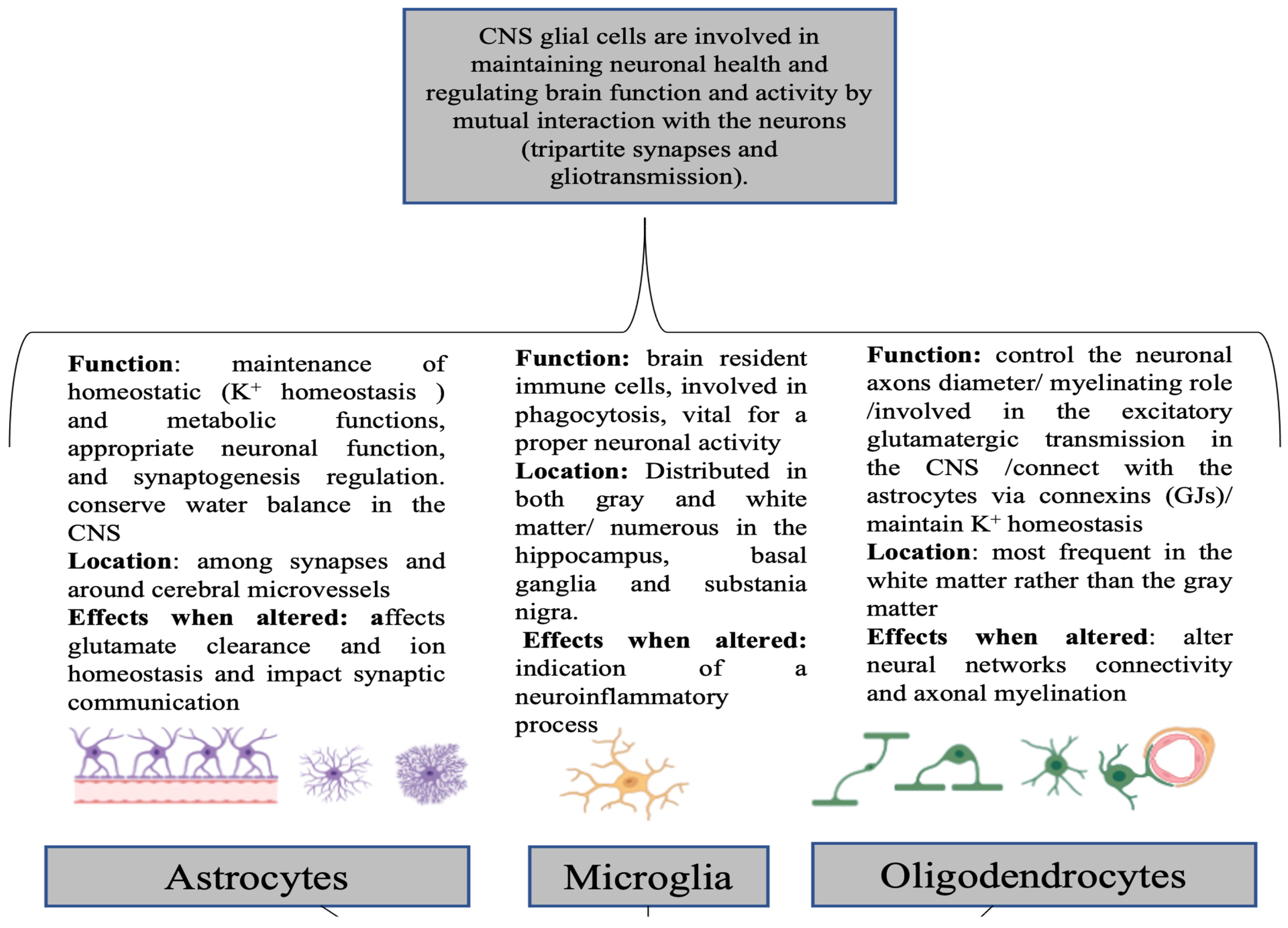
Figure 1. CNS glial cells: general overview of their location, main functions, and impact after dysfunction. (Created using BioRender.com. Maya N. Abou Chahla (2024)).
Neuroglia 06 00024 g001
Figure 2. Recent data about alterations linking SB to some mental disorders (MDD: major depressive disorder; BD: bipolar disorder; SUDs: substance use disorders).
Neuroglia 06 00024 g002
Disclaimer/Publisher’s Note: The statements, opinions and data contained in all publications are solely those of the individual author(s) and contributor(s) and not of MDPI and/or the editor(s). MDPI and/or the editor(s) disclaim responsibility for any injury to people or property resulting from any ideas, methods, instructions or products referred to in the content.