Review

Illustrating the Pathogenesis and Therapeutic Approaches of Epilepsy by Targeting Angiogenesis, Inflammation, and Oxidative Stress

by
Lucy Mohapatra
1,*,
Deepak Mishra
1,
Alok Shiomurti Tripathi
2,
Sambit Kumar Parida
3 and
Narahari N. Palei
1,*
1
Amity Institute of Pharmacy, Amity University Uttar Pradesh, Lucknow Campus, Lucknow 226028, UP, India
2
Era College of Pharmacy, Era University, Lucknow 226003, UP, India
3
Amity Institute of Pharmacy, Amity University Rajasthan, RIICO Kant Kalwar Industrial Area, Jaipur-Delhi Highway (Main Road), Jaipur 303002, RJ, India
*
Authors to whom correspondence should be addressed.
Neuroglia 2025, 6(3), 26; https://doi.org/10.3390/neuroglia6030026
Submission received: 29 April 2025 / Revised: 4 July 2025 / Accepted: 7 July 2025 / Published: 11 July 2025

Abstract

Epilepsy is one of the most prevalent chronic medical conditions that really can affect individuals at any age. A broader study of the pathogenesis of the epileptic condition will probably serve as the cornerstone for the development of new antiepileptic remedies that aim to treat epilepsy symptomatically as well as prevent the epileptogenesis process or regulate its progression. Cellular changes in the brain include oxidative stress, neuroinflammation, inflammatory cell invasion, angiogenesis, and extracellular matrix associated changes. The extensive molecular profiling of epileptogenic tissue has revealed details on the molecular pathways that might start and sustain cellular changes. In healthy brains, epilepsy develops because of vascular disruptions, such as blood–brain barrier permeability and pathologic angiogenesis. Key inflammatory mediators are elevated during epileptic seizures, increasing the risk of recurrent seizures and resulting in secondary brain injury. Prostaglandins and cytokines are well-known inflammatory mediators in the brain and, after seizures, their production is increased. These inflammatory mediators may serve as therapeutic targets in the clinical research of novel antiepileptic medications. The functions of inflammatory mediators in epileptogenesis are covered in this review. Oxidative stress also plays a significant role in the pathogenesis of various neurological disorders, specifically epilepsy. Antioxidant therapy seems to be crucial for treating epileptic patients, as it prevents neuronal death by scavenging excess free radicals formed during the epileptic condition. The significance of antioxidants in mitochondrial dysfunction prevention and the relationship between oxidative stress and inflammation in epileptic patients are the major sections covered in this review.

1. Introduction

Epilepsy is a common neurological illness that frequently causes abnormal brain electrical conductivity [1]. It results in an imbalance of excitatory and inhibitory neurotransmitters inside the neuronal system, which then contributes to psychological symptoms and a lower standard of living. An acquired epilepsy can develop from a variety of brain traumas, including infections, strokes, tumors, traumatic brain injuries (TBIs), and other neurological diseases. Numerous structural, physiological, chemical, and molecular changes occur inside brain throughout the epileptogenic process, including frequent neuronal cell death and malfunctioning of neurotransmitter systems. These are caused due to major pathological mechanisms, such as angiogenic factors, inflammation, and oxidative stress [1]. Globally, epilepsy impacts individuals of every generation and gender. It is estimated to impact sixty-five million people globally and affect people of various age groups, gender, social status, and geographical regions. The combined overall incidence of epileptic cases was observed to be 61 per 1 million people annually in a review of the literature and meta-analysis of prevalence data [2]. Due to the greater occurrence of stroke, neurologic illnesses, and tumors within that age category, males are significantly more likely than females to have epilepsy, and the condition tends to spike in the elderly. In youngsters and adults, the focal seizures seem to be more frequent than generalized seizures [3].
It has already been demonstrated that epileptogenesis involves both pathological angiogenesis and blood–brain barrier (BBB) disruptions. Steadily increasing angiogenic processes forming the overly complicated and defective arteries are observed in persons with temporal lobe epilepsy. The significance of inflammatory components and mechanisms in initial seizure events led to the theory that all these processes determine the barrier for seizure formation, which encouraged further research into their potential role in epileptogenesis [4]. The control of biological functions, cell damage, and the pathophysiology of diseases of the CNS are all impacted via the formation of free radicals. Oxidative stress (OS) is suspected to engage in many contributing factors associated with epileptogenic processes [4]. There are numerous crucial inflammatory mediators, including those in central inflammation, the BBB, and peripheral inflammation, that may play a role in epilepsy and have the potential to serve as biomarkers and benchmarks for treatment strategies. According to research regarding epilepsy, reactive oxygen species (ROS) as well as the generation of OS are common complications of severe head trauma, which may develop in neurological complications, leading to the epileptic condition [5]. Current animal experimentation has focused on how OS and ROS production maintains status epilepticus (SE) and triggers sudden seizures in people with persistent epilepsy. The high prevalence of microvasculature in the brain is likely a result of long-term adaptation to enhance microcirculation throughout seizures [6]. In conjunction with angiopoietins 1 and 2 (Ang1 and Ang2), vascular endothelial growth factor (VEGF) is recognized to contribute to physiologic or post-ischemic angiogenesis. In animal models, transitory treatment of N-acetylcysteine (NAC), a source of glutathione, can activate Nrf2 [7]. Additionally, neuroprotection and enhancements in spatially cognitive functioning were seen, with positive impacts remaining after therapy [8]. This review attempted to examine the innovative therapeutic initiatives for epilepsy targeting oxidative stress, neuroinflammation, and vascular alterations in the CNS, with the hope that they may serve as key moments in patient care in the coming decades.

2. Contributing Etiology of Epilepsy

2.1. Genetics and Epilepsy

A variety of genes and their associated genetic changes have been found involved in a minor portion of a few idiopathic seizures (in most cases, genetic factors are still unknown) and other less prevalent types of seizures. Numerous copy number variations have been found at 2q24.2–q24.3, 7q11.22, 15q11.2–q13.3, and 16p13.11–p13.2, several of which compromise a number of genes, including NRXN1, AUTS2, NLGN1, CNTNAP2, GRIN2A, PRRT2, NIPA2, and BMP5, and are linked to neurodevelopmental disorders, including autism spectrum disorders and intellectual disabilities, according to extensive genome-wide studies on epilepsy [9].

2.2. Brain Tumor and Brain Injury Leading to Epilepsy

Epilepsy is frequently brought on by brain tumors, which have a 30% epilepsy incidence. However, brain tumors are seen in roughly 4% of epilepsy patients. The central sulcus, the temporal cortex, or supplemental regions of the brain are where epilepsy is most likely to develop.
Typically, brain injury (BI) is to blame in about 20% of occurrences of symptomatic seizures. Incidents of BI, which are described as caused by a blow to the skull that is characterized by brain functioning, such as the loss of consciousness, memory loss, confusion, and focal temporary neurological deficiency, should be differentiated from surgical brain injury, which manifests structural harm, such as brain contusion or intracranial hemorrhage [10].

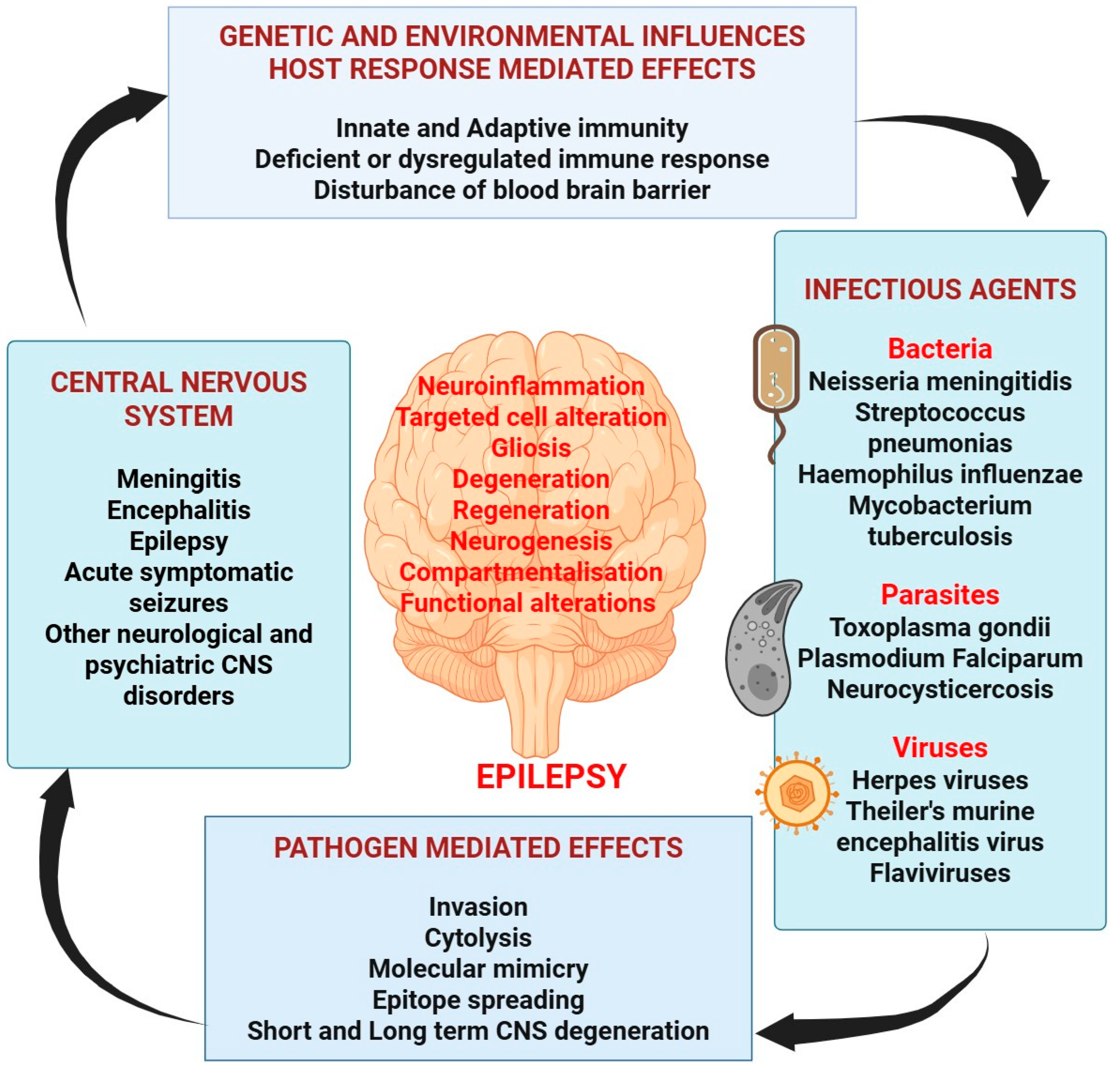
2.3. Infection and Epilepsy

Among the most frequent reasons for unprovoked symptomatic seizures accompanied by fever is meningitis. The stimulation of host pattern-recognition receptors, like TLR-4, which are known to start detrimental inflammatory responses when bacteria and their products interact, may be involved in the pathogenesis of epilepsy, as shown in Figure 1. HSV represents the most frequent cause of viral encephalitis and, as a result, HSE is frequently linked to this disease [11]. Certain amoebic brain infections, including Acanthamoeba species such as Naegleria fowleri, Balamuthia mandrillaris, and Sappinia pedata, are examples of amoeba species that may lead to severe conditions known as “Granulomatous Amoebic Encephalitis”, which impact major regions of the brain and may lead to an epileptic-like condition [12]. Additionally, there have been reports of elevated levels of T. gondii IgG in the wider populations of France (80%), the USA (20%), and Turkey (36%). According to a meta-analysis, there is a correlation between a greater rate of epilepsy and a higher incidence of toxoplasmosis, which might also suggest about the persistent Toxoplasma gondii, where there are incidents of cases of acute epilepsy [13].
The factors responsible for the cause of the epileptic condition include the different bacterial, parasitic, fungal, and viral CNS infectious species that can result in seizure and related disorders. Furthermore, the pathological processes of epilepsy due to brain infections, as well as the involvement of pathological microorganisms to explore the mechanisms of epileptogenesis, are stimulated by these agents by concentrating on the common inflammatory components held by these dysfunctions that are provoked by various etiological agents, eventually leading to the prevention of epilepsy. Infectious viruses with such a neurotropic tendency that invade the CNS and disrupt cognitive abilities, possibly through innate immunity-driven responses unique to the damaged brain tissue, can cause seizures. However, they may also be brought on by peripheral (non-CNS) disorder cell-mediated immune activity, which can lead to modifications in the BBB integrity brought on by proinflammatory cytokines, and subsequently increased neuronal excitability, among other changes.

2.4. Stroke and Epilepsy

Epilepsy is a significant risk after a stroke. Seizures and the possibility of having a stroke in old age are probably connected. Moreover, this could pertain to those who have geriatric incidence. Two to four percent of stroke victims were found to develop epilepsy during their lives in numerous significant and prospective studies [14]. According to studies, the risk of epilepsy following a stroke is 2.5% in Scandinavia over 5.5 years, 3.4% in the USA within 9 months of a stroke, and 32% in the Australian population within 26 months of initial stroke [14].

3. Understanding the Pathogenesis of Epilepsy

3.1. Role of Angiogenesis in Epilepsy

It has been demonstrated in the past ten years that the disturbance of the BBB and pathological vasculature both contribute to epigenetic changes. People with TLE have enhanced angiogenic activities that lead to the growth of abnormally complex and defective arteries. Irresolvable TLE pathology could be caused via neo-vessel rupture and BBB malfunction, which are brought about by leukocyte leaky vasculature and serum albumin absorption by neuronal cells [15]. Thus, it may be possible to attenuate epileptogenicity by using therapeutic techniques that address certain mechanisms that regulate angiogenesis and dilation of blood vessels. Depending upon those observations, we first discuss regular angiogenesis and the function of VEGF before moving on to dysfunctional vasculature related to epilepsy. Then, morphological microvascular and functioning microcirculatory alterations in epilepsy are covered [16].

3.1.1. Vascular Endothelial Growth Factor (VEGF)

Angiogenesis is a biological process in which new vasculature develops, with the help of angiogenetic substances like VEGF. VEGF-A/F or placental growth factor are two of the dimeric glycoproteins that make up that factor. Tyrosine kinase receptors VEGFR-1–3 are the three main VEGF receptors [17]. The various receptor categories that these molecules can interact with are diverse. Including its substrate VEGF-A, VEGFR-2 has mainly been explored. The markers in the brains of drug-resistant human TLE participants were profiled in a research study that looked at this VEGF signaling. VEGF and VEGFR-2 recently brought attention in relation to cerebrovascular alterations in epilepsy as a crucial part of the vasculature formation inside the CNS. After being produced, VEGF binds to VEGFR-2 on the endothelium and nerve cells to initiate paracrine signaling, which in turn promotes angiogenesis. Endothelial cells are stimulated to grow, migrate, and erupt during angiogenesis by the MEK-MAPK pathway, and then mature later [17,18].
Surprisingly, pericytes are a major producer of VEGF, and they may potentially trigger angiogenesis on their own. This helps in the generation of blood vessels with the adequate permeability and an appropriate response to vasoactive stimulation. Due to this endeavor to create a strong, carrier genesis, the mechanism of vascular maturation in the CNS is significantly more complicated. Since barrier genesis and endothelial multiplication both take place simultaneously and are triggered by multiple physiological factors, they cannot be regarded as two main pathways. Barrier genesis and endothelial cell proliferation and migration are thrown out of harmony through this VEGF production, which results in the development of matured, functioning, but leaky vasculature [19]. The role of angiogenesis regulation in the pathogenesis of epilepsy is depicted in Figure 2.
This figure illustrates the regulation of blood–brain barrier (BBB) permeability, and the role of various molecular pathways involved. At the top, the structure of the neurovascular unit is shown, comprising astrocytic foot, pericytes, and a neuron interacting with endothelial cells. Vascular endothelial growth factor (VEGF) is highlighted as a key factor involved in BBB modulation, influencing permeability through different signaling cascades. The presence of hypoxia leads to the activation of hypoxia-inducible factors (HIF), which, in turn, upregulates VEGF expression. VEGF activates downstream pathways, such as phosphatidylinositol 3-kinase (PI3K) and protein kinase B (AKT), which lead to nitric oxide (NO) production via endothelial nitric oxide synthase (eNOS) and modulate endothelial functions through the mechanistic target of rapamycin (mTOR) and ETS-related signaling. The figure also shows matrix metalloproteinases (MMP2 and MMP9) and their role in degrading components of the BBB, such as adherence junctions and tight junction proteins, like claudin-3, claudin-5, and occludin. Additionally, zonula occludens-1 (ZO-1) is depicted as a tight-junction-associated protein that plays a crucial role in maintaining BBB integrity. The interaction of neuronal activity with VEGF levels and subsequent BBB permeability modulation is emphasized, highlighting a dynamic regulatory relationship between neuronal and endothelial functions.

3.1.2. PDGF-β

Pericytes can synchronously communicate to numerous vascular endothelial cells, which sense hydrodynamic pressures inside the vessel and assist in the safeguarding of the CNS homeostasis because of its appearance and position. They show a crucial activity in barrier genesis. Regarding barrier genesis, there exists a homeostasis in the recycling of pericytes inside the walls of the vessel and underlying membrane under normal circumstances. PDGFR is a stromal cell protein that is secreted by fibroblasts, oligodendrocyte progenitor cells, and endothelium, in addition to pericytes. The lifecycle of pericytes is disturbed in SE, as evidenced by a reduction in the pericyte cell count and an unorganized structure. This implies that PDGF-β alone damages neurons or it aids in tissue regeneration by balancing the overall loss of pericytes and restoring the barrier properties [20]. Pericyte–glia injury development is associated with PDGF-β function in epilepsy. Prefrontal cortex removed from TLE patients had higher PDGFR-β sensitivity, demonstrating that PDGF-β does in fact contribute to the development of TLE. These findings show that elevated PDGFR-β production is a consequence of seizures. PDGFR might be antiepileptogenic by restoring the BBB or repairing neurodegeneration, or it might harm neurons and pericyte–glia [21].

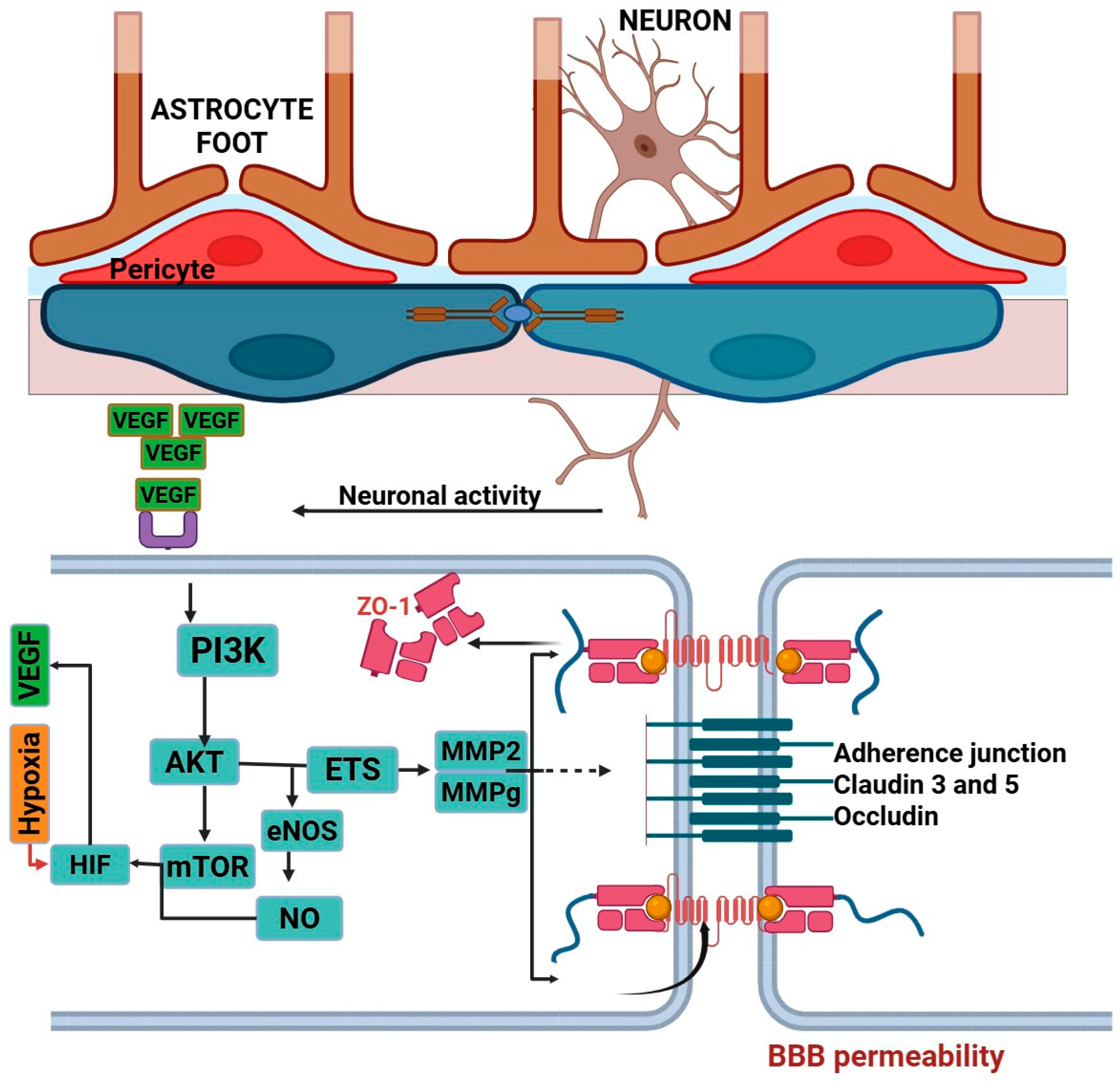
3.1.3. Blood–Brain Barrier

Endothelial cells, which possess intercellular tight and adhesion connections, without intercellular difficulty, and thus are distinguished by reduced pinocytotic function, make up the morphological barrier of the BBB. As a result, the BBB primarily serves as a barrier layer, restricting extracellular migration across brain endothelial cells. Claudins, occludin, and synaptic cell adhesion are just a few transmembrane protein components of tight junctions [22]. Catenin and cadherin are just two examples of the protein complexes that make up adhesion junctions. Increasing BBB permeability has been consistently proven to happen in seizures. There are diverse architectural abnormalities that are linked with this BBB permeability. Seizures cause BBB leaks in epilepsy and trigger a pathway involving NMDA signaling via cytosolic phospholipase A2, which increases MMP levels, diminishes junctional protein transcriptional activity, and promotes basement membrane disruption. Understanding and developing remedies in human epilepsy for the permeability of the BBB may be improved by in vivo imaging of the barrier stability [16].

3.2. Role of Inflammation in Epilepsy

An enhanced, durable, and chronic proinflammatory condition in the neural tissue’s microenvironment is related to epileptic progression coupled with moderate neuronal damage, gliosis, and microgliosis. Evidence, though, has also connected proinflammatory cytokines to neuronal loss and hippocampal neuronal death. For instance, multiple sclerosis is connected by the development of pure intra-cortical lesions, which shows significant inflammation in the cortical region, and individuals with MS are more likely to develop epilepsy. Comprehension of the neuroinflammatory mediators as well as receptors may elucidate their neuronal mechanical role in epigenetic changes [23].

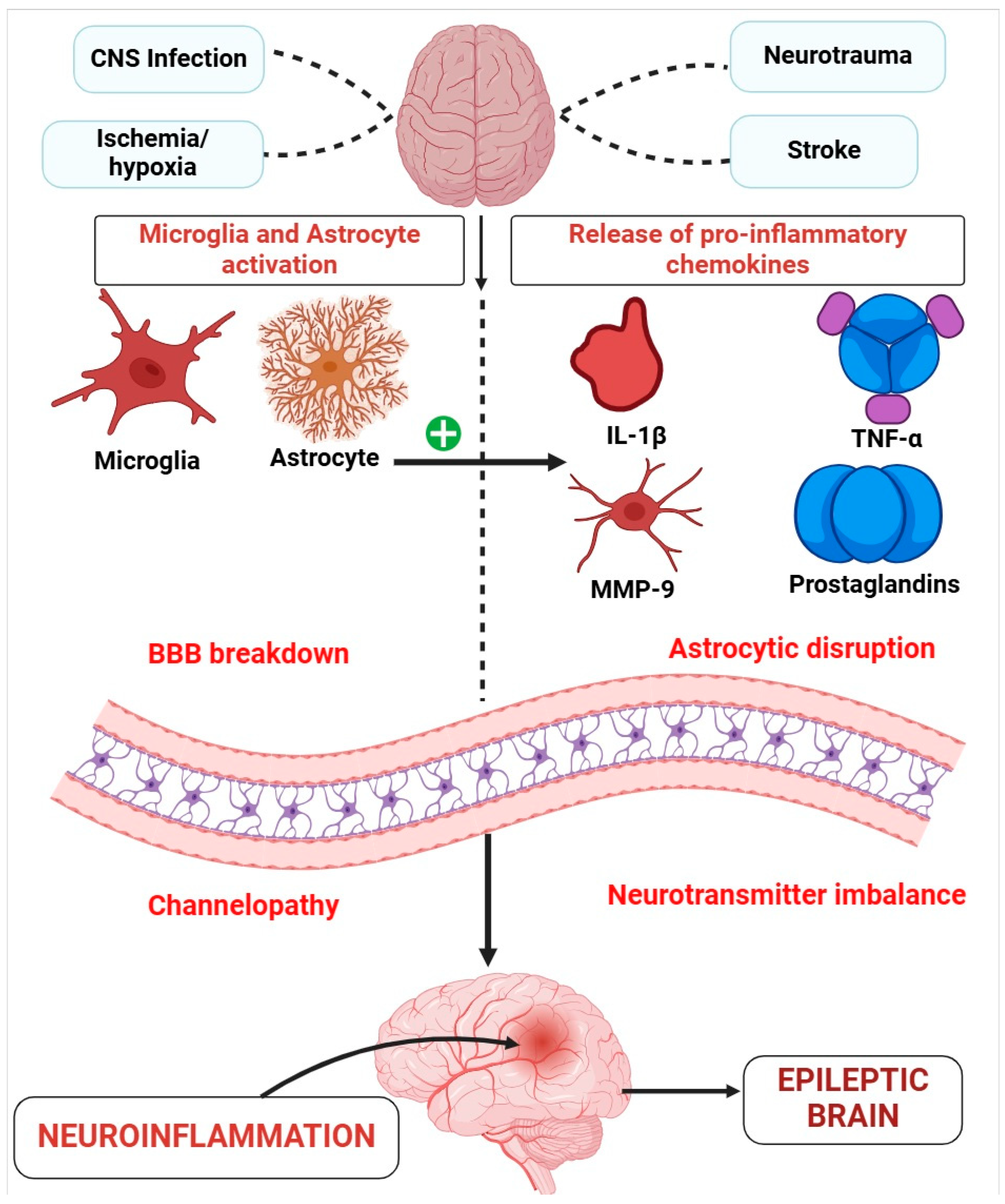
3.2.1. Glia, Neuroinflammation, and Epilepsy

There are many research studies that have concentrated on the stimulation of microglia and astrocytes and the substances these cells secrete. In both animal and human studies of epilepsy, the degree of astrocytes and microglial stimulation strongly relates to the longevity of the condition and the prevalence of random seizures [24]. In fact, a variety of epileptogenic insults cause astrocytes and glial cells to become quickly activated, creating and trying to release a variety of solubilized neurotoxic compounds, such as mediators of inflammatory cytokines, and the majority are blessed with neuroactive features, as shown in Figure 3. Notably, neuronal inflammation can result in peripheric immunity cells like leukocytes inside the brain’s parenchyma, a process that varies in intensity based on the kind of epilepsy. A harmful function of autoreactive lymphocytes in the elimination of neurological and glial cells in the known autoimmune type of epilepsy is further supported by research evidence [25].
There are various inciting epileptogenic events that can damage the central nervous system (CNS), such as CNS infection, ischemia/hypoxia, neurotrauma, and stroke. These events lead to injury in the brain. In response to CNS injury, microglia (the brain’s resident immune cells) and astrocytes (supportive glial cells in the brain) become activated. This activation leads to further signaling events that propagate inflammation. Activated microglia and astrocytes release proinflammatory molecules, including interleukin-1 beta (IL-1β), matrix metalloproteinase-9 (MMP-9), tumor necrosis factor-alpha (TNF-α), and prostaglandins. These inflammatory chemokines contribute to an inflammatory environment within the CNS. The proinflammatory environment can cause breakdown of the blood–brain barrier (BBB), leading to a loss of the selective permeability that normally protects the brain. Additionally, there is astrocytic disruption, which affects the normal functioning of astrocytes, essential for maintaining brain homeostasis. BBB breakdown and astrocytic disruption contribute to “channelopathy” (dysfunction of ion channels) and neurotransmitter imbalance. These changes disturb normal neuronal signaling and excitability. Ultimately, these processes converge, resulting in neuroinflammation and an environment conducive to seizures, leading to the development of an “epileptic brain”. This pathway suggests that interventions targeting neuroinflammation, BBB integrity, or specific inflammatory mediators might reduce or prevent the progression to epilepsy following CNS injury.

3.2.2. Chemokines and Epilepsy

There are several known pathways by which cytokines in the periphery can interact with the CNS, along with diffusion among organs other than the BBB, mostly by cranial nerves, through proinflammatory delivery vehicles, or by secreting active immune substances by BBB cells. It is significant to note that many cytokines can be produced and released by neural cells within the CNS, minimizing the need for cytokines to travel from the periphery to the brain. One hypothesis suggests that aberrant cytokine stimulation can induce prolonged alterations in the expression of immunological molecules that regulate neural connectivity and brain function [26]. Inflammatory molecules appear to become more and more prevalent and play an increasingly important role. Numerous studies have demonstrated that the cytokines are known to be generated in reaction to TBI, and increased levels of TNF-α and TGF-β, along with other cytokines, such as interleukin-1, interleukin-6, and interleukin-10, are some of those that have been frequently observed to be enhanced after TBI. It is possible that seizures will either be directly or indirectly caused by the chemokine and cytokine discharge brought on by TBI. Cytokines may interact with various immunological component groups in the CNS to exert their effects. The molecules Major Histocompatibility Complex Class I (MHC I) are members of one major group. These receptors are also present in neurons, in which they adversely control neuroplasticity necessary for activity-dependent synaptic pruning and junction formation. The capacity to form synapses is diminished in maternal immune activation due to a significant rise in MHC I expression levels in neurons in the CNS for offspring [27].
Glutamate supply at nerve terminals is increased by the proinflammatory cytokines, that is, IL-Iβ, which is produced in active astrocytes and microglia and increases glutamate outflow by astrocytes while decreasing glutamate re-uptake. This promotes neuron excitability. In one of the studies, it was demonstrated that the pathologic quantities of IL-1 in TLE could reduce GABA-mediated neural activity by 30% and result in seizure production due to neurons’ overactivity. The CSF of children with epilepsy was also found to contain much more cytokine IL-1 than the control group, indicating the cytokine’s critical function in the development of epilepsy [28].
Astrocytes and microglia are stimulated via the activation of TNF-α. To retain a specific amount of neuroexcitation input, microglia detect the concentrations of extracellular glutamate in the environment and release TNF-α when glutamate concentrations are minimal [29]. This upregulates synapses and keeps synaptic connections open. TNF-α not only increases the glutamate receptor concentration but it also triggers GABA receptor endocytosis, which lowers the inhibitory urge and results in appropriate alterations in responsiveness [29].
Prostaglandins (PGs) are mostly released by microglia and astrocytes, and many are produced by arachidonic acid by the enzyme’s cyclooxygenase-1 (COX-1) and COX-2, which are both induced to generate. The E-prostanoids (EP1, EP2, EP3, and EP4) receptors and PGE2 are connected. The function of COX-2 as a targeted therapy is still unknown, even though PGE2 production may be a valuable diagnostic strategy. Furthermore, Iwasa et al. found that blocking the secretion of PG reduced prolonged nerve cell death caused by PGD-2. Due to these contradictory findings, additional target sites within the PG synthesis and molecular mechanisms are being highlighted [30].
In the brain, cerebrum, cerebellum, microglia, and astrocytes predominantly release the protease enzyme MMP-9. In response to neuronal depolarization and elevating inflammatory mediators, such as IL-I and chemokines, the MMP-9 transcription rises. MMP-9 accelerates cell damage through a variety of processes, involving excitotoxicity, apoptosis, and the degradation of extracellular connections, hence higher MMP-9 overexpression also suggests a higher risk of epileptogenesis [31].
Glia and astrocytes are important types of cells that produce TLR1, TLR2, and TLR3, which control both adaptive and innate immune responses. The inflammatory mediators are secreted because of TLR activation, and these mediators are what cause epileptogenesis. Elimination of TLR3 suppressed seizures, TNF-α and IL-1 concentrations, microglial action, and enhanced life expectancies in an experimental animal model of epilepsy. TLR3 increased inflammatory mediators, including IFN-β, which led to hippocampus excitation. The fundamental significance of TLRs in brain inflammation and their key role in the restructuring of synaptic activity are highlighted in this study as prospective targeted therapies [31].

3.2.3. Infections as a Cause of Neuroinflammation in Epilepsy

Numerous bacterial infections in the CNS can cause immediate symptomatic seizures and, after that, epilepsy. They primarily affect the meninges and cerebral parenchyma. While bacterial infections in the CNS including meningitis can cause epilepsy, CNS infections leading to cysts formation are often linked to the onset of epilepsy in later stages. Since CSF, the BBB, and immunological features of the brain provide a special environment, CNS infections create specific challenges. Age and the prevalence of a genetic history of epilepsy appear to affect the chance of seizures arising, demonstrating that genetic mutations and neurological development are involved. Antibiotic treatment is typically not necessary at this point because most cases of epilepsy arise just after infection [32].
Additionally, fungal infections in the CNS may result in seizures. These were formerly a rather uncommon occurrence, but with the extensive utilization of steroids and cytotoxic drugs, CNS mycosis has become more common. This only accounts for a small minority of seizures in completely immunocompetent individuals. Infarctions and scar formation, hyphal vasculitis subacute or chronic basal meningitis causing parenchymal absences, myocardial infarction, and thrombosis can all be caused by fungus infections of the CNS. Seizures may occur at any stage of the infections, but those who survive tend to be at a high risk of late seizures. Encephalitis is characterized as the viral process of multiplication inside the brain that causes swelling of the parenchymal tissue of the brain. In contrast, viral infection can result in aseptic meningitis, a meningeal inflammation that does not impact the parenchyma. NMDAR-associated encephalitis is a condition that has many characteristics of viral encephalitis. Unprovoked seizures are 22 times more likely to occur after viral encephalitis exacerbated by acute seizures. The first five years following viral encephalitis are when this risk of seizures is most noticeable [33].

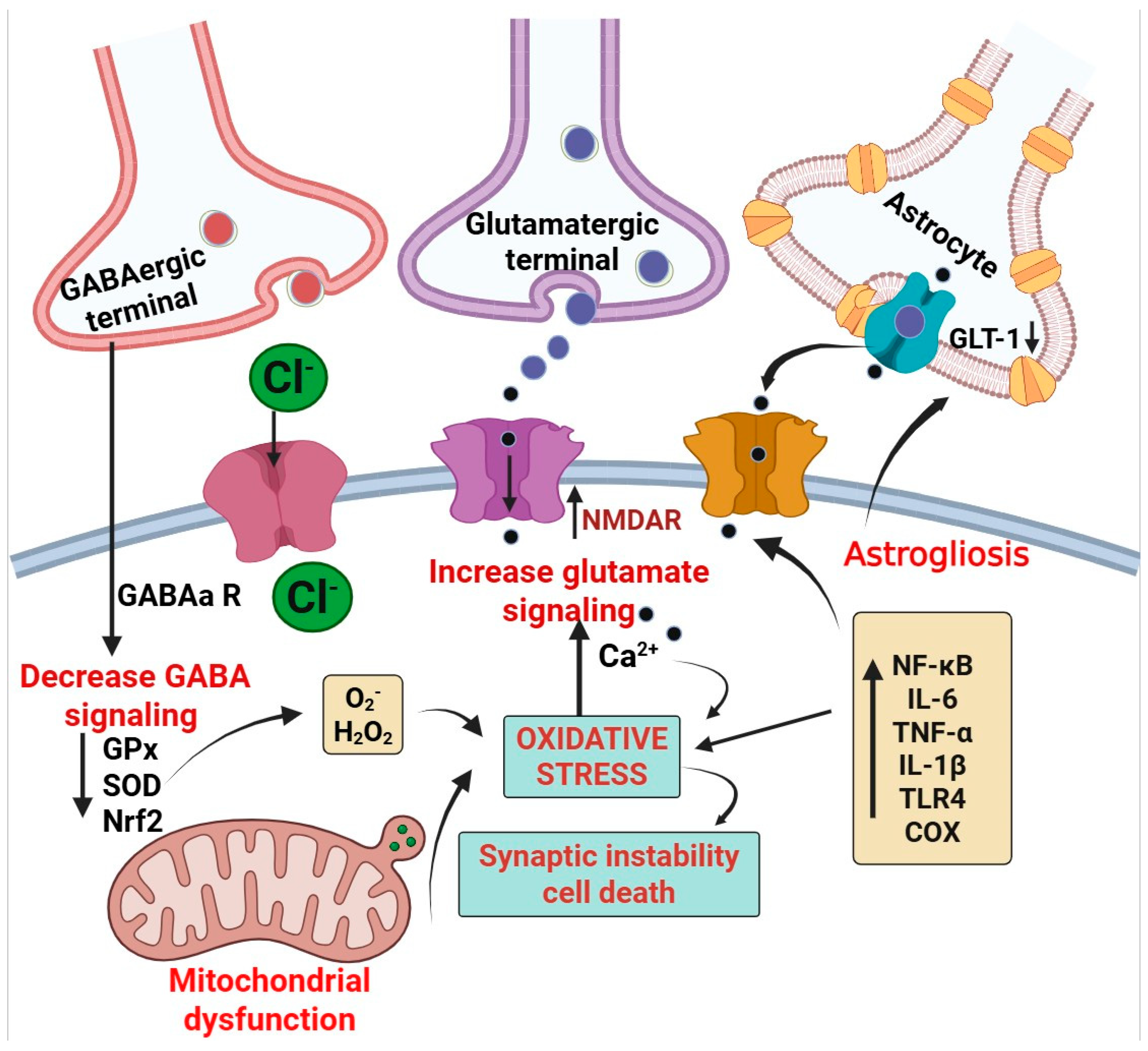
3.3. Role of Oxidative Stress in Epilepsy

Oxidative stress is a condition where an excess amount of ROS and free radicals are produced, upsetting the equilibrium among oxidative processes and biological antioxidant capacity, and having detrimental effects in vivo. Additionally, it has been shown in recent times that increased OS caused by excessive ROS and free radical generation accelerates aging as well as several other different illnesses. The method of keeping normal neuronal activity requires maintaining a low amount of ROS. Excess supply of ROS may have a negative outcome and result in extensive mitochondrial dysfunction, destruction of protein components, and serious harm to lipid membranes, all of which can cause cells to die [34].

3.3.1. Mitochondrial Oxidative Stress and Epilepsy

Superoxide (O2−) is produced in significant quantities as a byproduct of mitochondrial metabolism in various biochemical pathways, such as the mitochondrial oxidative phosphorylation and tricarboxylic acid (TCA) cycle, making mitochondria the predominant location of production of ROS inside the cell. The respiratory chain is widely acknowledged to be the important site of ROS in mitochondria. In fact, it has long been shown that the two respiratory chain complexes, complexes I (CI) and CIII, are responsible for the generation of oxygen. Peroxiredoxin 3 (Prdx3) and thioredoxin 2 (Trx2), thus, often perform an antioxidative action in mitochondria. Free radical production and scavenger processes tightly preserve the mtROS equilibrium physiologically. But after the onset of pathological conditions like epilepsy, an excessive amount of ROS production, particularly in the brain mitochondria, manifests and causes oxidative damage. Additionally, various enzymatic activities inside the mitochondria produce ROS, involving G-3-PDF, CYP-450, monoamine oxidase, and Cyt-b5 reductase. In fact, it is thought that mitochondrial proteins, lipids, and DNA are main targets of OS because of their accessibility to ROS, resulting in a “vicious loop” of damage from free radicals in the organelles, as evidently shown in Figure 4 [35].
This figure represents the interplay between GABAergic and glutamatergic signaling, oxidative stress, and neuroinflammation, with a focus on neuronal and astrocytic functions. The diagram shows GABAergic and glutamatergic terminals releasing their respective neurotransmitters. Reduced GABAergic signaling is indicated through decreased GABA-A receptor activation, which impairs inhibitory neurotransmission. Concurrently, increased glutamate release and signaling via N-methyl-D-aspartate receptors (NMDARs) lead to an influx of calcium ions (Ca2+), promoting excitotoxicity and oxidative stress. The mitochondrial dysfunction is highlighted, with reactive oxygen species (ROS) such as superoxide (O2−) and hydrogen peroxide (H2O2) exacerbating oxidative stress, accompanied by downregulated antioxidant responses involving glutathione peroxidase (GPx), superoxide dismutase (SOD), and nuclear factor erythroid 2-related factor 2 (Nrf2). Astrocytic dysfunction is represented by a decrease in glutamate transporter-1 (GLT-1), impairing glutamate clearance and contributing to excitotoxicity. Additionally, the figure depicts astrogliosis and increased proinflammatory signaling, involving pathways such as nuclear factor kappa B (NF-κB), interleukins (IL-6 and IL-1β), tumor necrosis factor-alpha (TNF-α), toll-like receptor 4 (TLR4), and cyclooxygenase (COX), collectively driving neuroinflammation, synaptic instability, and cell death.

3.3.2. Excitatory/Inhibitory (E/I) Imbalance: Relevance to Oxidative Stress and Epilepsy

A significant connection alteration that the neural network experiences during development carries the risk of causing its action to become unstable. Nerve cells are protected against hypo- or hyper-activation by homeostatic mechanisms during normal physiological circumstances from a neural perspective. Epilepsy entails a variety of abnormal changes that occur, including processes relating to ion channel physiology, synaptic vesicle (SV) discharge, and energetic metabolism, which cause a disbalance among both inhibition and excitation in particular areas of the brain. Synaptic morphology is affected by these modifications, including that of the total number of active regions and SVs in the quickly releasable pool [36]. The role of GABA (inhibitory neurotransmitter) and glutamate (excitatory neurotransmitter) is essential because the imbalance of E/I processes serves as the primary initiator in epileptic seizures. Elevated extracellular glutamate concentrations in the epileptic model and in the brains of epileptic participants are important findings that induced glutamate as a hyper-excitable phase of epilepsy. Astrocytes play a significant role in mediating epileptogenesis, particularly because of their close coupling via gap junctions, which makes it possible for them to respond quickly to stimuli when they are hyperactive. The connection between neurons and astrocytes, as an illustration, is a crucial component of glutamate physiology related to OS and epilepsy. Glutamate is taken up by astrocytes from the synapse, and in TLE, glutamate, which is an astrocyte mediator eliminated via glutamate transporter (GLT-1), is faulty, leading glutamate to build up and excite toxicity [36,37].

4. Angiogenesis, Oxidative Stress, and Inflammation Targeting Diagnostics

Assessment of the first seizure’s duration is critical if it persists at the time of medical surveillance. A prognosis of status epilepticus must be kept in mind if the seizure persists for more than twenty minutes. In all other instances, a practitioner must acquire historical and present details (by directly interrogating the participant, if possible) to establish the epileptic origin of the episode as well as whether it was an isolated event. The seizure is classified as unprovoked if it occurs without the interference of etiological factors or provoked if it is caused in close temporal association with toxic CNS damage with regard to the interval of time between a fundamental genetic predisposition or provoking medical syndrome. It is necessary to accurately represent the clinical characteristics of the episode, the findings of the neuropsychological tests, and the electrophysiological, analytical, and neuroimaging methods used to characterize the etiology and origins of the convulsions. Furthermore, accidental or traumatic brain injuries may occur in seizure activity. EEG, CT scan, MRI, and other diagnostic equipment are employed to confirm the diagnosis associated with angiogenesis, oxidative stress, and neuroinflammation [38]. Table 1 lists the different diagnostic tools for the detection of epilepsy.

5. Therapeutic Approaches for Epilepsy Targeting Angiogenesis, Inflammation, and Oxidative Stress

5.1. Angiogenesis Targeted Approach for the Treatment of Epilepsy

Investigating the underlying functions that result in the development of episodes of seizures after damage of the most vital organ, the brain, and better recognizing exactly how the triggered angiogenesis functions, which potentially leads to the development of disease-modifying medications that prolong the emergence of recurrent seizures, is important. One such treatment to be investigated is blocking angiogenesis. There are no FDA approved anti-angiogenic drugs for epilepsy, but few of them have gone through certain trials and have been found to be effective, such as sunitinib, 4-Aminopyridin (4-AMP), and levetiracetam [47].

5.1.1. Sunitinib

Sunitinib is a drug that acts as tyrosine receptor blocker specifically targeting VEGF that hinders the phosphorylation of the receptor and is now researched as a therapeutic alternative for advanced kidney cancer. It is interesting to note that the evident lack of angiogenesis in the rodents given sunitinib resulted in a lack of convulsive seizures as well as a prevention of hippocampus atrophy, suggesting a relation amongst cerebral atrophy, angiogenesis, and episode induction [48].

5.1.2. Aliskiren

The renin inhibitor aliskiren has illustrated neuroprotective characteristics in rat cortical neurons subjected to toxicity instigated by amyloid beta-peptide, credited to a reduction in renin liberation from amyloid beta, which may also correlate with the pathophysiology of epilepsy and cognitive impairments. The rectification of cognitive impairments through aliskiren has been evidenced in experimental models, pointing to an additional supportive role in the realm of epilepsy comorbidities [49].
Aliskiren has been demonstrated to have a beneficial effect in ischemic stroke conditions and in alleviating neurological outcomes following stroke, factors that are often associated with acquired epilepsy. Through the activation of the PI3K/Akt signaling pathway, the increase of beta-cell lymphoma 2 expression, and a decline of Bcl2-associated X protein expression, it displays antiapoptotic properties, reduced neurological impairments, and decreased infarcted volume [50].

5.1.3. Ephrin-A5/EphA4

It is conceivable that some pre-cultured ephrin-A5-Fc multimers in our work contribute significantly to EphA4 deregulation, and the ephrin-A5/EphA4 act over structural modification that may influence its activity. The activation of EphA4 after ephrin-A5-Fc stimulation may be impacted by the impact of gene modification or protein denaturation on the clustering interaction point. The ephrin/Eph complex may become inactive because of epileptogenesis due to mutation or repositioning at the interface. A major factor for the reduction of the blockage of the association for ephrin-A5/EphA4 complexity is the augmentation of hippocampal sensory zones and the emergence of new nerves, which also help to reduce SRS frequency and intensity. This mechanism might serve as a possible intervention for regulating episodes of seizure [51].

5.1.4. Levetiracetam (LEV)

LEV, a modulator of synaptic vesicle 2A (SV2A), reveals a broad spectrum of antiepileptogenic or disease-altering attributes in experimental models of kindling, genetic, and post-traumatic epilepsy (PTE), with findings emerging that propose its potential antiepileptogenic influence in two clinical trials, among which is a phase 2 investigation concerning TBI/PTE. The observed antiepileptogenic and disease-modifying effects in animal studies typically manifest at doses that are clinically relevant, corresponding to clinically significant plasma concentrations [52].
When 500 mg/kg of LEV was given 30 min after status epilepticus (SE) ended, the frequency and intensity of seizures were significantly reduced four weeks later. A reduction in BBB permeability, as evaluated between three hours and two days after SE, a reduction of the upregulation of angiogenic factors, neovascularization, microglial activation, and proinflammatory cytokines, and the inhibition of both cytotoxic and vasogenic edema were all responsible for this efficacious result in epilepsy [53].

5.2. Inflammation Targeted Approach for the Treatment of Epilepsy

Immunoregulatory drugs with anti-inflammatory implications have a proven history of becoming effective anticonvulsants. The finest medications for controlling seizures and various brain-related inflammatory conditions associated with epilepsies that commonly occur are ACTH and steroids. These drugs are complicated, and ill-defined mechanisms of action make it difficult to fully comprehend which critical pathways are involved in their anticonvulsant effects [54]. The ability of steroids to affect GABA and other neurotransmitters may play a role in acute seizure inhibition. Preclinical studies have identified some novel medications with disease-modified and neuroprotective characteristics, such as IL-1Ra/anakinra, as shown in Table 2, canakinumab, which exhibits the anti-IL-1α activity, infliximab, which leads to an anti-TNF-α antibodies mechanism, etanercept, which acts as a receptor fusion molecule, along with the EP2 receptor blocking properties with TLR3 or TLR4 antagonistic activity [55].

5.3. Oxidative Stress Targeted Approach for the Treatment of Epilepsy

There is some potential for using antioxidant therapies alone or in combination with currently available AEDs to treat neurological disorders like epilepsy. Even though this is a relatively new science, new compounds with superior pharmacodynamic features along with clinical trials are proceeding because of the substantial interest in treating epilepsy. The plant-based cannabinoid, i.e., cannabidiol, is one extremely promising chemical with antiepileptic and antioxidant properties. When it is administered along with anti-seizure drugs that have a mechanism residing in multiple targets, it thus shows stronger antioxidant defenses but also curbs the formation of ROS [56]. Although not an antioxidant molecule, the ketogenic diet (KD) acts as an efficient antioxidant characteristic for managing epilepsy. The KD has been used for decades to tackle pediatric seizures, but its underlying mechanisms of action, which also include antioxidant benefits, are still not understood and have just recently started to come into focus [57].
Table 2. Different antiepileptic drugs targeting inflammation, oxidative stress, and angiogenesis.
Sr. No.DrugsTargeted Seizure Mechanism of ActionEffects on Certain Markers References
Anti-inflammatory therapy
1.AdalimumabPartial convulsion accompanied by focal seizureIt is a monoclonal antibody that act as a TNF-α blocker It declines the TNF-α level during epilepsy and produces anti-inflammatory action[56]
2.AnakinraInfection-mediated febrile seizureInterleukin-1 receptor blockerDepletion in IL-1-mediated brain inflammation[58]
4.Canakinumab + Anakinra Generalized tonic clonic seizureMonoclonal Ab interleukin-1 receptor blockerDepletion in IL-1-mediated brain inflammation[59]
5.TocilizumabDuring acute epilepsy and SEMonoclonal antibody, which is an interleukin-6 antagonistIt decreases the interleukin-6 [60]
5.MinocyclineDrug-resistant seizureInterleukin-1β suppressant and blocks the stimulation of microglial cellsIt declines the interleukin-1β secretion from microglial cells[61]
6.AspirinRecurrent seizureCOX-PGE2 inhibitorIt promotes hippocampal neurogenesis by blocking the COX-PGE2 pathway[62]
7.VX09-765-401Partial seizure Interleukin-1β blockerNo evidence[63]
Antioxidant therapy
8.N-acetylcysteinePentylenetetrazole-induced epilepsyReduction of glutathione precursorMilder disruption of glutathione homeostasis[64]
9.Curcumin Pentylenetetrazole-induced epilepsyChelators of metals and elevated level of free radicalsInflammation-mediating gene transcription is reduced along with elevating the level of superoxide dismutase[65]
10.Cannabidiol Drug tolerance Adenosine uptake is inhibited and suppresses GRP55Degrades the concentration of ROS and a rise in a defense mechanism related to antioxidant properties[66]
11.Coenzyme Q10Pilocarpine-induced rat modelElevated level of TCA and enzymes of antioxidantsDeterioration of lipid peroxidation and elevates the level of growth-stimulating factor[67]
12.Naringenin Pilocarpine-induced rat modelFree radicals’ concentration is elevatedAntioxidant enzymes and glutathione concentrations are shown to be enhanced[68]
13.Sulforaphane Status epilepticusStimulation of NRF2 pathwayElevates the level of glutathione and depletes the level of malondialdehyde[69]
14.Vitamin ERefractory epilepsyElevates the level of peroxyl radicalsAntioxidant capacity as well as glutathione level is enhanced[70]
Angiogenesis targeted therapy
15.Anti-VEGF antibodyKainate-induced seizure in ratsFollowing kainate-induced seizures, administration of a neutralizing anti-VEGF antibody was performed, and it was found that this completely ceased tight junction protein breakdown and vascularizationAnti-VEGF activity utilized for improving vascularization inside brain[71]
16. Angiopoietin-1Pilocarpine-induced SE in ratsThe suppression of VEGF-induced vascular permeability did not impact VEGF’s neuroprotective effects. In order to reduce vascular anomalies in the epileptic brain, andiopoetin-1 may be used as a therapeutic approachVEGF is targeted and an angiogenic marker is regulated for the treatment of epilepsy[72]

6. Recent Advances Accomplished in the Treatment of Epilepsy Targeting Angiogenesis, Inflammation, and Oxidative Stress

6.1. Drug Repurposed for the Treatment of Epilepsy

In experimental models of acquired epilepsy, numerous pharmacological compounds utilized for various therapeutic aims have revealed antiepileptogenic or disease-modifying attributes, including those with favorable safety profiles. However, excluding vigabatrin, a considerable lack of translational research exists that aims to prevent or alter epilepsy through these potentially “repurposable” pharmacological agents [73]. The failure to clinically assess these medications may represent a missed opportunity for the advancement of preventive strategies against epilepsy. In this section, we systematically examine both animal and human evidence regarding the antiepileptogenic potential of these pharmacological agents. We outline the notable deficits in our insight related to each compound that must be addressed prior to the consideration of clinical translation, and we introduce a systematic framework for the preclinical evaluation of antiepileptogenesis for potentially repurposable compounds or their combinations in prospective research efforts [74]. The potential repurposed drugs for the treatment of epilepsy are listed in Table 3.

6.2. Antiepileptic Drugs in Different Phases of Clinical Trials

It is essential to know the current data of clinical trials targeting angiogenesis, inflammation, and oxidative stress for the treatment of epilepsy. Table 4 provides specific details about different clinical trial proceedings in the field of epilepsy with their status.

7. Conclusions

We elaborately discussed the roles of angiogenesis, inflammation, and oxidative stress in causing epilepsy and the various approaches for the treatment of such seizures. The development of new antiepileptic therapeutics that attempt to halt the epileptogenic process, alter the development of epilepsy, or treat seizures symptomatically will likely be dependent on better knowledge of the pathophysiology of epilepsy. Neo-vascularization has an impact on the hippocampus and other temporal components of the epileptogenic network that exhibit vast disparities in blood flow. Even if angiogenesis explains the neuroprotective pathways that emerge during seizures, it could also contribute to the BBB’s chronic leakage, which can have negative consequences, such as neurovascular uncoupling, inflammation, and excitability. Such non-invasive biomarkers are actively sought after through either brain imaging or serum testing. However, considering their importance in clinical diagnosis for preventive or disease-modifying clinical studies, they are not yet accessible. Mechanistic biomarkers of neuroinflammation might be very useful in this situation for determining the therapeutic efficacy of anti-inflammatory treatment. The death of neurons and epileptic convulsions are influenced by oxidative stress and mitochondrial malfunction. In several animal seizure studies, there are indications suggesting that antioxidant therapy may diminish damage brought on by oxidative free radicals. Recent research has suggested that the relationship between persistent oxidative stress and mitochondrial dysfunction may be vital to the epileptogenic process. To learn about the link between oxidative stress, seizures, and age, additional preclinical and clinical investigations are needed.

Author Contributions

Conceptualization of the overall article, L.M., A.S.T. and S.K.P.; methodology and data compiling, L.M. and D.M.; data curation and analysis, D.M. and A.S.T.; writing and drafting this article, L.M., N.N.P. and D.M.; final analysis and corrections, S.K.P. and A.S.T. All authors have read and agreed to the published version of the manuscript.

Funding

This research received no external funding.

Conflicts of Interest

The authors declare that they have no known competing financial interests or personal relationships that could have appeared to influence the work reported in this paper.

References

  1. Riva, A.; Golda, A.; Balagura, G.; Amadori, E.; Vari, M.S.; Piccolo, G.; Iacomino, M.; Lattanzi, S.; Salpietro, V.; Minetti, C.; et al. New trends and most promising therapeutic strategies for epilepsy treatment. Front. Neurol. 2021, 12, 753753. [Google Scholar] [CrossRef] [PubMed]
  2. Fiest, K.M.; Sauro, K.M.; Wiebe, S.; Patten, S.B.; Kwon, C.S.; Dykeman, J.; Pringsheim, T.; Lorenzetti, D.L.; Jetté, N. Prevalence and Incidence of Epilepsy: A Systematic Review and Meta-analysis of International Studies. Neurology 2017, 88, 296–303. [Google Scholar] [CrossRef]
  3. Beghi, E. The epidemiology of epilepsy. Neuroepidemiology 2020, 54, 185–191. [Google Scholar] [CrossRef] [PubMed]
  4. Sumadewi, K.T.; Harkitasari, S.; Tjandra, D.C. Biomolecular mechanisms of epileptic seizures and epilepsy: A review. Acta Epileptol. 2023, 5, 28. [Google Scholar] [CrossRef]
  5. Huang, L.; Xiao, W.; Wang, Y.; Li, J.; Gong, J.; Tu, E.; Long, L.; Xiao, B.; Yan, X.; Wan, L. Metabotropic glutamate receptors (mGluRs) in epileptogenesis: An update on abnormal mGluRs signaling and its therapeutic implications. Neural Regen. Res. 2024, 19, 360–368. [Google Scholar] [CrossRef] [PubMed]
  6. Berendt, M.; Farquhar, R.G.; Mandigers, P.J.J.; Pakozdy, A.; Bhatti, S.F.M.; De Risio, L.; Fischer, A.; Long, S.; Matiasek, K.; Muñana, K.; et al. International veterinary epilepsy task force consensus report on epilepsy definition, classification and terminology in companion animals. BMC Vet. Res. 2015, 11, 182. [Google Scholar] [CrossRef]
  7. Han, W.; Jiang, L.; Song, X.; Li, T.; Chen, H.; Cheng, L. VEGF modulates neurogenesis and microvascular remodeling in epileptogenesis after status epilepticus in immature rats. Front. Neurol. 2021, 12, 808568. [Google Scholar] [CrossRef]
  8. Vezzani, A.; Balosso, S.; Ravizza, T. Neuroinflammatory pathways as treatment targets and biomarkers in epilepsy. Nat. Rev. Neurol. 2019, 15, 459–472. [Google Scholar] [CrossRef]
  9. Perucca, P.; Bahlo, M.; Berkovic, S.F. The genetics of epilepsy. Annu. Rev. Genom. Hum. Genet. 2020, 21, 205–230. [Google Scholar] [CrossRef]
  10. Kazis, D.; Chatzikonstantinou, S.; Ciobica, A.; Kamal, F.Z.; Burlui, V.; Calin, G.; Mavroudis, I. Epidemiology, risk factors, and biomarkers of post-traumatic epilepsy: A comprehensive overview. Biomedicines 2024, 12, 410. [Google Scholar] [CrossRef]
  11. Khatab, A.A.; Soliman, M.A.; El-Dabaa, S.S.; Abd El-Naby, S.A. Probable relationship between Toxoplasma gondii and children with cryptogenic epilepsy. Menoufia Med. J. 2020, 33, 157–161. [Google Scholar]
  12. Mohapatra, L.; Tripathi, A.S.; Prajapati, B.G.; Alka Mishra, D.; Yasir, M.; Maurya, R.K. Granulomatous Amebic Encephalitis: Evolutionary Dynamics, Advances in Diagnostics and Therapeutic Interventions. In Rising Contagious Diseases: Basics, Management, and Treatments; John Wiley & Sons, Inc.: Hoboken, NJ, USA, 2024; pp. 378–395. [Google Scholar]
  13. Lekoubou, A.; Debroy, K.; Kwegyir-Aggrey, A.; Bonilha, L.; Kengne, A.P.; Chinchilli, V.M. Risk models to predict late-onset seizures after stroke: A systematic review. Epilepsy Behav. 2021, 121, 108003. [Google Scholar] [CrossRef]
  14. Tanaka, T.; Ihara, M.; Fukuma, K.; Mishra, N.K.; Koepp, M.J.; Guekht, A.; Ikeda, A. Pathophysiology, diagnosis, prognosis, and prevention of poststroke epilepsy: Clinical and research implications. Neurology 2024, 102, e209450. [Google Scholar] [CrossRef] [PubMed]
  15. Castro-Torres, R.D.; Ureña-Guerrero, M.E.; Morales-Chacón, L.M.; Lorigados-Pedre, L.; Estupiñan-Díaz, B.; Rocha, L.; Orozco-Suárez, S.; Rivera-Cervantes, M.C.; Alonso-Vanegas, M.; Beas-Zárate, C. New Aspects of VEGF, GABA, and Glutamate Signaling in the Neocortex of Human Temporal Lobe Pharmacoresistant Epilepsy Revealed by RT-qPCR Arrays. J. Mol. Neurosci. 2020, 70, 916–929. [Google Scholar] [CrossRef]
  16. Meijer, W.C.; Gorter, J.A. Role of blood–brain barrier dysfunction in the development of poststroke epilepsy. Epilepsia 2024, 65, 2519–2536. [Google Scholar] [CrossRef]
  17. Ogaki, A.; Ikegaya, Y.; Koyama, R. Vascular abnormalities and the role of vascular endothelial growth factors in the epileptic brain. Front. Pharmacol. 2020, 11, 20. [Google Scholar] [CrossRef] [PubMed]
  18. Ureña-Guerrero, M.E.; Castañeda-Cabral, J.L.; Rivera-Cervantes, M.C.; Macias-Velez, R.J.; Jarero-Basulto, J.J.; Gudiño-Cabrera, G.; Beas-Zárate, C. Neuroprotective and neurorestorative effects of Epo and VEGF: Perspectives for new therapeutic approaches to neurological diseases. Curr. Pharm. Des. 2020, 26, 1263–1276. [Google Scholar] [CrossRef]
  19. Guo, D.; Zhang, B.; Han, L.; Rensing, N.R.; Wong, M. Cerebral vascular and blood brain–barrier abnormalities in a mouse model of epilepsy and tuberous sclerosis complex. Epilepsia 2024, 65, 483–496. [Google Scholar] [CrossRef]
  20. Yamanaka, G.; Takata, F.; Kataoka, Y.; Kanou, K.; Morichi, S.; Dohgu, S.; Kawashima, H. The neuroinflammatory role of pericytes in epilepsy. Biomedicines 2021, 9, 759. [Google Scholar] [CrossRef]
  21. Kyyriäinen, J.; Ekolle Ndode-Ekane, X.; Pitkänen, A. Dynamics of PDGFRβ expression in different cell types after brain injury. Glia 2017, 65, 322–341. [Google Scholar] [CrossRef]
  22. Löscher, W.; Friedman, A. Structural, molecular, and functional alterations of the blood-brain barrier during epileptogenesis and epilepsy: A cause, consequence, or both? Int. J. Mol. Sci. 2020, 21, 591. [Google Scholar] [CrossRef] [PubMed]
  23. Meng, F.; Yao, L. The role of inflammation in epileptogenesis. Acta Epileptol. 2020, 2, 15. [Google Scholar] [CrossRef]
  24. Shen, W.; Pristov, J.B.; Nobili, P.; Nikolić, L. Can glial cells save neurons in epilepsy? Neural Regen. Res. 2023, 18, 1417–1422. [Google Scholar]
  25. Chen, P.; Chen, F.; Zhou, B. Understanding the role of glia-neuron communication in the pathophysiology of epilepsy: A review. J. Integr. Neurosci. 2022, 21, 102. [Google Scholar] [CrossRef] [PubMed]
  26. Wolinski, P.; Ksiazek-Winiarek, D.; Glabinski, A. Cytokines and neurodegeneration in epileptogenesis. Brain Sci. 2022, 12, 380. [Google Scholar] [CrossRef] [PubMed]
  27. Chen, Y.; Nagib, M.M.; Yasmen, N.; Sluter, M.N.; Littlejohn, T.L.; Yu, Y.; Jiang, J. Neuroinflammatory mediators in acquired epilepsy: An update. Inflamm. Res. 2023, 72, 683–701. [Google Scholar] [CrossRef]
  28. Löscher, W.; Howe, C.L. Molecular mechanisms in the genesis of seizures and epilepsy associated with viral infection. Front. Mol. Neurosci. 2022, 15, 870868. [Google Scholar] [CrossRef]
  29. Vezzani, A.; Ravizza, T.; Bedner, P.; Aronica, E.; Steinhäuser, C.; Boison, D. Astrocytes in the initiation and progression of epilepsy. Nat. Rev. Neurol. 2022, 18, 707–722. [Google Scholar] [CrossRef]
  30. Iwasa, K.; Yamamoto, S.; Yagishita, S.; Maruyama, K.; Yoshikawa, K. Excitotoxicity-induced prostaglandin D2 production induces sustained microglial activation and delayed neuronal death. J. Lipid Res. 2017, 58, 649–655. [Google Scholar] [CrossRef]
  31. Mukhtar, I. Inflammatory and immune mechanisms underlying epileptogenesis and epilepsy: From pathogenesis to treatment target. Seizure 2020, 82, 65–79. [Google Scholar] [CrossRef]
  32. Khan, S. Epilepsy; An Insight into Epileptogenic Potential of Infections and Antibiotics. Biomed. J. Sci. Tech. Res. 2023, 53, 44533–44540. [Google Scholar] [CrossRef]
  33. Tan, T.H.; Perucca, P.; O’Brien, T.J.; Kwan, P.; Monif, M. Inflammation, ictogenesis, and epileptogenesis: An exploration through human disease. Epilepsia 2021, 62, 303–324. [Google Scholar] [CrossRef] [PubMed]
  34. Borowicz-Reutt, K.K.; Czuczwar, S.J. Role of oxidative stress in epileptogenesis and potential implications for therapy. Pharmacol. Rep. 2020, 72, 1218–1226. [Google Scholar] [CrossRef]
  35. Mishra, D.; Mohapatra, L.; Tripathi, A.S.; Paswan, S.K. The influential responsibility of sirtuins in senescence and associated diseases: A review. J. Biochem. Mol. Toxicol. 2024, 38, e23812. [Google Scholar] [CrossRef]
  36. Lopatina, O.L.; Malinovskaya, N.A.; Komleva, Y.K.; Gorina, Y.V.; Shuvaev, A.N.; Olovyannikova, R.Y.; Belozor, O.S.; Belova, O.A.; Higashida, H.; Salmina, A.B. Excitation/inhibition imbalance and impaired neurogenesis in neurodevelopmental and neurodegenerative disorders. Rev. Neurosci. 2019, 30, 807–820. [Google Scholar] [CrossRef]
  37. Yang, N.; Guan, Q.W.; Chen, F.H.; Xia, Q.X.; Yin, X.X.; Zhou, H.H.; Mao, X.Y. Antioxidants targeting mitochondrial oxidative stress: Promising neuroprotectants for epilepsy. Oxidative Med. Cell. Longev. 2020, 2020, 6687185. [Google Scholar] [CrossRef] [PubMed]
  38. Ein Shoka, A.A.; Dessouky, M.M.; El-Sayed, A.; Hemdan, E.E. EEG seizure detection: Concepts, techniques, challenges, and future trends. Multimed. Tools Appl. 2023, 82, 42021–42051. [Google Scholar] [CrossRef]
  39. Reshma, S.; Megha, K.B.; Amir, S.; Rukhiya, S.; Mohanan, P.V. Blood brain barrier-on-a-chip to model neurological diseases. J. Drug Deliv. Sci. Technol. 2023, 80, 104174. [Google Scholar] [CrossRef]
  40. Werry, E.L.; Bright, F.M.; Piguet, O.; Ittner, L.M.; Halliday, G.M.; Hodges, J.R.; Kiernan, M.C.; Loy, C.T.; Kril, J.J.; Kassiou, M. Recent developments in TSPO PET imaging as a biomarker of neuroinflammation in neurodegenerative disorders. Int. J. Mol. Sci. 2019, 20, 3161. [Google Scholar] [CrossRef] [PubMed]
  41. Lach, P.; Klus, W.; Zajdel, K.; Szeleszczuk, A.; Komorowska, E.; Burda, K.; Kurowski, P. Neuroinflammation in epilepsy—Diagnostics and therapeutic perspectives. Curr. Pharmacol. Rep. 2022, 8, 31–35. [Google Scholar] [CrossRef]
  42. Yang, W.; Liu, R.; Yin, X.; Wu, K.; Yan, Z.; Wang, X.; Fan, G.; Tang, Z.; Li, Y.; Jiang, H. Novel near-infrared fluorescence probe for bioimaging and evaluating superoxide anion fluctuations in ferroptosis-mediated epilepsy. Anal. Chem. 2023, 95, 12240–12246. [Google Scholar] [CrossRef]
  43. Shao, C.; Yuan, J.; Liu, Y.; Qin, Y.; Wang, X.; Gu, J.; Chen, G.; Zhang, B.; Liu, H.K.; Zhao, J.; et al. Epileptic brain fluorescent imaging reveals apigenin can relieve the myeloperoxidase-mediated oxidative stress and inhibit ferroptosis. Proc. Natl. Acad. Sci. USA 2020, 117, 10155–10164. [Google Scholar] [CrossRef] [PubMed]
  44. Si, M.; Lv, L.; Shi, Y.; Li, Z.; Zhai, W.; Luo, X.; Zhang, L.; Qian, Y. Activatable Dual-Optical Molecular Probe for Bioimaging Superoxide Anion in Epilepsy. Anal. Chem. 2024, 96, 4632–4638. [Google Scholar] [CrossRef] [PubMed]
  45. Ponisio, M.R.; Zempel, J.M.; Day, B.K.; Eisenman, L.N.; Miller-Thomas, M.M.; Smyth, M.D.; Hogan, R.E. The role of SPECT and PET in epilepsy. Am. J. Roentgenol. 2021, 216, 759–768. [Google Scholar] [CrossRef]
  46. Liu, X.; Zhang, Y.; Zhao, Y.; Zhang, Q.; Han, F. The neurovascular unit dysfunction in the molecular mechanisms of epileptogenesis and targeted therapy. Neurosci. Bull. 2024, 40, 621–634. [Google Scholar] [CrossRef] [PubMed]
  47. Reiss, Y.; Bauer, S.; David, B.; Devraj, K.; Fidan, E.; Hattingen, E.; Liebner, S.; Melzer, N.; Meuth, S.G.; Rosenow, F.; et al. The neurovasculature as a target in temporal lobe epilepsy. Brain Pathol. 2023, 33, e13147. [Google Scholar] [CrossRef]
  48. Lui, A.; Vanleuven, J.; Perekopskiy, D.; Liu, D.; Xu, D.; Alzayat, O.; Elgokhy, T.; Do, T.; Gann, M.; Martin, R.; et al. FDA-approved kinase inhibitors in preclinical and clinical trials for neurological disorders. Pharmaceuticals 2022, 15, 1546. [Google Scholar] [CrossRef]
  49. Starace, V.; Battista, M.; Brambati, M.; Cavalleri, M.; Bertuzzi, F.; Amato, A.; Lattanzio, R.; Bandello, F.; Cicinelli, M.V. The role of inflammation and neurodegeneration in diabetic macular edema. Ther. Adv. Ophthalmol. 2021, 13, 25158414211055963. [Google Scholar] [CrossRef]
  50. Villapol, S.; Janatpour, Z.C.; Affram, K.O.; Symes, A.J. The renin angiotensin system as a therapeutic target in traumatic brain injury. Neurotherapeutics 2023, 20, 1565–1591. [Google Scholar] [CrossRef]
  51. Li, Y.; Su, P.; Chen, Y.; Nie, J.; Yuan, T.F.; Wong, A.H.; Liu, F. The Eph receptor A4 plays a role in demyelination and depression-related behavior. J. Clin. Investig. 2022, 132, e152187. [Google Scholar] [CrossRef]
  52. Contreras-García, I.J.; Cárdenas-Rodríguez, N.; Romo-Mancillas, A.; Bandala, C.; Zamudio, S.R.; Gómez-Manzo, S.; Hernández-Ochoa, B.; Mendoza-Torreblanca, J.G.; Pichardo-Macías, L.A. Levetiracetam mechanisms of action: From molecules to systems. Pharmaceuticals 2022, 15, 475. [Google Scholar] [CrossRef] [PubMed]
  53. Webb, C.A.; Wanbon, R.; Otto, E.D. Levetiracetam for status epilepticus in adults: A systematic review. Can. J. Hosp. Pharm. 2022, 75, 46. [Google Scholar] [CrossRef]
  54. Lagarde, S.; Villeneuve, N.; Trébuchon, A.; Kaphan, E.; Lepine, A.; McGonigal, A.; Roubertie, A.; Barthez, M.A.; Trommsdorff, V.; Lefranc, J.; et al. Anti–tumor necrosis factor alpha therapy (adalimumab) in Rasmussen’s encephalitis: An open pilot study. Epilepsia 2016, 57, 956–966. [Google Scholar] [CrossRef]
  55. Wiciński, M.; Puk, O.; Malinowski, B. Cenobamate: Neuroprotective potential of a new antiepileptic drug. Neurochem. Res. 2021, 46, 439–446. [Google Scholar] [CrossRef]
  56. Berman, E.; Noyman, I.; Medvedovsky, M.; Ekstein, D.; Eyal, S. Not your usual drug-drug interactions: Monoclonal antibody–based therapeutics may interact with antiseizure medications. Epilepsia 2022, 63, 271–289. [Google Scholar] [CrossRef] [PubMed]
  57. Łukawski, K.; Czuczwar, S.J. Oxidative stress and neurodegeneration in animal models of seizures and epilepsy. Antioxidants 2023, 12, 1049. [Google Scholar] [CrossRef]
  58. Lai, Y.C.; Muscal, E.; Wells, E.; Shukla, N.; Eschbach, K.; Hyeong Lee, K.; Kaliakatsos, M.; Desai, N.; Wickström, R.; Viri, M.; et al. Anakinra usage in febrile infection related epilepsy syndrome: An international cohort. Ann. Clin. Transl. Neurol. 2020, 7, 2467–2474. [Google Scholar] [CrossRef] [PubMed]
  59. Costagliola, G.; Depietri, G.; Michev, A.; Riva, A.; Foiadelli, T.; Savasta, S.; Bonuccelli, A.; Peroni, D.; Consolini, R.; Marseglia, G.L.; et al. Targeting inflammatory mediators in epilepsy: A systematic review of its molecular basis and clinical applications. Front. Neurol. 2022, 13, 741244. [Google Scholar] [CrossRef]
  60. Cantarín-Extremera, V.; Jiménez-Legido, M.; Duat-Rodríguez, A.; García-Fernández, M.; Ortiz-Cabrera, N.V.; Ruiz-Falcó-Rojas, M.L.; González-Gutiérrez-Solana, L. Tocilizumab in pediatric refractory status epilepticus and acute epilepsy: Experience in two patients. J. Neuroimmunol. 2020, 340, 577142. [Google Scholar] [CrossRef]
  61. Singh, T.; Thapliyal, S.; Bhatia, S.; Singh, V.; Singh, M.; Singh, H.; Kumar, A.; Mishra, A. Reconnoitering the transformative journey of minocycline from an antibiotic to an antiepileptic drug. Life Sci. 2022, 293, 120346. [Google Scholar] [CrossRef]
  62. Zhu, K.; Hu, M.; Yuan, B.; Liu, J.X.; Liu, Y. Aspirin attenuates spontaneous recurrent seizures in the chronically epileptic mice. Neurol. Res. 2017, 39, 744–757. [Google Scholar] [CrossRef] [PubMed]
  63. Vezzani, A.; Rüegg, S. Introduction to the 2nd meeting on Immunity and Inflammation in Epilepsy (IIE 2016). Epilepsia 2017, 58, 7–10. [Google Scholar] [CrossRef]
  64. Egilmez, C.B.; Pazarlar, B.A.; Erdogan, M.A.; Erbas, O. N-acetyl cysteine: A new look at its effect on PTZ-induced convulsions. Epilepsy Res. 2023, 193, 107144. [Google Scholar] [CrossRef]
  65. Rehman, S.; Ray, A. Neuroprotective medicinal plants: Focus on Curcuma longa. EC Pharmacol. Toxicol. 2020, 8, 16–30. [Google Scholar]
  66. Massey, S.; Quigley, A.; Rochfort, S.; Christodoulou, J.; Van Bergen, N.J. Cannabinoids and Genetic Epilepsy Models: A Review with Focus on CDKL5 Deficiency Disorder. Int. J. Mol. Sci. 2024, 25, 10768. [Google Scholar] [CrossRef] [PubMed]
  67. Pradhan, N.; Singh, C.; Singh, A. Coenzyme Q10 is a mitochondrial restorer for various brain disorders. Naunyn-Schmiedeberg’s Arch. Pharmacol. 2021, 394, 2197–2222. [Google Scholar] [CrossRef]
  68. Shrivastava, A.; Gupta, J.K.; Goyal, M.K. Flavonoids and antiepileptic drugs: A comprehensive review on their neuroprotective potentials. J. Med. Pharm. Allied Sci. 2022, 11, 4179–4186. [Google Scholar] [CrossRef]
  69. Folbergrová, J.; Ješina, P.; Otáhal, J. Protective effect of sulforaphane on oxidative stress and mitochondrial dysfunction associated with status epilepticus in immature rats. Mol. Neurobiol. 2023, 60, 2024–2035. [Google Scholar] [CrossRef]
  70. Yang, M.T.; Chou, I.C.; Wang, H.S. Role of vitamins in epilepsy. Epilepsy Behav. 2023, 139, 109062. [Google Scholar] [CrossRef]
  71. Shoja, A.; Sani, M.; Balaghirad, N.; Jafary, H.; Sagharichi, M.; Alipour, M.A.; Nazerian, Y.; Moghaddam, M.H.; Bayat, A.H.; Ashraf, H.; et al. Intrahippocampal transplantation of dental pulp stem cells improved memory function and reduced neuroinflamma-tion-induced cell death in the rat’s model of seizure. Res. Sq. 2023. [Google Scholar] [CrossRef]
  72. Jackson, L.; Eldahshan, W.; Fagan, S.C.; Ergul, A. Within the brain: The renin angiotensin system. Int. J. Mol. Sci. 2018, 19, 876. [Google Scholar] [CrossRef] [PubMed]
  73. Sanz, P.; Rubio, T.; Garcia-Gimeno, M.A. Neuroinflammation and Epilepsy: From Pathophysiology to Therapies Based on Repurposing Drugs. Int. J. Mol. Sci. 2024, 25, 4161. [Google Scholar] [CrossRef] [PubMed]
  74. Parreira, G.M.; de Oliveira, A.C.; de Oliveira Guarnieri, L.; Vieira, R.P. Drug Repurposing in CNS and Clinical Trials: Recent Achievements and Perspectives Focusing on Epilepsy and Related Comorbidities. In Frontiers in Clinical Drug Research-CNS and Neurological Disorders; Bentham Science Publishers Singapore Pte. Ltd.: Singapore, 2024; Volume 12, p. 171. [Google Scholar]
  75. Wang, L.; Ding, J.; Zhu, C.; Guo, B.; Yang, W.; He, W.; Li, X.; Wang, Y.; Li, W.; Wang, F.; et al. Semaglutide attenuates seizure severity and ameliorates cognitive dysfunction by blocking the NLR family pyrin domain containing 3 inflammasome in pentylenetetrazole-kindled mice. Int. J. Mol. Med. 2021, 48, 219. [Google Scholar] [CrossRef]
  76. Pawlik, M.J.; Miziak, B.; Walczak, A.; Konarzewska, A.; Chrościńska-Krawczyk, M.; Albrecht, J.; Czuczwar, S.J. Selected molecular targets for antiepileptogenesis. Int. J. Mol. Sci. 2021, 22, 9737. [Google Scholar] [CrossRef]
  77. Vishnoi, S.; Raisuddin, S.; Parvez, S. Glutamate excitotoxicity and oxidative stress in epilepsy: Modulatory role of melatonin. J. Environ. Pathol. Toxicol. Oncol. 2016, 35, 365–374. [Google Scholar] [CrossRef] [PubMed]
  78. Cumbres-Vargas, I.M.; Zamudio, S.R.; Pichardo-Macías, L.A.; Ramírez-San Juan, E. Thalidomide attenuates epileptogenesis and seizures by decreasing brain inflammation in lithium pilocarpine rat model. Int. J. Mol. Sci. 2023, 24, 6488. [Google Scholar] [CrossRef] [PubMed]
  79. Rayadurg, V.; Muthuchellappan, R.; Rao, U. Volatile anesthetic for the control of posthypoxic refractory myoclonic status. Indian J. Crit. Care Med. Peer-Rev. Off. Publ. Indian Soc. Crit. Care Med. 2016, 20, 485. [Google Scholar] [CrossRef]
  80. Mohammadi, E.; Nikbakht, F.; Barati, M.; Roghani, M.; Vazifekhah, S.; Khanizadeh, A.M.; Heidari, Z. Protective effect of N-acetyl cysteine on the mitochondrial dynamic imbalance in temporal lobe epilepsy: Possible role of mTOR. Neuropeptides 2022, 96, 102294. [Google Scholar] [CrossRef]
  81. Mohamed, A.M.; Ali, D.A.; Kolieb, E.; Abdelaziz, E.Z. Ceftriaxone and selenium mitigate seizures and neuronal injury in pentylenetetrazole-kindled rats: Oxidative stress and inflammatory pathway. Int. Immunopharmacol. 2023, 120, 110304. [Google Scholar] [CrossRef]
  82. Madireddy, S.; Madireddy, S. Therapeutic strategies to ameliorate neuronal damage in epilepsy by regulating oxidative stress, mitochondrial dysfunction, and neuroinflammation. Brain Sci. 2023, 13, 784. [Google Scholar] [CrossRef]
  83. Klein, P.; Friedman, A.; Hameed, M.Q.; Kaminski, R.M.; Bar-Klein, G.; Klitgaard, H.; Koepp, M.; Jozwiak, S.; Prince, D.A.; Rotenberg, A.; et al. Repurposed molecules for antiepileptogenesis: Missing an opportunity to prevent epilepsy? Epilepsia 2020, 61, 359–386. [Google Scholar] [CrossRef] [PubMed]
  84. Sun Yat-Sen Memorial Hospital of Sun Yat-Sen University. Ketogenic Diet in Pediatric Intractable Epilepsy. ClinicalTrials.gov Identifier NCT06310954. Available online: https://clinicaltrials.gov/study/NCT06310954 (accessed on 25 March 2024).
  85. Hospital, School of Medicine, Zhejiang University. Neural Autoantibody Prevalence in New-Onset Focal Seizures of Unknown Etiology. ClinicalTrials.gov Identifier: NCT06388161. Available online: https://clinicaltrials.gov/ct2/show/NCT06388161 (accessed on 25 March 2024).
  86. University of California, Davis. Imaging of Neuro-Inflammation and the Risk for Post-Traumatic Epilepsy. ClinicalTrials.gov Identifier NCT03999164. Available online: https://clinicaltrials.gov/study/NCT03999164 (accessed on 25 March 2024).
  87. Mostafa, B. Clinical Study Evaluating Safety of Pentoxifylline and Celecoxib in Patients with Grand-Mal Epilepsy Treated by Phenytoin Monotherapy. ClinicalTrials.gov Identifier NCT05637086. Updated 26 January 2024. Available online: https://clinicaltrials.gov/study/NCT05637086 (accessed on 20 July 2024).
  88. Yurtdaş, D.G. Effects of Low Glycemic Index Diet in Children with Drug-Resistant Epilepsy. ClinicalTrials.gov Identifier NCT06432231. Izmir Katip Celebi University. 2024. Available online: https://clinicaltrials.gov/study/NCT06432231 (accessed on 15 July 2024).
  89. University Hospital, Montpellier. Serum Profile of Inflammatory Factors, Immune and Angiogenic in Temporal Lobe Epilepsy. ClinicalTrials.gov Identifier NCT01563627. Available online: https://clinicaltrials.gov/study/NCT01563627 (accessed on 15 July 2024).
  90. Li, F. Xiangya Hospital of Central South University. Exploring the Preventive Effect of Mitochondrial Protective Agent Idebenone on Post-Stroke Epilepsy. ClinicalTrials.gov Identifier NCT05987397. ClinicalTrials.gov. Updated 10 August 2023. Available online: https://clinicaltrials.gov/ct2/show/NCT05987397 (accessed on 7 November 2024).
  91. Washington University School of Medicine. Sodium-Glucose Cotransporter-2 Inhibitors: A Potential Novel Treatment for Epilepsy. ClinicalTrials.gov Identifier NCT05512130. ClinicalTrials.gov. Updated 24 August 2022. Available online: https://clinicaltrials.gov/ct2/show/NCT05512130 (accessed on 7 November 2024).
Figure 1. Various etiological factors causing disruption of the central nervous system, leading to epilepsy via infection.
Neuroglia 06 00026 g001
Figure 2. Illustration of the process through which VEGF is generated and causes BBB leaking.
Neuroglia 06 00026 g002
Figure 3. Role of glia and chemokines in the progression of neuroinflammation in epilepsy.
Neuroglia 06 00026 g003
Figure 4. Oxidative stress involved in the pathogenesis of epilepsy.
Neuroglia 06 00026 g004
Table 1. Diagnostic methods with their levels of evidence and application in epilepsy.
Diagnostic MethodLevel of EvidenceApplicationReferences
Diagnosis of angiogenesis-associated pathological implications in epilepsy
Synchrotron radiation-based imagingThis technique allows for high-resolution, three-dimensional imaging of microvessels within the hippocampal area, highlighting considerable modifications in vascular architecture subsequent to seizure occurrences. Empirical observations have suggested an increase in vascular density together with activation of angiogenic factors like VEGF in individuals with temporal lobe epilepsy (TLE), reflecting a correlation with seizure frequency.Super-resolution-based inverse light penetration computed imaging (SR-based ILPCI) offers systematic and intricate representations of cerebrovascular architecture at the micron scale, achieved without the utilization of contrast-enhancing substances. [39]
Angiogenesis diagnostic chipAn extensive variety of genes, revealed through experimental investigations as being differentially expressed in tissues that undergo collateral development due to arterial occlusion, allows for the identification of single-nucleotide polymorphisms (SNPs) and other epigenetic alterations, including changes in DNA methylation patterns.The detection of abnormally low or high differential expressions of any combination of the candidate genes can be accomplished in tissues such as peripheral blood cells. The recognition of SNPs, alongside shifts in DNA methylation patterns and differences in expression levels corresponding to one or more of the candidate genes, implies an underlying genetic predisposition toward inferior compared to superior collateral development.[40]
Diagnosis of neuroinflammation in epilepsy
Positron emission tomography (PET)PET imaging, notably using translocator protein (TSPO) ligands such as [11C] DPA713, has highlighted an elevated uptake in epileptogenic zones, thereby suggesting the manifestation of neuroinflammation. Investigations reveal that 85.1% of patients diagnosed with focal epilepsy showed an increased accumulation of [11C] DPA713 in their lesions.Neuroimaging approaches assist in the exact localization of epileptogenic foci, particularly in situations of treatment resistance when conventional magnetic resonance imaging (MRI) may overlook abnormalities. An increase in TSPO binding has been correlated with microglial activation, thereby implying the significant role of inflammation in the pathophysiology of seizures.[41]
Electroencephalogram EEG conducted within twenty-four hours of a seizure has a greater chance of detecting epileptogenic disturbances than one performed later. On the other hand, any reduction in the EEG baseline activity that takes place up to forty-eight hours after a seizure may be momentary and must be cautiously treated.In particular for infants, the test should be completed within twenty-four hours of an episode.[42]
Brain CT scanPerformed even on those with focal seizures or neurological complications that have been found to be epileptogenic, and related anomalies can be seen in up to half of adults and up to one-third of children. When structural damage is indicated or when it is difficult to pinpoint the cause of the episode, a CT scan is mandatory. Among many other aspects, structural lesions include spatial traumas, neuronal hypoxia, neurological damage, and post-traumatic implications that may be indicated by prolonged consciousness loss and/or post-ictal impairments. [42]
Examination of CSFIn a bid to rule out a brain infection, a CSF examination is usually performed whenever a febrile seizure is occurring and associated by meningeal indicators. Despite the absence of meningeal inflammation indications, the CSF may be abnormal.Examining the CSF is only prescribed if a brain complication is suspected in adults and children, except in the case of infants younger than six months. Examination of the CSF is generally not suggested if the patient is not suffering from high fever.[42]
Diagnosis of oxidative stress markers in epilepsy
Near-infrared fluorescence probesA novel molecular sensor has been developed for the accurate monitoring of superoxide anions, oxygen anion (O2), within the framework of ferroptosis-associated epilepsy, demonstrating exceptional sensitivity and specificity. This probe has been proficiently employed in numerous epilepsy models, generating valuable insights regarding the oscillations of O2 throughout seizure occurrences.This activatable dual-optical molecular probe enables concurrent fluorescence imaging and chemiluminescence detection of O2 within neuronal cells, thereby enhancing the understanding of oxidative stress mechanisms in epilepsy.[43]
Two-photon fluorescence probe (named HCP)The authors have developed a robust two-photon fluorescence probe, termed HCP, which allows for the real-time observation of endogenous HClO signals produced by myeloperoxidase (MPO) in the brains of kainic acid (KA)-induced epileptic murine models, in which the chlorination of the quinolone fluorophore mediated by HClO results in an amplified fluorescence response.Notably, by leveraging HCP, the investigators have additionally established a high-throughput screening approach to rapidly identify potential antiepileptic compounds focused on reducing MPO-mediated oxidative stress.[44]
Activable dual optical molecular probeSuperoxide anion (O2) in epilepsy may be precisely imaged thanks to the activatable molecular probe CL-SA, opening the door to a better comprehension of the disease’s processes and possible treatment options.Overall, CL-SA gives us a useful instrument for O2 chemical and biological research, encouraging the study of O2 variations in epilepsy and offering a trustworthy way to investigate epilepsy diagnosis and treatment.[45]
Other diagnostic toolsNeuropsychological assessments using FMRI and SPECT cannot identify a first seizure episode from follow ups, according to the research, but can be proven effective in detection of seizure afterwards and can be based on case-by-case evidence. Neuroinflammation can be visualized properly using such techniques.The use of neuropsychological testing, functional MRI, SPECT, and PET scans in patients who have had their first epileptic seizure is not often advised, but it is possible in some circumstances. Lesions can be properly visualized using these techniques, which can help in early diagnosis of epilepsy and other associated neurological disorders.[46]
Table 3. Drugs repurposed for epilepsy with their projected mechanisms.
DrugsTherapeutic TargetClinically Used Mechanism of Action in EpilepsyReferences
SemaglutideInflammation and oxidative stressDiabetes MellitusBlocking the NLR family pyrin domain-containing 3 inflammasome. It reduces oxidative stress via modulating oxidative markers.[75]
CelecoxibInflammationNSAIDsBlocking of the cyclooxygenase 2 and HMGB1/TLR-4 pathways occurs.[76]
MelatoninOxidative stressDietary supplementMelatonin substantially scavenges reactive oxygen species, including hydroxyl radicals, peroxy radicals, peroxynitrite anions, and superoxide radicals, and concurrently stimulates the synthesis of superoxide dismutase and glutathione peroxidase, recognized as highly effective antioxidant enzymes.[77]
LosartanAngiogenesisAntihypertensiveBlocks angiotensin II receptors and has shown promise as an AEG drug.[76]
ThalidomideAngiogenesis and inflammationSedative and tranquilizerIn order to prevent apoptosis, vascular damage, and neuronal injury in the brain, thalidomide and its derivatives demonstrated action via activating the GABAergic system. Thalidomide also lowers IL-1β, TNF-α, and IL-6 levels.[78]
IsofluraneVasoprotectiveAnestheticsIsoflurane acts via altering thalamocortical pathways and amplifying inhibitory postsynaptic GABAA receptor-mediated currents. Additionally, it causes dose-dependent cerebral vasodilation, which raises intracranial pressure and cerebral blood flow. [79]
N-AcetylcysteineOxidative stressAntioxidantNAC acts via GSH depletion under oxidative stress that is averted by tissue replenishment. The direct scavenging of ROS in brain tissues takes place due to providing a sulfhydryl group.[80]
CeftriaxoneOxidative stressβ-lactam antibioticCeftriaxone-mediated restoration of GLT-1 expression and extracellular glutamate clearance also increases the activity of the system xc−antiporter, and ceftriaxone upregulates the expression of the antiporter directly via increased nuclear Nrf2 levels. These actions increase
intracellular GSH and decrease oxidative stress.
[81]
AtorvastatinInflammationAntihyperlipidemicStations have anti-inflammatory
effects, reducing brain penetration by monocytes and lymphocytes, reducing production of IL-1β, tumor necrosis factor (TNF), interferon-γ, and IL-6, and increasing production of IL-10, it has a free radical quenching effect, and impacts neurosteroid synthesis, all mechanisms relevant to epileptogenesis. However, only a small number of preclinical studies have evaluated antiepileptogenesis of statins, with mixed results.
[82]
FingolimodInflammation and oxidative stressMultiple sclerosisThe anti-inflammatory and antioxidant attributes of fingolimod (1 mg/kg ip) have been tied to the mitigation of social deficits, cognitive impairments, neuronal degeneration, and neuroinflammatory responses. Furthermore, it has been linked to the mitigation of the tripling of hippocampal levels of COX-2 and TNF.[83]
Table 4. Interventions in clinical trials for the treatment of epilepsy targeting angiogenesis, inflammation, and oxidative stress.
Clinical Trial IDIntervention/TreatmentSummary of Study StatusReferences
NCT06310954Ketogenic dietAims to evaluate the efficacy of a ketogenic diet in treating pediatric intractable epilepsy and to explore its relationship with changes in inflammatory markers.Recruiting[84]
NCT06388161Detectable autoimmune antibodies targeting inflammationDetectable serum neural autoantibodies, such as NMDAR, AMPAR1, AMPAR2, LON5, DPPX, GAD65, mGluR5, and MOG, will be evaluated for focal epileptic seizure.Recruiting[85]
NCT03999164Drug: [18F] DPA-714 positron emission tomography scanningPatients undergo PET scans of the brain at two weeks and two months after injury to measure neuroinflammation. The results of the PET scans will be analyzed and correlated with the risk of post-traumatic epilepsy.Phase I[86]
NCT05637086Pentoxifylline (400 mg) and Celecoxib (200 mg)They studied the pathogenesis of epilepsy involving multiple processes, including genetics, oxidative stress, ion channels, neuroinflammation, and cellular damage, through autophagy and apoptosis.Phase II[87]
NCT06432231Low glycemic index diet treatmentThe goal of this clinical trial was to evaluate the effectiveness of a low glycemic index diet (LGID) on seizure frequency, oxidative stress markers, and quality of life in children with drug-resistant epilepsy. They measured malondialdehyde and paraoxonase enzyme activityCompleted[88]
NCT01563627Blood sampling for drug resistance biomarkers. Device used: magnetic resonance imagingThe researchers demonstrated through their clinical and experimental analyses that the dysfunction of the blood–brain barrier (BBB) significantly contributes to the mechanisms underlying epileptogenesis and the occurrence of drug resistance, particularly in relation to inflammatory mediators, immune responses, and angiogenic factors in temporal lobe epilepsy.Completed[89]
NCT05987397Idebenone 30 mg for 14 daysIdebenone is to be administered for continuous 14 days in the post-stroke epileptic patients. The proportion of patients possessing epilepsy after stroke would be measured at different timeframes, i.e., at enrollment, after 24 weeks, and after 48 weeks, respectively.Phase 4[90]
NCT05512130Empagliflozin (25 mg daily for two weeks)Sodium-glucose cotransporter-2 inhibitors (SGLT2i), such as empagliflozin, have become important additions to the armamentarium for treating type 2 diabetes. SGLT2i decrease blood sugar by causing glucosuria, and they induce mild ketosis. These actions raise the possibility that SGLT2i can replace the MAD and LGIT as epilepsy treatments. [91]
Disclaimer/Publisher’s Note: The statements, opinions and data contained in all publications are solely those of the individual author(s) and contributor(s) and not of MDPI and/or the editor(s). MDPI and/or the editor(s) disclaim responsibility for any injury to people or property resulting from any ideas, methods, instructions or products referred to in the content.

