Article

Antioxidant System Disturbances, Bioenergetic Disruption, and Glial Reactivity Induced by Methylmalonic Acid in the Developing Rat Brain

by
Cristiano Antonio Dalpizolo
1,
Josyane de Andrade Silveira
1,
Manuela Bianchin Marcuzzo
1,
Vitor Gayger-Dias
1,
Vanessa-Fernanda Da Silva
1,
Camila Vieira Pinheiro
2,
Bruno Pereira dos Santos
3,
Tiago Franco de Oliveira
3,
Carlos-Alberto Gonçalves
1,4,5 and
Guilhian Leipnitz
1,2,4,5,6,*
1
Programa de Pós-Graduação em Ciências Biológicas: Bioquímica, Instituto de Ciências Básicas da Saúde, Universidade Federal do Rio Grande do Sul, 2600 Ramiro Barcelos Street—Attached, Porto Alegre 90035-003, RS, Brazil
2
Programa de Pós-Graduação em Ciências Biológicas: Fisiologia, Instituto de Ciências Básicas da Saúde, Universidade Federal do Rio Grande do Sul, 2600 Ramiro Barcelos Street—Attached, Porto Alegre 90035-003, RS, Brazil
3
Graduate Program in Health Sciences, Federal University of Health Sciences of Porto Alegre, 245 Sarmento Leite Street, Porto Alegre 90050-170, RS, Brazil
4
Programa de Pós-Graduação em Neurociências, Instituto de Ciências Básicas da Saúde, Universidade Federal do Rio Grande do Sul, 2600 Ramiro Barcelos Street—Attached, Porto Alegre 90035-003, RS, Brazil
5
Departamento de Bioquímica, Instituto de Ciências Básicas da Saúde, Universidade Federal de Rio Grande do Sul, 2600 Ramiro Barcelos Street—Attached, Porto Alegre 90035-003, RS, Brazil
6
Functional Pharmacology and Neuroscience, Department of Surgical Sciences, Uppsala University, 75124 Uppsala, Sweden
*
Author to whom correspondence should be addressed.
Neuroglia 2025, 6(3), 25; https://doi.org/10.3390/neuroglia6030025
Submission received: 30 April 2025 / Revised: 23 June 2025 / Accepted: 25 June 2025 / Published: 30 June 2025

Abstract

Background: Elevated levels of methylmalonic acid (MMA) are observed in the bodily fluids and tissues of patients with methylmalonic aciduria, a metabolic disorder characterized by manifestations such as vomiting, lethargy, muscle weakness, seizures, and coma. Objectives and Methods: To better understand the neuropathological mechanisms underlying this condition, we investigated the effects of intraperitoneal (i.p.) and intracerebroventricular (i.c.v.) administration of MMA on antioxidant defenses, citric acid cycle functioning, and glial reactivity in the cerebral cortex and striatum of Wistar rats. Amino acid levels were also quantified. Results: i.p. and i.c.v. administration of MMA decreased reduced glutathione levels and altered the activities of different antioxidant enzymes in the cortex and striatum. The activity of the citric acid cycle enzyme succinate dehydrogenase was diminished in both brain regions by i.p. and i.c.v. administration. Citrate synthase, isocitrate dehydrogenase, and malate dehydrogenase activities were further inhibited in the striatum. Furthermore, the i.p. administration increased glial fibrillary acidic protein (GFAP) and glucose transporter 1 (GLUT1) levels, whereas i.c.v. administration elevated GFAP and ionized calcium-binding adaptor molecule 1 (IBA1) levels in the striatum, suggesting glial activation. In contrast, no significant changes in glial markers were detected in the cortex. Moreover, synaptophysin levels remained unaltered in both regions. Finally, i.p. administration increased glutamate, glycine, and serine levels and reduced tyrosine concentrations in the striatum. Conclusions: Our findings indicate that oxidative stress, bioenergetic dysfunction, and glial reactivity induced by MMA may contribute to the neurological deficits observed in methylmalonic aciduria.

1. Introduction

Isolated methylmalonic aciduria is an organic aciduria caused by a deficiency in L-methylmalonyl-CoA mutase (mut0 and mut subtypes, respectively), methylmalonyl-CoA epimerase, or by defects in the synthesis of its active cofactor, 5′-deoxyadenosylcobalamin (subtypes cblA, cblB, cblC, and cblD MMA subtypes) [1,2,3]. The disorder disrupts the catabolic pathway of propionate, which is derived from isoleucine, threonine, valine, and methionine, as well as from odd-chain fatty acids, cholesterol esters, and propionic acid produced by the gut microbiota [1,2,3]. The biochemical profile is characterized by tissue accumulation and elevated urinary excretion of methylmalonic acid (MMA), 3-hydroxypropionic acid, methylcitric acid, tiglylglycine, propionyl glycine, and propionylcarnitine, along with lactic acidemia and hyperglycinemia. Collectively, methylmalonic acidurias affect approximately 1.14 per 100,000 newborns [4,5].
Clinical manifestations can emerge anytime from the neonatal period to adulthood [2,3]. Early-onset forms are more common, with patients typically presenting intermittent acute episodes characterized by anorexia, vomiting, respiratory distress, lethargy progressing to coma, seizures, severe metabolic acidosis, hyperammonemia, neutropenia, and thrombocytopenia [2,3]. Acute metabolic decompensation, often triggered by increased catabolic stress, exacerbates neurological symptoms and may result in irreversible central nervous system damage. In the chronic phase, the condition is marked by hypotonia, failure to thrive, and developmental and psychomotor delay. Individuals with methylmalonyl-CoA epimerase deficiency may be asymptomatic or exhibit ataxia, dysarthria, hypotonia, mild spastic paraparesis, and seizures [2]. Neuroimaging studies typically reveal basal ganglia lesions, acute cytotoxic edema, and chronic white matter abnormalities, including cortical atrophy [1,2]. The pathophysiology of methylmalonic aciduria is still a matter of intense investigation. Pioneering studies demonstrated that MMA impairs the transport of energetic substrates, reduces the activity of mitochondrial respiratory chain complexes, which results in impaired mitochondrial respiration, and increases both glucose consumption and lactate production. It is important to note that MMA is an inhibitor of succinate dehydrogenase (SDH) activity and of the mitochondrial dicarboxylate that transports succinate [3,6,7,8,9]. MMA was also shown to inhibit the activities of complexes I, I + III, and II + III, CO2 production from glucose and acetate, increase lactate production as well as glucose uptake in rat brain, indicating that aerobic glucose oxidation is affected [3,9]. These findings suggest the presence of bioenergetic dysfunction in the brain and other tissues and may explain the increased lactate levels observed in patients [2,3]. Additionally, MMA has been shown to induce oxidative stress via increased production of reactive oxygen species (ROS) and disruption of antioxidant defenses in rat brain [3,7,10]. This mechanism has also been associated with glial dysfunction in mice [3].
To better characterize the mechanisms involved in the neurological manifestations of methylmalonic aciduria, we investigated the effects of in vivo intracerebral and intraperitoneal (systemic) administration of MMA in the rat brain. We aimed to compare the vulnerability of the cerebral cortex and striatum, two regions particularly affected in methylmalonic aciduria, to the toxic effects of MMA using different animal models. Since mounting evidence shows that MMA induces oxidative stress and bioenergetic impairment [3,6,7,8,9,10], we investigated here the effects of MMA on antioxidant defenses, citric acid cycle (CAC) activity, and neural damage. The levels of the amino acids (threonine, tryptophan, valine, tyrosine, serine, proline, methionine, lysine, hydroxyproline, histidine, leucine, isoleucine, glutamine, alanine, arginine, asparagine, phenylalanine, glutamate, and aspartate) were also measured in the cerebral cortex and striatum by LC-MS/MS. This experimental design addresses gaps in the current understanding of how MMA contributes to brain dysfunction and may support future strategies for preventing or mitigating neurological injury in this disorder.

2. Materials and Methods

2.1. Animals and Reagents

Thirty-day-old male Wistar rats, weighing approximately 100–120 g, were obtained from the Centro de Reprodução e Experimentação de Animais de Laboratório (CREAL), ICBS, UFRGS, Porto Alegre, RS, Brazil. They were housed under a 12:12-h light/dark cycle (lights on 7:00 am–7:00 pm) at a controlled temperature of 22 ± 1 °C, with ad libitum access to water and 20% (w/w) protein commercial chow (SUPRA, São Leopoldo RS, Brazil). All procedures were conducted following Brazilian animal welfare legislation (Law 11,794/2008), the Guide for the Care and Use of Laboratory Animals (National Institutes of Health, publication no. 80–23, revised 2011), and Directive 2010/63/EU. The study protocol was approved by the Committee on the Ethical Use of Animals of UFRGS (project number 46592). All chemicals were purchased from Sigma-Aldrich Co. (St. Louis, MO, USA).
We utilized 30-day-old rats for both models described in the following sections because this period represents a critical period of neurodevelopment, corresponding to early adolescence in humans. It is characterized by fundamental processes such as intense synaptogenesis, neural circuit remodeling, mitochondrial maturation, and high cerebral energy demand, all of which are highly susceptible to metabolic and oxidative insults [11]. In addition, 30-day-old rats are considered young animals, and methylmalonic aciduria presentation occurs in young children [2,4].

2.2. Intraperitoneal Administration of MMA

At 30 days of age, the animals received three intraperitoneal (i.p.) injections of MMA (purity ≥ 99%; Sigma-Aldrich, St. Louis, MO, USA) or NaCl (a first dose of 10 μmol/g followed by two doses of 5 μmol/g body weight) with 90-min intervals between each injection. The MMA solution was freshly prepared immediately before administration, with the pH adjusted to 7.4. Rats were euthanized 60 min after the last injection, and the cerebral cortex and striatum were dissected for biochemical analyses. This experimental protocol, including the doses and the time intervals between administrations, was adapted from a previous study that investigated the effects of organic acids structurally related to MMA [12]. No animal suffering (respiratory distress and convulsions) or mortality was observed.

2.3. Intracerebroventricular Administration of MMA

Thirty-day-old rats received a bilateral intracerebroventricular (i.c.v.) injection of 2 μL of 8 M MMA (16 μmol) or 8 M NaCl. We used this dose of MMA because it was shown to cause no animal suffering (respiratory distress and convulsions) or mortality and was adequate to disturb critical CNS systems in a previous study [6]. The coordinates used for the injections were: 0.6 mm posterior to the bregma, 1.1 mm lateral to the midline, and 3.2 mm ventral from the dura [13]. Animals were euthanized 30 min after the injection. The cerebral cortex and striatum were dissected and stored at −80 °C until biochemical analysis.

2.4. Antioxidant Defenses

The cerebral cortex and striatum were homogenized (1:10 w/v) in 20 mM sodium phosphate buffer (pH 7.4) containing 140 mM KCl. After centrifugation at 750× g for 10 min at 4 °C, the supernatants were collected and used for the analysis of antioxidant defenses.
Reduced glutathione (GSH) concentrations: The evaluation of GSH levels was performed according to Browne and Armstrong [14]. One hundred microliters of tissue supernatants were treated with 2% metaphosphoric acid (1:1) and centrifuged for 10 min at 7000× g for deproteination. An aliquot of supernatant (30 μL containing approximately 0.4 mg of protein) was added to a medium containing 185 μL of 100 mM sodium phosphate buffer, pH 8.0, with 5 mM EDTA and 15 μL of o-phthaldialdehyde (1 mg/mL in methanol) and incubated in a dark room for 15 min at room temperature. The fluorescence was then measured at 350 (excitation) and 420 (emission) nm on a SpectraMax M5 plate reader (Molecular Devices, Sunnyvale, CA, USA). A calibration curve was prepared using a GSH standard solution (0.001–1 mM), and the results were expressed as nmol/mg protein.
Superoxide dismutase (SOD) activity: SOD activity was measured according to Marklund [15], based on the autoxidation of pyrogallol mediated by superoxide anion. As superoxide is the SOD substrate, pyrogallol autoxidation is inhibited by this enzyme. The monitoring of pyrogallol absorbance at 25 °C at 420 nm is used to indirectly measure SOD activity. The reaction medium contained tissue supernatants (approximately 45 μg of protein), 50 mM Tris buffer, pH 8.2, with 1 mM EDTA, 80 U/mL CAT, and 0.8 mM pyrogallol. A calibration curve was performed using purified SOD. The results were expressed as U/mg protein.
Catalase (CAT) activity: CAT activity was assayed according to Aebi [16], following the decrease of absorbance at 25 °C at 240 nm due to H2O2 decomposition. The reaction medium contained tissue supernatants (approximately 3 μg of protein), 10 mM potassium phosphate buffer, pH 7.0, 0.1% Triton X-100, and 20 mM H2O2. The results were expressed as U/mg protein.
Glutathione peroxidase (GPx) activity: GPx activity was evaluated according to Wendel [17], using tert-butyl hydroperoxide and NADPH as substrates. NADPH oxidation was monitored at 25 °C at 340 nm. The reaction medium contained tissue supernatants (approximately 75 μg protein), 100 mM potassium phosphate buffer, pH 7.0, with 1 mM EDTA, 0.4 mM sodium azide, 2 mM GSH, 0.1 U glutathione reductase/mL, 0.1 mM NADPH, and 0.5 mM tert-butyl hydroperoxide. The results were expressed as U/mg protein.
Glutathione S-transferase (GST): GST activity was determined by the method described by Mannervik and Guthenberg [18], using 1-chloro-2,4-dinitrobenzene and monitoring the formation of dinitrophenyl-S-glutathione at 25 °C at 340 nm. The reaction medium contained tissue supernatants (approximately 60 μg of protein), 50 mM potassium phosphate buffer, pH 6.5, 1 mM GSH, and 1 mM 1-chloro-2,4-dinitrobenzene. The results were expressed as U/mg protein.
Glutathione reductase (GR) activity: GR activity was measured using the method described by Carlberg and Mannervik [19], monitoring the oxidation of NADPH at 25 °C at 340 nm. The reaction medium contained tissue supernatants (approximately 90 μg of protein), 200 mM sodium phosphate buffer, pH 7.5, 6.3 mM EDTA, 1 mM oxidized glutathione (GSSG), and 0.1 mM NADPH. The results were expressed as U/mg protein.
Glucose-6-phosphate dehydrogenase (G6PDH) activity: G6PDH activity was evaluated by the method of Clark and Leong [20], which is based on the formation of NADPH at 25 °C at 340 nm. The reaction medium contained tissue supernatants (approximately 45 μg of protein), 100 mM Tris-HCl buffer, pH 7.5, 1.0 mM MgCl2, 0.05 mM NADP+, and 0.1 mM glucose-6-phosphate. The results were expressed as U/mg protein.
All methods are described in detail in Grings et al. (2017) [21].

2.5. Bioenergetics

The cerebral cortex and striatum were homogenized (1:10 w/v) in SET buffer (250 mM sucrose, 2.0 mM EDTA, and 10 mM Trizma base), pH 7.4. The homogenates were centrifuged at 800× g for 10 min at 4 °C, and the resulting supernatants were used to assess CAC enzyme activities. Citrate synthase (CS) activity was determined by measuring the reduction of 5,5′-dithiobis-(2-nitrobenzoic acid) at 412 nm [22]. Succinate dehydrogenase (SDH) activity was measured by monitoring the reduction of 2,6-dichlorophenol-indophenol at 600 nm [23], whereas malate dehydrogenase (MDH) activity was assessed by detecting the fluorescence of NADH at 366 nm (excitation) and 450 nm (emission) [24].

2.6. Western Blotting

The brain structures were homogenized (1:5 w/v) in RIPA buffer containing 1 mM sodium orthovanadate and 1% protease inhibitor cocktail. The homogenate was centrifuged at 3600× g for 10 min at 4 °C, and the protein concentration was determined by the method of Lowry et al. (1951) [25]. The assay is detailed in Grings et al. (2017) [21]. Samples were run in triplicate (30 µg of total protein) and separated by electrophoresis in SDS-PAGE (12%) and transferred to nitrocellulose membranes (0.45 µm) using a semi-dry transfer system (Bio-Rad Laboratories Inc., Hercules, CA, USA). Membranes were blocked for 2 h at room temperature with a solution containing 2% bovine serum albumin. The following primary antibodies were used in 4 °C overnight incubations: anti-GFAP (1:1000, G9269, Sigma-Aldrich, St. Louis, MO, USA), anti-vimentin (1:1000, ab8978, Abcam, Cambridge, UK), anti-GLUT1 (1:1000, MA5-31960, Thermo Fisher Scientific, Waltham, MA, USA), anti-synaptophysin (1:5000, MAB329, Millipore, Burlington, MA, USA), anti-KIR4.1 (1:1000, sc-23637, Santa Cruz Biotechnology, Dallas, TX, USA), anti-β-actin (1:30,000, HRP-60008, Proteintech, Rosemont, IL, USA). The following secondary antibodies were used in room temperature 2 h incubations: anti-mouse IgG (1:10000, E-AB 1001, Elabscience, Houston, TX, USA) and anti-rabbit IgG (1:10000, E-AB-1058, Elabscience, Houston, TX, USA). Membrane images were cropped to highlight the bands of interest. Immunoreactivity was detected by employing a chemiluminescence (ECL) detection substrate (Clarity Western ECL Substrate, Bio-Rad Laboratories Inc., Hercules, CA, USA), and chemiluminescence signals were captured using a charge-coupled device (CCD) camera (ImageQuant™ LAS 4000, GE Health Care, Piscataway, NJ, USA). Some proteins were marked on the same membrane before stripping. The optical densities (OD) of the images were calculated with ImageJ software (Version 1.54p, NIH, Bethesda, MD, USA), and the results were normalized using anti-β-Actin as a housekeeping protein.

2.7. Determination of Amino Acids Using LC-MS/MS Analysis

An aliquot of 50 μL of brain tissue homogenate (100 mg tissue/mL) and 150 μL of acetonitrile were added to a 1.5 mL polypropylene microtube. The sample was vortexed and centrifuged at 9000 rpm for 6 min. A 100 μL aliquot of the supernatant was transferred to a new microtube and mixed with 100 μL of 50 mM ammonium acetate buffer. Approximately 100 μL of this mixture was transferred to a vial, and 3 μL was injected into the liquid chromatograph–tandem mass spectrometry (LC-MS/MS) system. The analysis was performed in a Nexera-i LC-2040C Plus system coupled to an LC-MS-8045 triple quadrupole mass spectrometer (Shimadzu, Kyoto, Japan). Chromatographic separation was carried out on an NST column (250 × 4.6 mm, 5 μm particle size) (São Paulo, Brazil), with a total run of 25 min. Water (A) and methanol (B), both fortified with 0.1% formic acid, were used as mobile phases. Data acquisition was conducted in multiple reaction monitoring mode under positive electrospray ionization, using one transition for quantification and at least one transition for qualification.

2.8. Statistical Analysis

Data were expressed as means ± standard deviation (SD). Assays were performed in duplicate or triplicate, and the mean was used for statistical analysis. There were no blinding procedures for the experiments. The sample size was initially calculated with Minitab 16 software for experiments with two groups. We assumed a target power of 0.8, an SD of 10% and a difference of 35%. The data used for the Minitab parameters were assumed in accordance with previous studies from our group [26,27]. It should be noted that these calculations were used to estimate the sample size, but in some experiments, we used an N slightly different from the one determined with Minitab software, but still in accordance with previous works [26,27]. Data were analyzed using Student’s t-test. Differences between groups were rated significant at p < 0.05. All analyses were carried out in a compatible PC using the Statistical Package for the Social Sciences (SPSS v1) software.

3. Results

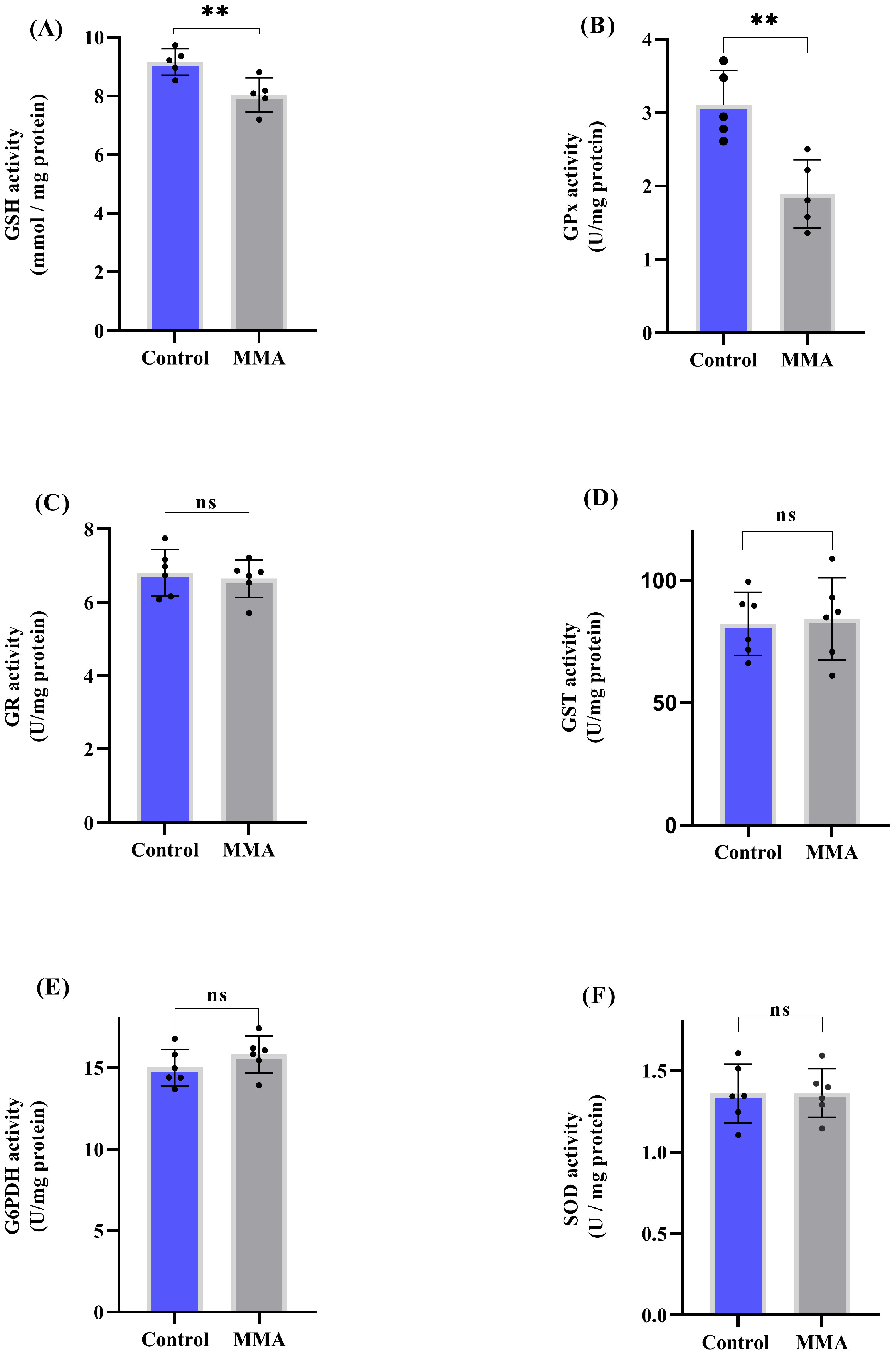
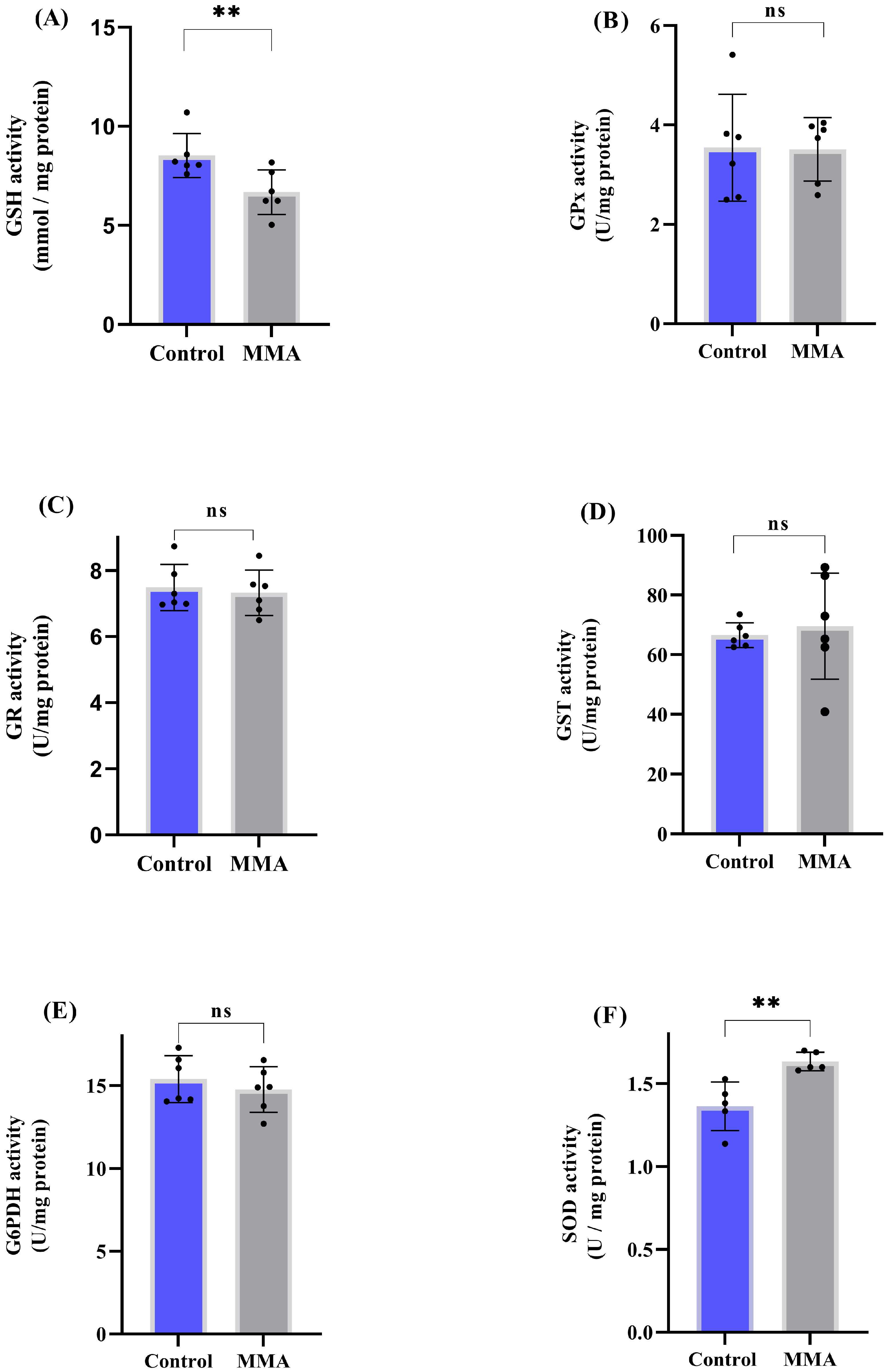
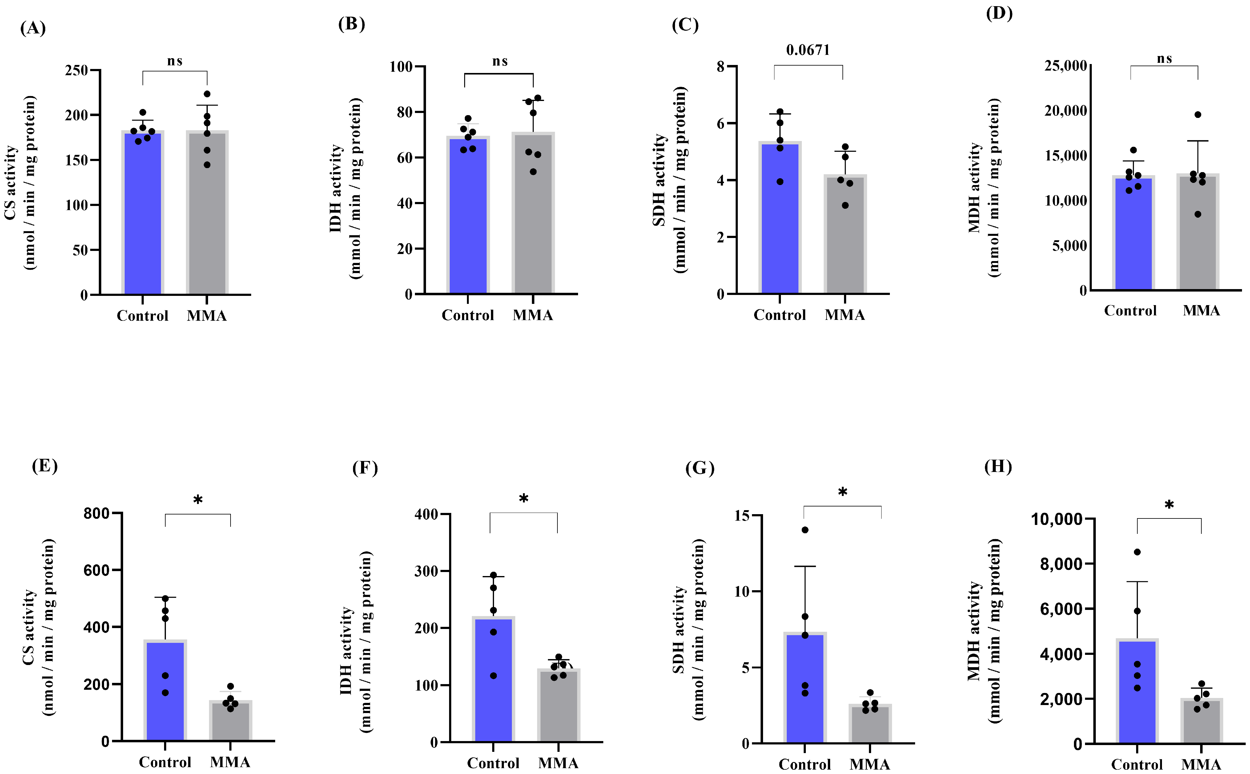
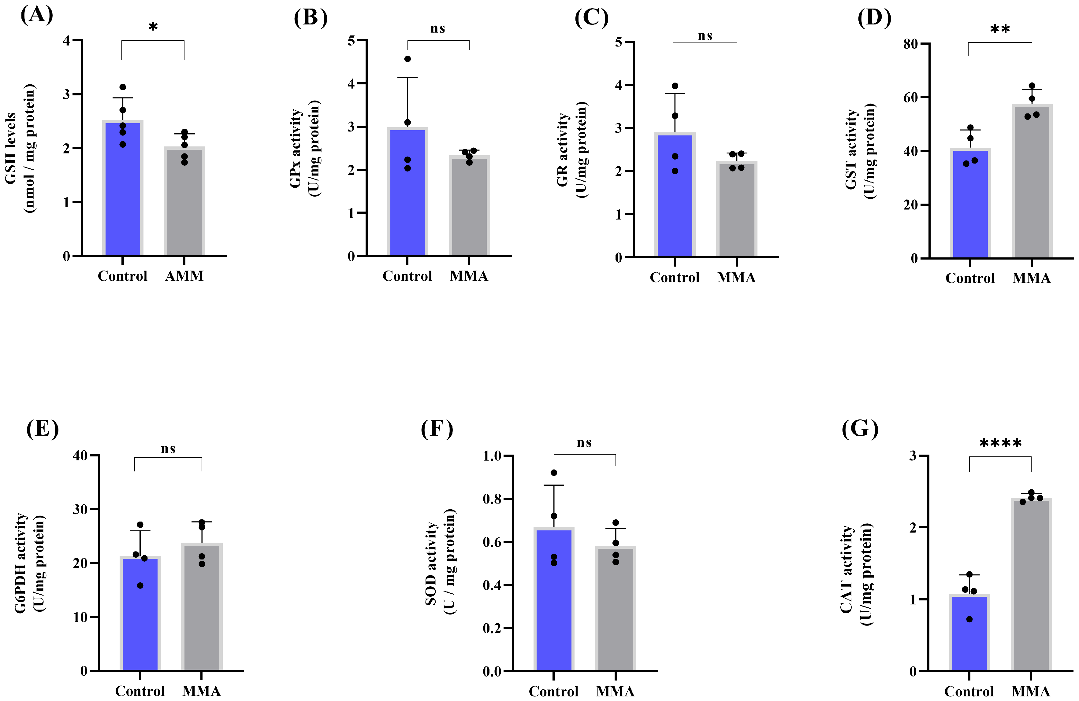
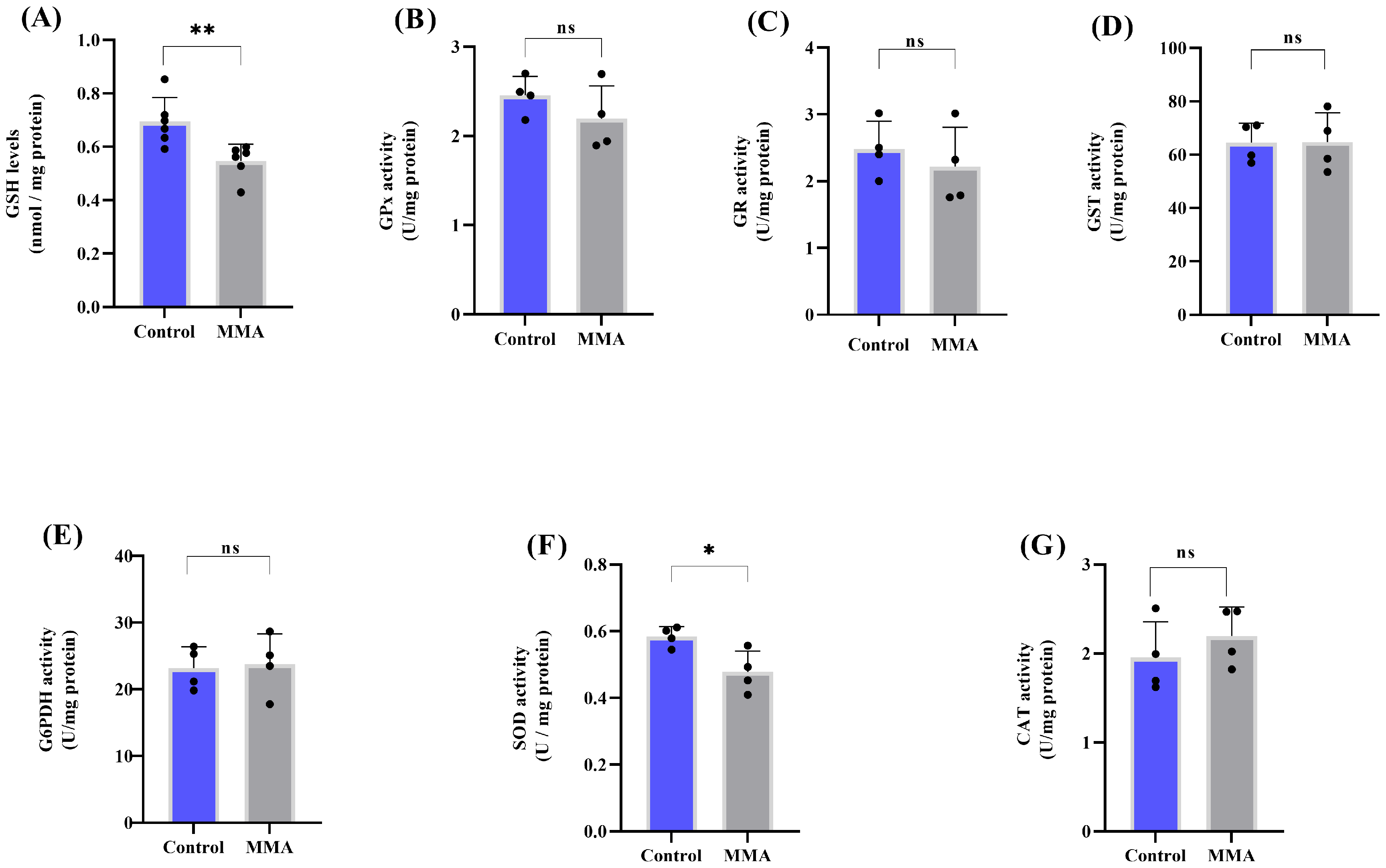
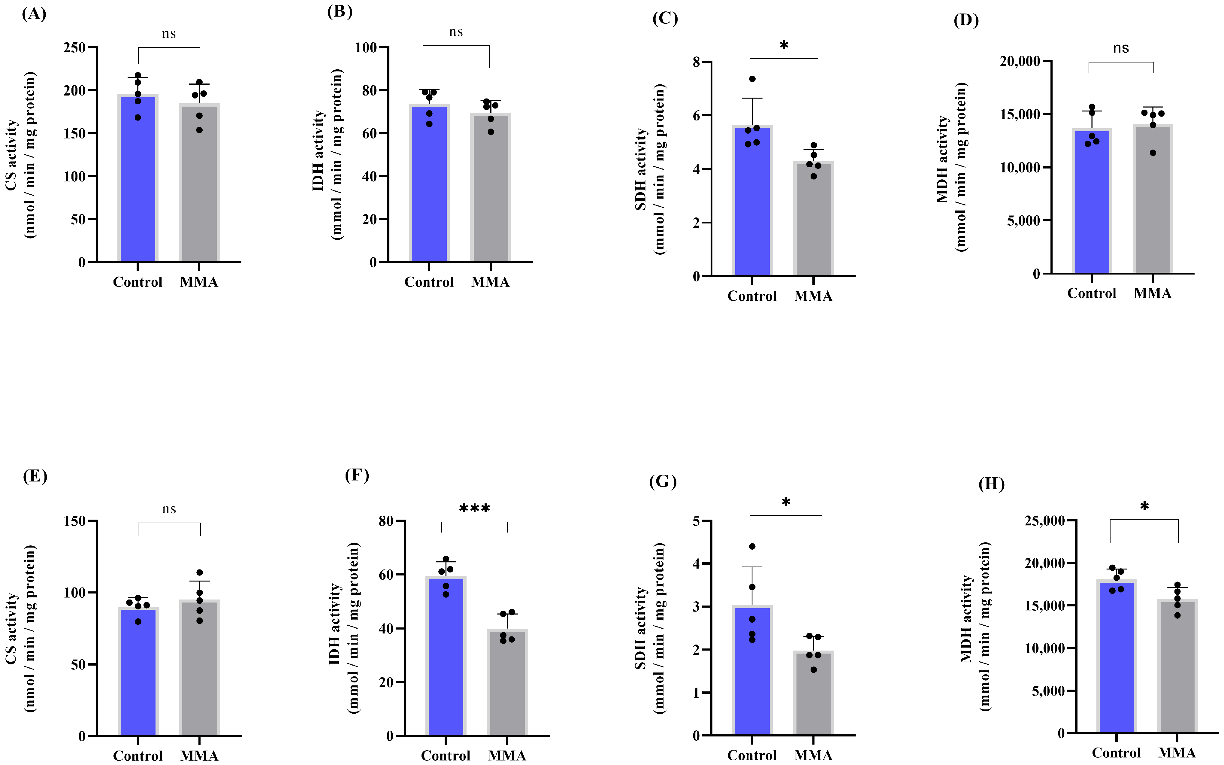
3.1. MMA Administration Disturbs Redox Status and Energy Metabolism in Rat Brain

We initially evaluated the effects of i.p. administration of MMA on antioxidant defenses in the cerebral cortex and striatum of rats. MMA significantly decreased GSH levels (p < 0.01) (Figure 1A) and GPx activity (p < 0.01). Figure 1B in the cerebral cortex. However, the activities of GR, GST, G6PDH and SOD remained unchanged in this brain region (Figure 1C–F). In the striatum, MMA decreased GSH levels (p < 0.01) (Figure 2A) and increased SOD activity (p < 0.01) (Figure 2F). In contrast, no significant changes were observed in the other antioxidant enzymatic activities in the striatal tissue (Figure 2B–E).
Next, energy metabolism was assessed in the rat brain by measuring the activities of CAC enzymes CS, IDH, SDH, and MDH. In the cerebral cortex, MMA induced a non-significant trend toward reduced SDH activity (p = 0.0671) (Figure 3C), while the activities of the other enzymes remained unchanged (Figure 3A,B,D). In the striatum, however, MMA significantly reduced the activity of all CAC enzymes evaluated (p < 0.05) (Figure 3E–H).
We also investigated the effects of i.c.v. injection of MMA on antioxidant defenses and CAC enzymes. As shown in Figure 4, MMA significantly reduced GSH levels (p < 0.05) and increased GST (p < 0.01) and CAT (p < 0.0001) activities in the cerebral cortex. In the striatum, MMA decreased GSH levels (p < 0.05) and reduced SOD activity (p < 0.05) (Figure 5).
As for the CAC enzymes, MMA inhibited SDH activity in the cerebral cortex (p < 0.05) (Figure 6C). The other activities remained unchanged (Figure 6A,B,D). In the striatum, the activities of IDH (p < 0.001), MDH (p < 0.05), and SDH (p < 0.05) were significantly reduced (Figure 6F–H).

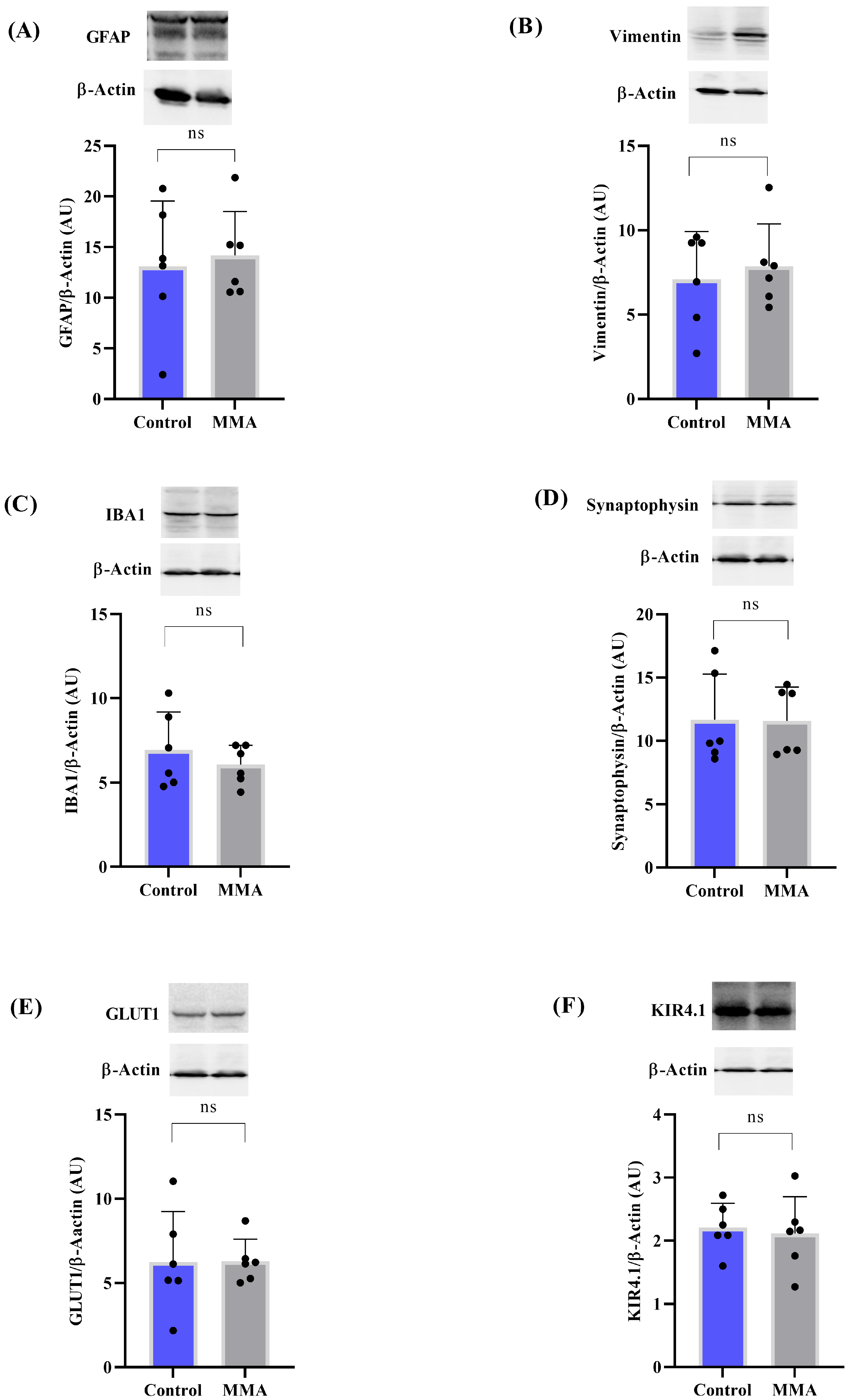
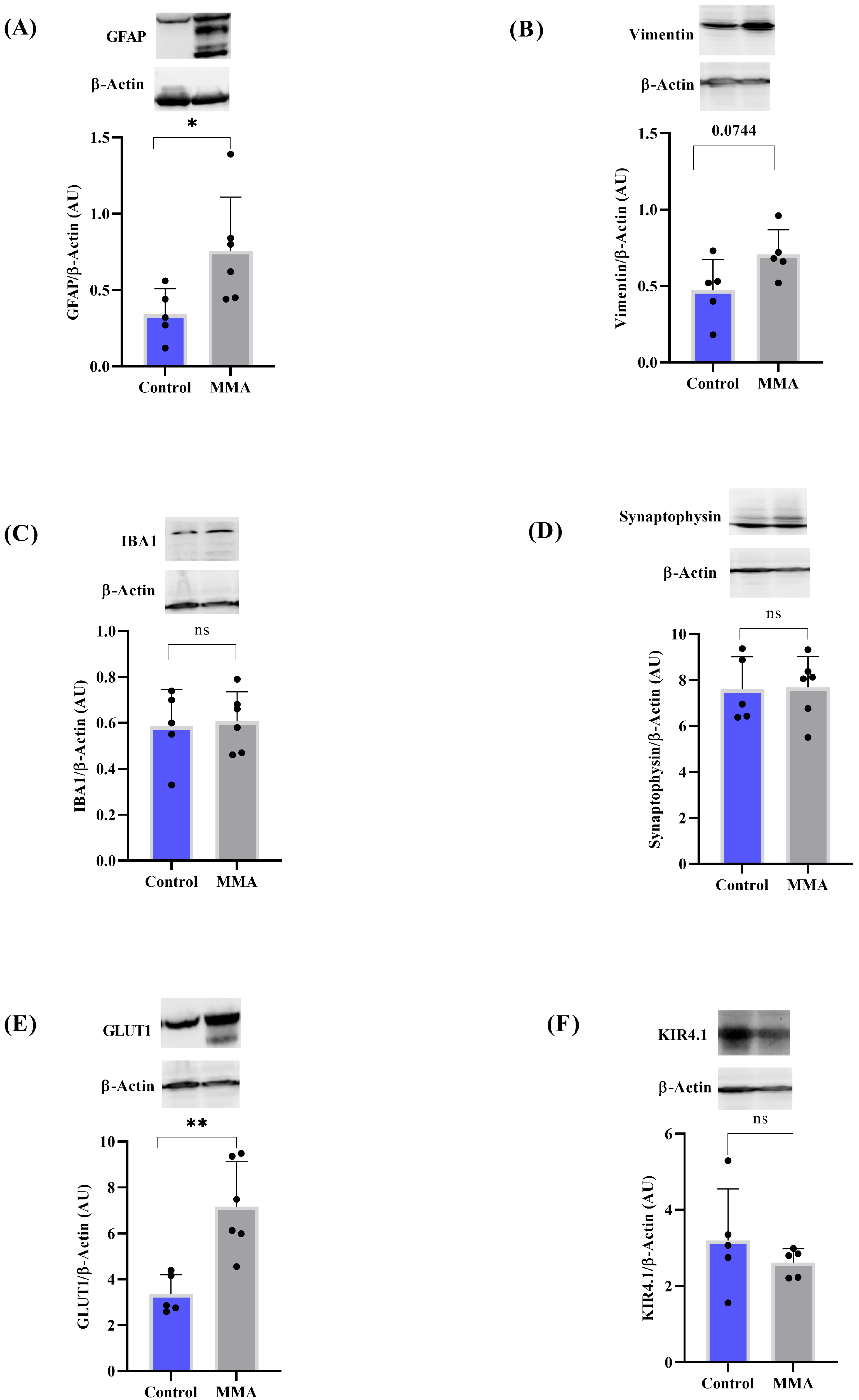
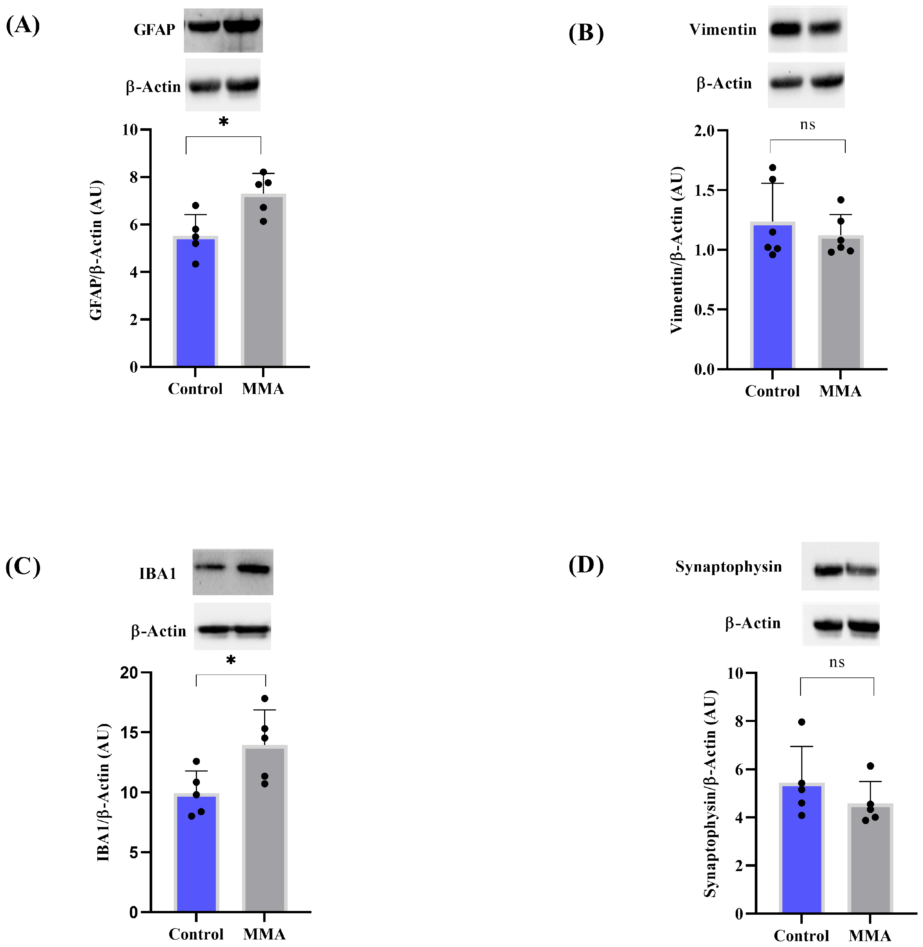
3.2. MMA Induces Glial Reactivity in the Striatum of Rats

We further evaluated markers of glial reactivity and synaptotoxicity, as these processes are commonly observed in various neurodegenerative conditions [28,29]. As shown in Figure 7, i.p. administration of MMA did not induce any significant changes in these markers in the cortex. However, in the striatum, MMA increased GFAP (p < 0.05) and GLUT1 (p < 0.01) levels (Figure 8). A strong trend toward increased vimentin expression was also observed (p = 0.0744) (Figure 8B). In contrast, IBA1, synaptophysin, and KIR4.1 levels remained unaltered in both brain regions (Figure 7 and Figure 8).
When MMA was administered via i.c.v. injection, there was a significant increase in GFAP and IBA1 levels in the striatum (Figure 9A,C), whereas synaptophysin and vimentin levels were not significantly altered (Figure 9B,D). No changes were observed in the cerebral cortex (Figure 10).

3.3. MMA Changes Amino Acid Profile

Table 1 summarizes the effects of i.p. administration of MMA on amino acid levels. In the striatum, MMA significantly increased the levels of glutamate (p < 0.05) and serine (p < 0.05), and decreased tyrosine (p < 0.05). A strong trend toward elevated glycine levels was also observed (p = 0.087). The levels of the other amino acids, including threonine, tryptophan, valine, proline, methionine, lysine, hydroxyproline, histidine, leucine, isoleucine, glutamine, alanine, arginine, asparagine, phenylalanine, and aspartate, remained unchanged. No alterations were detected in the cerebral cortex.

4. Discussion

Patients with methylmalonic aciduria present with acute and persistent neurological symptoms [3], the pathophysiology of which is not fully established. Our findings demonstrate that MMA administration disrupts antioxidant defenses, impairs CAC activity, and induces glial reactivity in the rat brain. Importantly, these effects were observed following both systemic and intracerebral administration of MMA, supporting the notion that MMA crosses the blood-brain barrier and contributes to neural injury.
Variable alterations were observed in the antioxidant defenses. Systemic administration of MMA decreased GSH levels and GPx activity in the cerebral cortex, whereas intracerebral administration also decreased GSH but increased GST and CAT activities in the same region. In the striatum, systemic injection of MMA led to a decrease in GSH concentrations and an increase in SOD activity, while intracerebral infusion of MMA resulted in decreased GSH and SOD activity. These differential effects are likely related to the concentrations of MMA achieved in each brain structure and may also depend on the route of administration used.
Despite regional and route-specific differences, our results indicate that GSH metabolism is markedly affected by MMA, as demonstrated by alterations in GSH levels and the activities of enzymes involved in its metabolism. These effects may be mediated by reactive species generated as a consequence of MMA exposure. In this context, previous studies have shown that MMA induces oxidative stress in different animal models, as well as in cells and biological fluids from patients [3,10]. While the inhibition of antioxidant enzymes is likely a result of oxidative damage caused by ROS, the increased activity of some enzymes may reflect a compensatory upregulation in response to oxidative stress [30]. Although the precise mechanisms underlying these opposing effects remain unclear, it is plausible that the concentration of MMA in the cellular microenvironment modulates the enzymatic responses. Consistently, previous reports have demonstrated that metabolite accumulation in other inherited metabolic disorders can exert distinct effects depending on the brain region analyzed [26,31].
MMA also disrupted CAC functioning in the striatum, indicating bioenergetic impairment. Systemic administration of MMA led to a reduction in CS, SDH, IDH, and MDH activities, whereas intracerebral administration decreased the activities of IDH, SDH, and MDH. These findings corroborate previous works suggesting that aerobic glucose oxidation is affected [10]. They are also in agreement with data reporting decreased expression and activity of Mdh2 in mitochondria isolated from the liver of a mouse model of MMA (Mut-ko/ki) [32]. Taken together, these data suggest that MDH is particularly susceptible to MMA-induced dysfunction. However, the study in Mut-ko/ki mice did not report changes in CS or IDH activity, which may reflect tissue-specific differences in enzyme regulation between the liver and brain [33,34]. It is also important to consider that Mut-ko/ki mice exhibit chronically elevated levels of MMA, whereas our models involve an acute increase in MMA concentration, which may account for some of the observed discrepancies.
We cannot rule out that 2-methylcitrate and malonate also mediate complex II activity inhibition, as Okun et al. [35] showed that MMA loading in striatal neurons causes the formation of these acids. Therefore, we speculate that, in our animal models, the CAC impairment may be due to a synergic effect caused by MMA, malonate, and 2-methylcitrate. Interestingly, the same study suggested that oxalacetate regeneration is impaired by these acids since 2-methylcitrate is formed from propionyl-CoA and oxalacetate, while MMA inhibits the malate shuttle and pyruvate carboxylase. Thus, it is conceivable that these mechanisms, which may lead to gluconeogenesis impairment and ketogenesis activation, also occur in our models.
The exact source of ROS generated by MMA was not established in our study. However, in agreement with previous reports, we demonstrated that MMA inhibits SDH activity in the rat brain [3,10]. Within this context, it has been shown that mitochondrial complex II can contribute to ROS production, particularly under conditions of impaired electron flow [35,36]. In addition, other studies have reported that MMA inhibits additional respiratory chain complexes in the rat brain, which may further enhance ROS generation and exacerbate oxidative stress [37].
It is also important to consider that several studies have demonstrated the involvement of NMDA receptor activation in the neurotoxic effects of MMA [6,38,39,40]. Activation of NMDA receptors may stimulate inducible nitric oxide synthase and promote calcium influx, which in turn activates cyclooxygenases and lipoxygenases, leading to increased ROS production [41]. In line with this mechanism, we observed increased levels of the NMDA receptor agonist glutamate, as well as of the co-agonists glycine and serine. Glycine levels are elevated, possibly by methylmalonylation and consequent inhibition of the glycine cleavage pathway [42], whereas glutamate may be derived from α-ketoglutarate that is accumulated due to CAC impairment. However, further investigations to determine the metabolism of glutamate in the brain are needed since recent works demonstrated no alterations in the pool of this amino acid in MUT HEK293 cells [43] and patient-derived neurons [44]. As for serine, it was shown that the de novo synthesis and transport of this amino acid increased in MUT HEK293 cells due to enrichment of proteins involved in serine metabolism [43]. Although we did not establish the sources of these amino acids, our findings suggest that the increase in these amino acids induced by MMA may contribute to NMDA receptor overactivation and subsequent ROS generation.
MMA also reduced tyrosine levels in the striatum of rats. A decrease in this amino acid in the brain has been associated with reduced catecholamine synthesis, which may help explain the neurological dysfunction observed in methylmalonic aciduria [45]. Impairment of dopaminergic neurotransmission, particularly in the striatum, potentially contributes to motor dysfunction. Supporting this hypothesis, previous studies have shown that tyrosine and phenylalanine depletion impairs memory and cognitive performance [46].
Our findings showed elevated levels of GFAP, a marker of mature astrocytes, and Iba1, a marker of microglial cells, in the striatum, suggesting that MMA triggers glial reactivity in this brain region. Activation of astrocytes and microglia can function either as pro-inflammatory, causing the release of molecules that trigger neuroinflammatory response, or as anti-inflammatory [47]. Although we did not determine which glial phenotype is involved in the MMA effects shown here, previous studies have demonstrated that MMA increases pro-inflammatory cytokine levels and causes oxidative stress in vitro in C6 astroglial cells and in vivo in rat brain [39,48]. Furthermore, earlier data suggest that accumulation of toxic metabolites in glutaric aciduria type I, a metabolic disorder related to methylmalonic aciduria, can alter astrocyte phenotype and initiate neuronal degeneration [2,49,50]. Similar findings indicative of inflammation were also observed in the plasma of patients [51] as well as in models of other inherited metabolic disorders characterized by oxidative stress and bioenergetic dysfunction [52,53,54]. Therefore, we speculate that the activation of glia mediated by MMA observed in our present study elicits a pro-inflammatory response.
Notably, MMA also increased GLUT-1 levels in rat striatum, a glucose transporter isoform expressed in the brain endothelium cells and astrocytes [55]. This finding further supports the view that MMA induces glial reactivity and may reflect an enhanced glucose uptake by astrocytes. Such an effect may represent a compensatory metabolic response to counteract energy disruption in these cells. In this regard, GLUT-1 expression is frequently upregulated under metabolic stress conditions such as hypoxia or mitochondrial dysfunction, aiming to enhance glucose uptake [56]. Additionally, glucose entry may sustain the pentose phosphate pathway, generating NADPH essential for redox support of neurons [57]. Increasing GLUT-1 may help restore the bioenergetic status impaired by MMA and boost NADPH, thus enhancing GSH recycling reduced by MMA.
Some limitations of our experimental models should be acknowledged. These models were developed in rats without genetic mutations affecting MMA metabolism, which means that MMA is still metabolized to some extent. However, it is important to emphasize that acute metabolic decompensation is a common feature in patients with methylmalonic aciduria, leading to a marked accumulation of metabolites in biological fluids and tissues, followed by a partial decline that remains above normal levels [2]. Based on this, we propose that our rat models partially replicate the metabolic state observed during acute decompensation episodes. On the other hand, our models fail to evaluate the chronic effects caused by sustained high levels of MMA observed in patients. Thus, further studies should be conducted in genetic and chronic MMA exposure-based animal models. Such models are also important to elucidate novel mechanisms that seem to be independent of MMA accumulation [58]. Another limitation of our study is that we did not measure the MMA levels that were achieved in each brain structure. Nevertheless, it should be noted that other organic acids structurally similar to MMA injected systemically into rats achieved low concentrations in the brain [12]. Thus, we speculate that even if MMA reached low amounts in the brain of rats, it was sufficient to cause neurotoxicity.
Our results indicate that the striatum is more vulnerable than the cerebral cortex to the deleterious effects provoked by MMA. This differential vulnerability may be explained by a number of factors. Previous studies have shown that the striatum contains high levels of cyclophilin-D, which may promote mitochondrial permeability transition, and that mitochondrial membrane potential in the striatum is more sensitive to calcium [59,60]. In addition, the presence of massive glutamatergic inputs in the striatum [61] reinforces the hypothesis that MMA-induced toxic effects are mediated by NMDA receptor activation, which leads to increased ROS generation. Consistent with this, high levels of iron are also found in the striatum, which possibly further contributes to ROS formation [62].
In conclusion, our findings show that MMA induces bioenergetic disruption and disturbs the antioxidant defenses, especially those related to GSH homeostasis. We also highlight that glial cells are severely affected by MMA, even though these cells have a more powerful antioxidant system than neurons [51]. These data reveal different neurotoxic mechanisms elicited by MMA, with emphasis on glial response activation, that may explain the neuropathophysiology of isolated methylmalonic aciduria.

5. Conclusions

In summary, our study provides evidence that MMA induces oxidative stress, bioenergetic impairment, inflammation, and glial reactivity in the rat brain, particularly through disturbances in GSH metabolism and CAC enzyme activity. Both systemic and intracerebral administration of MMA triggered similar neurochemical alterations, reinforcing the neurotoxic potential of this metabolite. We also identified increased levels of amino acids associated with NMDA receptor activation, suggesting a link between excitotoxicity and oxidative damage. Furthermore, the observed glial activation, marked by elevated GFAP, IBA1, and GLUT1 levels, underscores the involvement of astrocytes and microglia in MMA neurotoxicity. Additional studies are essential to confirm whether glial reactivity and disturbances in amino acid metabolism may be considered novel targets for therapeutic investigation. Together, these data contribute to the understanding of the multifactorial mechanisms underlying the neurological dysfunction in this disorder and support the role of glial cells as central mediators of MMA-induced neurotoxicity.

Author Contributions

Conceptualization, C.A.D., J.d.A.S., M.B.M., T.F.d.O., C.-A.G. and G.L.; methodology, C.A.D., J.d.A.S., M.B.M., V.G.-D., V.-F.D.S., C.V.P. and B.P.d.S.; formal analysis, C.A.D., J.d.A.S., M.B.M., T.F.d.O., C.-A.G. and G.L.; investigation, C.A.D., J.d.A.S., M.B.M., T.F.d.O., C.-A.G. and G.L.; resources, T.F.d.O., C.-A.G. and G.L.; data curation, C.A.D., J.d.A.S., M.B.M., T.F.d.O., C.-A.G. and G.L.; writing C.A.D., T.F.d.O., C.-A.G. and G.L.; supervision, T.F.d.O., C.-A.G. and G.L. All authors have read and agreed to the published version of the manuscript.

Funding

This research was funded by CNPq, grant number 402440/2021-8; CNPq, grant number 312141/2020-3; and the National Institute of Brain Health, INSC, grant number 406020/2022-1. A full waiver was granted by the journal.

Institutional Review Board Statement

The study was conducted in accordance with the Declaration of Helsinki and approved by the Ethics Committee on the Use of Animals (CEUA) from UFRGS (protocol code project numbers 32807, approved in August 2017, and 44267, approved in April 2024).

Informed Consent Statement

Not applicable.

Data Availability Statement

Dataset available on request from the authors due to privacy.

Conflicts of Interest

The authors declare no conflicts of interest.

Abbreviations

The following abbreviations are used in this manuscript:
CACCitric acid cycle
CATCatalase
CSCitrate synthase
G6PDHGlucose-6-phosphate dehydrogenase
GFAPGlial fibrillary acid protein
GLUT1Glucose transporter 1
GPxGlutathione peroxidase
GRGlutathione reductase
GSHGlutathione
GSTGlutathione-S-transferase
IBA1Ionized calcium-binding adapter molecule 1
IDHIsocitrate Dehydrogenase
KIR4.1Inwardly rectifying potassium channel
MDHMalate dehydrogenase
MMAMethylmalonic acid
NADPHNicotinamide adenine dinucleotide phosphate
NMDAN-methyl-D-aspartate
ROSReactive oxygen species
SDHSuccinate dehydrogenase
SODSuperoxide Dismutase

References

  1. Chen, T.; Gao, Y.; Zhang, S.; Wang, Y.; Sui, C.; Yang, L. Methylmalonic acidemia: Neurodevelopment and neuroimaging. Front. Neurosci. 2023, 17, 1110942. [Google Scholar] [CrossRef]
  2. Manoli, I.; Sloan, J.L.; Venditti, C.P. Isolated Methylmalonic Acidemia. In GeneReviews®; University of Washington: Seattle, WA, USA, 2005. [Google Scholar]
  3. Head, P.E.; Meier, J.L.; Venditti, C.P. New insights into the pathophysiology of methylmalonic acidemia. J. Inherit. Metab. Dis. 2023, 46, 436–449. [Google Scholar] [CrossRef]
  4. Jin, L.; Han, X.; He, F.; Zhang, C. Prevalence of methylmalonic acidemia among newborns and the clinical-suspected population: A meta-analysis. J. Matern.-Fetal Neonatal Med. 2021, 35, 8952–8967. [Google Scholar] [CrossRef]
  5. Fraser, J.L.; Venditti, C.P. Methylmalonic and propionic acidemias: Clinical management update. Curr. Opin. Pediatr. 2016, 28, 682–693. [Google Scholar] [CrossRef]
  6. Fernandes, C.G.; Borges, C.G.; Seminotti, B.; Amaral, A.U.; Knebel, L.A.; Eichler, P.; de Oliveira, A.B.; Leipnitz, G.; Wajner, M. Experimental evidence that methylmalonic acid provokes oxidative damage and compromises antioxidant defenses in nerve terminal and striatum of young rats. Cell. Mol. Neurobiol. 2011, 31, 775–785. [Google Scholar] [CrossRef]
  7. Chandler, R.J.; Zerfas, P.M.; Shanske, S.; Sloan, J.; Hoffmann, V.; DiMauro, S.; Venditti, C.P. Mitochondrial dysfunction in mut methylmalonic acidemia. FASEB J. 2009, 23, 1252–1261. [Google Scholar] [CrossRef]
  8. Schumann, A.; Brutsche, M.; Havermans, M.; Grünert, S.C.; Kölker, S.; Groß, O.; Hannibal, L. The impact of metabolic stressors on mitochondrial homeostasis in a renal epithelial cell model of methylmalonic aciduria. Sci. Rep. 2023, 13, 7677. [Google Scholar] [CrossRef]
  9. Melo, D.R.; Kowaltowski, A.J.; Wajner, M.; Castilho, R.F. Mitochondrial energy metabolism in neurodegeneration associated with methylmalonic acidemia. J. Bioenerg. Biomembr. 2011, 43, 39–46. [Google Scholar] [CrossRef]
  10. Richard, E.; Gallego-Villar, L.; Rivera-Barahona, A.; Oyarzábal, A.; Pérez, B.; Rodríguez-Pombo, P.; Desviat, L.R. Altered Redox Homeostasis in Branched-Chain Amino Acid Disorders, Organic Acidurias, and Homocystinuria. Oxidative Med. Cell. Longev. 2018, 2018, 1246069. [Google Scholar] [CrossRef]
  11. Semple, B.D.; Blomgren, K.; Gimlin, K.; Ferriero, D.M.; Noble-Haeusslein, L.J. Brain development in rodents and humans: Identifying benchmarks of maturation and vulnerability to injury across species. Prog. Neurobiol. 2013, 106–107, 1–16. [Google Scholar] [CrossRef]
  12. da Rosa, M.S.; Seminotti, B.; Amaral, A.U.; Fernandes, C.G.; Gasparotto, J.; Moreira, J.C.; Gelain, D.P.; Wajner, M.; Leipnitz, G. Redox homeostasis is compromised in vivo by the metabolites accumulating in 3-hydroxy-3-methylglutaryl-CoA lyase deficiency in rat cerebral cortex and liver. Free Radic. Res. 2013, 47, 1066–1075. [Google Scholar] [CrossRef] [PubMed]
  13. Paxinos, G.; Watson, C. The Rat Brain in Stereotaxic Coordinates, 7th ed.; Academic Press: Cambridge, MA, USA, 2013. [Google Scholar]
  14. Browne, R.W.; Armstrong, D. Reduced glutathione and glutathione disulfide. Methods Mol. Biol. 1998, 108, 347–352. [Google Scholar] [PubMed]
  15. Marklund, S.L. Pyrogallol autooxidation. In Handbook of Methods for Oxygen Radical Research; CRC Press: Boca Raton, FL, USA, 1985; pp. 243–247. [Google Scholar]
  16. Aebi, H. Catalase in vitro. In Methods in Enzymology; Academic Press: Cambridge, MA, USA, 1984; Volume 105, pp. 121–126. [Google Scholar]
  17. Wendel, A. Glutathione peroxidase. In Methods in Enzymology; Academic Press: Cambridge, MA, USA, 1985; Volume 77, pp. 325–333. [Google Scholar]
  18. Mannervik, B.; Guthenberg, C. Glutathione transferase (human placenta). In Methods in Enzymology; Academic Press: Cambridge, MA, USA, 1981; Volume 77, pp. 231–235. [Google Scholar] [CrossRef]
  19. Carlberg, I.; Mannervik, B. Glutathione reductase. In Methods in Enzymology; Academic Press: Cambridge, MA, USA, 1985; Volume 113, pp. 484–490. [Google Scholar]
  20. Leong, S.F.; Clark, J.B. Regional development of glutamate dehydrogenase in the at brain. J. Neurochem. 1984, 43, 106–111. [Google Scholar] [CrossRef]
  21. Grings, M.; Moura, A.P.; Parmeggiani, B.; Pletsch, J.T.; Cardoso, G.M.F.; August, P.M.; Matté, C.; Wyse, A.T.S.; Wajner, M.; Leipnitz, G. Bezafibrate prevents mitochondrial dysfunction, antioxidant system disturbance, glial reactivity and neuronal damage induced by sulfite administration in striatum of rats: Implications for a possible therapeutic strategy for sulfite oxidase deficiency. Biochim. Biophys. Acta Mol. Basis Dis. 2017, 1863, 2135–2148. [Google Scholar] [CrossRef]
  22. Shepherd, D.; Garland, P.B. Citrate synthase from rat liver:[EC 4.1. 3.7 Citrate oxaloacetate-lyase (CoA-acetylating)]. In Methods in Enzymology; Academic Press: Cambridge, MA, USA, 1969; Volume 13, pp. 11–16. [Google Scholar]
  23. Fischer, J.C.; Ruitenbeek, W.; Berden, J.A.; Trijbels, J.F.; Veerkamp, J.H.; Stadhouders, A.M.; Janssen, A.J. Differential investigation of the capacity of succinate oxidation in human skeletal muscle. Clin. Chim. Acta. 1985, 153, 23–36. [Google Scholar] [CrossRef]
  24. Kitto, G.B. Intra-and extramitochondrial malate dehydrogenases from chicken and tuna heart:[EC 1.1. 1.37 l-Malate: NAD oxidoreductase]. In Methods in Enzymology; Academic Press: Cambridge, MA, USA, 1969; Volume 13, pp. 106–116. [Google Scholar]
  25. Lowry, O.H.; Rosebrough, N.J.; Farr, A.L.; Randall, R.J. Protein measurement with the Folin phenol reagent. J. Biol. Chem. 1951, 193, 265–275. [Google Scholar] [CrossRef]
  26. Silveira, J.A.; Marcuzzo, M.B.; da Rosa, J.S.; Kist, N.S.; Hoffmann, C.I.H.; Carvalho, A.S.; Ribeiro, R.T.; Quincozes-Santos, A.; Netto, C.A.; Wajner, M.; et al. 3-Hydroxy-3-Methylglutaric Acid Disrupts Brain Bioenergetics, Redox Homeostasis, and Mitochondrial Dynamics and Affects Neurodevelopment in Neonatal Wistar Rats. Biomedicines 2024, 12, 1563. [Google Scholar] [CrossRef]
  27. Glänzel, N.M.; Parmeggiani, B.; Grings, M.; Seminotti, B.; Brondani, M.; Bobermin, L.D.; Ribeiro, C.A.J.; Quincozes-Santos, A.; Vockley, J.; Leipnitz, G. Myelin Disruption, Neuroinflammation, and Oxidative Stress Induced by Sulfite in the Striatum of Rats Are Mitigated by the pan-PPAR agonist Bezafibrate. Cells 2023, 12, 1557. [Google Scholar] [CrossRef]
  28. Dejanovic, B.; Sheng, M.; Hanson, J.E. Targeting synapse function and loss for treatment of neurodegenerative diseases. Nat. Rev. Drug Discov. 2024, 23, 23–42. [Google Scholar] [CrossRef]
  29. Gildea, H.K.; Liddelow, S.A. Mechanisms of astrocyte aging in reactivity and disease. Mol. Neurodegener. 2025, 20, 21. [Google Scholar] [CrossRef]
  30. Seminotti, B.; Grings, M.; Tucci, P.; Leipnitz, G.; Saso, L. Nuclear Factor Erythroid-2-Related Factor 2 Signaling in the Neuropathophysiology of Inherited Metabolic Disorders. Front. Cell. Neurosci. 2021, 15, 785057. [Google Scholar] [CrossRef] [PubMed]
  31. Zemniaçak, Â.B.; Ribeiro, R.T.; Pinheiro, C.V.; de Azevedo Cunha, S.; Tavares, T.Q.; Castro, E.T.; Leipnitz, G.; Wajner, M.; Amaral, A.U. In Vivo Intracerebral Administration of α-Ketoisocaproic Acid to Neonate Rats Disrupts Brain Redox Homeostasis and Promotes Neuronal Death, Glial Reactivity, and Myelination Injury. Mol. Neurobiol. 2024, 61, 2496–2513. [Google Scholar] [CrossRef] [PubMed]
  32. Wongkittichote, P.; Cunningham, G.; Summar, M.L.; Pumbo, E.; Forny, P.; Baumgartner, M.R.; Chapman, K.A. Tricarboxylic acid cycle enzyme activities in a mouse model of methylmalonic aciduria. Mol. Genet. Metab. 2019, 128, 444–451. [Google Scholar] [CrossRef]
  33. Zhang, Y.; Brasher, A.L.; Park, N.R.; Taylor, H.A.; Kavazis, A.N.; Hood, W.R. High activity before breeding improves reproductive performance by enhancing mitochondrial function and biogenesis. J. Exp. Biol. 2018, 221 Pt 7, jeb177469. [Google Scholar] [CrossRef]
  34. Schild, L.; Jaroscakova, I.; Lendeckel, U.; Wolf, G.; Keilhoff, G. Neuronal nitric oxide synthase controls enzyme activity pattern of mitochondria and lipid metabolism. FASEB J. 2005, 20, 145–147. [Google Scholar] [CrossRef]
  35. Okun, J.G.; Hörster, F.; Farkas, L.M.; Feyh, P.; Hinz, A.; Sauer, S.; Hoffmann, G.F.; Unsicker, K.; Mayatepek, E.; Kölker, S. Neurodegeneration in methylmalonic aciduria involves inhibition of complex II and the tricarboxylic acid cycle, and synergistically acting excitotoxicity. J. Biol. Chem. 2002, 277, 14674–14680. [Google Scholar] [CrossRef]
  36. Hadrava Vanova, K.; Kraus, M.; Neuzil, J.; Rohlena, J. Mitochondrial complex II and reactive oxygen species in disease and therapy. Redox Rep. Commun. Free Radic. Res. 2020, 25, 26–32. [Google Scholar] [CrossRef]
  37. Brusque, A.M.; Borba Rosa, R.; Schuck, P.F.; Dalcin, K.B.; Ribeiro, C.A.; Silva, C.G.; Wannmacher, C.M.; Dutra-Filho, C.S.; Wyse, A.T.; Briones, P.; et al. Inhibition of the mitochondrial respiratory chain complex activities in rat cerebral cortex by methylmalonic acid. Neurochem. Int. 2002, 40, 593–601. [Google Scholar] [CrossRef]
  38. de Mattos-Dutra, A.; Meirelles, R.; Bevilaqua da Rocha, B.; Kommers, T.; Wofchuk, S.T.; Wajner, M.; Pessoa-Pureur, R. Methylmalonic and propionic acids increase the in vitro incorporation of 32P into cytoskeletal proteins from cerebral cortex of young rats through NMDA glutamate receptors. Brain Res. 2000, 856, 111–118. [Google Scholar] [CrossRef]
  39. Ribeiro, L.R.; Della-Pace, I.D.; de Oliveira Ferreira, A.P.; Funck, V.R.; Pinton, S.; Bobinski, F.; de Oliveira, C.V.; da Silva Fiorin, F.; Duarte, M.M.; Furian, A.F.; et al. Chronic administration of methylmalonate on young rats alters neuroinflammatory markers and spatial memory. Immunobiology 2013, 218, 1175–1183. [Google Scholar] [CrossRef]
  40. Royes, L.F.; Fighera, M.R.; Furian, A.F.; Oliveira, M.S.; da Silva, L.G.; Malfatti, C.R.; Schneider, P.H.; Braga, A.L.; Wajner, M.; Mello, C.F. Creatine protects against the convulsive behavior and lactate production elicited by the intrastriatal injection of methylmalonate. Neuroscience 2003, 118, 1079–1090. [Google Scholar] [CrossRef] [PubMed]
  41. Greene, J.G.; Greenamyre, J.T. Bioenergetics and glutamate excitotoxicity. Prog. Neurobiol. 1996, 48, 613–634. [Google Scholar] [CrossRef] [PubMed]
  42. Head, P.E.; Myung, S.; Chen, Y.; Schneller, J.L.; Wang, C.; Duncan, N.; Hoffman, P.; Chang, D.; Gebremariam, A.; Gucek, M.; et al. Aberrant methylmalonylation underlies methylmalonic acidemia and is attenuated by an engineered sirtuin. Sci. Transl. Med. 2022, 14, eabn4772. [Google Scholar] [CrossRef]
  43. Anzmann, A.F.; Pinto, S.; Busa, V.; Carlson, J.; McRitchie, S.; Sumner, S.; Pandey, A.; Vernon, H.J. Multi-omics studies in cellular models of methylmalonic acidemia and propionic acidemia reveal dysregulation of serine metabolism. Biochim. Biophys. Acta Mol. Basis Dis. 2019, 1865, 165538. [Google Scholar] [CrossRef]
  44. Denley, M.C.S.; Straub, M.S.; Marcionelli, G.; Güra, M.A.; Penton, D.; Delvendahl, I.; Poms, M.; Vekeriotaite, B.; Cherkaoui, S.; Conte, F.; et al. Mitochondrial dysfunction drives a neuronal exhaustion phenotype in methylmalonic aciduria. Commun. Biol. 2025, 8, 410. [Google Scholar] [CrossRef]
  45. Fernstrom, J.D.; Fernstrom, M.H. Tyrosine, phenylalanine, and catecholamine synthesis and function in the brain. J. Nutr. 2007, 137, 1539S–1548S. [Google Scholar] [CrossRef] [PubMed]
  46. Linssen, A.M.; Riedel, W.J.; Sambeth, A. Effects of tyrosine/phenylalanine depletion on electrophysiological correlates of memory in healthy volunteers. J. Psychopharmacol. 2011, 25, 230–238. [Google Scholar] [CrossRef]
  47. Kempuraj, D.; Dourvetakis, K.D.; Cohen, J.; Valladares, D.S.; Joshi, R.S.; Kothuru, S.P.; Anderson, T.; Chinnappan, B.; Cheema, A.K.; Klimas, N.G.; et al. Neurovascular unit, neuroinflammation and neurodegeneration markers in brain disorders. Front. Cell. Neurosci. 2024, 18, 1491952. [Google Scholar] [CrossRef]
  48. de Souza Almeida, R.R.; Bobermin, L.D.; Parmeggiani, B.; Wartchow, K.M.; Souza, D.O.; Gonçalves, C.A.; Wajner, M.; Leipnitz, G.; Quincozes-Santos, A. Methylmalonic acid induces inflammatory response and redox homeostasis disruption in C6 astroglial cells: Potential glioprotective roles of melatonin and resveratrol. Amino Acids 2022, 54, 1505–1517. [Google Scholar] [CrossRef]
  49. Olivera, S.; Fernandez, A.; Latini, A.; Rosillo, J.C.; Casanova, G.; Wajner, M.; Cassina, P.; Barbeito, L. Astrocytic proliferation and mitochondrial dysfunction induced by accumulated glutaric acidemia I (GAI) metabolites: Possible implications for GAI pathogenesis. Neurobiol. Dis. 2008, 32, 528–534. [Google Scholar] [CrossRef]
  50. Olivera-Bravo, S.; Ribeiro, C.A.; Isasi, E.; Trías, E.; Leipnitz, G.; Díaz-Amarilla, P.; Woontner, M.; Beck, C.; Goodman, S.I.; Souza, D.; et al. Striatal neuronal death mediated by astrocytes from the Gcdh-/- mouse model of glutaric acidemia type I. Hum. Mol. Genet. 2015, 24, 4504–4515. [Google Scholar] [CrossRef]
  51. Li, Q.; Jin, H.; Liu, Y.; Rong, Y.; Yang, T.; Nie, X.; Song, W. Determination of Cytokines and Oxidative Stress Biomarkers in Cognitive Impairment Induced by Methylmalonic Acidemia. Neuroimmunomodulation 2021, 28, 178–186. [Google Scholar] [CrossRef]
  52. Glänzel, N.M.; Grings, M.; da Rosa-Junior, N.T.; de Carvalho, L.M.C.; Mohsen, A.W.; Wipf, P.; Wajner, M.; Vockley, J.; Leipnitz, G. The mitochondrial-targeted reactive species scavenger JP4-039 prevents sulfite-induced alterations in antioxidant defenses, energy transfer, and cell death signaling in striatum of rats. J. Inherit. Metab. Dis. 2021, 44, 481–491. [Google Scholar] [CrossRef]
  53. da Rosa, M.S.; da Rosa-Junior, N.T.; Parmeggiani, B.; Glänzel, N.M.; de Moura Alvorcem, L.; Ribeiro, R.T.; Grings, M.; Wajner, M.; Leipnitz, G. 3-Hydroxy-3-Methylglutaric Acid Impairs Redox and Energy Homeostasis, Mitochondrial Dynamics, and Endoplasmic Reticulum-Mitochondria Crosstalk in Rat Brain. Neurotox. Res. 2020, 37, 314–325. [Google Scholar] [CrossRef] [PubMed]
  54. Seminotti, B.; Amaral, A.U.; Ribeiro, R.T.; Rodrigues, M.D.N.; Colín-González, A.L.; Leipnitz, G.; Santamaría, A.; Wajner, M. Oxidative Stress, Disrupted Energy Metabolism, and Altered Signaling Pathways in Glutaryl-CoA Dehydrogenase Knockout Mice: Potential Implications of Quinolinic Acid Toxicity in the Neuropathology of Glutaric Acidemia Type I. Mol. Neurobiol. 2016, 53, 6459–6475. [Google Scholar] [CrossRef] [PubMed]
  55. Vannucci, S.J. Developmental expression of GLUT1 and GLUT3 glucose transporters in rat brain. J. Neurochem. 1994, 62, 240–246. [Google Scholar] [CrossRef]
  56. Thieren, L.; Zanker, H.S.; Droux, J.; Dalvi, U.; Wyss, M.T.; Waag, R.; Germain, P.-L.; von Ziegler, L.M.; Looser, Z.J.; Hösli, L.; et al. Astrocytic GLUT1 deletion in adult mice enhances glucose metabolism and resilience to stroke. Nat. Commun. 2025, 16, 4190. [Google Scholar] [CrossRef] [PubMed]
  57. Almeida, A.; Jimenez-Blasco, D.; Bolaños, J.P. Cross-talk between energy and redox metabolism in astrocyte-neuron functional cooperation. Essays Biochem. 2023, 67, 17–26. [Google Scholar] [CrossRef]
  58. Luciani, A.; Schumann, A.; Berquez, M.; Chen, Z.; Nieri, D.; Failli, M.; Debaix, H.; Festa, B.P.; Tokonami, N.; Raimondi, A.; et al. Impaired mitophagy links mitochondrial disease to epithelial stress in methylmalonyl-CoA mutase deficiency. Nat. Commun. 2020, 11, 970. [Google Scholar] [CrossRef]
  59. Brustovetsky, N.; Brustovetsky, T.; Purl, K.J.; Capano, M.; Crompton, M.; Dubinsky, J.M. Increased Susceptibility of Striatal Mitochondria to Calcium-Induced Permeability Transition. J. Neurosci. 2003, 23, 4858–4867. [Google Scholar] [CrossRef]
  60. Pickrell, A.M.; Fukui, H.; Wang, X.; Pinto, M.; Moraes, T.C. The Striatum Is Highly Susceptible to Mitochondrial Oxidative Phosphorylation Dysfunctions. J. Neurosci. 2011, 31, 9895–9904. [Google Scholar] [CrossRef] [PubMed]
  61. Reig, R.; Silberberg, G. Distinct. Corticostriatal and Intracortical Pathways Mediate Bilateral Sensory Responses in the Striatum. Cereb. Cortex 2016, 26, 4405–4415. [Google Scholar] [CrossRef] [PubMed]
  62. Halliwell, B.; Gutteridge, J.M.C. Free Radicals in Biology and Medicine, 5th ed.; Oxford Academic: Oxford, UK, 2015. [Google Scholar] [CrossRef]
Figure 1. Intraperitoneal administration of methylmalonic acid (MMA) disrupts the cortical antioxidant system. Levels of glutathione (GSH) (A) and activities of glutathione peroxidase (GPx) (B), glutathione reductase (GR) (C), glutathione S-transferase (GST) (D), glucose-6-phosphate dehydrogenase (E), and superoxide dismutase (F) were measured. Values are means ± SD (N = 5–6). ** p < 0.01, compared to rats receiving NaCl (control group). ns = not significant (Student’s t-test).
Neuroglia 06 00025 g001
Figure 2. Intraperitoneal administration of methylmalonic acid (MMA) disrupts the striatal antioxidant system. Levels of GSH (A) and activities of glutathione peroxidase (GPx) (B), glutathione reductase (GR) (C), glutathione S-transferase (GST) (D), glucose-6-phosphate dehydrogenase (E), and superoxide dismutase (F) were measured. Values are means ± SD (N = 5–6). ** p < 0.01, compared to rats receiving NaCl (control group). ns = not significant (Student’s t-test).
Neuroglia 06 00025 g002
Figure 3. Intraperitoneal administration of methylmalonic acid (MMA) impairs the cortical and striatal citric acid cycle functioning. Citrate synthase (A), isocitrate dehydrogenase (IDH) (B), succinate dehydrogenase (SDH) (C), and malate dehydrogenase (MDH) (D) activities were measured in the cortex (AD) and striatum (EH). Values are means ± SD (N = 5–6). ns = not significant (Student’s t-test). Intraperitoneal administration of methylmalonic acid (MMA) disrupts the striatal citric acid cycle (CAC) functioning. Citrate synthase (A), isocitrate dehydrogenase (IDH) (B), succinate dehydrogenase (SDH) (C), and malate dehydrogenase (MDH) (D) activities were measured. Values are means ± SD (N = 5–6). * p < 0.05, compared to rats receiving NaCl (control group). ns = not significant (Student’s t-test).
Neuroglia 06 00025 g003
Figure 4. Intracerebroventricular administration of methylmalonic acid (MMA) disrupts the cortical antioxidant system. Levels of reduced glutathione (GSH) (A) and activities of glutathione peroxidase (GPx) (B), glutathione reductase (C), glutathione S-transferase (GST) (D), glucose-6-phosphate dehydrogenase (E), superoxide dismutase (F), and catalase (G) were measured. Values are means ± SD (N = 4–5). * p < 0.05, ** p < 0.01, **** p < 0.0001, compared to rats receiving NaCl (control group). ns = not significant (Student’s t-test).
Neuroglia 06 00025 g004
Figure 5. Intracerebroventricular administration of methylmalonic acid (MMA) disrupts the striatal antioxidant system. Levels of reduced glutathione (GSH) (A) and activities of glutathione peroxidase (GPx) (B), glutathione reductase (C), glutathione S-transferase (GST) (D), glucose-6-phosphate dehydrogenase (E), superoxide dismutase (F), and catalase (G) were measured. Values are means ± SD (N = 4–6). * p < 0.05, ** p < 0.01, compared to rats receiving NaCl (control group). ns = not significant (Student’s t-test).
Neuroglia 06 00025 g005
Figure 6. Intracerebroventricular administration of methylmalonic acid (MMA) disrupts the cortical and striatal citric acid cycle functioning. Citrate synthase (A), isocitrate dehydrogenase (IDH) (B), succinate dehydrogenase (SDH) (C), and malate dehydrogenase (MDH) (D), activities were measured. Values are means ± SD (N = 5). * p < 0.05, compared to rats receiving NaCl (control group). ns = not significant (Student’s t-test). Intracerebroventricular administration of methylmalonic acid (MMA) disrupts the striatal citric acid cycle functioning. Citrate synthase (E), isocitrate dehydrogenase (IDH) (F), succinate dehydrogenase (SDH) (G), and malate dehydrogenase (MDH) (H), activities were measured. Values are means ± SD (N = 5). * p < 0.05, *** p < 0.001, compared to rats receiving NaCl (control group). ns = not significant (Student’s t-test).
Neuroglia 06 00025 g006
Figure 7. Intraperitoneal administration of methylmalonic acid (MMA) does not alter neural damage markers in the cerebral cortex. GFAP (A), vimentin (B), IBA1 (C), synaptophysin (D), GLUT1 (E), and KIR4.1 (F) were measured. Representative images of the blots are shown. Data are presented as arbitrary units (AU). Values are means ± SD (N = 6). ns = not significant (Student’s t-test).
Neuroglia 06 00025 g007
Figure 8. Intraperitoneal administration of methylmalonic acid (MMA) increases GFAP and GLUT1 in the striatum. GFAP (A), vimentin (B), IBA1 (C), synaptophysin (D), GLUT1 (E), and KIR4.1 (F) were measured. Representative images of the blots are shown. Data are presented as arbitrary units (AU). Values are means ± SD (N = 5–6). * p < 0,05, ** p < 0.01, compared to rats receiving NaCl (control group). ns = not significant (Student’s t-test).
Neuroglia 06 00025 g008
Figure 9. Intracerebroventricular administration of methylmalonic acid (MMA) increases GFAP and IBA1 levels in the striatum. GFAP (A), vimentin (B), IBA1 (C), and synaptophysin (D) were measured. Representative images of the blots are shown. Data are presented as arbitrary units (AU). Values are means ± SD (N = 5–6). * p < 0.05, compared to rats receiving NaCl (control group). ns = not significant (Student’s t-test).
Neuroglia 06 00025 g009
Figure 10. Intracerebroventricular administration of methylmalonic acid (MMA) does not alter neural damage markers in the cerebral cortex. GFAP (A), vimentin (B), IBA1 (C), and synaptophysin (D) were measured. Representative images of the blots are shown. Data are presented as arbitrary units (AU). Values are means ± SD (N = 5–6). ns = not significant (Student’s t-test).
Neuroglia 06 00025 g010
Table 1. Effect of intraperitoneal administration of methylmalonic acid (MMA) on amino acid concentrations in the cerebral cortex and striatum of rats.
Cerebral CortexStriatum
AMINO ACIDControl (ng/mg)MMA (ng/mg)p ValueControl (ng/mg)MMA (ng/mg)p Value
Threonine4.05 ± 0.474.93 ± 2.910.38623.93 ± 0.854.14 ± 0.490.6037
Tryptophan5.22 ± 1.054.05 ± 1.100.33865.00 ± 1.244.62 ± 0.700.5202
Valine1.65 ± 0.341.77 ± 0.440.61461.45 ± 0.231.55 ± 0.360.5956
Tyrosine100 ± 20.2105 ± 32.60.774091.0 ± 15.987.1 ± 12.40.0472 *
Serine1.34 ± 0.081.38 ± 0.400.78431.23 ± 0.211.43 ± 0.150.0492 *
Proline0.62 ± 0.100.84 ± 0.720.54600.60 ± 0.140.62 ± 0.120.7249
Methionine3.30 ± 0.543.64 ± 1.230.62983.72 ± 0.703.60 ± 0.630.7751
Lysine0.62 ± 0.150.72 ± 0.270.58180.63 ± 0.160.62 ± 0.090.9634
Hydroxyproline0.77 ± 0.090.88 ± 0.240.97470.49 ± 0.100.68 ± 0.110.7827
Histidine19.1 ± 5.1220.8 ± 8.400.389320.0 ± 4.2822.4 ± 2.570.2770
Leucine5.63 ± 0.836.19 ± 1.880.58106.55 ± 1.276.38 ± 1.600.8328
Isoleucine2.65 ± 0.462.87 ± 0.710.53342.49 ± 0.372.52 ± 0.500.8908
Glutamine26.9 ± 8.9029.0 ± 14.40.852424.2 ± 6.0725.0 ± 4.110.7827
Alanine4.60 ± 0.533.92 ± 0.740.71203.20 ± 0.363.67 ± 0.560.3739
Arginine4.76 ± 1.245.44 ± 2.730.28166.46 ± 1.046.61 ± 1.340.9289
Asparagine0.70 ± 0.120.66 ± 0.220.72630.56 ± 0.09 0.60 ± 0.070.3584
Phenylalanine349 ± 60.4384 ± 68.90.3659354 ± 75.8356 ± 66.50.9684
Glycine1.37 ± 0.201.52 ± 0.340.90651.15 ± 0.16 1.32 ± 0.140.0087
Glutamic acid77.8 ± 16.480.6 ± 20.40.796354.3 ± 7.53 65.0 ± 6.140.0217 *
Aspartic acid42.4 ± 7.7840.7 ± 12.20.778138.6 ± 7.3943.9 ± 8.340.2739
Values are means ± SD (N = 5–6). * p < 0.05, compared to rats receiving NaCl (control group) (Student’s t-test).
Disclaimer/Publisher’s Note: The statements, opinions and data contained in all publications are solely those of the individual author(s) and contributor(s) and not of MDPI and/or the editor(s). MDPI and/or the editor(s) disclaim responsibility for any injury to people or property resulting from any ideas, methods, instructions or products referred to in the content.

