Next Issue
Volume 2, December
Previous Issue
Volume 2, June


Methods Protoc., Volume 2, Issue 3 (September 2019) – 24 articles

Cover Story (view full-size image): 
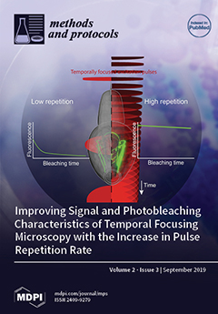
Wide-field temporal focused two-photon microscopy enables high-speed volumetric imaging via the simultaneous acquisition of a large sample area with high lateral and axial resolution. Implementations to date have been held back due to the requirement for high fluence laser sources for the excitation of a large area, which typically operated at a low repetition. Achieving high signal intensity in this configuration necessitates increasing the energy per pulse; however, this exacerbates photobleaching in a power-law fashion. We demonstrate that the increase in the repetition rate of the laser system, within thermal constraints compatible with live imaging, strongly enhances fluorescence signal intensity without accelerating photobleaching. We apply these findings to the volumetric imaging of the nematode C. elegans sensory neurons. View this paper.
  • Issues are regarded as officially published after their release is announced to the table of contents alert mailing list.
  • You may sign up for e-mail alerts to receive table of contents of newly released issues.
  • PDF is the official format for papers published in both, html and pdf forms. To view the papers in pdf format, click on the "PDF Full-text" link, and use the free Adobe Reader to open them.
Order results
Result details
Section
Select all
Export citation of selected articles as:
11 pages, 1225 KiB  
Article
Dynamic Macronutrient Meal-Equivalent Menu Method: Towards Individual Nutrition Intervention Programs
by Ana Teresa Limon-Miro, Veronica Lopez-Teros and Humberto Astiazaran-Garcia
Methods Protoc. 2019, 2(3), 78; https://doi.org/10.3390/mps2030078 - 05 Sep 2019
Cited by 6 | Viewed by 4304
Abstract
Nutrition interventions should reflect the state of the art in science and dietetics to meet each patient’s requirements. Incorporating new knowledge into individualized food-based nutrition interventions is a major challenge, and health care professionals constantly search for novel approaches through specific and standardized [...] Read more.
Nutrition interventions should reflect the state of the art in science and dietetics to meet each patient’s requirements. Incorporating new knowledge into individualized food-based nutrition interventions is a major challenge, and health care professionals constantly search for novel approaches through specific and standardized methods. The dynamic macronutrient meal-equivalent menu method involves individuals making informed food choices that match their requirements, schedule, and food availability and affinity, helping them maintain a sense of control and motivation to adhere to a nutrition intervention program. This protocol includes the steps required to prepare a nutrition plan containing equivalent meal options consistent with the patient’s needs and preferences. Standard food servings are planned according to population specific dietary guidelines and individual characteristics. Servings are distributed at required mealtimes, and are all equivalent in energy and macronutrient content, providing every patient with interchangeable choices within each mealtime. This empowers individuals to select foods in a guided format whilst adhering to a dietary plan. Acceptable variations for calculated energy and macronutrient content are as follows: protein ±1 g/day, fat ±1 g/day, carbohydrate ±2 g/day, and energy ±15 kcal/day. Following this method, health care professionals can develop individualized nutrition intervention programs that may improve patients’ adherence, nutritional status, and health. Full article
Show Figures

Figure 1

11 pages, 617 KiB  
Protocol
An Enhanced Self-Care Protocol for People Affected by Moderate to Severe Lymphedema
by Janet Douglass, Hayley E. Mableson, Sarah Martindale and Louise A. Kelly-Hope
Methods Protoc. 2019, 2(3), 77; https://doi.org/10.3390/mps2030077 - 04 Sep 2019
Cited by 16 | Viewed by 6050
Abstract
Lymphedema is a chronic skin disease that has many causes and leads to significant disfigurement and disability worldwide. Recommendations for lymphedema self-care vary by setting and the World Health Organization guidelines for people affected by lymphatic filariasis- and podoconiosis-related lymphedema are centered around [...] Read more.
Lymphedema is a chronic skin disease that has many causes and leads to significant disfigurement and disability worldwide. Recommendations for lymphedema self-care vary by setting and the World Health Organization guidelines for people affected by lymphatic filariasis- and podoconiosis-related lymphedema are centered around a basic daily hygiene regimen. Research on cancer-related lymphedema in developed country settings suggests that deep-breathing exercises and self-massage can improve lymphedema status, but these exercises are not routinely taught to people affected by lymphedema in developing country settings. To determine if the activities proven in cancer-related lymphedema can improve outcomes for people affected by lymphatic filariasis- or podoconiosis-related lymphedema, an enhanced self-care protocol for lower limb lymphedema was developed and trialed in Nilphamari District in Bangladesh and Simada Woreda in Ethiopia. Enhanced self-care activities were chosen on the basis that they would not add financial burden to patients or their families and included recommendations to perform deep-breathing exercises and self-massage, drink clean water, and eat fresh fruits and vegetables. The enhanced-care protocol was developed in collaboration with implementing partners in both countries and may be applicable in other populations affected by lower-limb lymphedema. Trial methods and results will be submitted for peer reviewed publication. Current recommendations for lymphedema self-care may be less effective for people with more advanced disease and new or cross-cutting methods are needed to improve outcomes for these populations. Full article
Show Figures

Figure 1

10 pages, 3120 KiB  
Protocol
Quantifying Fluorescently Labeled Ceramide Levels in Human Sarcoma Cell Lines in Response to a Sphingomyelin Synthase Inhibitor
by Srinath Pashikanti, Farjana Afrin, Trevor C. Meldrum, John L. Stegelmeier, Adriene Pavek, Yashar A. Habashi, Kaniz Fatema and Jared J. Barrott
Methods Protoc. 2019, 2(3), 76; https://doi.org/10.3390/mps2030076 - 31 Aug 2019
Cited by 1 | Viewed by 4675
Abstract
Sphingolipid metabolism is an important process in sustaining the growth needs of rapidly dividing cancer cells. Enzymes that synthesize sphingolipids have become attractive targets in cancer pharmacology. Ceramide is a precursor for synthesizing sphingolipids such as sphingomyelin, sphingosine-1-phosphate, and glucosylceramide. Sphingomyelin synthase (SMS) [...] Read more.
Sphingolipid metabolism is an important process in sustaining the growth needs of rapidly dividing cancer cells. Enzymes that synthesize sphingolipids have become attractive targets in cancer pharmacology. Ceramide is a precursor for synthesizing sphingolipids such as sphingomyelin, sphingosine-1-phosphate, and glucosylceramide. Sphingomyelin synthase (SMS) is the enzyme that transfers a phosphatidylcholine to ceramide to generate sphingomyelin. To test the inhibition of SMS, scientists assess the buildup of ceramide in the cell, which is cytotoxic. Because ceramide is a small lipid molecule, there are limited tools like antibodies to detect its presence. Alternatively, designated machines for small-molecule separation coupled with mass spectrometry detection can be used; however, these can be cost-prohibitive. We used a commercially available NBD-ceramide to apply to human cancer cell lines in the presence or absence of a known SMS inhibitor, jaspine B. After short incubation times, we were able to collect cell lysates and using solvent extraction methods, run the cellular material on a thin-layer chromatography plate to determine the levels of intact fluorescently labeled ceramide. Brighter fluorescence on the TLC plate correlated to greater SMS inhibition. Small molecules can then be screened quantifiably to determine the biological impact of inhibiting the sphingolipid metabolism pathways involving ceramide. Full article
Show Figures

Figure 1

10 pages, 3382 KiB  
Protocol
Large Field Alpha Irradiation Setup for Radiobiological Experiments
by Stefan J. Roobol, Jasper J.M. Kouwenberg, Antonia G. Denkova, Roland Kanaar and Jeroen Essers
Methods Protoc. 2019, 2(3), 75; https://doi.org/10.3390/mps2030075 - 28 Aug 2019
Cited by 5 | Viewed by 2593
Abstract
The use of alpha particles irradiation in clinical practice has gained interest in the past years, for example with the advance of radionuclide therapy. The lack of affordable and easily accessible irradiation systems to study the cell biological impact of alpha particles hampers [...] Read more.
The use of alpha particles irradiation in clinical practice has gained interest in the past years, for example with the advance of radionuclide therapy. The lack of affordable and easily accessible irradiation systems to study the cell biological impact of alpha particles hampers broad investigation. Here we present a novel alpha particle irradiation set-up for uniform irradiation of cell cultures. By combining a small alpha emitting source and a computer-directed movement stage, we established a new alpha particle irradiation method allowing more advanced biological assays, including large-field local alpha particle irradiation and cell survival assays. In addition, this protocol uses cell culture on glass cover-slips which allows more advanced microscopy, such as super-resolution imaging, for in-depth analysis of the DNA damage caused by alpha particles. This novel irradiation set-up provides the possibility to perform reproducible, uniform and directed alpha particle irradiation to investigate the impact of alpha radiation on the cellular level. Full article
Show Figures

Figure 1

9 pages, 1541 KiB  
Article
An Assay to Study Intra-Chromosomal Deletions in Yeast
by Bailey E. Lucas, Matthew T. McPherson, Tila M. Hawk, Lexia N. Wilson, Jacob M. Kroh, Kyle G. Hickman, Sean R. Fitzgerald, W. Miguel Disbennett, P. Daniel Rollins, Hannah M. Hylton, Mohammed A. Baseer, Paige N. Montgomery, Jian-Qiu Wu and Ruben C. Petreaca
Methods Protoc. 2019, 2(3), 74; https://doi.org/10.3390/mps2030074 - 26 Aug 2019
Cited by 2 | Viewed by 3019
Abstract
An accurate DNA damage response pathway is critical for the repair of DNA double-strand breaks. Repair may occur by homologous recombination, of which many different sub-pathways have been identified. Some recombination pathways are conservative, meaning that the chromosome sequences are preserved, and others [...] Read more.
An accurate DNA damage response pathway is critical for the repair of DNA double-strand breaks. Repair may occur by homologous recombination, of which many different sub-pathways have been identified. Some recombination pathways are conservative, meaning that the chromosome sequences are preserved, and others are non-conservative, leading to some alteration of the DNA sequence. We describe an in vivo genetic assay to study non-conservative intra-chromosomal deletions at regions of non-tandem direct repeats in Schizosaccharomyces pombe. This assay can be used to study both spontaneous breaks arising during DNA replication and induced double-strand breaks created with the S. cerevisiae homothallic endonuclease (HO). The preliminary genetic validation of this assay shows that spontaneous breaks require rad52+ but not rad51+, while induced breaks require both genes, in agreement with previous studies. This assay will be useful in the field of DNA damage repair for studying mechanisms of intra-chromosomal deletions. Full article
Show Figures

Figure 1

20 pages, 3480 KiB  
Article
The Effects of Chloride Flux on Drosophila Heart Rate
by Catherine E. Stanley, Alex S. Mauss, Alexander Borst and Robin L. Cooper
Methods Protoc. 2019, 2(3), 73; https://doi.org/10.3390/mps2030073 - 22 Aug 2019
Cited by 11 | Viewed by 3628
Abstract
Approaches are sought after to regulate ionotropic and chronotropic properties of the mammalian heart. Electrodes are commonly used for rapidly exciting cardiac tissue and resetting abnormal pacing. With the advent of optogenetics and the use of tissue-specific expression of light-activated channels, cardiac cells [...] Read more.
Approaches are sought after to regulate ionotropic and chronotropic properties of the mammalian heart. Electrodes are commonly used for rapidly exciting cardiac tissue and resetting abnormal pacing. With the advent of optogenetics and the use of tissue-specific expression of light-activated channels, cardiac cells cannot only be excited but also inhibited with ion-selective conductance. As a proof of concept for the ability to slow down cardiac pacing, anion-conducting channelrhodopsins (GtACR1/2) and the anion pump halorhodopsin (eNpHR) were expressed in hearts of larval Drosophila and activated by light. Unlike body wall muscles in most animals, the equilibrium potential for Cl is more positive as compared to the resting membrane potential in larval Drosophila. As a consequence, upon activating the two forms of GtACR1 and 2 with low light intensity the heart rate increased, likely due to depolarization and opening of voltage-gated Ca2+ channels. However, with very intense light activation the heart rate ceases, which may be due to Cl shunting to the reversal potential for chloride. Activating eNpHR hyperpolarizes body wall and cardiac muscle in larval Drosophila and rapidly decreases heart rate. The decrease in heart rate is related to light intensity. Intense light activation of eNpHR stops the heart from beating, whereas lower intensities slowed the rate. Even with upregulation of the heart rate with serotonin, the pacing of the heart was slowed with light. Thus, regulation of the heart rate in Drosophila can be accomplished by activating anion-conducting channelrhodopsins using light. These approaches are demonstrated in a genetically amenable insect model. Full article
Show Figures

Figure 1

14 pages, 530 KiB  
Article
Comparison of Sample Preparation Methods for Multielements Analysis of Olive Oil by ICP-MS
by Fadwa Damak, Maki Asano, Koji Baba, Mohamed Ksibi and Kenji Tamura
Methods Protoc. 2019, 2(3), 72; https://doi.org/10.3390/mps2030072 - 19 Aug 2019
Cited by 9 | Viewed by 6061
Abstract
Elemental analysis of olive oils by Inductively Coupled Plasma Mass Spectrometry (ICP-MS) is challenging because of the high organic load in olive oil samples and the low analyte concentrations. However, conflicting operating procedures in the preparation of oils prior to analysis by ICP-MS [...] Read more.
Elemental analysis of olive oils by Inductively Coupled Plasma Mass Spectrometry (ICP-MS) is challenging because of the high organic load in olive oil samples and the low analyte concentrations. However, conflicting operating procedures in the preparation of oils prior to analysis by ICP-MS have been reported to overcome these difficulties. This study compared three methods of inorganic elements’ extraction from olive oils: The two commonly used microwave-assisted, acid digestion, and liquid–liquid, ultrasound-assisted extraction methods; and an optimized method: The combined microwave digestion-evaporation. Overall, microwave digestion-based methods did not compare opportunely, and ultrasound-assisted extraction was found to provide the best accord between simplicity of use, detection limits and precision improvement. The detection limits were in the range of 0.3–160 µg·kg-1, 0.012–190 µg·kg−1 and 0.00061–1.5 µg·kg−1, while repeatabilities were in the range of 5–21%, 5.4–99% and 5.1–40% for the microwave digestion, the combined digestion-evaporation and the ultrasound assisted extraction, respectively. The ultrasound-assisted extraction is therefore recommended as a preparation method for olive oils prior to analysis by ICP-MS. The broader range of elements that can be accurately detected is expected to help increase the discriminatory power and performance of geographical traceability models. Full article
Show Figures

Figure 1

15 pages, 4399 KiB  
Protocol
Method to Convert Stem Cells into Cancer Stem Cells
by Said M. Afify, Ling Chen, Ting Yan, Anna Sanchez Calle, Neha Nair, Chikae Murakami, Maram H. Zahra, Nobuhiro Okada, Yoshiaki Iwasaki, Akimasa Seno and Masahura Seno
Methods Protoc. 2019, 2(3), 71; https://doi.org/10.3390/mps2030071 - 16 Aug 2019
Cited by 12 | Viewed by 3599
Abstract
The cancer stem cell (CSC) hypothesis suggests that tumors are sustained exclusively by a small population of the cells with stem cell properties. CSCs have been identified in most tumors and are responsible for the initiation, recurrence, and resistance of different cancers. In [...] Read more.
The cancer stem cell (CSC) hypothesis suggests that tumors are sustained exclusively by a small population of the cells with stem cell properties. CSCs have been identified in most tumors and are responsible for the initiation, recurrence, and resistance of different cancers. In vitro CSC models will be of great help in revisiting the mechanism of cancer development, as well as the tumor microenvironment and the heterogeneity of cancer and metastasis. Our group recently described the generation of CSCs from induced pluripotent stem cells (iPSCs), which were reprogrammed from normal cells, and/or embryonic stem cells (ESCs). This procedure will improve the understanding of the essential niche involved in cancer initiation. The composition of this cancer-inducing niche, if identified, will let us know how normal cells convert to malignant in the body and how, in turn, cancer prevention could be achieved. Further, once developed, CSCs demonstrate the ability to differentiate into endothelial cells, cancer-associated fibroblasts, and other phenotypes establishing the CSC niche. These will be good materials for developing novel cancer treatments. In this protocol, we describe how to handle mouse iPSCs/ESCs and how to choose the critical time for starting the conversion into CSCs. This CSC generation protocol is essential for understanding the role of CSC in cancer initiation and progress. Full article
Show Figures

Figure 1

8 pages, 2367 KiB  
Protocol
Synthesis Chalones and Their Isomerization into Flavanones and Azaflavanones
by Djenisa H. A. Rocha, Patrícia A. A. M. Vaz, Diana C. G. A. Pinto and Artur M. S. Silva
Methods Protoc. 2019, 2(3), 70; https://doi.org/10.3390/mps2030070 - 15 Aug 2019
Cited by 17 | Viewed by 3595
Abstract
Flavanones [2-aryl-2,3-dihydrochromen-4(1H)ones] and 2-aryl-2,3-dihydroquinolin-4(1H)-ones are valuable precursors in the synthesis of important pharmacological scaffolds, so efficient methodologies towards their synthesis are important in the medicinal chemistry context. Their synthesis also involves theoretical concepts such as aldol condensation, isomerization, and [...] Read more.
Flavanones [2-aryl-2,3-dihydrochromen-4(1H)ones] and 2-aryl-2,3-dihydroquinolin-4(1H)-ones are valuable precursors in the synthesis of important pharmacological scaffolds, so efficient methodologies towards their synthesis are important in the medicinal chemistry context. Their synthesis also involves theoretical concepts such as aldol condensation, isomerization, and catalysis that make it useful in an undergraduate organic chemistry laboratory. The use of both microwave irradiation as a source of energy to promote reactions and efficient catalysts are considered within green chemistry principles, mostly because the reaction yields are improved and reaction time decreased. In this paper, the efficiency of microwave irradiation use in the synthesis of chalcone derivatives and efficient catalyst systems to promote their isomerization into flavanones and 2-aryl-2,3-dihydroquinolin-4(1H)-ones is demonstrated. Full article
(This article belongs to the Collection Green Chemistry)
Show Figures

Figure 1

7 pages, 2757 KiB  
Protocol
Solvent-Free Synthesis of 2,5-Bis((dimethylamino)methylene)cyclopentanone
by Inês S. Martins and Jaime A. S. Coelho
Methods Protoc. 2019, 2(3), 69; https://doi.org/10.3390/mps2030069 - 12 Aug 2019
Cited by 1 | Viewed by 3365
Abstract
Available protocols for the synthesis of ketocyanine dyes precursor 2,5-bis((dimethylamino)methylene)cyclopentanone are not straightforward and the reported yields are low to moderate. The important feature in the synthesis of this product through organocatalyzed condensation of cyclopentanone and N,N-Dimethylformamide dimethyl acetal is [...] Read more.
Available protocols for the synthesis of ketocyanine dyes precursor 2,5-bis((dimethylamino)methylene)cyclopentanone are not straightforward and the reported yields are low to moderate. The important feature in the synthesis of this product through organocatalyzed condensation of cyclopentanone and N,N-Dimethylformamide dimethyl acetal is the removal of methanol produced during the reaction. By studying the reaction profile, in particular the selectivity for the formation of mono- and bis-condensation products, a high yield of the desired product can be obtained through an operationally simple and solvent-free protocol. Full article
(This article belongs to the Collection Green Chemistry)
Show Figures

Figure 1

15 pages, 2705 KiB  
Protocol
A Crude Extract Preparation and Optimization from a Genomically Engineered Escherichia coli for the Cell-Free Protein Synthesis System: Practical Laboratory Guideline
by Jeehye Kim, Caroline E. Copeland, Sahana R. Padumane and Yong-Chan Kwon
Methods Protoc. 2019, 2(3), 68; https://doi.org/10.3390/mps2030068 - 09 Aug 2019
Cited by 20 | Viewed by 7115
Abstract
With the advancement of synthetic biology, the cell-free protein synthesis (CFPS) system has been receiving the spotlight as a versatile toolkit for engineering natural and unnatural biological systems. The CFPS system reassembles the materials necessary for transcription and translation and recreates the in [...] Read more.
With the advancement of synthetic biology, the cell-free protein synthesis (CFPS) system has been receiving the spotlight as a versatile toolkit for engineering natural and unnatural biological systems. The CFPS system reassembles the materials necessary for transcription and translation and recreates the in vitro protein synthesis environment by escaping a physical living boundary. The cell extract plays an essential role in this in vitro format. Here, we propose a practical protocol and method for Escherichia coli-derived cell extract preparation and optimization, which can be easily applied to both commercially available and genomically engineered E. coli strains. The protocol includes: (1) The preparation step for cell growth and harvest, (2) the thorough step-by-step procedures for E. coli cell extract preparation including the cell wash and lysis, centrifugation, runoff reaction, and dialysis, (3) the preparation for the CFPS reaction components and, (4) the quantification of cell extract and cell-free synthesized protein. We anticipate that the protocol in this research will provide a simple preparation and optimization procedure of a highly active E. coli cell extract. Full article
(This article belongs to the Special Issue Cell-Free Synthetic Biology)
Show Figures

Figure 1

9 pages, 6354 KiB  
Benchmark
Process Intensification for the Synthesis of 6-Allyl-6-azabicyclo[3.1.0]hex-3-en-2-ol from 1-Allylpyridinium Salt Using a Continuous UV-Light Photoflow Approach
by Milene A. G. Fortunato, Chi-Phong Ly, Filipa Siopa and Carlos A. M. Afonso
Methods Protoc. 2019, 2(3), 67; https://doi.org/10.3390/mps2030067 - 05 Aug 2019
Cited by 6 | Viewed by 3030
Abstract
A new home-made UV photochemical reactor (95 cm of irradiation zone) consisting of a 12 parallel quartz tubes flow reactor, PQT6 (95 cm under irradiation and an internal diameter of 0.6 cm) was assembled to perform photochemical transformations in continuous-flow. PQT6 was evaluated [...] Read more.
A new home-made UV photochemical reactor (95 cm of irradiation zone) consisting of a 12 parallel quartz tubes flow reactor, PQT6 (95 cm under irradiation and an internal diameter of 0.6 cm) was assembled to perform photochemical transformations in continuous-flow. PQT6 was evaluated for the photoreaction of 1-allylpyridinium bromide (1a) to 6-allyl-6-azabicyclo[3.1.0]hex-3-en-2-ol (2a), in a continuous process. This technology provides reduced reaction times, continuous production of 2a, and a productivity of 129 mg h−1, corresponding to 1.94 g of isolated 2a after 15 h of irradiation. Full article
(This article belongs to the Collection Green Chemistry)
Show Figures

Figure 1

18 pages, 1241 KiB  
Protocol
An Effective Protocol for Proteome Analysis of Medaka (Oryzias latipes) after Acute Exposure to Ionizing Radiation
by Yeni Pérez-Gélvez, Shem Unger, Gerardo Gutiérrez-Sánchez, Robert Bridger, Olin E. Rhodes, Jr. and Carl Bergmann
Methods Protoc. 2019, 2(3), 66; https://doi.org/10.3390/mps2030066 - 30 Jul 2019
Cited by 4 | Viewed by 3740
Abstract
All terrestrial organisms are subject to evolutionary pressures associated with natural sources of ionizing radiation (IR). The legacy of human-induced IR associated with energy, weapons production, medicine, and research has changed the distribution and magnitude of these evolutionary pressures. To date, no study [...] Read more.
All terrestrial organisms are subject to evolutionary pressures associated with natural sources of ionizing radiation (IR). The legacy of human-induced IR associated with energy, weapons production, medicine, and research has changed the distribution and magnitude of these evolutionary pressures. To date, no study has systematically examined the effects of environmentally relevant doses of radiation exposure across an organismal proteome. This void in knowledge has been due, in part, to technological deficiencies that have hampered quantifiable environmentally relevant IR doses and sensitive detection of proteomic responses. Here, we describe a protocol that addresses both needs, combining quantifiable IR delivery with a reliable method to yield proteomic comparisons of control and irradiated Medaka fish. Exposures were conducted at the Savannah River Ecology Laboratory (SREL, in Aiken, SC), where fish were subsequently dissected into three tissue sets (carcasses, organs and intestines) and frozen until analysis. Tissue proteins were extracted, resolved by Sodium Dodecyl Sulfate-Polyacrylamide Gel Electrophoresis (SDS-PAGE), and each sample lane was divided into ten equal portions. Following in-gel tryptic digestion, peptides released from each gel portion were identified and quantified by Liquid Chromatography-Mass Spectrometry (LC-MS/MS) to obtain the most complete, comparative study to date of proteomic responses to environmentally relevant doses of IR. This method provides a simple approach for use in ongoing epidemiologic studies of chronic exposure to environmentally relevant levels of IR and should also serve well in physiological, developmental, and toxicological studies. Full article
Show Figures

Graphical abstract

13 pages, 2969 KiB  
Article
Improving Signal and Photobleaching Characteristics of Temporal Focusing Microscopy with the Increase in Pulse Repetition Rate
by Viktoras Lisicovas, Bala Murali Krishna Mariserla, Chakradhar Sahoo, Reuben T. Harding, Michael K. L. Man, E Laine Wong, Julien Madéo and Keshav M. Dani
Methods Protoc. 2019, 2(3), 65; https://doi.org/10.3390/mps2030065 - 28 Jul 2019
Viewed by 3767
Abstract
Wide-field temporal focused (WF-TeFo) two-photon microscopy allows for the simultaneous imaging of a large planar area, with a potential order of magnitude enhancement in the speed of volumetric imaging. To date, low repetition rate laser sources with over half a millijoule per pulse [...] Read more.
Wide-field temporal focused (WF-TeFo) two-photon microscopy allows for the simultaneous imaging of a large planar area, with a potential order of magnitude enhancement in the speed of volumetric imaging. To date, low repetition rate laser sources with over half a millijoule per pulse have been required in order to provide the high peak power densities for effective two-photon excitation over the large area. However, this configuration suffers from reduced signal intensity due to the low repetition rate, saturation effects due to increased excitation fluences, as well as faster photobleaching of the fluorescence probe. In contrast, with the recent advent of high repetition rate, high pulse energy laser systems could potentially provide the advantages of high repetition rate systems that are seen in traditional two-photon microscopes, while minimizing the negatives of high fluences in WF-TeFo setups to date. Here, we use a 100 microjoule/high repetition rate (50–100 kHz) laser system to investigate the performance of a WF-TeFo two-photon microscope. While using micro-beads as a sample, we demonstrate a proportionate increase in signal intensity with repetition rate, at no added cost in photobleaching. By decreasing pulse intensity, via a corresponding increase in repetition rate to maintain fluorescence signal intensity, we find that the photobleaching rate is reduced by ~98.4%. We then image live C. elegans at a high repetition rate for 25 min. as a proof-of-principle. Lastly, we identify the steady state temperature increase as the limiting process in further increasing the repetition rate, and we estimate that repetition rate in the range between 0.5 and 5 MHz is ideal for live imaging with a simple theoretical model. With new generation low-cost fiber laser systems offering high pulse energy/high repetition rates in what is essentially a turn-key solution, we anticipate increased adoption of this microscopy technique by the neuroscience community. Full article
(This article belongs to the Special Issue Technical Advances in Light Microscopy)
Show Figures

Graphical abstract

9 pages, 1272 KiB  
Protocol
Solvation Free Energy Simulation for Rosmarinic Acid Extraction from Orthosiphon stamineus
by Cher Haan Lau and Lee Suan Chua
Methods Protoc. 2019, 2(3), 64; https://doi.org/10.3390/mps2030064 - 27 Jul 2019
Cited by 4 | Viewed by 2942
Abstract
This study was aimed to extract rosmarinic acid from Orthosiphon stamineus Benth. (Lamiaceae) in high yield. The mixture of chloroform–ethyl acetate (70:30) was chosen as the solvent system because rosmarinic acid gave the lowest solvation free energy in that solvent system [...] Read more.
This study was aimed to extract rosmarinic acid from Orthosiphon stamineus Benth. (Lamiaceae) in high yield. The mixture of chloroform–ethyl acetate (70:30) was chosen as the solvent system because rosmarinic acid gave the lowest solvation free energy in that solvent system based on the computational solubility prediction. The crude extract of the plant was fractionated by C18 reversed phase absorbent to recover rosmarinic acid. The content of rosmarinic acid was increased from 4.0% w/w to 6.7% w/w after fractionation. The radical scavenging activity of rosmarinic acid rich fraction (IC50 = 38.3 μg/mL) was higher than the crude extract (IC50 = 58.85 μg/mL) based on the DPPH assay. Several phytochemicals were also identified based on the detection of fragment ions of target compounds. Fractions 1 to 3 could be combined to be a rosmarinic acid rich fraction. Simultaneously, the combination of fractions 4 to 6 could obtain a plant fraction rich in rosmarinic acid, sinensetin and eupatorin, whereas fractions 7 to 9 could be combined as a sinensetin rich fraction. The preparation of known phytochemical profile of O. stamineus fraction is highly required for value added product formulation and pharmacological studies, particularly for anti-diabetes and kidney related diseases which had previously been reported attributed to this herbal plant. This is the first study using solvation free energy to predict the suitable solvent system for rosmarinic acid extraction from highly complex herbal sample using the technology of solid phase extraction. The use of solvation free energy simulation is convenient and reliable before wet experiments for time and cost saving. Full article
(This article belongs to the Special Issue Mass Spectrometry-Based Omics of Soil and Plant Microbiomes)
Show Figures

Graphical abstract

14 pages, 4509 KiB  
Article
One-Step ARMS-PCR for the Detection of SNPs—Using the Example of the PADI4 Gene
by Sabrina Ehnert, Caren Linnemann, Bianca Braun, Josephine Botsch, Karolin Leibiger, Philipp Hemmann and Andreas K. Nussler
Methods Protoc. 2019, 2(3), 63; https://doi.org/10.3390/mps2030063 - 25 Jul 2019
Cited by 15 | Viewed by 6507
Abstract
In eukaryotes, cellular functions are tightly controlled by diverse post-translational modifications (PTMs) of proteins. One such PTM affecting many proteins is the deimination of arginine to citrulline. This process, called citrullination is catalyzed by a group of hydrolases called protein arginine deiminases (PADs), [...] Read more.
In eukaryotes, cellular functions are tightly controlled by diverse post-translational modifications (PTMs) of proteins. One such PTM affecting many proteins is the deimination of arginine to citrulline. This process, called citrullination is catalyzed by a group of hydrolases called protein arginine deiminases (PADs), of which five isoforms have been identified. Hypercitrullination, as a result of increased PAD expression or activity, is associated with autoimmune diseases e.g., rheumatoid arthritis, lupus, Alzheimer’s disease, ulcerative colitis, multiple sclerosis, and certain cancers. Three common single nucleotide polymorphisms (SNPs) in the PADI4 gene have been described, namely rs874881, rs11203366, and rs11203367, which are thought to affect PAD4 expression and activity. We here compared the suitability of four methods for the screening of SNPs in the PADI4 gene: (i) SYBR-green based real-time polymerase chain reaction followed by high resolution melting curve analysis (HRM-PCR); (ii) PCR followed by detection of restriction fragment length polymorphisms (PCR-RFLP); (iii) conventional tetra-primer amplification refractory mutation system PCR (ARMS-PCR); and (iv) real-time PCR based on the one-step ARMS-PCR. Of these, ARMS-PCR proved to be the most suitable method regarding handling, duration, and cost of experiments. Using the method with SYBR-green based real-time PCR reagents further diminished handling steps and thus potential sources of error. Full article
Show Figures

Figure 1

14 pages, 578 KiB  
Technical Note
Robust Normalization of Luciferase Reporter Data
by Andrea Repele and Manu
Methods Protoc. 2019, 2(3), 62; https://doi.org/10.3390/mps2030062 - 25 Jul 2019
Cited by 4 | Viewed by 4296
Abstract
Transient Luciferase reporter assays are widely used in the study of gene regulation and intracellular cell signaling. In order to control for sample-to-sample variation in luminescence arising from variability in transfection efficiency and other sources, an internal control reporter is co-transfected with the [...] Read more.
Transient Luciferase reporter assays are widely used in the study of gene regulation and intracellular cell signaling. In order to control for sample-to-sample variation in luminescence arising from variability in transfection efficiency and other sources, an internal control reporter is co-transfected with the experimental reporter. The luminescence of the experimental reporter is normalized against the control by taking the ratio of the two. Here we show that this method of normalization, “ratiometric”, performs poorly when the transfection efficiency is low and leads to biased estimates of relative activity. We propose an alternative methodology based on linear regression that is much better suited for the normalization of reporter data, especially when transfection efficiency is low. We compare the ratiometric method against three regression methods on both simulated and empirical data. Our results suggest that robust errors-in-variables (REIV) regression performs the best in normalizing Luciferase reporter data. We have made the R code for Luciferase data normalization using REIV available on GitHub. Full article
Show Figures

Figure 1

7 pages, 803 KiB  
Protocol
An Inexpensive Staining Alternative for Gelatin Zymography Gels
by Christian Wechselberger, Christian Doppler and David Bernhard
Methods Protoc. 2019, 2(3), 61; https://doi.org/10.3390/mps2030061 - 24 Jul 2019
Cited by 5 | Viewed by 4993
Abstract
Zymography is a widely used electrophoretic method to determine proteolytic activities in samples from various sources. The method is based on copolymerizing a suitable protein substrate within a sodium dodecyl sulfate-polyacrylamide gel. Following electrophoretic separation of the protease containing samples and a suitable [...] Read more.
Zymography is a widely used electrophoretic method to determine proteolytic activities in samples from various sources. The method is based on copolymerizing a suitable protein substrate within a sodium dodecyl sulfate-polyacrylamide gel. Following electrophoretic separation of the protease containing samples and a suitable incubation period, degradation of the substrate can be visualized through staining with Coomassie blue. Sites of proteolysis become visible as white bands on a dark blue background. However, this staining protocol requires considerable amounts of ethanol and acetic acid to remove unbound dye molecules. In this report, we describe a new staining protocol using Ponceau S which offers substantial advantages in terms of assay usability and cost reduction, especially when performing large quantities of zymograms or in resource-limited settings. Fast and reproducible staining of zymograms with our protocol is demonstrated, and reliable quantitation of proteolytic activity in comparison to the standard Coomassie staining procedure is shown. Full article
Show Figures

Figure 1

13 pages, 1562 KiB  
Article
A UV/Vis Spectroscopy-Based Assay for Monitoring of Transformations Between Nucleosides and Nucleobases
by Felix Kaspar, Robert T. Giessmann, Niels Krausch, Peter Neubauer, Anke Wagner and Matthias Gimpel
Methods Protoc. 2019, 2(3), 60; https://doi.org/10.3390/mps2030060 - 15 Jul 2019
Cited by 19 | Viewed by 5408
Abstract
Efficient reaction monitoring is crucial for data acquisition in kinetic and mechanistic studies. However, for conversions of nucleosides to their corresponding nucleobases, as observed in enzymatically catalyzed nucleoside phosphorylation reactions, the current analytical arsenal does not meet modern requirements regarding cost, speed of [...] Read more.
Efficient reaction monitoring is crucial for data acquisition in kinetic and mechanistic studies. However, for conversions of nucleosides to their corresponding nucleobases, as observed in enzymatically catalyzed nucleoside phosphorylation reactions, the current analytical arsenal does not meet modern requirements regarding cost, speed of analysis and high throughput. Herein, we present a UV/Vis spectroscopy-based assay employing an algorithm for spectral unmixing in a 96-well plate format. The algorithm relies on fitting of reference spectra of nucleosides and their bases to experimental spectra and allows determination of nucleoside/nucleobase ratios in solution with high precision. The experimental procedure includes appropriate dilution of a sample into aqueous alkaline solution, transfer to a multi-well plate, measurement of a UV/Vis spectrum and subsequent in silico spectral unmixing. This enables data collection in a high-throughput fashion and reduces costs compared to state-of-the-art HPLC analyses by approximately 5-fold while being 20-fold faster and offering comparable precision. Additionally, the method is robust regarding dilution and sample transfer errors as it only considers spectral form and not absolute intensity. It can be applied to all natural nucleosides and nucleobases and even unnatural ones as demonstrated by several examples. Full article
Show Figures

Graphical abstract

13 pages, 2109 KiB  
Article
Highly Sensitive Shack–Hartmann Wavefront Sensor: Application to Non-Transparent Tissue Mimic Imaging with Adaptive Light-Sheet Fluorescence Microscopy
by Javier Morgado Brajones, Gregory Clouvel, Guillaume Dovillaire, Xavier Levecq and Corinne Lorenzo
Methods Protoc. 2019, 2(3), 59; https://doi.org/10.3390/mps2030059 - 11 Jul 2019
Cited by 4 | Viewed by 3629
Abstract
High-quality in-depth imaging of three-dimensional samples remains a major challenge in modern microscopy. Selective plane illumination microscopy (SPIM) is a widely used technique that enables imaging of living tissues with subcellular resolution. However, scattering, absorption, and optical aberrations limit the depth at which [...] Read more.
High-quality in-depth imaging of three-dimensional samples remains a major challenge in modern microscopy. Selective plane illumination microscopy (SPIM) is a widely used technique that enables imaging of living tissues with subcellular resolution. However, scattering, absorption, and optical aberrations limit the depth at which useful imaging can be done. Adaptive optics (AOs) is a method capable of measuring and correcting aberrations in different kinds of fluorescence microscopes, thereby improving the performance of the optical system. We have incorporated a wavefront sensor adaptive optics scheme to SPIM (WAOSPIM) to correct aberrations induced by optically-thick samples, such as multi-cellular tumor spheroids (MCTS). Two-photon fluorescence provides us with a tool to produce a weak non-linear guide star (NGS) in any region of the field of view. The faintness of NGS; however, led us to develop a high-sensitivity Shack–Hartmann wavefront sensor (SHWS). This paper describes this newly developed SHWS and shows the correction capabilities of WAOSPIM using NGS in thick, inhomogeneous samples like MCTS. We report improvements of up to 79% for spatial frequencies corresponding to cellular and subcellular size features. Full article
(This article belongs to the Special Issue Technical Advances in Light Microscopy)
Show Figures

Figure 1

10 pages, 2826 KiB  
Technical Note
Validity of Ultrasound Imaging Versus Magnetic Resonance Imaging for Measuring Anterior Thigh Muscle, Subcutaneous Fat, and Fascia Thickness
by Filippo Mechelli, Lars Arendt-Nielsen, Maria Stokes and Sandra Agyapong-Badu
Methods Protoc. 2019, 2(3), 58; https://doi.org/10.3390/mps2030058 - 10 Jul 2019
Cited by 20 | Viewed by 4007
Abstract
The aim of the present study was to determine the validity of ultrasound (US) imaging versus magnetic resonance imaging (MRI) for measuring anterior thigh muscle, subcutaneous adipose tissue (SAT), and fascia thickness. Twenty healthy, moderately active participants (aged 49.1 ± 9.74 (36–64) years), [...] Read more.
The aim of the present study was to determine the validity of ultrasound (US) imaging versus magnetic resonance imaging (MRI) for measuring anterior thigh muscle, subcutaneous adipose tissue (SAT), and fascia thickness. Twenty healthy, moderately active participants (aged 49.1 ± 9.74 (36–64) years), underwent imaging of the anterior thigh, using ultrasound and MRI modalities on the same day. Images were analyzed offline to assess the level of agreement between US and MRI measurements. Pearson’s correlation coefficient showed an excellent relationship between US imaging and MRI for measuring muscle (r = 0.99, p < 0.01), SAT (r = 0.99, p < 0.01), and non-contractile tissue (SAT combined with perimuscular fascia) thickness (r = 0.99, p < 0.01). Perimuscular fascia thickness measurement showed a poor correlation between modalities (r = 0.39, p < 0.01). Intra-class correlation coefficients (ICC3,1) also showed excellent correlation of the measurements with ICC = 0.99 for muscle thickness, SAT, and non-contractile tissue, but not for perimuscular fascia, which showed poor agreement ICC = 0.36. Bland and Altman plots demonstrated excellent agreement between US imaging and MRI measurements. Criterion validity was demonstrated for US imaging against MRI, for measuring thickness of muscle and SAT, but not perimuscular fascia alone on the anterior thigh. The US imaging technique is therefore applicable for research and clinical purposes for muscle and SAT. Full article
Show Figures

Figure 1

12 pages, 2206 KiB  
Letter
Enhanced Light Sheet Elastic Scattering Microscopy by Using a Supercontinuum Laser
by Diego Di Battista, David Merino, Giannis Zacharakis, Pablo Loza-Alvarez and Omar E. Olarte
Methods Protoc. 2019, 2(3), 57; https://doi.org/10.3390/mps2030057 - 05 Jul 2019
Cited by 10 | Viewed by 4979
Abstract
Light sheet fluorescence microscopy techniques have revolutionized biological microscopy enabling low-phototoxic long-term 3D imaging of living samples. Although there exist many light sheet microscopy (LSM) implementations relying on fluorescence, just a few works have paid attention to the laser elastic scattering source of [...] Read more.
Light sheet fluorescence microscopy techniques have revolutionized biological microscopy enabling low-phototoxic long-term 3D imaging of living samples. Although there exist many light sheet microscopy (LSM) implementations relying on fluorescence, just a few works have paid attention to the laser elastic scattering source of contrast available in every light sheet microscope. Interestingly, elastic scattering can potentially disclose valuable information from the structure and composition of the sample at different spatial scales. However, when coherent scattered light is detected with a camera sensor, a speckled intensity is generated on top of the native imaged features, compromising their visibility. In this work, we propose a novel light sheet based optical setup which implements three strategies for dealing with speckles of elastic scattering images: (i) polarization filtering; (ii) reducing the temporal coherence of the excitation laser light; and, (iii) reducing the spatial coherence of the light sheet. Finally, we show how these strategies enable pristine light-sheet elastic-scattering imaging of structural features in challenging biological samples avoiding the deleterious effects of speckle, and without relying on, but complementing, fluorescent labelling. Full article
(This article belongs to the Special Issue Technical Advances in Light Microscopy)
Show Figures

Graphical abstract

15 pages, 7403 KiB  
Protocol
Protocol for the Design and Assembly of a Light Sheet Light Field Microscope
by Jorge Madrid-Wolff and Manu Forero-Shelton
Methods Protoc. 2019, 2(3), 56; https://doi.org/10.3390/mps2030056 - 04 Jul 2019
Cited by 6 | Viewed by 9217
Abstract
Light field microscopy is a recent development that makes it possible to obtain images of volumes with a single camera exposure, enabling studies of fast processes such as neural activity in zebrafish brains at high temporal resolution, at the expense of spatial resolution. [...] Read more.
Light field microscopy is a recent development that makes it possible to obtain images of volumes with a single camera exposure, enabling studies of fast processes such as neural activity in zebrafish brains at high temporal resolution, at the expense of spatial resolution. Light sheet microscopy is also a recent method that reduces illumination intensity while increasing the signal-to-noise ratio with respect to confocal microscopes. While faster and gentler to samples than confocals for a similar resolution, light sheet microscopy is still slower than light field microscopy since it must collect volume slices sequentially. Nonetheless, the combination of the two methods, i.e., light field microscopes that have light sheet illumination, can help to improve the signal-to-noise ratio of light field microscopes and potentially improve their resolution. Building these microscopes requires much expertise, and the resources for doing so are limited. Here, we present a protocol to build a light field microscope with light sheet illumination. This protocol is also useful to build a light sheet microscope. Full article
(This article belongs to the Special Issue Technical Advances in Light Microscopy)
Show Figures

Figure 1

8 pages, 811 KiB  
Protocol
Recovery and Detection of Enteric Viruses from Non-Traditional Irrigation Water Sources
by Brienna L. Anderson-Coughlin and Kalmia E. Kniel
Methods Protoc. 2019, 2(3), 55; https://doi.org/10.3390/mps2030055 - 30 Jun 2019
Cited by 7 | Viewed by 3485
Abstract
The variability of environmental water samples impacts the allowance of one method to be universally ideal for all water types and volumes. Surface and reclaimed waters can be used for crop irrigation and may be referred to as non-traditional irrigation waters as these [...] Read more.
The variability of environmental water samples impacts the allowance of one method to be universally ideal for all water types and volumes. Surface and reclaimed waters can be used for crop irrigation and may be referred to as non-traditional irrigation waters as these water types may be associated with a higher risk of microbial contamination compared to groundwater. These waters are typically more microbially and chemically complex than groundwater and have a higher risk of viral contamination. To detect viruses in these water types, an infinite number of variations can be made to traditional recovery methods. This protocol was developed based on a commonly used virus adsorption and elution (VIRADEL) method. Additional steps were included to simplify and efficiently reduce particulates in the viral concentrate and remove DNA from eluted nucleic acids prior to detection. Method alterations allow for volumes up to 40 liters to be processed with consistent recovery of enteric viruses including Aichi virus, hepatitis A virus, and noroviruses belonging to genogroups GI and GII. No inhibition was observed among either surface or reclaimed water samples. This protocol could be utilized in the monitoring of a wide array of irrigation water sources throughout irrigation processes. Full article
Show Figures

Figure 1

Previous Issue
Next Issue
Back to TopTop