


Methods Protoc. 2026, 9(3), 77; https://doi.org/10.3390/mps9030077 (registering DOI) - 13 May 2026
Abstract
The HIV-1 genome is initially transcribed as a single primary RNA that undergoes extensive splicing to produce over 40 linear and 15 circular RNA (circRNA) isoforms sharing common sequences. Conventional methods for circRNA detection, such as Northern blotting and hybridization-based assays, are inadequate
[...] Read more.
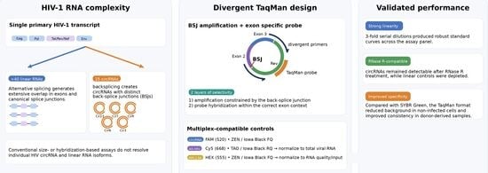
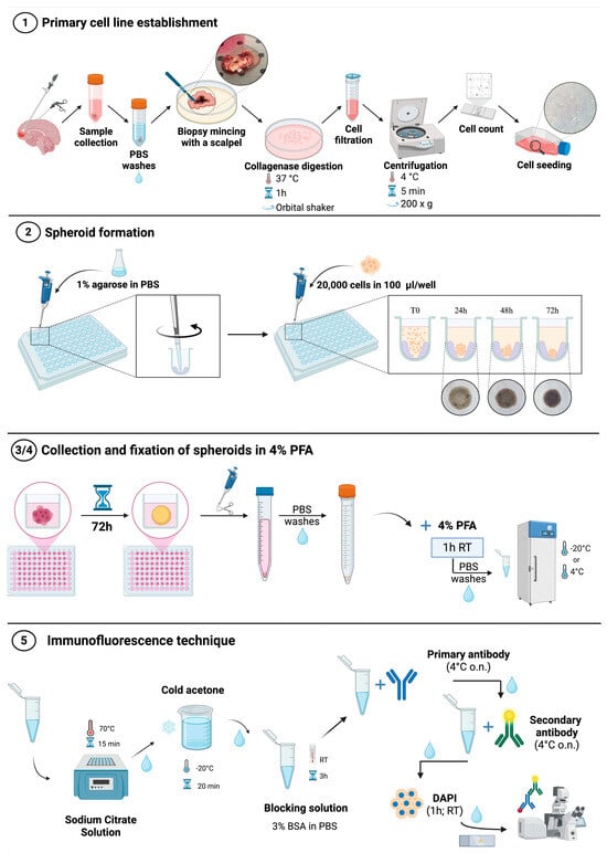
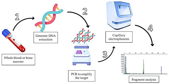
The HIV-1 genome is initially transcribed as a single primary RNA that undergoes extensive splicing to produce over 40 linear and 15 circular RNA (circRNA) isoforms sharing common sequences. Conventional methods for circRNA detection, such as Northern blotting and hybridization-based assays, are inadequate for distinguishing specific circRNA isoforms when multiple circular and linear species originate from the same transcript. We previously identified 15 HIV-1 circRNAs generated by backsplicing and demonstrated that some enhance viral replication by sequestering cellular miRNAs. PCR-based approaches using divergent primers (RT-qPCR) offer greater specificity for detecting individual circular RNAs under these conditions. Building on this, we have developed a TaqMan qPCR assay capable of specifically detecting 14 HIV circRNA isoforms using backsplicing junction-directed divergent primers coupled to a hydrolysis probe for signal confirmation. Compared with matched SYBR Green assays, the TaqMan platform showed lower background in non-infected controls and reduced variance across donor-derived samples. This method provides a robust platform for selective and qualitative analysis of HIV-1 circRNAs.
Full article
(This article belongs to the Section Molecular and Cellular Biology)
►
Show Figures
Graphical abstract


