Next Issue
Volume 11, May
Previous Issue
Volume 11, March


Bioengineering, Volume 11, Issue 4 (April 2024) – 112 articles

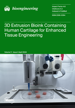
Cover Story (view full-size image): Addressing the challenge of articular cartilage repair amidst the growing prevalence of osteoarthritis, this study introduces a novel approach utilizing an alginate-based bioink integrated with a human decellularized articular cartilage matrix for 3D bioprinting. The investigation aims to evaluate the bioink's capacity for supporting chondrocyte viability and promoting chondrogenic differentiation in a bid to engineer cartilage tissue effectively. By leveraging the structural and biological advantages of alginate and the physiological relevance of human-derived cartilage matrix, this research contributes to the advancement of tissue engineering methodologies. View this paper
  • Issues are regarded as officially published after their release is announced to the table of contents alert mailing list.
  • You may sign up for e-mail alerts to receive table of contents of newly released issues.
  • PDF is the official format for papers published in both, html and pdf forms. To view the papers in pdf format, click on the "PDF Full-text" link, and use the free Adobe Reader to open them.
Order results
Result details
Section
Select all
Export citation of selected articles as:
12 pages, 1226 KiB  
Review
Fabrication of Artificial Nerve Conduits Used in a Long Nerve Gap: Current Reviews and Future Studies
by Ryosuke Kakinoki, Yukiko Hara, Koichi Yoshimoto, Yukitoshi Kaizawa, Kazuhiko Hashimoto, Hiroki Tanaka, Takaya Kobayashi, Kazuhiro Ohtani, Takashi Noguchi, Ryosuke Ikeguchi, Masao Akagi and Koji Goto
Bioengineering 2024, 11(4), 409; https://doi.org/10.3390/bioengineering11040409 - 22 Apr 2024
Viewed by 1629
Abstract
There are many commercially available artificial nerve conduits, used mostly to repair short gaps in sensory nerves. The stages of nerve regeneration in a nerve conduit are fibrin matrix formation between the nerve stumps joined to the conduit, capillary extension and Schwann cell [...] Read more.
There are many commercially available artificial nerve conduits, used mostly to repair short gaps in sensory nerves. The stages of nerve regeneration in a nerve conduit are fibrin matrix formation between the nerve stumps joined to the conduit, capillary extension and Schwann cell migration from both nerve stumps, and, finally, axon extension from the proximal nerve stump. Artificial nerves connecting transected nerve stumps with a long interstump gap should be biodegradable, soft and pliable; have the ability to maintain an intrachamber fibrin matrix structure that allows capillary invasion of the tubular lumen, inhibition of scar tissue invasion and leakage of intratubular neurochemical factors from the chamber; and be able to accommodate cells that produce neurochemical factors that promote nerve regeneration. Here, we describe current progress in the development of artificial nerve conduits and the future studies needed to create nerve conduits, the nerve regeneration of which is compatible with that of an autologous nerve graft transplanted over a long nerve gap. Full article
(This article belongs to the Special Issue Innovations in Nerve Regeneration)
Show Figures

Figure 1

12 pages, 1150 KiB  
Article
How the Effect of Virtual Reality on Cognitive Functioning Is Modulated by Gender Differences
by Stefania Righi, Gioele Gavazzi, Viola Benedetti, Giulia Raineri and Maria Pia Viggiano
Bioengineering 2024, 11(4), 408; https://doi.org/10.3390/bioengineering11040408 - 21 Apr 2024
Cited by 1 | Viewed by 1520
Abstract
Virtual reality (VR) can be a promising tool to simulate reality in various settings but the real impact of this technology on the human mental system is still unclear as to how VR might (if at all) interfere with cognitive functioning. Using a [...] Read more.
Virtual reality (VR) can be a promising tool to simulate reality in various settings but the real impact of this technology on the human mental system is still unclear as to how VR might (if at all) interfere with cognitive functioning. Using a computer, we can concentrate, enter a state of flow, and still maintain control over our surrounding world. Differently, VR is a very immersive experience which could be a challenge for our ability to allocate divided attention to the environment to perform executive functioning tasks. This may also have a different impact on women and men since gender differences in both executive functioning and the immersivity experience have been referred to by the literature. The present study aims to investigate cognitive multitasking performance as a function of (1) virtual reality and computer administration and (2) gender differences. To explore this issue, subjects were asked to perform simultaneous tasks (span forward and backward, logical–arithmetic reasoning, and visuospatial reasoning) in virtual reality via a head-mounted display system (HDMS) and on a personal computer (PC). Our results showed in virtual reality an overall impairment of executive functioning but a better performance of women, compared to men, in visuospatial reasoning. These findings are consistent with previous studies showing a detrimental effect of virtual reality on cognitive functioning. Full article
Show Figures

Figure 1

12 pages, 2446 KiB  
Article
Short-Term l-arginine Treatment Mitigates Early Damage of Dermal Collagen Induced by Diabetes
by Irena Miler, Mihailo D. Rabasovic, Sonja Askrabic, Andreas Stylianou, Bato Korac and Aleksandra Korac
Bioengineering 2024, 11(4), 407; https://doi.org/10.3390/bioengineering11040407 - 21 Apr 2024
Viewed by 1528
Abstract
Changes in the structural properties of the skin due to collagen alterations are an important factor in diabetic skin complications. Using a combination of photonic methods as an optic diagnostic tool, we investigated the structural alteration in rat dermal collagen I in diabetes, [...] Read more.
Changes in the structural properties of the skin due to collagen alterations are an important factor in diabetic skin complications. Using a combination of photonic methods as an optic diagnostic tool, we investigated the structural alteration in rat dermal collagen I in diabetes, and after short-term l-arginine treatment. The multiplex approach shows that in the early phase of diabetes, collagen fibers are partially damaged, resulting in the heterogeneity of fibers, e.g., “patchy patterns” of highly ordered/disordered fibers, while l-arginine treatment counteracts to some extent the conformational changes in collagen-induced by diabetes and mitigates the damage. Raman spectroscopy shows intense collagen conformational changes via amides I and II in diabetes, suggesting that diabetes-induced structural changes in collagen originate predominantly from individual collagen molecules rather than supramolecular structures. There is a clear increase in the amounts of newly synthesized proline and hydroxyproline after treatment with l-arginine, reflecting the changed collagen content. This suggests that it might be useful for treating and stopping collagen damage early on in diabetic skin. Our results demonstrate that l-arginine attenuates the early collagen I alteration caused by diabetes and that it could be used to treat and prevent collagen damage in diabetic skin at a very early stage. Full article
(This article belongs to the Section Biomedical Engineering and Biomaterials)
Show Figures

Graphical abstract

21 pages, 3240 KiB  
Article
Deep Transfer Learning Using Real-World Image Features for Medical Image Classification, with a Case Study on Pneumonia X-ray Images
by Chanhoe Gu and Minhyeok Lee
Bioengineering 2024, 11(4), 406; https://doi.org/10.3390/bioengineering11040406 - 20 Apr 2024
Cited by 1 | Viewed by 1738
Abstract
Deep learning has profoundly influenced various domains, particularly medical image analysis. Traditional transfer learning approaches in this field rely on models pretrained on domain-specific medical datasets, which limits their generalizability and accessibility. In this study, we propose a novel framework called real-world feature [...] Read more.
Deep learning has profoundly influenced various domains, particularly medical image analysis. Traditional transfer learning approaches in this field rely on models pretrained on domain-specific medical datasets, which limits their generalizability and accessibility. In this study, we propose a novel framework called real-world feature transfer learning, which utilizes backbone models initially trained on large-scale general-purpose datasets such as ImageNet. We evaluate the effectiveness and robustness of this approach compared to models trained from scratch, focusing on the task of classifying pneumonia in X-ray images. Our experiments, which included converting grayscale images to RGB format, demonstrate that real-world-feature transfer learning consistently outperforms conventional training approaches across various performance metrics. This advancement has the potential to accelerate deep learning applications in medical imaging by leveraging the rich feature representations learned from general-purpose pretrained models. The proposed methodology overcomes the limitations of domain-specific pretrained models, thereby enabling accelerated innovation in medical diagnostics and healthcare. From a mathematical perspective, we formalize the concept of real-world feature transfer learning and provide a rigorous mathematical formulation of the problem. Our experimental results provide empirical evidence supporting the effectiveness of this approach, laying the foundation for further theoretical analysis and exploration. This work contributes to the broader understanding of feature transferability across domains and has significant implications for the development of accurate and efficient models for medical image analysis, even in resource-constrained settings. Full article
(This article belongs to the Special Issue Artificial Intelligence in Advanced Medical Imaging - 2nd Edition)
Show Figures

Figure 1

11 pages, 4904 KiB  
Article
Breaking the Limit of Cardiovascular Regenerative Medicine: Successful 6-Month Goat Implant in World’s First Ascending Aortic Replacement Using Biotube Blood Vessels
by Kazuki Mori, Tadashi Umeno, Takayuki Kawashima, Tomoyuki Wada, Takuro Genda, Masanagi Arakura, Yoshifumi Oda, Takayuki Mizoguchi, Ryosuke Iwai, Tsutomu Tajikawa, Yasuhide Nakayama and Shinji Miyamoto
Bioengineering 2024, 11(4), 405; https://doi.org/10.3390/bioengineering11040405 - 20 Apr 2024
Cited by 2 | Viewed by 1222
Abstract
This study investigated six-month outcomes of first models of ascending aortic replacement. The molds used to produce the Biotube were implanted subcutaneously in goats. After 2–3 months, the molds were explanted to obtain the Biotubes (inner diameter, 12 mm; wall thickness, 1.5 mm). [...] Read more.
This study investigated six-month outcomes of first models of ascending aortic replacement. The molds used to produce the Biotube were implanted subcutaneously in goats. After 2–3 months, the molds were explanted to obtain the Biotubes (inner diameter, 12 mm; wall thickness, 1.5 mm). Next, we performed ascending aortic replacement using the Biotube in five allogenic goats. At 6 months, the animals underwent computed tomography (CT) and histologic evaluation. As a comparison, we performed similar surgeries using glutaraldehyde-fixed autologous pericardial rolls or pig-derived heterogenous Biotubes. At 6 months, CT revealed no aneurysmalization of the Biotube or pseudoaneurysm formation. The histologic evaluation showed development of endothelial cells, smooth muscle cells, and elastic fibers along the Biotube. In the autologous pericardium group, there was no evidence of new cell development, but there was calcification. The histologic changes observed in the heterologous Biotube group were similar to those in the allogenic Biotube group. However, there was inflammatory cell infiltration in some heterologous Biotubes. Based on the above, we could successfully create the world’s first Biotube-based ascending aortic replacement models. The present results indicate that the Biotube may serve as a scaffold for aortic tissue regeneration. Full article
(This article belongs to the Section Regenerative Engineering)
Show Figures

Figure 1

10 pages, 1864 KiB  
Article
An Investigation of Running Kinematics with Recovered Anterior Cruciate Ligament Reconstruction on a Treadmill and In-Field Using Inertial Measurement Units: A Preliminary Study
by Matteo Hill, Pierre Kiesewetter, Thomas L. Milani and Christian Mitschke
Bioengineering 2024, 11(4), 404; https://doi.org/10.3390/bioengineering11040404 - 19 Apr 2024
Viewed by 1329
Abstract
Anterior cruciate ligament reconstruction (ACLR) may affect movement even years after surgery. The purpose of this study was to determine possible interlimb asymmetries due to ACLR when running on a treadmill and in field conditions, with the aim of contributing to the establishment [...] Read more.
Anterior cruciate ligament reconstruction (ACLR) may affect movement even years after surgery. The purpose of this study was to determine possible interlimb asymmetries due to ACLR when running on a treadmill and in field conditions, with the aim of contributing to the establishment of objective movement assessment in real-world settings; moreover, we aimed to gain knowledge on recovered ACLR as a biomechanical risk factor. Eight subjects with a history of unilateral ACLR 5.4 ± 2.8 years after surgery and eight healthy subjects ran 1 km on a treadmill and 1 km on a concrete track. The ground contact time and triaxial peak tibial accelerations were recorded using inertial measurement units. Interlimb differences within subjects were tested and compared between conditions. There were no significant differences between limbs in the ACLR subjects or in healthy runners for any of the chosen parameters on both running surfaces. However, peak tibial accelerations were higher during field running (p-values < 0.01; Cohen’s d effect sizes > 0.8), independent of health status. To minimize limb loading due to higher impacts during field running, this should be considered when choosing a running surface, especially in rehabilitation or when running with a minor injury or health issues. Full article
(This article belongs to the Special Issue Biomechanics of Human Movement and Its Clinical Applications)
Show Figures

Figure 1

17 pages, 7407 KiB  
Case Report
Biomechanical Considerations in the Orthodontic Treatment of a Patient with Stabilised Stage IV Grade C Generalised Periodontitis: A Case Report
by Fung Hou Kumoi Mineaki Howard Sum, Zhiyi Shan, Yat Him Dave Chan, Ryan Julian Dick Hei Chu, George Pelekos and Tsang Tsang She
Bioengineering 2024, 11(4), 403; https://doi.org/10.3390/bioengineering11040403 - 19 Apr 2024
Viewed by 1462
Abstract
Orthodontic treatment of periodontally compromised patients presents unique challenges, including controlling periodontal inflammation, applying appropriate force, designing an effective dental anchorage, and maintaining treatment results. Deteriorated periodontal support leads to alterations in the biological responses of teeth to mechanical forces, and thus orthodontists [...] Read more.
Orthodontic treatment of periodontally compromised patients presents unique challenges, including controlling periodontal inflammation, applying appropriate force, designing an effective dental anchorage, and maintaining treatment results. Deteriorated periodontal support leads to alterations in the biological responses of teeth to mechanical forces, and thus orthodontists must take greater care when treating patients with periodontal conditions than when treating those with a good periodontal status. In this article, we report the case of a 59-year-old woman with stabilised Stage IV grade C generalised periodontitis characterised by pathological tooth migration (PTM). The assessment, planning, and treatment of this patient with orthodontic fixed appliances is described. Moreover, the anchorage planning and biomechanical considerations are detailed. Specific orthodontic appliances were employed in this case to produce force systems for achieving precise tooth movement, which included a cantilever, mini-screws, and a box loop. Careful application of those appliances resulted in satisfactory aesthetic and functional orthodontic outcomes in the patient. This case highlights the importance of multidisciplinary collaboration in the treatment of patients with severe periodontitis and the potential for tailored biomechanical approaches in orthodontic treatment to furnish good outcomes. Full article
(This article belongs to the Special Issue Application of Bioengineering to Clinical Orthodontics)
Show Figures

Figure 1

19 pages, 10631 KiB  
Article
Dimensionality Matters: Exploiting UV-Photopatterned 2D and Two-Photon-Printed 2.5D Contact Guidance Cues to Control Corneal Fibroblast Behavior and Collagen Deposition
by Cas van der Putten, Gozde Sahin, Rhiannon Grant, Mirko D’Urso, Stefan Giselbrecht, Carlijn V. C. Bouten and Nicholas A. Kurniawan
Bioengineering 2024, 11(4), 402; https://doi.org/10.3390/bioengineering11040402 - 19 Apr 2024
Cited by 1 | Viewed by 1908
Abstract
In the event of disease or injury, restoration of the native organization of cells and extracellular matrix is crucial for regaining tissue functionality. In the cornea, a highly organized collagenous tissue, keratocytes can align along the anisotropy of the physical microenvironment, providing a [...] Read more.
In the event of disease or injury, restoration of the native organization of cells and extracellular matrix is crucial for regaining tissue functionality. In the cornea, a highly organized collagenous tissue, keratocytes can align along the anisotropy of the physical microenvironment, providing a blueprint for guiding the organization of the collagenous matrix. Inspired by this physiological process, anisotropic contact guidance cues have been employed to steer the alignment of keratocytes as a first step to engineer in vitro cornea-like tissues. Despite promising results, two major hurdles must still be overcome to advance the field. First, there is an enormous design space to be explored in optimizing cellular contact guidance in three dimensions. Second, the role of contact guidance cues in directing the long-term deposition and organization of extracellular matrix proteins remains unknown. To address these challenges, here we combined two microengineering strategies—UV-based protein patterning (2D) and two-photon polymerization of topographies (2.5D)—to create a library of anisotropic contact guidance cues with systematically varying height (H, 0 µm ≤ H ≤ 20 µm) and width (W, 5 µm ≤ W ≤ 100 µm). With this unique approach, we found that, in the short term (24 h), the orientation and morphology of primary human fibroblastic keratocytes were critically determined not only by the pattern width, but also by the height of the contact guidance cues. Upon extended 7-day cultures, keratocytes were shown to produce a dense, fibrous collagen network along the direction of the contact guidance cues. Moreover, increasing the heights also increased the aligned fraction of deposited collagen and the contact guidance response of cells, all whilst the cells maintained the fibroblastic keratocyte phenotype. Our study thus reveals the importance of dimensionality of the physical microenvironment in steering both cellular organization and the formation of aligned, collagenous tissues. Full article
(This article belongs to the Special Issue Bioengineering and the Eye—2nd Edition)
Show Figures

Graphical abstract

16 pages, 1642 KiB  
Article
Assessment of Microvascular Hemodynamic Adaptations in Finger Flexors of Climbers
by Blai Ferrer-Uris, Albert Busquets, Faruk Beslija and Turgut Durduran
Bioengineering 2024, 11(4), 401; https://doi.org/10.3390/bioengineering11040401 - 19 Apr 2024
Viewed by 1333
Abstract
Climbing performance is greatly dependent on the endurance of the finger flexors which, in turn, depends on the ability to deliver and use oxygen within the muscle. Near-infrared spectroscopy (NIRS) and diffuse correlation spectroscopy (DCS) have provided new possibilities to explore these phenomena [...] Read more.
Climbing performance is greatly dependent on the endurance of the finger flexors which, in turn, depends on the ability to deliver and use oxygen within the muscle. Near-infrared spectroscopy (NIRS) and diffuse correlation spectroscopy (DCS) have provided new possibilities to explore these phenomena in the microvascular environment. The aim of the present study was to explore climbing-related microvascular adaptations through the comparison of the oxygen concentration and hemodynamics of the forearm between climbers and non-climber active individuals during a vascular occlusion test (VOT). Seventeen climbers and fifteen non-climbers joined the study. Through NIRS and DCS, the oxyhemoglobin (O2Hb) and deoxyhemoglobin (HHb) concentrations, tissue saturation index (TSI), and blood flow index (BFI) were obtained from the flexor digitorum profundus during the VOT. During the reactive hyperemia, climbers presented greater blood flow slopes (p = 0.043, d = 0.573), as well as greater O2Hb maximum values (p = 0.001, d = 1.263) and HHb minimum values (p = 0.009, d = 0.998), than non-climbers. The superior hemodynamics presented by climbers could indicate potential training-induced structural and functional adaptations that could enhance oxygen transportation to the muscle, and thus enhance muscle endurance and climbing performance. Full article
Show Figures

Figure 1

21 pages, 1468 KiB  
Article
Ergonomic Analysis of Dental Work in Different Oral Quadrants: A Motion Capture Preliminary Study among Endodontists
by Sophie Feige, Fabian Holzgreve, Laura Fraeulin, Christian Maurer-Grubinger, Werner Betz, Christina Erbe, Albert Nienhaus, David A. Groneberg and Daniela Ohlendorf
Bioengineering 2024, 11(4), 400; https://doi.org/10.3390/bioengineering11040400 - 19 Apr 2024
Cited by 1 | Viewed by 1646
Abstract
Background: Dentists, including endodontists, frequently experience musculoskeletal disorders due to unfavourable working postures. Several measures are known to reduce the ergonomic risk; however, there are still gaps in the research, particularly in relation to dental work in the different oral regions (Quadrants 1–4). [...] Read more.
Background: Dentists, including endodontists, frequently experience musculoskeletal disorders due to unfavourable working postures. Several measures are known to reduce the ergonomic risk; however, there are still gaps in the research, particularly in relation to dental work in the different oral regions (Quadrants 1–4). Methods: In this study (of a pilot character), a total of 15 dentists (8 male and 7 female) specialising in endodontics were measured while performing root canal treatments on a phantom head. These measurements took place in a laboratory setting using an inertial motion capture system. A slightly modified Rapid Upper Limb Assessment (RULA) coding system was employed for the analysis of kinematic data. The significance level was set at p = 0.05. Results: The ergonomic risk for the entire body was higher in the fourth quadrant than in the first quadrant for 80% of the endodontists and higher than in the second quadrant for 87%. For 87% of the endodontists, the ergonomic risk for the right side of the body was significantly higher in the fourth quadrant compared to the first and second quadrant. The right arm was stressed more in the lower jaw than in the upper jaw, and the neck also showed a greater ergonomic risk in the fourth quadrant compared to the first quadrant. Conclusion: In summary, both the total RULA score and scores for the right- and lefthand sides of the body ranged between 5 and 6 out of a possible 7 points. Considering this considerable burden, heightened attention, especially to the fourth quadrant with a significantly higher ergonomic risk compared to Quadrants 1 and 2, may be warranted. Full article
Show Figures

Figure 1

19 pages, 5200 KiB  
Article
Precision Identification of Locally Advanced Rectal Cancer in Denoised CT Scans Using EfficientNet and Voting System Algorithms
by Chun-Yu Lin, Jacky Chung-Hao Wu, Yen-Ming Kuan, Yi-Chun Liu, Pi-Yi Chang, Jun-Peng Chen, Henry Horng-Shing Lu and Oscar Kuang-Sheng Lee
Bioengineering 2024, 11(4), 399; https://doi.org/10.3390/bioengineering11040399 - 19 Apr 2024
Cited by 1 | Viewed by 1618
Abstract
Background and objective: Local advanced rectal cancer (LARC) poses significant treatment challenges due to its location and high recurrence rates. Accurate early detection is vital for treatment planning. With magnetic resonance imaging (MRI) being resource-intensive, this study explores using artificial intelligence (AI) to [...] Read more.
Background and objective: Local advanced rectal cancer (LARC) poses significant treatment challenges due to its location and high recurrence rates. Accurate early detection is vital for treatment planning. With magnetic resonance imaging (MRI) being resource-intensive, this study explores using artificial intelligence (AI) to interpret computed tomography (CT) scans as an alternative, providing a quicker, more accessible diagnostic tool for LARC. Methods: In this retrospective study, CT images of 1070 T3–4 rectal cancer patients from 2010 to 2022 were analyzed. AI models, trained on 739 cases, were validated using two test sets of 134 and 197 cases. By utilizing techniques such as nonlocal mean filtering, dynamic histogram equalization, and the EfficientNetB0 algorithm, we identified images featuring characteristics of a positive circumferential resection margin (CRM) for the diagnosis of locally advanced rectal cancer (LARC). Importantly, this study employs an innovative approach by using both hard and soft voting systems in the second stage to ascertain the LARC status of cases, thus emphasizing the novelty of the soft voting system for improved case identification accuracy. The local recurrence rates and overall survival of the cases predicted by our model were assessed to underscore its clinical value. Results: The AI model exhibited high accuracy in identifying CRM-positive images, achieving an area under the curve (AUC) of 0.89 in the first test set and 0.86 in the second. In a patient-based analysis, the model reached AUCs of 0.84 and 0.79 using a hard voting system. Employing a soft voting system, the model attained AUCs of 0.93 and 0.88, respectively. Notably, AI-identified LARC cases exhibited a significantly higher five-year local recurrence rate and displayed a trend towards increased mortality across various thresholds. Furthermore, the model’s capability to predict adverse clinical outcomes was superior to those of traditional assessments. Conclusion: AI can precisely identify CRM-positive LARC cases from CT images, signaling an increased local recurrence and mortality rate. Our study presents a swifter and more reliable method for detecting LARC compared to traditional CT or MRI techniques. Full article
(This article belongs to the Special Issue Application of Deep Learning in Medical Diagnosis)
Show Figures

Graphical abstract

13 pages, 1278 KiB  
Article
Effects of Action Observation Plus Motor Imagery Administered by Immersive Virtual Reality on Hand Dexterity in Healthy Subjects
by Paola Adamo, Gianluca Longhi, Federico Temporiti, Giorgia Marino, Emilia Scalona, Maddalena Fabbri-Destro, Pietro Avanzini and Roberto Gatti
Bioengineering 2024, 11(4), 398; https://doi.org/10.3390/bioengineering11040398 - 19 Apr 2024
Viewed by 1278
Abstract
Action observation and motor imagery (AOMI) are commonly delivered through a laptop screen. Immersive virtual reality (VR) may enhance the observer’s embodiment, a factor that may boost AOMI effects. The study aimed to investigate the effects on manual dexterity of AOMI delivered through [...] Read more.
Action observation and motor imagery (AOMI) are commonly delivered through a laptop screen. Immersive virtual reality (VR) may enhance the observer’s embodiment, a factor that may boost AOMI effects. The study aimed to investigate the effects on manual dexterity of AOMI delivered through immersive VR compared to AOMI administered through a laptop. To evaluate whether VR can enhance the effects of AOMI, forty-five young volunteers were enrolled and randomly assigned to the VR-AOMI group, who underwent AOMI through immersive VR, the AOMI group, who underwent AOMI through a laptop screen, or the control group, who observed landscape video clips. All participants underwent a 5-day treatment, consisting of 12 min per day. We investigated between and within-group differences after treatments relative to functional manual dexterity tasks using the Purdue Pegboard Test (PPT). This test included right hand (R), left hand (L), both hands (B), R + L + B, and assembly tasks. Additionally, we analyzed kinematics parameters including total and sub-phase duration, peak and mean velocity, and normalized jerk, during the Nine-Hole Peg Test to examine whether changes in functional scores may also occur through specific kinematic patterns. Participants were assessed at baseline (T0), after the first training session (T1), and at the end of training (T2). A significant time by group interaction and time effects were found for PPT, where both VR-AOMI and AOMI groups improved at the end of training. Larger PPT-L task improvements were found in the VR-AOMI group (d: 0.84, CI95: 0.09–1.58) compared to the AOMI group from T0 to T1. Immersive VR used for the delivery of AOMI speeded up hand dexterity improvements. Full article
(This article belongs to the Special Issue Bioengineering of the Motor System)
Show Figures

Graphical abstract

12 pages, 1521 KiB  
Article
Micro-Computed Tomography Analysis of Peri-Implant Bone Defects Exposed to a Peri-Implantitis Microcosm, with and without Bone Substitute, in a Rabbit Model: A Pilot Study
by Camila Panes, Iván Valdivia-Gandur, Carlos Veuthey, Vanessa Sousa, Mariano del Sol and Víctor Beltrán
Bioengineering 2024, 11(4), 397; https://doi.org/10.3390/bioengineering11040397 - 19 Apr 2024
Viewed by 1416
Abstract
Peri-implantitis is an inflammatory condition characterized by inflammation in the peri-implant connective tissue and a progressive loss of supporting bone; it is commonly associated with the presence of biofilms on the surface of the implant, which is an important factor in the development [...] Read more.
Peri-implantitis is an inflammatory condition characterized by inflammation in the peri-implant connective tissue and a progressive loss of supporting bone; it is commonly associated with the presence of biofilms on the surface of the implant, which is an important factor in the development and progression of the disease. The objective of this study was to evaluate, using micro-CT, the bone regeneration of surgically created peri-implant defects exposed to a microcosm of peri-implantitis. Twenty-three adult New Zealand white rabbits were included in the study. Bone defects of 7 mm diameter were created in both tibiae, and a cap-shaped titanium device was placed in the center, counter-implanted with a peri-implantitis microcosm. The bone defects received a bone substitute and/or a resorbable synthetic PLGA membrane, according to random distribution. Euthanasia was performed 15 and 30 days postoperatively. Micro-CT was performed on all samples to quantify bone regeneration parameters. Bone regeneration of critical defects occurred in all experimental groups, with a significantly greater increase in cases that received bone graft treatment (p < 0.0001), in all measured parameters, at 15 and 30 days. No significant differences were observed in the different bone neoformation parameters between the groups that did not receive bone grafts (p > 0.05). In this experimental model, the presence of peri-implantitis microcosms was not a determining factor in the bone volume parameter, both in the groups that received regenerative treatment and in those that did not. Full article
(This article belongs to the Special Issue Biomaterials for Bone Repair and Regeneration)
Show Figures

Figure 1

12 pages, 738 KiB  
Review
A Systematic Review of Bone Bruise Patterns following Acute Anterior Cruciate Ligament Tears: Insights into the Mechanism of Injury
by Sueen Sohn, Saad Mohammed AlShammari, Byung Jun Hwang and Man Soo Kim
Bioengineering 2024, 11(4), 396; https://doi.org/10.3390/bioengineering11040396 - 19 Apr 2024
Viewed by 1248
Abstract
(1) Background: The purpose of this systematic review was to determine the prevalence of bone bruises in patients with anterior cruciate ligament (ACL) injuries and the location of the bruises relative to the tibia and femur. Understanding the relative positions of these bone [...] Read more.
(1) Background: The purpose of this systematic review was to determine the prevalence of bone bruises in patients with anterior cruciate ligament (ACL) injuries and the location of the bruises relative to the tibia and femur. Understanding the relative positions of these bone bruises could enhance our comprehension of the knee loading patterns that occur during an ACL injury. (2) Methods: The MEDLINE, EMBASE, and the Cochrane Library databases were searched for studies that evaluated the presence of bone bruises following ACL injuries. Study selection, data extraction, and a systematic review were performed. (3) Results: Bone bruises were observed in 3207 cases (82.8%) at the lateral tibia plateau (LTP), 1608 cases (41.5%) at the medial tibia plateau (MTP), 2765 cases (71.4%) at the lateral femoral condyle (LFC), and 1257 cases (32.4%) at the medial femoral condyle (MFC). Of the 30 studies, 11 were able to assess the anterior to posterior direction. The posterior LTP and center LFC were the most common areas of bone bruises. Among the 30 studies, 14 documented bone bruises across all four sites (LTP, MTP, LFC, and MFC). The most common pattern was bone bruises appearing at the LTP and LFC. (4) Conclusions: The most frequently observed pattern of bone bruises was restricted to the lateral aspects of both the tibia and femur. In cases where bone bruises were present on both the lateral and medial sides, those on the lateral side exhibited greater severity. The positioning of bone bruises along the front–back axis indicated a forward shift of the tibia in relation to the femur during ACL injuries. Full article
(This article belongs to the Special Issue Biomechanics of Sports Injuries)
Show Figures

Figure 1

12 pages, 2260 KiB  
Article
Tensile Yield Strain of Human Cortical Bone from the Femoral Diaphysis Is Constant among Healthy Adults and across the Anatomical Quadrants
by Massimiliano Baleani, Paolo Erani, Alice Acciaioli and Enrico Schileo
Bioengineering 2024, 11(4), 395; https://doi.org/10.3390/bioengineering11040395 - 19 Apr 2024
Viewed by 1366
Abstract
The literature suggests that the yield strain of cortical bone is invariant to its stiffness (elastic modulus) and strength (yield stress). However, data about intra-individual variations, e.g., the influence of different collagen/mineral organisations observed in bone aspects withstanding different habitual loads, are lacking. [...] Read more.
The literature suggests that the yield strain of cortical bone is invariant to its stiffness (elastic modulus) and strength (yield stress). However, data about intra-individual variations, e.g., the influence of different collagen/mineral organisations observed in bone aspects withstanding different habitual loads, are lacking. The hypothesis that the yield strain of human cortical bone tissue, retrieved from femoral diaphyseal quadrants subjected to different habitual loads, is invariant was tested. Four flat dumbbell-shaped specimens were machined from each quadrant of the proximal femoral diaphysis of five adult donors for a total of 80 specimens. Two extensometers attached to the narrow specimen region were used to measure deformation during monotonic tensile testing. The elastic modulus (linear part of the stress–strain curve) and yield strain/stress at a 0.2% offset were obtained. Elastic modulus and yield stress values were, respectively, in the range of 12.2–20.5 GPa and 75.9–136.6 MPa and exhibited a positive linear correlation. All yield strain values were in the narrow range of 0.77–0.87%, regardless of the stiffness and strength of the tissue and the anatomical quadrant. In summary, the results corroborate the hypothesis that tensile yield strain in cortical bone is invariant, irrespective also of the anatomical quadrant. The mean yield strain value found in this study is similar to what was reported by inter-species and evolution studies but slightly higher than previous reports in humans, possibly because of the younger age of our subjects. Further investigations are needed to elucidate a possible dependence of yield strain on age. Full article
(This article belongs to the Section Biomechanics and Sports Medicine)
Show Figures

Graphical abstract

12 pages, 6255 KiB  
Article
Finite Element Analysis of Fixed Orthodontic Retainers
by Sebastian Hetzler, Stefan Rues, Andreas Zenthöfer, Peter Rammelsberg, Christopher J. Lux and Christoph J. Roser
Bioengineering 2024, 11(4), 394; https://doi.org/10.3390/bioengineering11040394 - 18 Apr 2024
Cited by 1 | Viewed by 1492
Abstract
The efficacy of retainers is a pivotal concern in orthodontic care. This study examined the biomechanical behaviour of retainers, particularly the influence of retainer stiffness and tooth resilience on force transmission and stress distribution. To do this, a finite element model was created [...] Read more.
The efficacy of retainers is a pivotal concern in orthodontic care. This study examined the biomechanical behaviour of retainers, particularly the influence of retainer stiffness and tooth resilience on force transmission and stress distribution. To do this, a finite element model was created of the lower jaw from the left to the right canine with a retainer attached on the oral side. Three levels of tooth resilience and variable retainer bending stiffness (influenced by retainer type, retainer diameter, and retainer material) were simulated. Applying axial or oblique (45° tilt) loads on a central incisor, the force transmission increased from 2% to 65% with increasing tooth resilience and retainer stiffness. Additionally, a smaller retainer diameter reduced the uniformity of the stress distribution in the bonding interfaces, causing concentrated stress peaks within a small field of the bonding area. An increase in retainer stiffness and in tooth resilience as well as a more oblique load direction all lead to higher overall stress in the adhesive bonding area associated with a higher risk of retainer bonding failure. Therefore, it might be recommended to avoid the use of retainers that are excessively stiff, especially in cases with high tooth resilience. Full article
(This article belongs to the Special Issue Application of Bioengineering to Clinical Orthodontics)
Show Figures

Figure 1

19 pages, 2977 KiB  
Article
Debulking of the Femoral Stem in a Primary Total Hip Joint Replacement: A Novel Method to Reduce Stress Shielding
by Gulshan Sunavala-Dossabhoy, Brent M. Saba and Kevin J. McCarthy
Bioengineering 2024, 11(4), 393; https://doi.org/10.3390/bioengineering11040393 - 18 Apr 2024
Viewed by 1453
Abstract
In current-generation designs of total primary hip joint replacement, the prostheses are fabricated from alloys. The modulus of elasticity of the alloy is substantially higher than that of the surrounding bone. This discrepancy plays a role in a phenomenon known as stress shielding, [...] Read more.
In current-generation designs of total primary hip joint replacement, the prostheses are fabricated from alloys. The modulus of elasticity of the alloy is substantially higher than that of the surrounding bone. This discrepancy plays a role in a phenomenon known as stress shielding, in which the bone bears a reduced proportion of the applied load. Stress shielding has been implicated in aseptic loosening of the implant which, in turn, results in reduction in the in vivo life of the implant. Rigid implants shield surrounding bone from mechanical loading, and the reduction in skeletal stress necessary to maintain bone mass and density results in accelerated bone loss, the forerunner to implant loosening. Femoral stems of various geometries and surface modifications, materials and material distributions, and porous structures have been investigated to achieve mechanical properties of stems closer to those of bone to mitigate stress shielding. For improved load transfer from implant to femur, the proposed study investigated a strategic debulking effort to impart controlled flexibility while retaining sufficient strength and endurance properties. Using an iterative design process, debulked configurations based on an internal skeletal truss framework were evaluated using finite element analysis. The implant models analyzed were solid; hollow, with a proximal hollowed stem; FB-2A, with thin, curved trusses extending from the central spine; and FB-3B and FB-3C, with thick, flat trusses extending from the central spine in a balanced-truss and a hemi-truss configuration, respectively. As outlined in the International Organization for Standardization (ISO) 7206 standards, implants were offset in natural femur for evaluation of load distribution or potted in testing cylinders for fatigue testing. The commonality across all debulked designs was the minimization of proximal stress shielding compared to conventional solid implants. Stem topography can influence performance, and the truss implants with or without the calcar collar were evaluated. Load sharing was equally effective irrespective of the collar; however, the collar was critical to reducing the stresses in the implant. Whether bonded directly to bone or cemented in the femur, the truss stem was effective at limiting stress shielding. However, a localized increase in maximum principal stress at the proximal lateral junction could adversely affect cement integrity. The controlled accommodation of deformation of the implant wall contributes to the load sharing capability of the truss implant, and for a superior biomechanical performance, the collared stem should be implanted in interference fit. Considering the results of all implant designs, the truss implant model FB-3C was the best model. Full article
(This article belongs to the Special Issue Novel and Advanced Technologies for Orthopaedic Implant)
Show Figures

Graphical abstract

20 pages, 6271 KiB  
Article
Evaluation of Deep Learning Model Architectures for Point-of-Care Ultrasound Diagnostics
by Sofia I. Hernandez Torres, Austin Ruiz, Lawrence Holland, Ryan Ortiz and Eric J. Snider
Bioengineering 2024, 11(4), 392; https://doi.org/10.3390/bioengineering11040392 - 18 Apr 2024
Viewed by 1236
Abstract
Point-of-care ultrasound imaging is a critical tool for patient triage during trauma for diagnosing injuries and prioritizing limited medical evacuation resources. Specifically, an eFAST exam evaluates if there are free fluids in the chest or abdomen but this is only possible if ultrasound [...] Read more.
Point-of-care ultrasound imaging is a critical tool for patient triage during trauma for diagnosing injuries and prioritizing limited medical evacuation resources. Specifically, an eFAST exam evaluates if there are free fluids in the chest or abdomen but this is only possible if ultrasound scans can be accurately interpreted, a challenge in the pre-hospital setting. In this effort, we evaluated the use of artificial intelligent eFAST image interpretation models. Widely used deep learning model architectures were evaluated as well as Bayesian models optimized for six different diagnostic models: pneumothorax (i) B- or (ii) M-mode, hemothorax (iii) B- or (iv) M-mode, (v) pelvic or bladder abdominal hemorrhage and (vi) right upper quadrant abdominal hemorrhage. Models were trained using images captured in 27 swine. Using a leave-one-subject-out training approach, the MobileNetV2 and DarkNet53 models surpassed 85% accuracy for each M-mode scan site. The different B-mode models performed worse with accuracies between 68% and 74% except for the pelvic hemorrhage model, which only reached 62% accuracy for all model architectures. These results highlight which eFAST scan sites can be easily automated with image interpretation models, while other scan sites, such as the bladder hemorrhage model, will require more robust model development or data augmentation to improve performance. With these additional improvements, the skill threshold for ultrasound-based triage can be reduced, thus expanding its utility in the pre-hospital setting. Full article
(This article belongs to the Special Issue Machine-Learning-Driven Medical Image Analysis)
Show Figures

Figure 1

16 pages, 3827 KiB  
Article
Thermal Characterization and Preclinical Feasibility Verification of an Accessible, Carbon Dioxide-Based Cryotherapy System
by Yixin Hu, Naomi Gordon, Katherine Ogg, Dara L. Kraitchman, Nicholas J. Durr and Bailey Surtees
Bioengineering 2024, 11(4), 391; https://doi.org/10.3390/bioengineering11040391 - 18 Apr 2024
Viewed by 1283
Abstract
To investigate the potential of an affordable cryotherapy device for the accessible treatment of breast cancer, the performance of a novel carbon dioxide-based device was evaluated through both benchtop testing and an in vivo canine model. This novel device was quantitatively compared to [...] Read more.
To investigate the potential of an affordable cryotherapy device for the accessible treatment of breast cancer, the performance of a novel carbon dioxide-based device was evaluated through both benchtop testing and an in vivo canine model. This novel device was quantitatively compared to a commercial device that utilizes argon gas as the cryogen. The thermal behavior of each device was characterized through calorimetry and by measuring the temperature profiles of iceballs generated in tissue phantoms. A 45 min treatment in a tissue phantom from the carbon dioxide device produced a 1.67 ± 0.06 cm diameter lethal isotherm that was equivalent to a 7 min treatment from the commercial argon-based device, which produced a 1.53 ± 0.15 cm diameter lethal isotherm. An in vivo treatment was performed with the carbon dioxide-based device in one spontaneously occurring canine mammary mass with two standard 10 min freezes. Following cryotherapy, this mass was surgically resected and analyzed for necrosis margins via histopathology. The histopathology margin of necrosis from the in vivo treatment with the carbon dioxide device at 14 days post-cryoablation was 1.57 cm. While carbon dioxide gas has historically been considered an impractical cryogen due to its low working pressure and high boiling point, this study shows that carbon dioxide-based cryotherapy may be equivalent to conventional argon-based cryotherapy in size of the ablation zone in a standard treatment time. The feasibility of the carbon dioxide device demonstrated in this study is an important step towards bringing accessible breast cancer treatment to women in low-resource settings. Full article
(This article belongs to the Special Issue Novel, Low Cost Technologies for Cancer Diagnostics and Therapeutics)
Show Figures

Figure 1

15 pages, 2250 KiB  
Article
DTONet a Lightweight Model for Melanoma Segmentation
by Shengnan Hao, Hongzan Wang, Rui Chen, Qinping Liao, Zhanlin Ji, Tao Lyu and Li Zhao
Bioengineering 2024, 11(4), 390; https://doi.org/10.3390/bioengineering11040390 - 18 Apr 2024
Viewed by 1427
Abstract
With the further development of neural networks, automatic segmentation techniques for melanoma are becoming increasingly mature, especially under the conditions of abundant hardware resources. This allows for the accuracy of segmentation to be improved by increasing the complexity and computational capacity of the [...] Read more.
With the further development of neural networks, automatic segmentation techniques for melanoma are becoming increasingly mature, especially under the conditions of abundant hardware resources. This allows for the accuracy of segmentation to be improved by increasing the complexity and computational capacity of the model. However, a new problem arises when it comes to actual applications, as there may not be the high-end hardware available, especially in hospitals and among the general public, who may have limited computing resources. In response to this situation, this paper proposes a lightweight deep learning network that can achieve high segmentation accuracy with minimal resource consumption. We introduce a network called DTONet (double-tailed octave network), which was specifically designed for this purpose. Its computational parameter count is only 30,859, which is 1/256th of the mainstream UNet model. Despite its reduced complexity, DTONet demonstrates superior performance in terms of accuracy, with an IOU improvement over other similar models. To validate the generalization capability of this model, we conducted tests on the PH2 dataset, and the results still outperformed existing models. Therefore, the proposed DTONet network exhibits excellent generalization ability and is sufficiently outstanding. Full article
(This article belongs to the Special Issue Application of Deep Learning in Medical Diagnosis)
Show Figures

Figure 1

14 pages, 2488 KiB  
Article
Altered Functional Connectivity of Temporoparietal Lobe in Obstructive Sleep Apnea: A Resting-State fNIRS Study
by Fang Xiao, Minghui Liu, Yalin Wang, Ligang Zhou, Jingchun Luo, Chen Chen and Wei Chen
Bioengineering 2024, 11(4), 389; https://doi.org/10.3390/bioengineering11040389 - 18 Apr 2024
Viewed by 1592
Abstract
Obstructive Sleep Apnea (OSA), a sleep disorder with high prevalence, is normally accompanied by affective, autonomic, and cognitive abnormalities, and is deemed to be linked to functional brain alterations. To investigate alterations in brain functional connectivity properties in patients with OSA, a comparative [...] Read more.
Obstructive Sleep Apnea (OSA), a sleep disorder with high prevalence, is normally accompanied by affective, autonomic, and cognitive abnormalities, and is deemed to be linked to functional brain alterations. To investigate alterations in brain functional connectivity properties in patients with OSA, a comparative analysis of global and local topological properties of brain networks was conducted between patients with OSA and healthy controls (HCs), utilizing functional near-infrared spectroscopy (fNIRS) imaging. A total of 148 patients with OSA and 150 healthy individuals were involved. Firstly, quantitative alterations in blood oxygen concentration, changes in functional connectivity, and variations in graph theory-based network topological characteristics were assessed. Then, with Mann–Whitney statistics, this study compared whether there are significant differences in the above characteristics between patients with OSA and HCs. Lastly, the study further examined the correlation between the altered characteristics and the apnea hypopnea index (AHI) using linear regression. Results revealed a higher mean and standard deviation of hemoglobin concentration in the superior temporal gyrus among patients with OSA compared to HCs. Resting-state functional connectivity (RSFC) exhibited a slight increase between the superior temporal gyrus and other specific areas in patients with OSA. Notably, neither patients with OSA nor HCs demonstrated significant small-world network properties. Patients with OSA displayed an elevated clustering coefficient (p < 0.05) and local efficiency (p < 0.05). Additionally, patients with OSA exhibited a tendency towards increased nodal betweenness centrality (p < 0.05) and degree centrality (p < 0.05) in the right supramarginal gyrus, as well as a trend towards higher betweenness centrality (p < 0.05) in the right precentral gyrus. The results of multiple linear regressions indicate that the influence of the AHI on RSFC between the right precentral gyrus and right superior temporal gyrus (p < 0.05), as well as between the right precentral gyrus and right supramarginal gyrus (p < 0.05), are statistically significant. These findings suggest that OSA may compromise functional brain connectivity and network topological properties in affected individuals, serving as a potential neurological mechanism underlying the observed abnormalities in brain function associated with OSA. Full article
(This article belongs to the Section Biosignal Processing)
Show Figures

Figure 1

22 pages, 4938 KiB  
Article
Functional and Molecular Analysis of Human Osteoarthritic Chondrocytes Treated with Bone Marrow-Derived MSC-EVs
by Annachiara Scalzone, Clara Sanjurjo-Rodríguez, Rolando Berlinguer-Palmini, Anne M. Dickinson, Elena Jones, Xiao-Nong Wang and Rachel E. Crossland
Bioengineering 2024, 11(4), 388; https://doi.org/10.3390/bioengineering11040388 - 17 Apr 2024
Cited by 2 | Viewed by 2434
Abstract
Osteoarthritis (OA) is a degenerative joint disease, causing impaired mobility. There are currently no effective therapies other than palliative treatment. Mesenchymal stromal cells (MSCs) and their secreted extracellular vesicles (MSC-EVs) have shown promise in attenuating OA progression, promoting chondral regeneration, and modulating joint [...] Read more.
Osteoarthritis (OA) is a degenerative joint disease, causing impaired mobility. There are currently no effective therapies other than palliative treatment. Mesenchymal stromal cells (MSCs) and their secreted extracellular vesicles (MSC-EVs) have shown promise in attenuating OA progression, promoting chondral regeneration, and modulating joint inflammation. However, the precise molecular mechanism of action driving their beneficial effects has not been fully elucidated. In this study, we analyzed MSC-EV-treated human OA chondrocytes (OACs) to assess viability, proliferation, migration, cytokine and catabolic protein expression, and microRNA and mRNA profiles. We observed that MSC-EV-treated OACs displayed increased metabolic activity, proliferation, and migration compared to the controls. They produced decreased proinflammatory (Il-8 and IFN-γ) and increased anti-inflammatory (IL-13) cytokines, and lower levels of MMP13 protein coupled with reduced expression of MMP13 mRNA, as well as negative microRNA regulators of chondrogenesis (miR-145-5p and miR-21-5p). In 3D models, MSC-EV-treated OACs exhibited enhanced chondrogenesis-promoting features (elevated sGAG, ACAN, and aggrecan). MSC-EV treatment also reversed the pathological impact of IL-1β on chondrogenic gene expression and extracellular matrix component (ECM) production. Finally, MSC-EV-treated OACs demonstrated the enhanced expression of genes associated with cartilage function, collagen biosynthesis, and ECM organization and exhibited a signature of 24 differentially expressed microRNAs, associated with chondrogenesis-associated pathways and ECM interactions. In conclusion, our data provide new insights on the potential mechanism of action of MSC-EVs as a treatment option for early-stage OA, including transcriptomic analysis of MSC-EV-treated OA, which may pave the way for more targeted novel therapeutics. Full article
(This article belongs to the Section Regenerative Engineering)
Show Figures

Figure 1

13 pages, 4023 KiB  
Article
Could Dental Material Reuse Play a Significant Role in Preservation of Raw Materials, Water, Energy, and Costs? Microbiological Analysis of New versus Reused Healing Abutments: An In Vitro Study
by Roberto Burioni, Lucia Silvestrini, Bianca D’Orto, Giulia Tetè, Matteo Nagni, Elisabetta Polizzi and Enrico Felice Gherlone
Bioengineering 2024, 11(4), 387; https://doi.org/10.3390/bioengineering11040387 - 16 Apr 2024
Cited by 1 | Viewed by 1146
Abstract
Aim: The objective of this in vitro study was to compare reused and sterilized versus new healing abutments to assess whether a decontamination and sterilization process performed on resued healing abutments was sufficient to remove residual proteins. The two groups were comparable with [...] Read more.
Aim: The objective of this in vitro study was to compare reused and sterilized versus new healing abutments to assess whether a decontamination and sterilization process performed on resued healing abutments was sufficient to remove residual proteins. The two groups were comparable with respect to patient safety. Materials and methods: During the period from September 2022 to October 2023, healing abutment screws were selected and divided into two groups according to whether they were new or previously used in patients. The samples were subjected to a decontamination and sterilization protocol, and results from sample sterility evaluation and assessment of surface protein levels were recorded. Results: The obtained results revealed a significant difference in the OD562 nm values between new and reused healing abutment samples. The assay demonstrates how treated healing abutments were still contaminated by residual proteins. Conclusion: Within the limitations of the present study, although from an infectious point of view sterilization results in the total eradication of pathogens, surface proteins remain on reused healing abutments. Full article
(This article belongs to the Special Issue Dental Implant Reconstruction and Biomechanical Evaluation)
Show Figures

Figure 1

17 pages, 5127 KiB  
Article
Balance Evaluation Based on Walking Experiments with Exoskeleton Interference
by Liping Wang, Xin Li, Yiying Peng, Jianda Han and Juanjuan Zhang
Bioengineering 2024, 11(4), 386; https://doi.org/10.3390/bioengineering11040386 - 16 Apr 2024
Viewed by 1230
Abstract
The impairment of walking balance function seriously affects human health and will lead to a significantly increased risk of falling. It is important to assess and improve the walking balance of humans. However, existing evaluation methods for human walking balance are relatively subjective, [...] Read more.
The impairment of walking balance function seriously affects human health and will lead to a significantly increased risk of falling. It is important to assess and improve the walking balance of humans. However, existing evaluation methods for human walking balance are relatively subjective, and the selected metrics lack effectiveness and comprehensiveness. We present a method to construct a comprehensive evaluation index of human walking balance. We used it to generate personal and general indexes. We first pre-selected some preliminary metrics of walking balance based on theoretical analysis. Seven healthy subjects walked with exoskeleton interference on a treadmill at 1.25 m/s while their ground reaction force information and kinematic data were recorded. One subject with Charcot–Marie–Tooth walked at multiple speeds without the exoskeleton while the same data were collected. Then, we picked a number of effective evaluation metrics based on statistical analysis. We finally constructed the Walking Balance Index (WBI) by combining multiple metrics using principal component analysis. The WBI can distinguish walking balance among different subjects and gait conditions, which verifies the effectiveness of our method in evaluating human walking balance. This method can be used to evaluate and further improve the walking balance of humans in subsequent simulations and experiments. Full article
(This article belongs to the Special Issue Bioengineering of the Motor System)
Show Figures

Figure 1

11 pages, 1891 KiB  
Article
Application of the Single Source—Detector Separation Algorithm in Wearable Neuroimaging Devices: A Step toward Miniaturized Biosensor for Hypoxia Detection
by Thien Nguyen, Soongho Park, Jinho Park, Asma Sodager, Tony George and Amir Gandjbakhche
Bioengineering 2024, 11(4), 385; https://doi.org/10.3390/bioengineering11040385 - 16 Apr 2024
Cited by 1 | Viewed by 1600
Abstract
Most currently available wearable devices to noninvasively detect hypoxia use the spatially resolved spectroscopy (SRS) method to calculate cerebral tissue oxygen saturation (StO2). This study applies the single source—detector separation (SSDS) algorithm to calculate StO2. Near-infrared spectroscopy (NIRS) data [...] Read more.
Most currently available wearable devices to noninvasively detect hypoxia use the spatially resolved spectroscopy (SRS) method to calculate cerebral tissue oxygen saturation (StO2). This study applies the single source—detector separation (SSDS) algorithm to calculate StO2. Near-infrared spectroscopy (NIRS) data were collected from 26 healthy adult volunteers during a breath-holding task using a wearable NIRS device, which included two source—detector separations (SDSs). These data were used to derive oxyhemoglobin (HbO) change and StO2. In the group analysis, both HbO change and StO2 exhibited significant change during a breath-holding task. Specifically, they initially decreased to minimums at around 10 s and then steadily increased to maximums, which were significantly greater than baseline levels, at 25–30 s (p-HbO < 0.001 and p-StO2 < 0.05). However, at an individual level, the SRS method failed to detect changes in cerebral StO2 in response to a short breath-holding task. Furthermore, the SSDS algorithm is more robust than the SRS method in quantifying change in cerebral StO2 in response to a breath-holding task. In conclusion, these findings have demonstrated the potential use of the SSDS algorithm in developing a miniaturized wearable biosensor to monitor cerebral StO2 and detect cerebral hypoxia. Full article
(This article belongs to the Special Issue Neuroimaging Techniques for Wearable Devices in Bioengineering)
Show Figures

Graphical abstract

11 pages, 1901 KiB  
Article
A Clinical Trial Evaluating the Efficacy of Deep Learning-Based Facial Recognition for Patient Identification in Diverse Hospital Settings
by Ayako Sadahide, Hideki Itoh, Ken Moritou, Hirofumi Kameyama, Ryoya Oda, Hitoshi Tabuchi and Yoshiaki Kiuchi
Bioengineering 2024, 11(4), 384; https://doi.org/10.3390/bioengineering11040384 - 15 Apr 2024
Viewed by 1693
Abstract
Background: Facial recognition systems utilizing deep learning techniques can improve the accuracy of facial recognition technology. However, it remains unclear whether these systems should be available for patient identification in a hospital setting. Methods: We evaluated a facial recognition system using deep learning [...] Read more.
Background: Facial recognition systems utilizing deep learning techniques can improve the accuracy of facial recognition technology. However, it remains unclear whether these systems should be available for patient identification in a hospital setting. Methods: We evaluated a facial recognition system using deep learning and the built-in camera of an iPad to identify patients. We tested the system under different conditions to assess its authentication scores (AS) and determine its efficacy. Our evaluation included 100 patients in four postures: sitting, supine, and lateral positions, with and without masks, and under nighttime sleeping conditions. Results: Our results show that the unmasked certification rate of 99.7% was significantly higher than the masked rate of 90.8% (p < 0.0001). In addition, we found that the authentication rate exceeded 99% even during nighttime sleeping. Furthermore, the facial recognition system was safe and acceptable for patient identification within a hospital environment. Even for patients wearing masks, we achieved a 100% success rate for authentication regardless of illumination if they were sitting with their eyes open. Conclusions: This is the first systematical study to evaluate facial recognition among hospitalized patients under different situations. The facial recognition system using deep learning for patient identification shows promising results, proving its safety and acceptability, especially in hospital settings where accurate patient identification is crucial. Full article
(This article belongs to the Special Issue Artificial Intelligence (AI) in Biomedicine)
Show Figures

Graphical abstract

15 pages, 3732 KiB  
Article
Primary Stability of Implants Inserted into Polyurethane Blocks: Micro-CT and Analysis In Vitro
by Chadi Dura Haddad, Ludovica Andreatti, Igor Zelezetsky, Davide Porrelli, Gianluca Turco, Lorenzo Bevilacqua and Michele Maglione
Bioengineering 2024, 11(4), 383; https://doi.org/10.3390/bioengineering11040383 - 15 Apr 2024
Viewed by 1326
Abstract
The approach employed for the site preparation of the dental implant is a variable factor that affects the implant’s primary stability and its ability to integrate with the surrounding bone. The main objective of this in vitro study is to evaluate the influence [...] Read more.
The approach employed for the site preparation of the dental implant is a variable factor that affects the implant’s primary stability and its ability to integrate with the surrounding bone. The main objective of this in vitro study is to evaluate the influence of different techniques used to prepare the implant site on the primary stability of the implant in two different densities of artificial bone. Materials and Methods: A total of 150 implant sites were prepared in rigid polyurethane blocks to simulate two distinct bone densities of 15 pounds per cubic foot (PCF) and 30 PCF, with a 1-mm-thick simulated cortex. The implant sites were equally distributed among piezoelectric surgery (PES), traditional drills (TD), and black ruby magnetic mallet inserts (MM). Two methods have been employed to evaluate the implant’s primary stability, Osstell and micro-tomography. Results: In the present study, we observed significant variations in the implant stability quotient (ISQ) values. More precisely, our findings indicate that the ISQ values were generally higher for 30 PCF compared to 15 PCF. In terms of the preparation technique, PES exhibited the greatest ISQ values, followed by MM, and finally TD. These findings corresponded for both bone densities of 30 PCF (PES 75.6 ± 1.73, MM 69.8 ± 1.91, and TD 65.8 ± 1.91) and 15 PCF (PES 72.3 ± 1.63, MM 62.4 ± 1.77, and TD 60.6 ± 1.81). By utilizing Micro-CT scans, we were able to determine the ratio of the implant occupation to the preparation site. Furthermore, we could calculate the maximum distance between the implant and the wall of the preparation site. The findings demonstrated that PES had a higher ratio of implant to preparation site occupation, followed by TD, and then the MM, at a bone density of 30 PCF (PES 96 ± 1.95, TD 94 ± 1.88, and MM 90.3 ± 2.11). Nevertheless, there were no statistically significant differences in the occupation ratio among these three approaches in the bone density of 15 PCF (PES 89.6 ± 1.22, TD 90 ± 1.31, and MM 88.4 ± 1.17). Regarding the maximum gap between the implant and the site preparation, the smallest gaps were seen when TD were used, followed by MM, and finally by PES, either in a bone density 15 PCF (PES 318 ± 21, TD 238 ± 17, and MM 301 ± 20 μm) or in a bone density 30 PCF (PES 299 ± 20, TD 221 ± 16, and MM 281 ± 19 μm). A statistical analysis using ANOVA revealed these differences to be significant, with p-values of < 0.05. Conclusion: The outcomes of this study indicate that employing the PES technique and osteo-densification with MM during implant insertion may enhance the primary stability and increase the possibility of early implant loading. Full article
Show Figures

Figure 1

15 pages, 6261 KiB  
Article
Limb Volume Measurements: A Comparison of Circumferential Techniques and Optoelectronic Systems against Water Displacement
by Giovanni Farina, Manuela Galli, Leonardo Borsari, Andrea Aliverti, Ioannis Th. Paraskevopoulos and Antonella LoMauro
Bioengineering 2024, 11(4), 382; https://doi.org/10.3390/bioengineering11040382 - 15 Apr 2024
Cited by 1 | Viewed by 1360
Abstract
Background. Accurate measurements of limb volumes are important for clinical reasons. We aimed to assess the reliability and validity of two centimetric and two optoelectronic techniques for limb volume measurements against water volumetry, defined as the gold standard. Methods. Five different measurement methods [...] Read more.
Background. Accurate measurements of limb volumes are important for clinical reasons. We aimed to assess the reliability and validity of two centimetric and two optoelectronic techniques for limb volume measurements against water volumetry, defined as the gold standard. Methods. Five different measurement methods were executed on the same day for each participant, namely water displacement, fixed-height (circumferences measured every 5 (10) cm for the upper (lower limb) centimetric technique, segmental centimetric technique (circumferences measured according to proportional height), optoelectronic plethysmography (OEP, based on a motion analysis system), and IGOODI Gate body scanner technology (which creates an accurate 3D avatar). Results. A population of 22 (15 lower limbs, 11 upper limbs, 8 unilateral upper limb lymphoedema, and 6 unilateral lower limb lymphoedema) participants was selected. Compared to water displacement, the fixed-height centimetric method, the segmental centimetric method, the OEP, and the IGOODI technique resulted in mean errors of 1.2, 0.86, −16.0, and 0.71%, respectively. The corresponding slopes (and regression coefficients) of the linear regression lines were 1.0002 (0.98), 1.0047 (0.99), 0.874 (0.94) and 0.9966 (0.99). Conclusion. The centimetric methods and the IGOODI system are accurate in measuring limb volume with an error of <2%. It is important to evaluate new objective and reliable techniques to improve diagnostic and follow-up possibilities. Full article
(This article belongs to the Special Issue Optical Techniques for Biomedical Engineering)
Show Figures

Figure 1

19 pages, 1491 KiB  
Article
Development of a Human B7-H3-Specific Antibody with Activity against Colorectal Cancer Cells through a Synthetic Nanobody Library
by Jingxian Li, Bingjie Zhou, Shiting Wang, Jiayi Ouyang, Xinyi Jiang, Chenglin Wang, Teng Zhou, Ke-wei Zheng, Junqing Wang and Jiaqi Wang
Bioengineering 2024, 11(4), 381; https://doi.org/10.3390/bioengineering11040381 - 15 Apr 2024
Viewed by 1968
Abstract
Nanobodies have emerged as promising tools in biomedicine due to their single-chain structure and inherent stability. They generally have convex paratopes, which potentially prefer different epitope sites in an antigen compared to traditional antibodies. In this study, a synthetic phage display nanobody library [...] Read more.
Nanobodies have emerged as promising tools in biomedicine due to their single-chain structure and inherent stability. They generally have convex paratopes, which potentially prefer different epitope sites in an antigen compared to traditional antibodies. In this study, a synthetic phage display nanobody library was constructed and used to identify nanobodies targeting a tumor-associated antigen, the human B7-H3 protein. Combining next-generation sequencing and single-clone validation, two nanobodies were identified to specifically bind B7-H3 with medium nanomolar affinities. Further characterization revealed that these two clones targeted a different epitope compared to known B7-H3-specific antibodies, which have been explored in clinical trials. Furthermore, one of the clones, dubbed as A6, exhibited potent antibody-dependent cell-mediated cytotoxicity (ADCC) against a colorectal cancer cell line with an EC50 of 0.67 nM, upon conversion to an Fc-enhanced IgG format. These findings underscore a cost-effective strategy that bypasses the lengthy immunization process, offering potential rapid access to nanobodies targeting unexplored antigenic sites. Full article
(This article belongs to the Section Nanobiotechnology and Biofabrication)
Show Figures

Figure 1

14 pages, 3647 KiB  
Article
Characteristics of Far-Infrared Ray Emitted from Functional Loess Bio-Balls and Its Effect on Improving Blood Flow
by Yeon Jin Choi, Woo Cheol Choi, Gye Rok Jeon, Jae Ho Kim, Min Seok Kim and Jae Hyung Kim
Bioengineering 2024, 11(4), 380; https://doi.org/10.3390/bioengineering11040380 - 15 Apr 2024
Viewed by 1156
Abstract
XRD diffraction and IR absorption were investigated for raw loess powder and heat-treated loess powder. Raw loess retains its useful minerals, but loses their beneficial properties when calcined at 850 °C and 1050 °C. To utilize the useful minerals, loess balls were made [...] Read more.
XRD diffraction and IR absorption were investigated for raw loess powder and heat-treated loess powder. Raw loess retains its useful minerals, but loses their beneficial properties when calcined at 850 °C and 1050 °C. To utilize the useful minerals, loess balls were made using a low-temperature wet-drying method. The radiant energy and transmittance were measured for the loess balls. Far-infrared ray (FIR) emitted from loess bio-balls is selectively absorbed as higher vibrational energy by water molecules. FIR can raise the body’s core temperature, thereby improving blood flow through the body’s thermoregulatory mechanism. In an exploratory study with 40 participants, when the set temperature of the loess ball mat was increased from 25 °C to 50 °C, blood flow increased by 39.01%, from 37.48 mL/min to 52.11 mL/min, in the left middle finger; in addition, it increased by 39.62%, from 37.15 mL/min to 51.87 mL/min, in the right middle finger. The FIR emitted from loess balls can be widely applied, in various forms, to diseases related to blood flow, such as cold hands and feet, diabetic foot, muscle pain, and menstrual pain. Full article
(This article belongs to the Section Biomedical Engineering and Biomaterials)
Show Figures

Figure 1

Previous Issue
Next Issue
Back to TopTop