Next Issue
Volume 9, May
Previous Issue
Volume 9, March


Biomolecules, Volume 9, Issue 4 (April 2019) – 43 articles

Cover Story (view full-size image): 
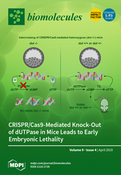
Genome integrity is essential for proper development and viability. dUTPase (the protein product of the dut gene) plays a key role in preserving DNA by removing dUTP from the cellular pool and contributes to dTTP de novo biosynthesis (by producing the dUMP substrate for thymidylate synthase (TS)). CRISPR/Cas9 transgenic mouse studies provided major insights into the physiological role of dUTPase in mammalian embryonic development. Biallelic dUTPase knock-out (dut -/-) embryos were still viable at the blastocyst stage but died shortly after implantation. Wild type and heterozygote animals showed the same healthy phenotype. Results suggest that zygotic dUTPase may be dispensable for pre-implantation embryonic stages but the lack of the enzyme lethally perturbs later embryonic development. View this paper.
  • Issues are regarded as officially published after their release is announced to the table of contents alert mailing list.
  • You may sign up for e-mail alerts to receive table of contents of newly released issues.
  • PDF is the official format for papers published in both, html and pdf forms. To view the papers in pdf format, click on the "PDF Full-text" link, and use the free Adobe Reader to open them.
Order results
Result details
Section
Select all
Export citation of selected articles as:
13 pages, 1983 KiB  
Article
Mechanism of Cell Wall Polysaccharides Modification in Harvested ‘Shatangju’ Mandarin (Citrus reticulate Blanco) Fruit Caused by Penicillium italicum
by Taotao Li, Dingding Shi, Qixian Wu, Chunxiao Yin, Fengjun Li, Youxia Shan, Xuewu Duan and Yueming Jiang
Biomolecules 2019, 9(4), 160; https://doi.org/10.3390/biom9040160 - 24 Apr 2019
Cited by 24 | Viewed by 3934
Abstract
Modification of cell wall polysaccharide in the plant plays an important role in response to fungi infection. However, the mechanism of fungi infection on cell wall modification need further clarification. In this study, the effects of Penicillium italicum inoculation on ‘shatangju’ mandarin disease [...] Read more.
Modification of cell wall polysaccharide in the plant plays an important role in response to fungi infection. However, the mechanism of fungi infection on cell wall modification need further clarification. In this study, the effects of Penicillium italicum inoculation on ‘shatangju’ mandarin disease development and the potential mechanism of cell wall polysaccharides modification caused by P. italicum were investigated. Compared to the control fruit, P. italicum infection modified the cell wall polysaccharides, indicated by water-soluble pectin (WSP), acid-soluble pectin (ASP), hemicellulose and lignin contents change. P. italicum infection enhanced the activities of polygalacturonase (PG), pectin methylesterase (PME), and the expression levels of xyloglucanendotransglucosylase/hydrolase (XTH) and expansin, which might contribute to cell wall disassembly and cellular integrity damage. Additionally, higher accumulation of reactive oxygen species (ROS) via decreasing antioxidant metabolites and the activities of antioxidant enzymes including superoxide dismutase (SOD), catalase (CAT), and ascorbate peroxidase (APX) also contributed to the cell wall polysaccharides modification. Meanwhile, the gene expression levels of hydroxyproline-rich glycoprotein (HRGP) and germin-like protein (GLP) were inhibited by pathogen infection. Altogether, these findings suggested that cell wall degradation/modification caused by non-enzymatic and enzymatic factors was an important strategy for P. italicum to infect ‘shatangju’ mandarin. Full article
Show Figures

Figure 1

20 pages, 2878 KiB  
Article
Curcuma raktakanda Induces Apoptosis and Suppresses Migration in Cancer Cells: Role of Reactive Oxygen Species
by Shruti Mishra, Sumit Singh Verma, Vipin Rai, Nikee Awasthee, Jayadev S. Arya, Kaustabh K. Maiti and Subash C. Gupta
Biomolecules 2019, 9(4), 159; https://doi.org/10.3390/biom9040159 - 23 Apr 2019
Cited by 28 | Viewed by 4381
Abstract
Although over 100 species of Curcuma are reported, only Curcuma longa is extensively studied. Curcuma raktakanda, a poorly studied species, is most commonly distributed in the Kerala state of India. For the first time, we examined the efficacy of different fractions (acetone, [...] Read more.
Although over 100 species of Curcuma are reported, only Curcuma longa is extensively studied. Curcuma raktakanda, a poorly studied species, is most commonly distributed in the Kerala state of India. For the first time, we examined the efficacy of different fractions (acetone, hexane, and ethyl acetate) of C. raktakanda against glioma, cervical, and breast cancer cell lines. As determined by mitochondrial reductase activity assay, the viability of cancer cells was decreased in a concentration-dependent manner by the three fractions. The half maximal inhibitory concentration (IC-50) values after the treatment of C-6 glioma cells for 48 h was found to be 32.97 µg/mL (acetone extract), 40.63 µg/mL (hexane extract), and 51.65 µg/mL (ethyl acetate extract). Of the three fractions, the acetone fraction was more effective. The long-term colony formation of cancer cells was significantly suppressed by the acetone fraction. Analyses using DAPI (4′,6-diamidino-2-phenylindole) staining, AO/PI (acridine orange/propidium iodide) staining, DNA laddering, and sub-G1 population revealed that the acetone extract induced apoptosis in glioma cells. The extract induced reactive oxygen species generation and suppressed the expression of cell survival proteins. The migration of cancer cells was also suppressed by the acetone extract. The gas chromatography-mass spectrometry (GC-MS) analysis indicated that tetracontane, dotriacontane, hexatriacontane, pentacosane, hexacosane, and eicosane are the major components in the acetone extract. Collectively, the extract from C. raktakanda exhibited anti-carcinogenic activities in cancer cells. We are exploring whether the phytoconstituents, individually, or collectively contribute to the anti-cancer activities of C. raktakanda. Full article
(This article belongs to the Special Issue Antitumor Agents from Natural Sources)
Show Figures

Figure 1

17 pages, 1372 KiB  
Review
Mouse Models for Exploring the Biological Consequences and Clinical Significance of PIK3CA Mutations
by Camilla B. Mitchell and Wayne A. Phillips
Biomolecules 2019, 9(4), 158; https://doi.org/10.3390/biom9040158 - 23 Apr 2019
Cited by 11 | Viewed by 4324
Abstract
The phosphatidylinositol 3-kinase (PI3K) pathway is involved in a myriad of cellular signalling pathways that regulate cell growth, metabolism, proliferation and survival. As a result, alterations in the PI3K pathway are frequently associated with human cancers. Indeed, PIK3CA—the gene encoding the p110α [...] Read more.
The phosphatidylinositol 3-kinase (PI3K) pathway is involved in a myriad of cellular signalling pathways that regulate cell growth, metabolism, proliferation and survival. As a result, alterations in the PI3K pathway are frequently associated with human cancers. Indeed, PIK3CA—the gene encoding the p110α catalytic subunit of PI3K—is one of the most commonly mutated human oncogenes. PIK3CA mutations have also been implicated in non-malignant conditions including congenital overgrowth syndromes and vascular malformations. In order to study the role of PIK3CA mutations in driving tumorigenesis and tissue overgrowth and to test potential therapeutic interventions for these conditions, model systems are essential. In this review we discuss the various mouse models currently available for preclinical studies into the biological consequences and clinical significance of PIK3CA mutations. Full article
(This article belongs to the Special Issue Phosphoinositide 3-kinase, a Field in Transition)
Show Figures

Graphical abstract

11 pages, 243 KiB  
Article
Association between Interleukin 35 Gene Single Nucleotide Polymorphisms and Systemic Lupus Erythematosus in a Chinese Han Population
by Shi-Yang Guan, Li-Na Liu, Yan-Mei Mao, Chan-Na Zhao, Qian Wu, Yi-Lin Dan, Napoleon Bellua Sam and Hai-Feng Pan
Biomolecules 2019, 9(4), 157; https://doi.org/10.3390/biom9040157 - 22 Apr 2019
Cited by 9 | Viewed by 3719
Abstract
Interleukin-35 (IL-35) exerts crucial roles in the pathogenesis and development of systemic lupus erythematosus (SLE), in this study we aim to explore the associations between IL-35 gene polymorphisms and the susceptibility, clinical features and plasma IL-35 levels of SLE patients, respectively. 490 SLE [...] Read more.
Interleukin-35 (IL-35) exerts crucial roles in the pathogenesis and development of systemic lupus erythematosus (SLE), in this study we aim to explore the associations between IL-35 gene polymorphisms and the susceptibility, clinical features and plasma IL-35 levels of SLE patients, respectively. 490 SLE patients and 489 healthy controls were recruited in our study. The correlations between the polymorphisms of seven SNPs of IL-35 encoding gene and the susceptibility, main clinical manifestations of SLE were evaluated, respectively. Plasma IL-35 levels were assessed in 76 SLE patients, and the associations between plasma IL-35 levels and the polymorphisms of genotyped SNPs were explored. There were significant associations between the polymorphisms of rs4740 and the occurrence of renal disorder, hematological disorder in SLE patients, respectively (p = 0.001; p = 0.001). In addition, there were no significant associations observed between the genotype frequencies of genotyped SNPs and the risk of SLE, plasma IL-35 levels, respectively. The polymorphism of rs4740 of IL-35 encoding gene is associated with the occurrence of renal disorder and hematological disorder of SLE patients. Full article
(This article belongs to the Special Issue Molecular Basis of Autoimmunity Diseases)
15 pages, 1719 KiB  
Article
Global Transcriptomic Profile Analysis of Genes Involved in Lignin Biosynthesis and Accumulation Induced by Boron Deficiency in Poplar Roots
by Wan-Long Su, Na Liu, Li Mei, Jie Luo, Yi-Jie Zhu and Zhu Liang
Biomolecules 2019, 9(4), 156; https://doi.org/10.3390/biom9040156 - 19 Apr 2019
Cited by 16 | Viewed by 3692
Abstract
To uncover the transcriptomic mechanism of lignin accumulation caused by boron deficiency (BD), Nanlin895 (Populus × euramericana “Nanlin895”) was subjected to control (CK, 0.25 mg·L−1) and BD (0 mg·L−1) treatments for 3 days. RNA-Seq was carried out to [...] Read more.
To uncover the transcriptomic mechanism of lignin accumulation caused by boron deficiency (BD), Nanlin895 (Populus × euramericana “Nanlin895”) was subjected to control (CK, 0.25 mg·L−1) and BD (0 mg·L−1) treatments for 3 days. RNA-Seq was carried out to survey the expression patterns of the lignin-regulated biosynthetic genes in response to BD. The results showed that 5946 genes were identified as differentially expressed genes (DEGs), 2968 (44.2%) of which were upregulated and 3318 (55.8%) of which were downregulated in response to BD. Among them, the expression of lignin monomer biosynthetic (PAL, CCR, CAD, COMT, F5H, PER/LAC) and modulated genes, for example, transcription factors (MYBs) and hormone signal regulating genes (GIDs, histidine kinase 1, coronatine-insensitive protein 1), were upregulated, and some hormone signal regulating genes, such as AUXs and BR-related (sterol methyltransferases), were downregulated under BD treatment. There are also some genes that were screened as candidates for an association with wood formation, which will be used for the further analysis of the function of lignin formation. These results provide an important theoretical basis and reference data in plant for further research on the mechanism of lignin accumulation under BD. Full article
Show Figures

Figure 1

15 pages, 4105 KiB  
Article
In Vitro Anticancer Properties of Copper Metallodendrimers
by Marcin Hołota, Jakub Magiera, Sylwia Michlewska, Małgorzata Kubczak, Natalia Sanz del Olmo, Sandra García-Gallego, Paula Ortega, Francisco Javier de la Mata, Maksim Ionov and Maria Bryszewska
Biomolecules 2019, 9(4), 155; https://doi.org/10.3390/biom9040155 - 18 Apr 2019
Cited by 22 | Viewed by 3763
Abstract
Newly synthesized carbosilane copper dendrimers (CCD) with chloride and nitrate surface groups seem to be good candidates to be used as gene and drug carriers in anti-cancer therapy, due to their properties such as size and surface charge. Copper attached to the nanoparticles [...] Read more.
Newly synthesized carbosilane copper dendrimers (CCD) with chloride and nitrate surface groups seem to be good candidates to be used as gene and drug carriers in anti-cancer therapy, due to their properties such as size and surface charge. Copper attached to the nanoparticles is an important element of many biological processes and recently their anti-cancer properties have been widely examined. Zeta size and potential, transmission electron microscopy (TEM), circular dichroism (CD), analysis of haemolytic activity, and fluorescence anisotropy techniques were used to characterize copper dendrimers. Additionally, their cytotoxic properties toward normal (PBMC) and cancer (1301; HL-60) cells were examined. All tested dendrimers were more cytotoxic against cancer cells in comparison with normal cells. Full article
Show Figures

Graphical abstract

18 pages, 2720 KiB  
Article
Targeted Delivery of Nanoparticulate Cytochrome C into Glioma Cells Through the Proton-Coupled Folate Transporter
by Yuriy V. Kucheryavykh, Josue Davila, Jescelica Ortiz-Rivera, Mikhael Inyushin, Luis Almodovar, Miguel Mayol, Moraima Morales-Cruz, Alejandra Cruz-Montañez, Vanessa Barcelo-Bovea, Kai Griebenow and Lilia Y. Kucheryavykh
Biomolecules 2019, 9(4), 154; https://doi.org/10.3390/biom9040154 - 18 Apr 2019
Cited by 13 | Viewed by 4383
Abstract
In this study, we identified the proton-coupled folate transporter (PCFT) as a route for targeted delivery of drugs to some gliomas. Using the techniques of confocal imaging, quantitative reverse transcription-polymerase chain reaction (qRT-PCR), and small interfering (siRNA) knockdown against the PCFT, we demonstrated [...] Read more.
In this study, we identified the proton-coupled folate transporter (PCFT) as a route for targeted delivery of drugs to some gliomas. Using the techniques of confocal imaging, quantitative reverse transcription-polymerase chain reaction (qRT-PCR), and small interfering (siRNA) knockdown against the PCFT, we demonstrated that Gl261 and A172 glioma cells, but not U87 and primary cultured astrocytes, express the PCFT, which provides selective internalization of folic acid (FA)-conjugated cytochrome c-containing nanoparticles (FA-Cyt c NPs), followed by cell death. The FA-Cyt c NPs (100 µg/mL), had no cytotoxic effects in astrocytes but caused death in glioma cells, according to their level of expression of PCFT. Whole-cell patch clamp recording revealed FA-induced membrane currents in FA-Cyt c NPs-sensitive gliomas, that were reduced by siRNA PCFT knockdown in a similar manner as by application of FA-Cyt c NPs, indicating that the PCFT is a route for internalization of FA-conjugated NPs in these glioma cells. Analysis of human glioblastoma specimens revealed that at least 25% of glioblastomas express elevated level of either PCFT or folate receptor (FOLR1). We conclude that the PCFT provides a mechanism for targeted delivery of drugs to some gliomas as a starting point for the development of efficient methods for treating gliomas with high expression of PCFT and/or FOLR1. Full article
(This article belongs to the Special Issue Nanoparticles for Cancer Therapy)
Show Figures

Figure 1

11 pages, 674 KiB  
Review
The PTEN–PI3K Axis in Cancer
by Antonella Papa and Pier Paolo Pandolfi
Biomolecules 2019, 9(4), 153; https://doi.org/10.3390/biom9040153 - 17 Apr 2019
Cited by 185 | Viewed by 9448
Abstract
The PI3K–AKT–mTOR signal transduction pathway regulates a variety of biological processes including cell growth, cell cycle progression and proliferation, cellular metabolism, and cytoskeleton reorganization. Fine-tuning of the phosphatidylinositol 3-kinase (PI3K) pathway signaling output is essential for the maintenance of tissue homeostasis and uncontrolled [...] Read more.
The PI3K–AKT–mTOR signal transduction pathway regulates a variety of biological processes including cell growth, cell cycle progression and proliferation, cellular metabolism, and cytoskeleton reorganization. Fine-tuning of the phosphatidylinositol 3-kinase (PI3K) pathway signaling output is essential for the maintenance of tissue homeostasis and uncontrolled activation of this cascade leads to a number of human pathologies including cancer. Inactivation of the tumor suppressor phosphatase and tensin homologue deleted on Chromosome 10 (PTEN) and/or activating mutations in the proto-typical lipid kinase PI3K have emerged as some of the most frequent events associated with human cancer and as a result the PI3K pathway has become a highly sought-after target for cancer therapies. In this review we summarize the essential role of the PTEN–PI3K axis in controlling cellular behaviors by modulating activation of key proto-oncogenic molecular nodes and functional targets. Further, we highlight important functional redundancies and peculiarities of these two critical enzymes that over the last few decades have become a central part of the cancer research field and have instructed hundreds of pre-clinical and clinical trials to better cancer treatments. Full article
(This article belongs to the Special Issue Phosphoinositide 3-kinase, a Field in Transition)
Show Figures

Figure 1

14 pages, 1723 KiB  
Article
Membrane Localization of HspA1A, a Stress Inducible 70-kDa Heat-Shock Protein, Depends on Its Interaction with Intracellular Phosphatidylserine
by Andrei D. Bilog, Larissa Smulders, Ryan Oliverio, Cedra Labanieh, Julianne Zapanta, Robert V. Stahelin and Nikolas Nikolaidis
Biomolecules 2019, 9(4), 152; https://doi.org/10.3390/biom9040152 - 17 Apr 2019
Cited by 17 | Viewed by 5358
Abstract
HspA1A is a cytosolic molecular chaperone essential for cellular homeostasis. HspA1A also localizes at the plasma membrane (PM) of tumor and stressed cells. However, it is currently unknown how this cytosolic protein translocates to the PM. Taking into account that HspA1A interacts with [...] Read more.
HspA1A is a cytosolic molecular chaperone essential for cellular homeostasis. HspA1A also localizes at the plasma membrane (PM) of tumor and stressed cells. However, it is currently unknown how this cytosolic protein translocates to the PM. Taking into account that HspA1A interacts with lipids, including phosphatidylserine (PS), and that lipids recruit proteins to the PM, we hypothesized that the interaction of HspA1A with PS allows the chaperone to localize at the PM. To test this hypothesis, we subjected cells to mild heat-shock and the PM-localized HspA1A was quantified using confocal microscopy and cell surface biotinylation. These experiments revealed that HspA1A’s membrane localization increased during recovery from non-apoptotic heat-shock. Next, we selectively reduced PS targets by overexpressing the C2 domain of lactadherin (Lact-C2), a known PS-biosensor, and determined that HspA1A’s membrane localization was greatly reduced. In contrast, the reduction of PI(4,5)P2 availability by overexpression of the PLCδ-PH biosensor had minimal effects on HspA1A’s PM-localization. Implementation of a fluorescent PS analog, TopFluor-PS, established that PS co-localizes with HspA1A. Collectively, these results reveal that HspA1A’s PM localization and anchorage depend on its selective interaction with intracellular PS. This discovery institutes PS as a new and dynamic partner in the cellular stress response. Full article
Show Figures

Figure 1

12 pages, 3186 KiB  
Article
Regulation of the Expression of DAPK1 by SUMO Pathway
by Qingshui Wang, Xiuli Zhang, Ling Chen, Shuyun Weng, Yun Xia, Yan Ye, Ke Li, Ziqiang Liao, Pengchen Chen, Khaldoon Alsamman, Chen Meng, Craig Stevens, Ted R. Hupp and Yao Lin
Biomolecules 2019, 9(4), 151; https://doi.org/10.3390/biom9040151 - 17 Apr 2019
Cited by 6 | Viewed by 3527
Abstract
Death Associated Protein Kinase 1 (DAPK1) is an important signaling kinase mediating the biological effect of multiple natural biomolecules such as IFN-γ, TNF-α, curcumin, etc. DAPK1 is degraded through both ubiquitin-proteasomal and lysosomal degradation pathways. To investigate the crosstalk between these two DAPK1 [...] Read more.
Death Associated Protein Kinase 1 (DAPK1) is an important signaling kinase mediating the biological effect of multiple natural biomolecules such as IFN-γ, TNF-α, curcumin, etc. DAPK1 is degraded through both ubiquitin-proteasomal and lysosomal degradation pathways. To investigate the crosstalk between these two DAPK1 degradation pathways, we carried out a screen using a set of ubiquitin E2 siRNAs at the presence of Tuberous Sclerous 2 (TSC2) and identified that the small ubiquitin-like molecule (SUMO) pathway is able to regulate the protein levels of DAPK1. Inhibition of the SUMO pathway enhanced DAPK1 protein levels and the minimum domain of DAPK1 protein required for this regulation is the kinase domain, suggesting that the SUMO pathway regulates DAPK1 protein levels independent of TSC2. Suppression of the SUMO pathway did not enhance DAPK1 protein stability. In addition, mutation of the potential SUMO conjugation sites on DAPK1 kinase domain did not alter its protein stability or response to SUMO pathway inhibition. These data suggested that the SUMO pathway does not regulate DAPK1 protein degradation. The exact molecular mechanism underlying this regulation is yet to be discovered. Full article
Show Figures

Figure 1

28 pages, 1636 KiB  
Review
Intracellular Peptides in Cell Biology and Pharmacology
by Christiane B. de Araujo, Andrea S. Heimann, Ricardo A. Remer, Lilian C. Russo, Alison Colquhoun, Fábio L. Forti and Emer S. Ferro
Biomolecules 2019, 9(4), 150; https://doi.org/10.3390/biom9040150 - 16 Apr 2019
Cited by 37 | Viewed by 5755
Abstract
Intracellular peptides are produced by proteasomes following degradation of nuclear, cytosolic, and mitochondrial proteins, and can be further processed by additional peptidases generating a larger pool of peptides within cells. Thousands of intracellular peptides have been sequenced in plants, yeast, zebrafish, rodents, and [...] Read more.
Intracellular peptides are produced by proteasomes following degradation of nuclear, cytosolic, and mitochondrial proteins, and can be further processed by additional peptidases generating a larger pool of peptides within cells. Thousands of intracellular peptides have been sequenced in plants, yeast, zebrafish, rodents, and in human cells and tissues. Relative levels of intracellular peptides undergo changes in human diseases and also when cells are stimulated, corroborating their biological function. However, only a few intracellular peptides have been pharmacologically characterized and their biological significance and mechanism of action remains elusive. Here, some historical and general aspects on intracellular peptides’ biology and pharmacology are presented. Hemopressin and Pep19 are examples of intracellular peptides pharmacologically characterized as inverse agonists to cannabinoid type 1 G-protein coupled receptors (CB1R), and hemopressin fragment NFKF is shown herein to attenuate the symptoms of pilocarpine-induced epileptic seizures. Intracellular peptides EL28 (derived from proteasome 26S protease regulatory subunit 4; Rpt2), PepH (derived from Histone H2B type 1-H), and Pep5 (derived from G1/S-specific cyclin D2) are examples of peptides that function intracellularly. Intracellular peptides are suggested as biological functional molecules, and are also promising prototypes for new drug development. Full article
(This article belongs to the Special Issue The Broader Cellular Impact of Proteasome-CSN-eIf3 (PCI) Complexes)
Show Figures

Figure 1

22 pages, 3457 KiB  
Communication
Mining for Oxysterols in Cyp7b1−/− Mouse Brain and Plasma: Relevance to Spastic Paraplegia Type 5
by Anna Meljon, Peter J. Crick, Eylan Yutuc, Joyce L. Yau, Jonathan R. Seckl, Spyridon Theofilopoulos, Ernest Arenas, Yuqin Wang and William J. Griffiths
Biomolecules 2019, 9(4), 149; https://doi.org/10.3390/biom9040149 - 13 Apr 2019
Cited by 12 | Viewed by 5564
Abstract
Deficiency in cytochrome P450 (CYP) 7B1, also known as oxysterol 7α-hydroxylase, in humans leads to hereditary spastic paraplegia type 5 (SPG5) and in some cases in infants to liver disease. SPG5 is medically characterized by loss of motor neurons in the corticospinal tract. [...] Read more.
Deficiency in cytochrome P450 (CYP) 7B1, also known as oxysterol 7α-hydroxylase, in humans leads to hereditary spastic paraplegia type 5 (SPG5) and in some cases in infants to liver disease. SPG5 is medically characterized by loss of motor neurons in the corticospinal tract. In an effort to gain a better understanding of the fundamental biochemistry of this disorder, we have extended our previous profiling of the oxysterol content of brain and plasma of Cyp7b1 knockout (-/-) mice to include, amongst other sterols, 25-hydroxylated cholesterol metabolites. Although brain cholesterol levels do not differ between wild-type (wt) and knockout mice, we find, using a charge-tagging methodology in combination with liquid chromatography–mass spectrometry (LC–MS) and multistage fragmentation (MSn), that there is a build-up of the CYP7B1 substrate 25-hydroxycholesterol (25-HC) in Cyp7b1-/- mouse brain and plasma. As reported earlier, levels of (25R)26-hydroxycholesterol (26-HC), 3β-hydroxycholest-5-en-(25R)26-oic acid and 24S,25-epoxycholesterol (24S,25-EC) are similarly elevated in brain and plasma. Side-chain oxysterols including 25-HC, 26-HC and 24S,25-EC are known to bind to INSIG (insulin-induced gene) and inhibit the processing of SREBP-2 (sterol regulatory element-binding protein-2) to its active form as a master regulator of cholesterol biosynthesis. We suggest the concentration of cholesterol in brain of the Cyp7b1-/- mouse is maintained by balancing reduced metabolism, as a consequence of a loss in CYP7B1, with reduced biosynthesis. The Cyp7b1-/- mouse does not show a motor defect; whether the defect in humans is a consequence of less efficient homeostasis of cholesterol in brain has yet to be uncovered. Full article
(This article belongs to the Special Issue Lipidomics)
Show Figures

Figure 1

19 pages, 1253 KiB  
Article
Promoter Hypermethylation of Tumor-Suppressor Genes p16INK4a, RASSF1A, TIMP3, and PCQAP/MED15 in Salivary DNA as a Quadruple Biomarker Panel for Early Detection of Oral and Oropharyngeal Cancers
by Chamikara Liyanage, Asanga Wathupola, Sanjayan Muraleetharan, Kanthi Perera, Chamindie Punyadeera and Preethi Udagama
Biomolecules 2019, 9(4), 148; https://doi.org/10.3390/biom9040148 - 12 Apr 2019
Cited by 47 | Viewed by 8083
Abstract
Silencing of tumor-suppressor genes (TSGs) by DNA promoter hypermethylation is an early event in carcinogenesis; hence, TSGs may serve as early tumor biomarkers. We determined the promoter methylation levels of p16INK4a, RASSF1A, TIMP3, and PCQAP/MED15 TSGs in salivary DNA [...] Read more.
Silencing of tumor-suppressor genes (TSGs) by DNA promoter hypermethylation is an early event in carcinogenesis; hence, TSGs may serve as early tumor biomarkers. We determined the promoter methylation levels of p16INK4a, RASSF1A, TIMP3, and PCQAP/MED15 TSGs in salivary DNA from oral cancer (OC) and oropharyngeal cancer (OPC) patients, using methylation-specific PCR coupled with densitometry analysis. We assessed the association between DNA methylation of individual TSGs with OC and OPC risk factors. The performance and the clinical validity of this quadruple-methylation marker panel were evaluated in discriminating OC and OPC patients from healthy controls using the CombiROC web tool. Our study reports that RASSF1A, TIMP3, and PCQAP/MED15 TSGs were significantly hypermethylated in OC and OPC cases compared to healthy controls. DNA methylation levels of TSGs were significantly augmented by smoking, alcohol use, and betel quid chewing, indicating the fact that frequent exposure to risk factors may drive oral and oropharyngeal carcinogenesis through TSG promoter hypermethylation. Also, this quadruple-methylation marker panel of p16INK4a, RASSF1A, TIMP3, and PCQAP/MED15 TSGs demonstrated excellent diagnostic accuracy in the early detection of OC at 91.7% sensitivity and 92.3% specificity and of OPC at 99.8% sensitivity and 92.1% specificity from healthy controls. Full article
(This article belongs to the Special Issue Biomarkers for Cancer)
Show Figures

Graphical abstract

6 pages, 222 KiB  
Editorial
Intrinsically Disordered Proteins in Chronic Diseases
by Prakash Kulkarni and Vladimir N. Uversky
Biomolecules 2019, 9(4), 147; https://doi.org/10.3390/biom9040147 - 11 Apr 2019
Cited by 50 | Viewed by 4689
Abstract
It is now increasingly evident that a large fraction of the human proteome comprises proteins that, under physiological conditions, lack fixed, ordered 3D structures as a whole or have segments that are not likely to form a defined 3D structure [...] Full article
(This article belongs to the Special Issue Intrinsically Disordered Proteins and Chronic Diseases)
22 pages, 2126 KiB  
Review
Recent Advances in Computational Protocols Addressing Intrinsically Disordered Proteins
by Supriyo Bhattacharya and Xingcheng Lin
Biomolecules 2019, 9(4), 146; https://doi.org/10.3390/biom9040146 - 11 Apr 2019
Cited by 48 | Viewed by 7608
Abstract
Intrinsically disordered proteins (IDP) are abundant in the human genome and have recently emerged as major therapeutic targets for various diseases. Unlike traditional proteins that adopt a definitive structure, IDPs in free solution are disordered and exist as an ensemble of conformations. This [...] Read more.
Intrinsically disordered proteins (IDP) are abundant in the human genome and have recently emerged as major therapeutic targets for various diseases. Unlike traditional proteins that adopt a definitive structure, IDPs in free solution are disordered and exist as an ensemble of conformations. This enables the IDPs to signal through multiple signaling pathways and serve as scaffolds for multi-protein complexes. The challenge in studying IDPs experimentally stems from their disordered nature. Nuclear magnetic resonance (NMR), circular dichroism, small angle X-ray scattering, and single molecule Förster resonance energy transfer (FRET) can give the local structural information and overall dimension of IDPs, but seldom provide a unified picture of the whole protein. To understand the conformational dynamics of IDPs and how their structural ensembles recognize multiple binding partners and small molecule inhibitors, knowledge-based and physics-based sampling techniques are utilized in-silico, guided by experimental structural data. However, efficient sampling of the IDP conformational ensemble requires traversing the numerous degrees of freedom in the IDP energy landscape, as well as force-fields that accurately model the protein and solvent interactions. In this review, we have provided an overview of the current state of computational methods for studying IDP structure and dynamics and discussed the major challenges faced in this field. Full article
(This article belongs to the Special Issue Intrinsically Disordered Proteins and Chronic Diseases)
Show Figures

Figure 1

14 pages, 897 KiB  
Review
Essential Kinases and Transcriptional Regulators and Their Roles in Autoimmunity
by Ya Nan Deng, Joseph A. Bellanti and Song Guo Zheng
Biomolecules 2019, 9(4), 145; https://doi.org/10.3390/biom9040145 - 10 Apr 2019
Cited by 16 | Viewed by 4363
Abstract
Kinases and transcriptional regulators are fundamental components of cell signaling that are expressed on many types of immune cells which are involved in secretion of cytokines, cell proliferation, differentiation, and apoptosis. Both play important roles in biological responses in health as well as [...] Read more.
Kinases and transcriptional regulators are fundamental components of cell signaling that are expressed on many types of immune cells which are involved in secretion of cytokines, cell proliferation, differentiation, and apoptosis. Both play important roles in biological responses in health as well as in illnesses such as the autoimmune diseases which comprise at least 80 disorders. These diseases are caused by complex genetic and environmental interactions that lead to a breakage of immunologic tolerance and a disruption of the balance between self-reactive cells and regulatory cells. Kinases or transcriptional regulatory factors often have an abnormal expression in the autoimmune cells that participate in the pathogenesis of autoimmune disease. These abnormally expressed kinases or transcriptional regulators can over-activate the function of self-reactive cells to produce inflammatory cytokines or down-regulate the activity of regulatory cells, thus causing autoimmune diseases. In this review we introduce five kinds of kinase and transcriptional regulator related to autoimmune diseases, namely, members of the Janus kinase (JAK) family (JAK3 and/or tyrosine kinase 2 (TYK2)), fork head box protein 3 (Foxp3), the retinoic acid-related orphan receptor gamma t (RORγt), and T-box expressed in T cells (T-bet) factors. We also provide a mechanistic insight into how these kinases and transcriptional regulators affect the function of the immune cells related to autoimmune diseases, as well as a description of a current drug design targeting these kinases and transcriptional regulators. Understanding their exact role helps offer new therapies for control of the inflammatory responses that could lead to clinical improvement of the autoimmune diseases. Full article
(This article belongs to the Special Issue Molecular Basis of Autoimmunity Diseases)
Show Figures

Figure 1

15 pages, 2540 KiB  
Article
Host-Microbiome Synergistic Control on Sphingolipid Metabolism by Mechanotransduction in Model Arthritis
by Xiaoyuan Zhou, Valentina Devescovi, Yuanhua Liu, Jennifer E. Dent and Christine Nardini
Biomolecules 2019, 9(4), 144; https://doi.org/10.3390/biom9040144 - 09 Apr 2019
Cited by 6 | Viewed by 3363
Abstract
Chronic inflammatory autoimmune disorders are systemic diseases with increasing incidence and still lack a cure. More recently, attention has been placed in understanding gastrointestinal (GI) dysbiosis and, although important progress has been made in this area, it is currently unclear to what extent [...] Read more.
Chronic inflammatory autoimmune disorders are systemic diseases with increasing incidence and still lack a cure. More recently, attention has been placed in understanding gastrointestinal (GI) dysbiosis and, although important progress has been made in this area, it is currently unclear to what extent microbiome manipulation can be used in the treatment of autoimmune disorders. Via the use of appropriate models, rheumatoid arthritis (RA), a well-known exemplar of such pathologies, can be exploited to shed light on the currently overlooked effects of existing therapies on the GI microbiome. In this direction, we here explore the crosstalk between the GI microbiome and the host immunity in model arthritis (collagen induced arthritis, CIA). By exploiting omics from samples of limited invasiveness (blood and stools), we assess the host-microbiome responses to standard therapy (methotrexate, MTX) combined with mechanical subcutaneous stimulation (MS) and to mechanical stimulation alone. When MS is involved, results reveal the sphingolipid metabolism as the trait d’union among known hallmarks of (model) RA, namely: Imbalance in the S1P-S1PR1 axis, expansion of Prevotella sp., and invariant Natural Killer T (iNKT)-penia, thus offering the base of a rationale to mechanically modulate this pathway as a therapeutic target in RA. Full article
(This article belongs to the Special Issue Mechanobiology in Health and Disease)
Show Figures

Figure 1

11 pages, 1097 KiB  
Article
Impact of DARC rs12075 Variants on Liver Fibrosis Progression in Patients with Chronic Hepatitis C: A Retrospective Study
by María Ángeles Jiménez-Sousa, Ana Zaida Gómez-Moreno, Daniel Pineda-Tenor, Juan José Sánchez-Ruano, Tomas Artaza-Varasa, María Martin-Vicente, Amanda Fernández-Rodríguez, Isidoro Martínez and Salvador Resino
Biomolecules 2019, 9(4), 143; https://doi.org/10.3390/biom9040143 - 09 Apr 2019
Cited by 6 | Viewed by 3175
Abstract
The Duffy antigen receptor for chemokines (DARC) rs12075 polymorphism regulates leukocyte trafficking and proinflammatory chemokine homeostasis. Hepatitis C virus (HCV)-mediated liver fibrosis is associated with an uncontrolled inflammatory response. In this study, we evaluate the association between the DARC rs12075 polymorphism [...] Read more.
The Duffy antigen receptor for chemokines (DARC) rs12075 polymorphism regulates leukocyte trafficking and proinflammatory chemokine homeostasis. Hepatitis C virus (HCV)-mediated liver fibrosis is associated with an uncontrolled inflammatory response. In this study, we evaluate the association between the DARC rs12075 polymorphism and liver stiffness progression in HCV-infected patients. We carried out a retrospective cohort study (repeated measures design) in 208 noncirrhotic patients with chronic hepatitis C (CHC) who had at least two liver stiffness measurements (LSM) with a separation of at least 12 months. We used generalized linear models to analyze the association between DARC rs12075 polymorphism and outcome variables. During a follow-up of 46.6 months, the percentage of patients with stages of fibrosis F0/F1 decreased (p < 0.001), while LSM values and the percentage of patients with cirrhosis increased (p < 0.001). This pattern of changes was maintained in each of the groups of patients analyzed according to their rs12075 genotypes (AA or AG/GG). However, the variations in liver stiffness characteristics were lower in patients with the rs12075 AG/GG genotype (AG/GG versus AA). Thereby, in the adjusted analysis, patients with the rs12075 AG/GG genotype had a lower risk of an increased value of LSM2/LSM1 arithmetic mean ratio (AMR = 0.83; p = 0.001) and of an increase in LSM ≥ 5 kPa (odds ratio (OR) = 0.28; p = 0.009). Besides, patients with rs12075 AG/GG had a lower risk of cirrhosis progression (OR = 0.24; p = 0.009). No significant associations were found for an increase in LSM ≥ 10 kPa. We found an association between the DARC rs12075 single nucleotide polymorphism (SNP) and CHC progression. Specifically, patients with the DARC rs12075 AG/GG genotype had a lower risk of liver fibrosis progression and development of cirrhosis. Full article
(This article belongs to the Section Molecular Medicine)
Show Figures

Figure 1

48 pages, 5225 KiB  
Review
Receptor Ligands as Helping Hands to L-DOPA in the Treatment of Parkinson’s Disease
by Fabio Del Bello, Mario Giannella, Gianfabio Giorgioni, Alessandro Piergentili and Wilma Quaglia
Biomolecules 2019, 9(4), 142; https://doi.org/10.3390/biom9040142 - 09 Apr 2019
Cited by 19 | Viewed by 7217
Abstract
Levodopa (LD) is the most effective drug in the treatment of Parkinson’s disease (PD). However, although it represents the “gold standard” of PD therapy, LD can cause side effects, including gastrointestinal and cardiovascular symptoms as well as transient elevated liver enzyme levels. Moreover, [...] Read more.
Levodopa (LD) is the most effective drug in the treatment of Parkinson’s disease (PD). However, although it represents the “gold standard” of PD therapy, LD can cause side effects, including gastrointestinal and cardiovascular symptoms as well as transient elevated liver enzyme levels. Moreover, LD therapy leads to LD-induced dyskinesia (LID), a disabling motor complication that represents a major challenge for the clinical neurologist. Due to the many limitations associated with LD therapeutic use, other dopaminergic and non-dopaminergic drugs are being developed to optimize the treatment response. This review focuses on recent investigations about non-dopaminergic central nervous system (CNS) receptor ligands that have been identified to have therapeutic potential for the treatment of motor and non-motor symptoms of PD. In a different way, such agents may contribute to extending LD response and/or ameliorate LD-induced side effects. Full article
(This article belongs to the Special Issue Advances in Parkinson's Disease Drugs)
Show Figures

Figure 1

28 pages, 3159 KiB  
Review
Unilateral Ureteral Obstruction as a Model to Investigate Fibrosis-Attenuating Treatments
by Elena Martínez-Klimova, Omar Emiliano Aparicio-Trejo, Edilia Tapia and José Pedraza-Chaverri
Biomolecules 2019, 9(4), 141; https://doi.org/10.3390/biom9040141 - 08 Apr 2019
Cited by 174 | Viewed by 14572
Abstract
Renal fibrosis is the common pathway for most forms of progressive renal disease. The Unilateral Ureteral Obstruction (UUO) model is used to cause renal fibrosis, where the primary feature of UUO is tubular injury as a result of obstructed urine flow. Furthermore, experimental [...] Read more.
Renal fibrosis is the common pathway for most forms of progressive renal disease. The Unilateral Ureteral Obstruction (UUO) model is used to cause renal fibrosis, where the primary feature of UUO is tubular injury as a result of obstructed urine flow. Furthermore, experimental UUO in rodents is believed to mimic human chronic obstructive nephropathy in an accelerated manner. Renal fibrosis is the common pathway for most forms of progressive renal disease. Removing the obstruction may not be sufficient to reverse fibrosis, so an accompanying treatment may be of benefit. In this review, we have done a revision on treatments shown to ameliorate fibrosis in the context of the UUO experimental model. The treatments inhibit the production of fibrotic and inflammatory proteins such as Transforming Growth Factor β1 (TGF-β1), Tumor Necrosis Factor α (TNF-α), collagen and fibronectin, Heat Shock Protein 47 (HSP47), suppress the proliferation of fibroblasts, prevent epithelial-to-mesenchymal transition, reduce oxidative stress, inhibit the action of the Nuclear Factor κB (NF-κB), reduce the phosphorylation of mothers against decapentaplegic homolog (SMAD) family members 2 and 3 (Smad2/3) or Mitogen-Activated Protein Kinases (MAPKs), inhibit the activation of the renin-angiotensin system. Summaries of the UUO experimental methods and alterations observed in the UUO experiments are included. Full article
(This article belongs to the Special Issue Molecular Mechanisms of Kidney Injury and Repair)
Show Figures

Figure 1

12 pages, 2845 KiB  
Article
Organellar DNA Polymerases in Complex Plastid-Bearing Algae
by Yoshihisa Hirakawa and Arisa Watanabe
Biomolecules 2019, 9(4), 140; https://doi.org/10.3390/biom9040140 - 07 Apr 2019
Cited by 10 | Viewed by 4876
Abstract
DNA replication in plastids and mitochondria is generally regulated by nucleus-encoded proteins. In plants and red algae, a nucleus-encoded enzyme called POP (plant and protist organellar DNA polymerase) is involved in DNA replication in both organelles by virtue of its dual localization. POPs [...] Read more.
DNA replication in plastids and mitochondria is generally regulated by nucleus-encoded proteins. In plants and red algae, a nucleus-encoded enzyme called POP (plant and protist organellar DNA polymerase) is involved in DNA replication in both organelles by virtue of its dual localization. POPs are family A DNA polymerases, which include bacterial DNA polymerase I (PolI). POP homologs have been found in a wide range of eukaryotes, including plants, algae, and non-photosynthetic protists. However, the phylogeny and subcellular localizations of POPs remain unclear in many algae, especially in secondary and tertiary plastid-bearing groups. In this study, we report that chlorarachniophytes possess two evolutionarily distinct POPs, and fluorescent protein-tagging experiments demonstrate that they are targeted to the secondary plastids and mitochondria, respectively. The timing of DNA replication is different between the two organelles in the chlorarachniophyte Bigelowiella natans, and this seems to be correlated to the transcription of respective POP genes. Dinoflagellates also carry two distinct POP genes, possibly for their plastids and mitochondria, whereas haptophytes and ochrophytes have only one. Therefore, unlike plants, some algal groups are likely to have evolved multiple DNA polymerases for various organelles. This study provides a new insight into the evolution of organellar DNA replication in complex plastid-bearing organisms. Full article
(This article belongs to the Special Issue Evolutionary and Molecular Aspects of Plastid Endosymbioses)
Show Figures

Figure 1

13 pages, 5702 KiB  
Article
Freeze Tolerance in Sculpins (Pisces; Cottoidea) Inhabiting North Pacific and Arctic Oceans: Antifreeze Activity and Gene Sequences of the Antifreeze Protein
by Aya Yamazaki, Yoshiyuki Nishimiya, Sakae Tsuda, Koji Togashi and Hiroyuki Munehara
Biomolecules 2019, 9(4), 139; https://doi.org/10.3390/biom9040139 - 06 Apr 2019
Cited by 6 | Viewed by 4259
Abstract
Many marine species inhabiting icy seawater produce antifreeze proteins (AFPs) to prevent their body fluids from freezing. The sculpin species of the superfamily Cottoidea are widely found from the Arctic to southern hemisphere, some of which are known to express AFP. Here we [...] Read more.
Many marine species inhabiting icy seawater produce antifreeze proteins (AFPs) to prevent their body fluids from freezing. The sculpin species of the superfamily Cottoidea are widely found from the Arctic to southern hemisphere, some of which are known to express AFP. Here we clarified DNA sequence encoding type I AFP for 3 species of 2 families (Cottidae and Agonidae) belonging to Cottoidea. We also examined antifreeze activity for 3 families and 32 species of Cottoidea (Cottidae, Agonidae, and Rhamphocottidae). These fishes were collected in 2013–2015 from the Arctic Ocean, Alaska, Japan. We could identify 8 distinct DNA sequences exhibiting a high similarity to those reported for Myoxocephalus species, suggesting that Cottidae and Agonidae share the same DNA sequence encoding type I AFP. Among the 3 families, Rhamphocottidae that experience a warm current did not show antifreeze activity. The species inhabiting the Arctic Ocean and Northern Japan that often covered with ice floe showed high activity, while those inhabiting Alaska, Southern Japan with a warm current showed low/no activity. These results suggest that Cottoidea acquires type I AFP gene before dividing into Cottidae and Agonidae, and have adapted to each location with optimal antifreeze activity level. Full article
(This article belongs to the Special Issue Antifreeze Protein: New Insight from Different Approaches)
Show Figures

Figure 1

19 pages, 1915 KiB  
Review
Essential Oils and Isolated Terpenes in Nanosystems Designed for Topical Administration: A Review
by Sheila P. de Matos, Helder F. Teixeira, Ádley A. N. de Lima, Valdir F. Veiga-Junior and Letícia S. Koester
Biomolecules 2019, 9(4), 138; https://doi.org/10.3390/biom9040138 - 05 Apr 2019
Cited by 79 | Viewed by 9883
Abstract
Essential oils are natural products with a complex composition. Terpenes are the most common class of chemical compounds present in essential oils. Terpenes and the essential oils containing them are widely used and investigated by their pharmacological properties and permeation-enhancing ability. However, many [...] Read more.
Essential oils are natural products with a complex composition. Terpenes are the most common class of chemical compounds present in essential oils. Terpenes and the essential oils containing them are widely used and investigated by their pharmacological properties and permeation-enhancing ability. However, many terpenes and essential oils are sensitive to environmental conditions, undergoing volatilization and chemical degradation. In order to overcome the chemical instability of some isolated terpenes and essential oils, the encapsulation of these compounds in nanostructured systems (polymeric, lipidic, or molecular complexes) has been employed. In addition, nanoencapsulation can be of interest for pharmaceutical applications due to its capacity to improve the bioavailability and allow the controlled release of drugs. Topical drug administration is a convenient and non-invasive administration route for both local and systemic drug delivery. The present review focuses on describing the current status of research concerning nanostructured delivery systems containing isolated terpenes and/or essential oils designed for topical administration and on discussing the use of terpenes and essential oils either for their biological activities or as permeation enhancers in pharmaceutic formulations. Full article
(This article belongs to the Special Issue Evaluation and formulation of Bioactive Terpenes)
Show Figures

Figure 1

13 pages, 14433 KiB  
Article
Preventive Effect of Anji White Tea Flavonoids on Alcohol-Induced Gastric Injury through Their Antioxidant Effects in Kunming Mice
by Bihui Liu, Xingxing Feng, Jing Zhang, Yang Wei and Xin Zhao
Biomolecules 2019, 9(4), 137; https://doi.org/10.3390/biom9040137 - 04 Apr 2019
Cited by 35 | Viewed by 5364
Abstract
Anji white tea (Camellia sinensis) is a traditional Chinese tea beverage, which is classified as green tea and contains an abundant amount of flavonoids. In this study, the preventive effect of Anji white tea flavonoids (AJWTFs) on ethanol/hydrochloric acid-induced gastric injury [...] Read more.
Anji white tea (Camellia sinensis) is a traditional Chinese tea beverage, which is classified as green tea and contains an abundant amount of flavonoids. In this study, the preventive effect of Anji white tea flavonoids (AJWTFs) on ethanol/hydrochloric acid-induced gastric injury in mice was evaluated. The serum and gastric tissues of mice were analyzed using a biochemical kit and by quantitative polymerase chain reaction (qPCR). Observation of the appearance of the stomach indicated that AJWTFs could effectively reduce the area of gastric injury caused by ethanol/hydrochloric acid, and the inhibition rate of AJWTF on gastric injury increased with an increase in AJWTF concentration. The Anji white tea flavonoids could also reduce the volume and pH of gastric juice in mice with gastric injury. Biochemical results showed that AJWTFs could increase the superoxide dismutase (SOD) and glutathione (GSH) activities, as well as decrease the malondialdehyde (MDA) level, in the serum and liver of mice with gastric injury. Pathological observation confirmed that AJWTFs could inhibit the tissue damage caused by ethanol/hydrochloric acid in the stomach of mice. Further qPCR experiments also showed that AJWTFs could inhibit the decreases in neuronal nitric oxide synthase (nNOS), endothelial nitric oxide synthase (eNOS), copper/zinc superoxide dismutase (Cu/Zn–SOD), manganese superoxide dismutase (Mn–SOD), catalase (CAT), and the increase in inducible nitric oxide synthase (iNOS) expression in the gastric tissue of mice caused by gastric injury. As observed, AJWTFs exerted a good preventive effect on alcohol-induced gastric injury in mice induced by ethanol/hydrochloric acid, and the effect is close to that of ranitidine. Anji white tea flavonoids present good antioxidant effect, which allows them to effectively prevent alcoholic gastric injury and be used as biologically active substances with a broad range of applications. Full article
Show Figures

Figure 1

14 pages, 2295 KiB  
Article
CRISPR/Cas9-Mediated Knock-Out of dUTPase in Mice Leads to Early Embryonic Lethality
by Hajnalka Laura Pálinkás, Gergely Attila Rácz, Zoltán Gál, Orsolya Ivett Hoffmann, Gergely Tihanyi, Gergely Róna, Elen Gócza, László Hiripi and Beáta G. Vértessy
Biomolecules 2019, 9(4), 136; https://doi.org/10.3390/biom9040136 - 04 Apr 2019
Cited by 15 | Viewed by 4700
Abstract
Sanitization of nucleotide pools is essential for genome maintenance. Deoxyuridine 5′-triphosphate nucleotidohydrolase (dUTPase) is a key enzyme in this pathway since it catalyzes the cleavage of 2′-deoxyuridine 5′-triphosphate (dUTP) into 2′-deoxyuridine 5′-monophosphate (dUMP) and inorganic pyrophosphate. Through its action dUTPase efficiently prevents uracil [...] Read more.
Sanitization of nucleotide pools is essential for genome maintenance. Deoxyuridine 5′-triphosphate nucleotidohydrolase (dUTPase) is a key enzyme in this pathway since it catalyzes the cleavage of 2′-deoxyuridine 5′-triphosphate (dUTP) into 2′-deoxyuridine 5′-monophosphate (dUMP) and inorganic pyrophosphate. Through its action dUTPase efficiently prevents uracil misincorporation into DNA and at the same time provides dUMP, the substrate for de novo thymidylate biosynthesis. Despite its physiological significance, knock-out models of dUTPase have not yet been investigated in mammals, but only in unicellular organisms, such as bacteria and yeast. Here we generate CRISPR/Cas9-mediated dUTPase knock-out in mice. We find that heterozygous dut +/– animals are viable while having decreased dUTPase levels. Importantly, we show that dUTPase is essential for embryonic development since early dut −/− embryos reach the blastocyst stage, however, they die shortly after implantation. Analysis of pre-implantation embryos indicates perturbed growth of both inner cell mass (ICM) and trophectoderm (TE). We conclude that dUTPase is indispensable for post-implantation development in mice. Full article
(This article belongs to the Section Cellular Biochemistry)
Show Figures

Figure 1

16 pages, 2296 KiB  
Article
LcNAC13 Physically Interacts with LcR1MYB1 to Coregulate Anthocyanin Biosynthesis-Related Genes during Litchi Fruit Ripening
by Guoxiang Jiang, Zhiwei Li, Yunbo Song, Hong Zhu, Sen Lin, Riming Huang, Yueming Jiang and Xuewu Duan
Biomolecules 2019, 9(4), 135; https://doi.org/10.3390/biom9040135 - 04 Apr 2019
Cited by 38 | Viewed by 5234
Abstract
Anthocyanin accumulation is crucial for the development of quality for most fruit. The mechanism underlying the regulation of anthocyanin biosynthesis by transcription factors in litchi fruit remains largely unknown. In this study, we isolated one NAC (NAM, ATAF1/2 and CUC2) TF gene, LcNAC13 [...] Read more.
Anthocyanin accumulation is crucial for the development of quality for most fruit. The mechanism underlying the regulation of anthocyanin biosynthesis by transcription factors in litchi fruit remains largely unknown. In this study, we isolated one NAC (NAM, ATAF1/2 and CUC2) TF gene, LcNAC13. Expression of LcNAC13 was upregulated as ripening proceeded, followed by the accumulation of anthocyanins. Electrophoretic mobility shift assay (EMSA) and transient expression assay showed that LcNAC13 could negatively regulate the expression of anthocyanin biosynthesis-related genes, including LcCHS1/2, LcCHI, LcF3H, LcF3’H, LcDFR, and LcMYB1. Furthermore, LcR1MYB1, as one R1-MYB type MYB, was identified to physically interact with LcNAC13 and reverse the effect of LcNAC13. Taken together, these results suggested that LcNAC13 and LcR1MYB1 may act together to antagonistically regulate anthocyanin biosynthesis during litchi fruit ripening, which helps to provide new insights into the regulatory networks of anthocyanin biosynthesis. Full article
Show Figures

Figure 1

17 pages, 2762 KiB  
Article
Evidence of Destabilization of the Human Thymidylate Synthase (hTS) Dimeric Structure Induced by the Interface Mutation Q62R
by Cecilia Pozzi, Ludovica Lopresti, Matteo Santucci, Maria Paola Costi and Stefano Mangani
Biomolecules 2019, 9(4), 134; https://doi.org/10.3390/biom9040134 - 03 Apr 2019
Cited by 3 | Viewed by 3647
Abstract
In human cells, thymidylate synthase (TS) provides the only source of 2′-deoxythymidyne-5′-monophosphate (dTMP), which is required for DNA biosynthesis. Because of its pivotal role, human TS (hTS) represents a validated target for anticancer chemotherapy. Nonetheless, the efficacy of drugs blocking the hTS active [...] Read more.
In human cells, thymidylate synthase (TS) provides the only source of 2′-deoxythymidyne-5′-monophosphate (dTMP), which is required for DNA biosynthesis. Because of its pivotal role, human TS (hTS) represents a validated target for anticancer chemotherapy. Nonetheless, the efficacy of drugs blocking the hTS active site has limitations due to the onset of resistance in cancer cells, requiring the identification of new strategies to effectively inhibit this enzyme. Human TS works as an obligate homodimer, making the inter-subunit interface an attractive targetable area. Here, we report the design and investigation of a new hTS variant, in which Gln62, located at the dimer interface, has been replaced by arginine in order to destabilize the enzyme quaternary assembly. The hTS Q62R variant has been characterized though kinetic assay, thermal denaturation analysis and X-ray crystallography. Our results provide evidence that hTS Q62R has a reduced melting temperature. The effective destabilization of the TS quaternary structure is also confirmed by structural analysis, showing that the introduced mutation induces a slight aperture of the hTS dimer. The generation of hTS variants having a more accessible interface area can facilitate the screening of interface-targeting molecules, providing key information for the rational design of innovative hTS interface inhibitors. Full article
(This article belongs to the Section Molecular Structure and Dynamics)
Show Figures

Figure 1

20 pages, 3288 KiB  
Article
Mutagenesis of DsbAss is Crucial for the Signal Recognition Particle Mechanism in Escherichia coli: Insights from Molecular Dynamics Simulations
by Faiza Gul Durrani, Roquyya Gul, Muhammad Usman Mirza, Naheed Nazly Kaderbhai, Matheus Froeyen and Mahjabeen Saleem
Biomolecules 2019, 9(4), 133; https://doi.org/10.3390/biom9040133 - 03 Apr 2019
Cited by 12 | Viewed by 4173
Abstract
The disulfide bond signal sequence (DsbAss) protein is characterized as an important virulence factor in gram-negative bacteria. This study aimed to analyze the “alanine” alteration in the hydrophobic (H) region of DsbAss and to understand the conformational DsbAss alteration(s) inside the fifty-four homolog [...] Read more.
The disulfide bond signal sequence (DsbAss) protein is characterized as an important virulence factor in gram-negative bacteria. This study aimed to analyze the “alanine” alteration in the hydrophobic (H) region of DsbAss and to understand the conformational DsbAss alteration(s) inside the fifty-four homolog (Ffh)-binding groove which were revealed to be crucial for translocation of ovine growth hormone (OGH) to the periplasmic space in Escherichia coli via the secretory (Sec) pathway. An experimental design was used to explore the hydrophobicity and alteration of alanine (Ala) to isoleucine (Ile) in the tripartite structure of DsbAss. As a result, two DsbAss mutants (Ala at positions -11 and -13) with same hydrophobicity of 1.539 led to the conflicting translocation of the active OGH gene. We performed molecular dynamics (MD) simulations and molecular mechanics generalized born surface area (MM-GBSA) binding free energy calculations to examine the interaction energetic and dynamic aspects of DsbAss/signal repetition particle 54 (SRP54) binding, which has a principle role in Escherichia coli Sec pathways. Although both DsbAss mutants retained helicity, the MD simulation analysis evidenced that altering Ala-13 changed the orientation of the signal peptide in the Ffh M binding domain groove, favored more stable interaction energies (MM-GBSA ΔGtotal = −140.62 kcal mol−1), and hampered the process of OGH translocation, while Ala-11 pointed outward due to unstable conformation and less binding energy (ΔGtotal = −124.24 kcal mol−1). Here we report the dynamic behavior of change of “alanine” in the H-domain of DsbAss which affects the process of translocation of OGH, where MD simulation and MM-GBSA can be useful initial tools to investigate the virulence of bacteria. Full article
(This article belongs to the Special Issue Protein Dynamics Simulations)
Show Figures

Figure 1

10 pages, 2796 KiB  
Review
Helical Twist and Rotational Forces in the Mitotic Spindle
by Iva M. Tolić, Maja Novak and Nenad Pavin
Biomolecules 2019, 9(4), 132; https://doi.org/10.3390/biom9040132 - 01 Apr 2019
Cited by 9 | Viewed by 7362
Abstract
The mitotic spindle segregates chromosomes into two daughter cells during cell division. This process relies on the precise regulation of forces acting on chromosomes as the cell progresses through mitosis. The forces in the spindle are difficult to directly measure using the available [...] Read more.
The mitotic spindle segregates chromosomes into two daughter cells during cell division. This process relies on the precise regulation of forces acting on chromosomes as the cell progresses through mitosis. The forces in the spindle are difficult to directly measure using the available experimental techniques. Here, we review the ideas and recent advances of how forces can be determined from the spindle shape. By using these approaches, it has been shown that tension and compression coexist along a single kinetochore fiber, which are balanced by a bridging fiber between sister kinetochore fibers. An extension of this approach to three dimensions revealed that microtubule bundles have rich shapes, and extend not simply like meridians on the Earth’s surface but, rather, twisted in a helical manner. Such complex shapes are due to rotational forces, which, in addition to linear forces, act in the spindle and may be generated by motor proteins such as kinesin-5. These findings open new questions for future studies, to understand the mechanisms of rotational forces and reveal their biological roles in cells. Full article
(This article belongs to the Special Issue Cytoskeleton and Regulation of Mitosis)
Show Figures

Figure 1

15 pages, 1442 KiB  
Review
Splicing Factors Have an Essential Role in Prostate Cancer Progression and Androgen Receptor Signaling
by Ken-ichi Takayama
Biomolecules 2019, 9(4), 131; https://doi.org/10.3390/biom9040131 - 01 Apr 2019
Cited by 38 | Viewed by 5811
Abstract
Although inhibition of the androgen–androgen receptor (AR) axis effectively represses the growth of prostate cancer, most of all cases eventually become castration-resistant prostate cancers (CRPCs). Enhancement of the expression of AR and its variants along with the downstream signals is important for disease [...] Read more.
Although inhibition of the androgen–androgen receptor (AR) axis effectively represses the growth of prostate cancer, most of all cases eventually become castration-resistant prostate cancers (CRPCs). Enhancement of the expression of AR and its variants along with the downstream signals is important for disease progression. AR-V7, a constitutive active form of AR, is generated as a result of RNA splicing. RNA splicing creates multiple transcript variants from one pre-messenger RNA (mRNA) by removing introns/exons to allow mRNA translation. The molecular mechanisms leading to marked increases of AR and generation of AR-V7 have been unclear. However, recent papers highlighted the roles of RNA splicing factors which promote AR expression and production of variants. Notably, a broad range of splicing components were aberrantly regulated in CRPC tissues. Interestingly, expression of various spliceosome genes is enhanced by RNA-binding protein splicing factor proline- and glutamine-rich (PSF/SFPQ), leading to changes in the expression of AR transcript variants. Moreover, inhibition of several splicing factors repressed tumor growth in vivo. Altered expression of splicing factors is correlated to biochemical recurrence in prostate cancer patients. Thus, these findings suggest that splicing factors would be a potential therapeutic target. This review focuses on the emerging roles of splicing factors in prostate cancer progression and AR signaling. Full article
(This article belongs to the Special Issue Hormone Receptors in Cancer)
Show Figures

Figure 1

Previous Issue
Next Issue
Back to TopTop