


Abstract
Probiotic strains with multifunctional skin-beneficial properties represent promising candidates for next-generation cosmetic ingredients. In this study, a systematic stepwise screening strategy was applied to an in-house bacterial library comprising 302 isolates to identify candidates with antioxidant, anti-inflammatory, antimicrobial, wound-healing, and skin barrier-supportive activities. From this library, 33 strains were selected based on preliminary assessments and subjected to comprehensive in vitro evaluation. Cell viability and cytotoxicity assays confirmed the safety of all selected strains in RAW264.7 macrophages and HaCaT keratinocytes. Several strains exhibited strong DPPH radical scavenging activity and significantly inhibited nitric oxide production in LPS-stimulated macrophages. Among the selected candidates, Lacticaseibacillus rhamnosus DM073 demonstrated the most potent anti-inflammatory activity, whereas Lactiplantibacillus plantarum DM043 exhibited the greatest wound-healing capacity. All five selected strains displayed antimicrobial activity against Cutibacterium acnes. Furthermore, selected strains, particularly Lactiplantibacillus plantarum DM175 and Ligilactobacillus salivarius DM079, enhanced the expression of skin barrier-related genes, including zonula occludens-1 (ZO-1), occludin (OCLN), claudin-1 (Cla-1), and filaggrin (FLG), and partially restored their expression under TNF-α/IFN-γ-induced inflammatory conditions. Selected strains also reduced the expression of inflammatory chemokines in stimulated keratinocytes. Collectively, these findings demonstrate that the selected probiotic strains possess complementary multifunctional activities associated with skin health. In particular, DM073 exhibited superior anti-inflammatory activity, DM043 showed strong wound-healing potential, and DM175 demonstrated remarkable skin barrier-supportive effects. These strain-specific properties support their potential application in the development of probiotic-derived postbiotic cosmetic ingredients for skin soothing, barrier reinforcement, skin recovery, and microbiome-friendly skincare formulations. Further studies are warranted to evaluate their efficacy and safety in advanced skin models and clinical cosmetic applications.
1. Introduction
The skin is the largest organ of the human body and serves as a primary barrier against environmental stressors, pathogens, and chemical irritants. Maintenance of skin homeostasis relies on a complex interaction among epidermal cells, immune responses, and the resident skin microbiota [1,2]. Disruption of this balance can contribute to various skin disorders, including acne, atopic dermatitis, psoriasis, and premature skin aging.
In particular, oxidative stress, chronic inflammation, microbial dysbiosis, and impaired epidermal barrier function are closely associated with the development of inflammatory skin disorders, acne pathogenesis, and premature skin aging [2,3].
Recent advances in microbiome research have highlighted the beneficial role of probiotics in maintaining host health beyond the gastrointestinal tract. Probiotic microorganisms can modulate immune responses, inhibit pathogenic microorganisms, enhance epithelial barrier function, and produce bioactive metabolites with antioxidant and anti-inflammatory activities [4,5]. These findings have stimulated growing interest in the application of probiotics and probiotic-derived products as functional ingredients in cosmetic and dermatological products [5,6]. Several studies have demonstrated that specific Lactobacillus and Bifidobacterium strains can alleviate skin inflammation, improve barrier integrity, accelerate wound healing, and regulate skin-associated microbial communities [4,6,7]. In addition, probiotic-derived metabolites and postbiotics have emerged as promising cosmetic ingredients due to their improved stability and safety profiles compared with live microorganisms.
Among the mechanisms underlying probiotic-mediated skin benefits, antioxidant and anti-inflammatory activities are particularly important. Excessive production of reactive oxygen species (ROS) induces oxidative damage to cellular components and activates inflammatory signaling pathways, ultimately leading to epidermal dysfunction and skin aging. Probiotic-derived metabolites and postbiotic bioactive components, including exopolysaccharides, peptides, and cell wall-associated molecules, have been reported to exhibit antioxidant and immunomodulatory activities [5,8]. Furthermore, inflammatory mediators such as nitric oxide (NO), tumor necrosis factor-alpha (TNF-α), interleukin (IL)-1β, and IL-6 contribute to chronic skin inflammation and disruption of epidermal barrier integrity [9,10]. Therefore, probiotic strains capable of suppressing oxidative stress and inflammatory responses may represent promising candidates for skin health applications [4,11].
Maintenance of epidermal barrier function is another critical aspect of skin health. Tight junction proteins, including zonula occludens-1 (ZO-1), occludin (OCLN), and claudin-1 (Cla-1), together with structural proteins such as filaggrin (FLG), play essential roles in regulating barrier permeability and epidermal integrity [12,13]. Reduced expression of these proteins has been associated with inflammatory skin disorders, including atopic dermatitis, and impaired barrier function [13,14]. Recent studies suggest that certain probiotic strains can enhance the expression of barrier-associated proteins and improve epithelial integrity under inflammatory conditions, indicating their potential utility in cosmetic and skin-health applications [15,16].
Although numerous probiotic strains have been reported to exhibit individual skin-beneficial properties, systematic screening approaches that simultaneously evaluate antioxidant, anti-inflammatory, antimicrobial, wound-healing, and barrier-supportive activities remain limited [4,7,17]. Moreover, comparative evaluation of a large probiotic strain library is necessary to identify superior candidates with multifunctional cosmetic potential [5,6].
Therefore, the objective of the present study was to systematically screen an in-house bacterial library comprising 302 isolates and identify strains with multifunctional skin-beneficial activities. This study aimed to identify bacterial strains with postbiotic potential for cosmetic applications by comprehensively evaluating antioxidant, anti-inflammatory, antimicrobial, wound-healing, and skin barrier-supportive properties. Through this comprehensive screening strategy, we aimed to provide scientific evidence supporting the development of next-generation postbiotic-based cosmetic ingredients for skin health applications. Furthermore, the identification of strain-specific functional characteristics may facilitate the development of targeted dermocosmetic formulations designed for skin barrier care, soothing applications, and microbiome-friendly skin care products.
2. Materials and Methods
2.1. Bacterial Strains and Preparation of Heat-Killed Bacterial Cells
An in-house bacterial library comprising 302 isolates, including 216 Lactobacillus isolates, 35 Bifidobacterium isolates, and 51 isolates belonging to other bacterial genera (including Streptococcus and Enterococcus), was established from diverse sources, including healthy human oral samples, human fecal samples, and fermented foods. From this library, 33 candidate strains were selected through preliminary functional screening. Among these candidates, five strains isolated from the healthy human oral cavity and showing superior anti-inflammatory, antioxidant, and antimicrobial activities were selected for further evaluation: Lacticaseibacillus paracasei DM162, Ligilactobacillus salivarius DM079, Lactiplantibacillus plantarum DM043, Lactiplantibacillus plantarum DM175, and Lacticaseibacillus rhamnosus DM073.
All strains were maintained as glycerol stocks (20%, v/v) at −80 °C until use and routinely cultured in de Man, Rogosa and Sharpe (MRS) broth (KisanBio, Seoul, Republic of Korea) at 37 °C for 18–24 h under anaerobic conditions. Bacterial cultures were adjusted to approximately 1 × 10⁹ CFU/mL prior to heat treatment. Bacterial cells were harvested by centrifugation at 4000× g for 10 min, washed twice with phosphate-buffered saline (PBS), and heat-killed at 100 °C for 30 min according to a previously described method [18], with minor modifications. Heat-killed bacterial suspensions were stored at 4 °C until use.
2.2. Cell Culture
HaCaT human keratinocytes and RAW264.7 murine macrophages were cultured in Dulbecco’s Modified Eagle Medium (DMEM; Corning, NY, USA) supplemented with 10% fetal bovine serum (FBS; Atlas Biologicals, Fort Collins, CO, USA) and 1% penicillin–streptomycin (Corning, NY, USA). Cells were maintained at 37 °C in a humidified atmosphere containing 5% CO2.
2.3. Cell Viability Assay (MTT Assay)
Cell viability was evaluated using the MTT assay. HaCaT keratinocytes and RAW264.7 macrophages were seeded into 96-well plates at a density of 3 × 10⁴ cells/well and incubated for 24 h. The cells were then treated with heat-killed bacterial preparations at a final concentration equivalent to 1 × 10⁸ CFU/mL for an additional 24 h. This concentration was selected because it is commonly used for evaluating the biological activities of probiotic-derived preparations in cell-based assays. Following treatment, MTT solution was added to each well at a final concentration of 0.5 mg/mL and incubated at 37 °C for 2 h. The resulting formazan crystals were dissolved in dimethyl sulfoxide (DMSO), and absorbance was measured at 570 nm using a SpectraMax iD3 microplate reader (Molecular Devices, San Jose, CA, USA). Cell viability was expressed as a percentage relative to the untreated control group.
2.4. Cytotoxicity Evaluation by LDH Release Assay
Cytotoxicity was additionally evaluated using a lactate dehydrogenase (LDH) release assay kit (Quanti-LDH™ PLUS Cytotoxicity Assay Kit, Colorimetric; ScienCell, Carlsbad, CA, USA) according to the manufacturer’s instructions. HaCaT keratinocytes and RAW264.7 macrophages were seeded into 96-well plates at a density of 3 × 10⁴ cells/well and incubated for 24 h. The cells were then treated with heat-killed bacterial preparations at a final concentration equivalent to 1 × 10⁸ CFU/mL for 24 h. Culture supernatants were subsequently collected, and LDH release was measured at 490 nm using a SpectraMax iD3 microplate reader (Molecular Devices, San Jose, CA, USA). Cytotoxicity was calculated relative to the untreated control group.
2.5. DPPH Radical Scavenging Activity Assay
The DPPH radical scavenging activity of probiotic culture supernatants was evaluated using an EZ-DPPH Antioxidant Assay Kit (cat. no. DG-DPH400; DoGenBio, Seoul, Republic of Korea). Briefly, bacterial cultures were centrifuged at 4000× g for 10 min and the culture supernatants were collected. The supernatants were mixed with DPPH working solution and incubated in the dark at room temperature for 10 min according to the manufacturer’s instructions. Uninoculated MRS broth was included as a blank control, and its background absorbance was subtracted from all measurements. Absorbance was measured at 517 nm using a SpectraMax iD3 microplate reader (Molecular Devices, San Jose, CA, USA), and the DPPH radical scavenging activity (%) was calculated relative to the untreated control.
2.6. Nitric Oxide (NO) Assay
The inhibitory effect of heat-killed bacterial preparations on nitric oxide (NO) production was evaluated using RAW264.7 macrophages stimulated with lipopolysaccharide (LPS). RAW264.7 macrophages were seeded into 96-well plates at a density of 5 × 10⁴ cells/well and incubated for 24 h. The cells were then pretreated with heat-killed bacterial preparations at a final concentration equivalent to 1 × 10⁸ CFU/mL for 1 h, followed by stimulation with LPS (1 μg/mL) for 24 h.
Nitric oxide production was determined by measuring nitrite accumulation in the culture supernatant using a Griess Reagent Assay Kit (Open PicoSens™, BIOMAX, Republic of Korea) according to the manufacturer’s instructions. Absorbance was measured at 540 nm using a SpectraMax iD3 microplate reader (Molecular Devices, San Jose, CA, USA).
2.7. Antimicrobial Activity Assay Against Cutibacterium Acnes
The antimicrobial activity of the selected probiotic strains against Cutibacterium acnes (KCTC 5012) was evaluated using an agar diffusion assay. C. acnes was anaerobically cultured for 24 h at 37 °C in ATCC Medium 2722 (Supplemented Tryptic Soy Broth, consisting of Trypticase Soy Broth supplemented with yeast extract, hemin, vitamin K1, and L-cysteine hydrochloride) and adjusted to a final concentration of approximately 1 × 10⁸ CFU/mL using a McFarland standard. The bacterial suspension was uniformly spread onto Supplemented Tryptic Soy agar plates (ATCC Medium 2722). Selected probiotic strains were cultured in MRS broth at 37 °C for 18–24 h to a final concentration of approximately 108–109 CFU/mL. Aliquots (40 μL) of each bacterial culture were applied to paper disks (8 mm diameter), which were then placed onto the agar surface. Plates were incubated anaerobically at 37 °C for 48 h, and the diameter of the inhibition zone (mm) was measured. Lacticaseibacillus rhamnosus GG (LGG) was used as the reference strain for comparison.
2.8. Wound Healing Assay
The wound healing activity of heat-killed bacterial preparations was evaluated using a scratch assay in HaCaT keratinocytes as previously described [19,20], with minor modifications. HaCaT keratinocytes were seeded into 12-well plates at a density of 1 × 10⁶ cells/well and cultured until reaching a confluent monolayer. A linear scratch was created using a sterile pipette tip, and detached cells were removed by washing with PBS. Cells were then treated with heat-killed bacterial preparations at a final concentration equivalent to 1 × 10⁸ CFU/mL in serum-reduced medium.
For inflammatory wound healing conditions, HaCaT keratinocytes were pretreated with heat-killed bacterial preparations at a final concentration equivalent to 1 × 10⁸ CFU/mL for 1 h, followed by stimulation with TNF-α/IFN-γ (10 ng/mL each). Wound closure was monitored at 0, 24, and 48 h using an IX51 inverted microscope (Olympus Corporation, Tokyo, Japan). The wound closure rate was calculated using the following equation:
where W0 represents the initial wound width and Wt represents the wound width at the indicated time point.
2.9. Reverse Transcription-Quantitative PCR (RT-qPCR)
HaCaT keratinocytes or RAW264.7 macrophages were seeded into culture plates at a density of 2.5 × 10⁵ cells/well and incubated for 24 h. The cells were then treated with heat-killed bacterial preparations at a final concentration equivalent to 1 × 10⁸ CFU/mL. For inflammatory conditions, HaCaT keratinocytes were pretreated with heat-killed bacterial preparations for 1 h, followed by stimulation with TNF-α and IFN-γ (10 ng/mL each).
Total RNA was extracted from treated cells according to the manufacturer’s instructions. Complementary DNA (cDNA) was synthesized using the PrimeScript™ RT Reagent Kit (Takara Bio Inc., Shiga, Japan). Reverse transcription-quantitative PCR (RT-qPCR) was performed using TOPreal™ qPCR 2× PreMIX (Enzynomics Co., Ltd., Daejeon, Republic of Korea) on a QuantStudio™ 3 Real-Time PCR System (Applied Biosystems, Foster City, CA, USA) for SYBR Green-based detection. Thermal cycling conditions were as follows: 95 °C for 10 min, followed by 40 cycles of 95 °C for 10 s, 60 °C for 15 s, and 72 °C for 20 s. Fluorescence data were collected at 72 °C, and melt curve analysis was conducted to verify amplification specificity.
The expression levels of inflammation-related genes, skin barrier-associated genes, and inflammatory chemokines, including IL-6, IL-1β, TNF-α, iNOS, COX-2, ZO-1, OCLN, Cla-1, FLG, RANTES (CCL5), TARC (CCL17), MDC (CCL22), and IL-8 (CXCL8), were analyzed. Gene expression levels were normalized to GAPDH as the housekeeping gene (using mouse- or human-specific GAPDH primers for RAW264.7 and HaCaT keratinocytes, respectively) and calculated using the 2−ΔΔCt method.
The primer sequences used in this study are listed in Table 1.
Table 1.
Primer sequences used for RT-qPCR analysis.
2.10. Statistical Analysis
All experiments were independently performed at least three times. Data are presented as the mean ± standard deviation (SD). Statistical analyses were performed using GraphPad Prism version 9 (GraphPad Software, San Diego, CA, USA). Differences among groups were analyzed using one-way analysis of variance (ANOVA) followed by Dunnett’s multiple comparison test. Differences were considered statistically significant at p < 0.05.
3. Results
3.1. Screening and Selection of Candidate Probiotic Strains for Cosmetic Applications
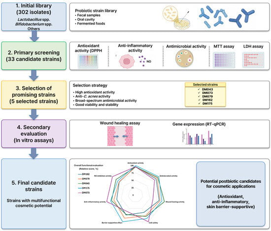
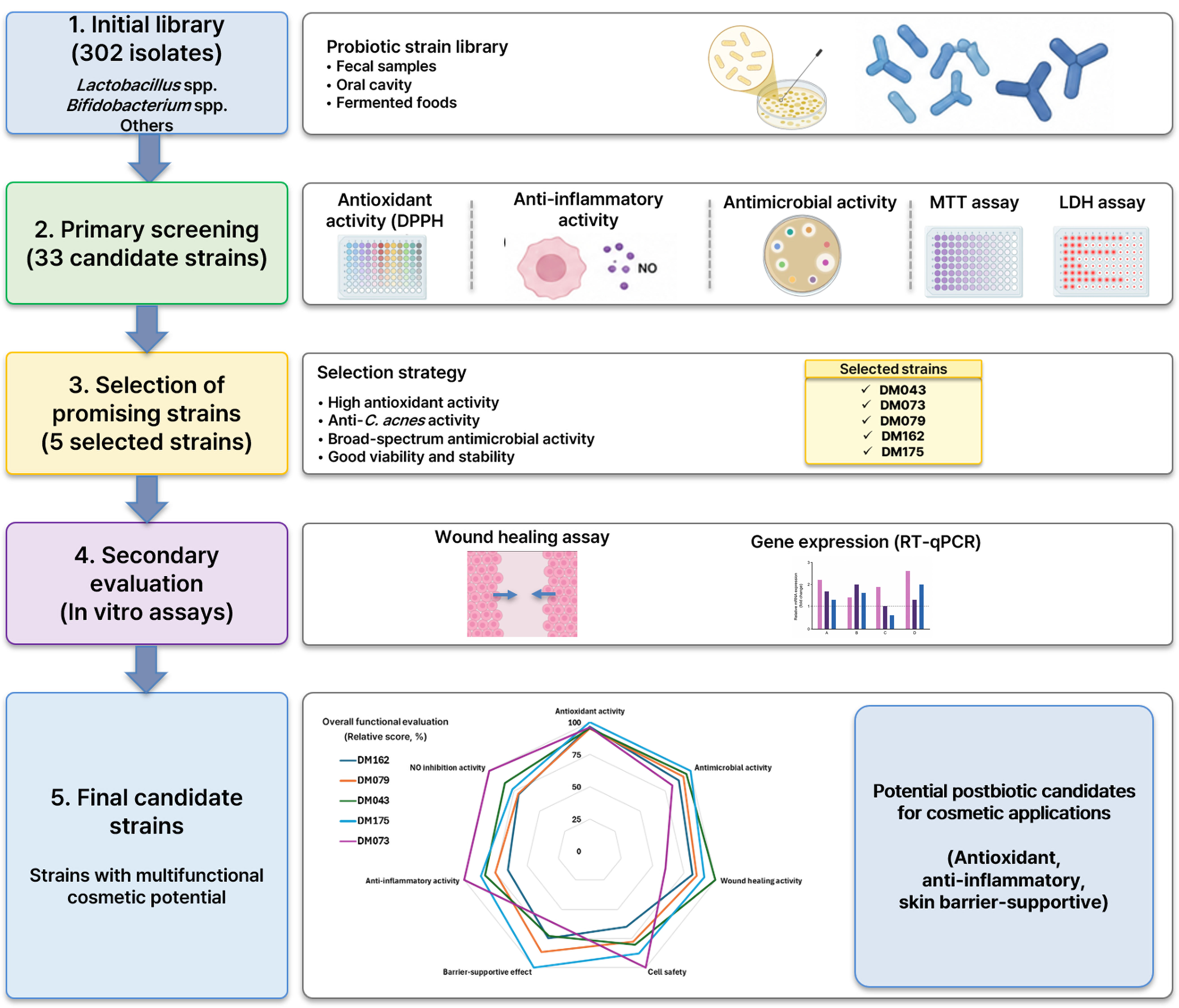
A total of more than 300 probiotic strains belonging to Lactobacillus spp. and Bifidobacterium spp. were screened to identify candidates with potential cosmetic applications. The bacterial library consisted of 302 isolates, including 216 Lactobacillus isolates, 35 Bifidobacterium isolates, and 51 isolates belonging to other bacterial genera, obtained from healthy human oral samples, human fecal samples, and fermented foods (Figure 1).
Figure 1.
Workflow for screening and selection of bacterial strains with postbiotic potential for cosmetic applications. An in-house bacterial library comprising 302 isolates from healthy human oral samples, human fecal samples, and fermented foods was subjected to primary screening based on DPPH radical scavenging, anti-inflammatory, antimicrobial, and safety evaluations (MTT and LDH assays). Five candidate strains were selected and subsequently evaluated for wound-healing activity and skin-related gene expression by RT-qPCR. The relative functional performance of selected strains is summarized in the radar chart, and the normalized scores used for radar plot construction are provided in Supplementary Table S1. DPPH radical scavenging activity was evaluated using culture supernatants, whereas anti-inflammatory, cytotoxicity, wound-healing, and gene expression analyses were performed using heat-killed bacterial preparations.
Primary screening was performed based on DPPH radical scavenging, anti-inflammatory, and antimicrobial activities. Strains exhibiting superior DPPH radical scavenging activity in the DPPH assay, strong inhibition of nitric oxide (NO) production, and broad-spectrum antimicrobial activity were selected as promising candidates. In addition, cell viability and strain stability were considered during the selection process.
Based on the primary screening results, five strains were selected for further evaluation: Lacticaseibacillus paracasei DM162, Ligilactobacillus salivarius DM079, Lactiplantibacillus plantarum DM043, Lactiplantibacillus plantarum DM175, and Lacticaseibacillus rhamnosus DM073. These strains were subsequently subjected to secondary evaluation, including MTT assay, LDH assay, wound healing assay, and RT-qPCR analysis to assess their safety and skin-related functional properties (Figure 1).
The overall functional performance of the selected strains was summarized using a radar chart, demonstrating strain-dependent differences in DPPH radical scavenging, anti-inflammatory, antimicrobial, wound healing, cell safety, and skin barrier-supportive activities (Figure 1).
3.2. Safety Screening of Heat-Killed Probiotic Strains in RAW264.7 and HaCaT Keratinocytes
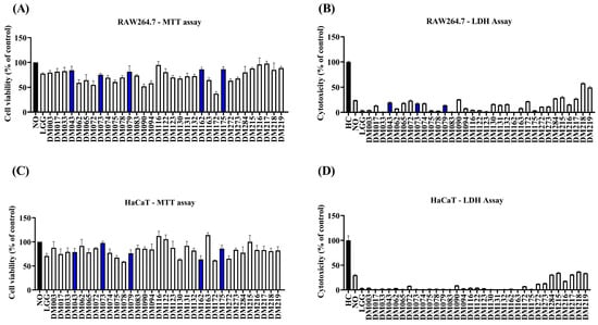
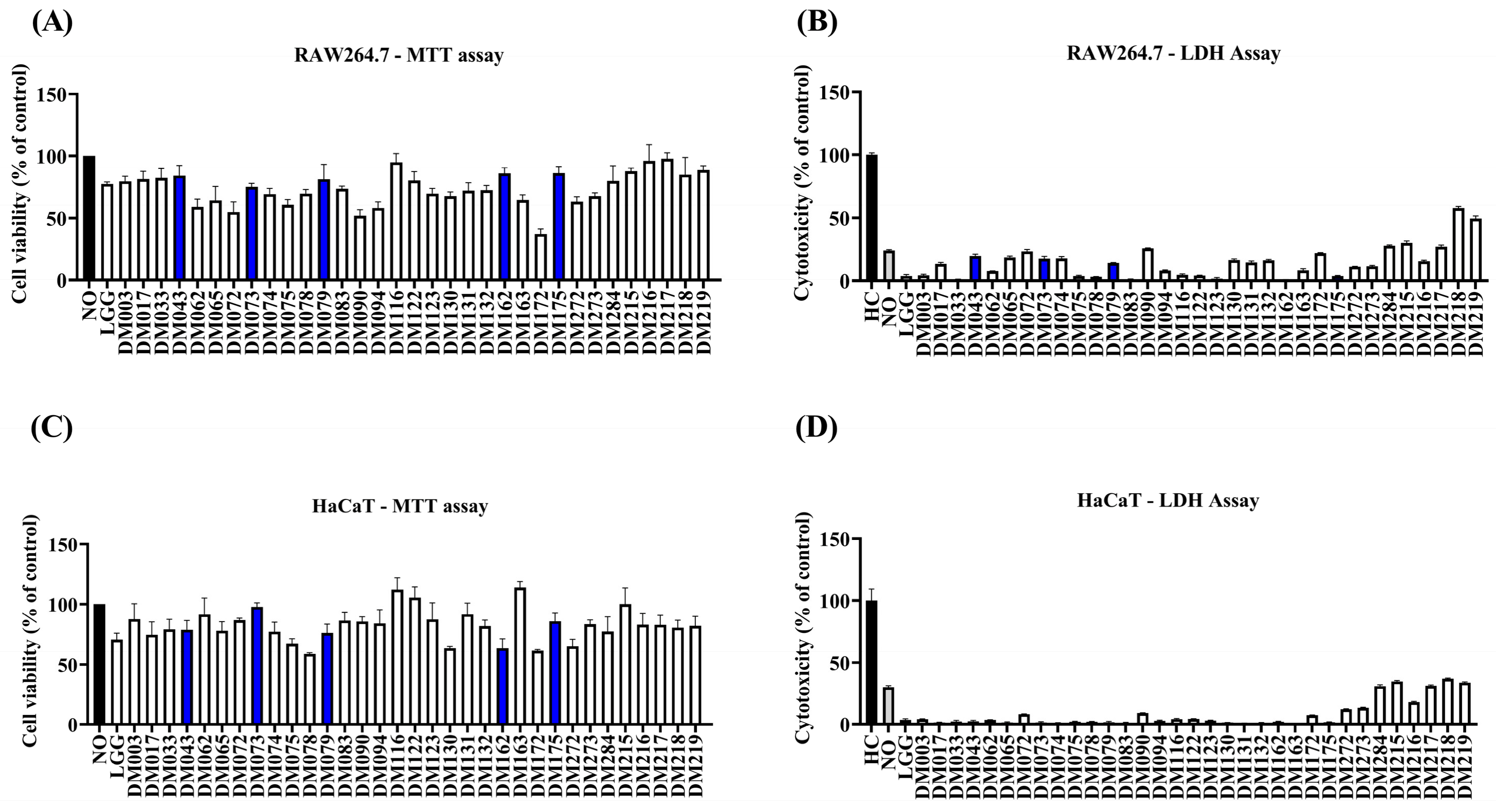
To identify probiotic strains with favorable safety profiles, cell viability and cytotoxicity were evaluated in RAW264.7 macrophages and HaCaT keratinocytes using MTT and LDH assays (Figure 2).
Figure 2.
Safety evaluation of heat-killed bacterial preparations in RAW264.7 macrophages and HaCaT keratinocytes. (A) Cell viability of RAW264.7 macrophages determined by the MTT assay. (B) Cytotoxicity of RAW264.7 macrophages determined by the LDH release assay. (C) Cell viability of HaCaT keratinocytes determined by the MTT assay. (D) Cytotoxicity of HaCaT keratinocytes determined by the LDH release assay. Black bars indicate the untreated control (MTT assay) or high control (LDH assay), gray bars indicate the low control (LDH assay), white bars indicate the candidate strains, and blue bars indicate the five selected strains (DM043, DM073, DM079, DM162, and DM175) selected for subsequent functional evaluation based on their favorable cell viability and low cytotoxicity.
Most strains maintained cell viability above 70% in both cell types, indicating low cytotoxic potential (Figure 2A,C). In addition, LDH release remained low for the majority of strains in both RAW264.7 and HaCaT keratinocytes (Figure 2B,D), suggesting minimal membrane damage following treatment with heat-killed bacterial preparations.
Based on the combined results of MTT and LDH assays, strains exhibiting high cell viability and low cytotoxicity were selected for subsequent functional screening. These selected strains were further evaluated for antioxidant, anti-inflammatory, antimicrobial, wound healing, and skin barrier-supportive activities.
3.3. DPPH Radical Scavenging Activity of Probiotic Culture Supernatants
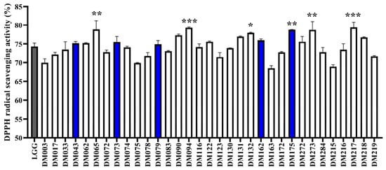
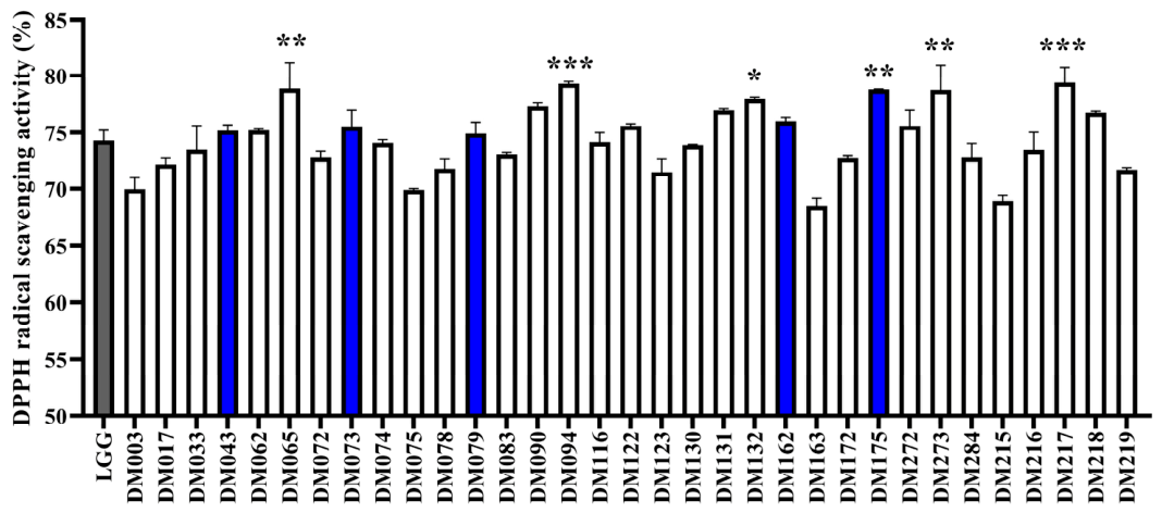
The DPPH radical scavenging activity of probiotic culture supernatants was evaluated using the DPPH assay (Figure 3). Overall, the tested strains exhibited substantial DPPH radical scavenging activity, ranging from approximately 68% to 80%, indicating strain-dependent differences in radical scavenging activity.
Figure 3.
DPPH radical scavenging activity of probiotic culture supernatants. DPPH radical scavenging activity was expressed as DPPH radical scavenging activity (%). The reference strain (LGG) is shown in gray, and the five selected candidate strains are highlighted in blue. Data are presented as mean ± SD. Statistical significance was determined using one-way ANOVA followed by Dunnett’s multiple comparison test relative to LGG (* p < 0.05, ** p < 0.01, *** p < 0.001).
Several strains demonstrated significantly higher DPPH radical scavenging activities than the reference strain LGG. In particular, DM062, DM094, DM162, DM175, DM217, and DM273 showed significantly higher DPPH radical scavenging activities compared with LGG (p < 0.05, p < 0.01, or p < 0.001). Among these strains, DM094 and DM217 exhibited the highest radical scavenging activities, approaching 80%.
These findings indicate that metabolites present in probiotic culture supernatants possess substantial DPPH radical scavenging activity. However, additional cellular and in vivo studies are required to determine whether these effects translate into protection against oxidative stress under physiological conditions. The observed strain-dependent differences suggest that DPPH radical scavenging activity may represent an important criterion for selecting probiotic strains with potential cosmetic applications.
3.4. Screening of Anti-Inflammatory Activity Using Nitric Oxide Inhibition Assay
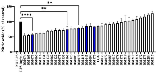
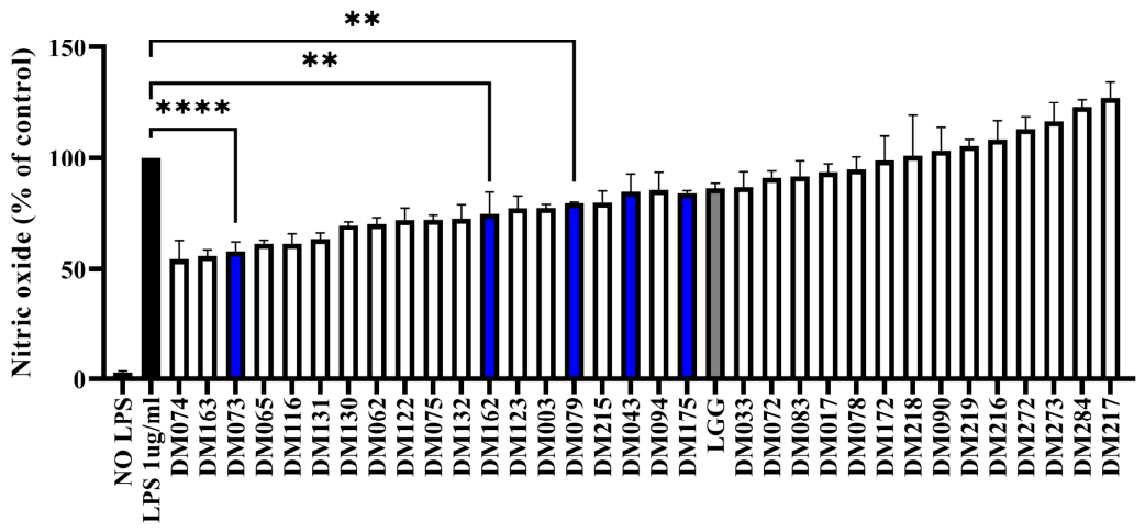
The anti-inflammatory activity of heat-killed probiotic strains was evaluated by measuring nitric oxide (NO) production in LPS-stimulated RAW264.7 macrophages (Figure 4). All tested strains reduced NO production to varying degrees compared with the LPS-treated control group, indicating strain-dependent anti-inflammatory activity.
Figure 4.
Inhibitory effects of heat-killed bacterial preparations on LPS-induced nitric oxide production in RAW264.7 macrophages. Nitric oxide production was quantified using the Griess assay and expressed as a percentage relative to the LPS-treated control group. Lower values indicate stronger inhibition of nitric oxide production. Strains are arranged in ascending order of relative nitric oxide levels. The LPS-treated control group is shown in black, the reference strain (LGG) is shown in gray, and the five selected candidate strains are highlighted in blue. Data are presented as the mean ± SD. Statistical significance was determined using one-way ANOVA followed by Dunnett’s multiple comparison test relative to the LPS-treated control group (** p < 0.01, **** p < 0.0001).
To facilitate comparison of anti-inflammatory potential, strains were ranked according to their relative NO production levels, with lower values indicating stronger inhibition of NO production. Several strains exhibited greater inhibitory effects on NO production than the reference strain LGG. Notably, DM073, DM065, DM132, DM162, DM079, DM043, and DM175 showed lower relative NO levels than LGG, suggesting superior anti-inflammatory activity.
Based on their favorable NO inhibitory activity together with DPPH radical scavenging activity, safety profile, and antimicrobial potential, DM073, DM162, DM079, DM043, and DM175 were selected for subsequent evaluation. These results demonstrate that selected heat-killed preparations effectively suppressed LPS-induced NO production in RAW264.7 macrophages, indicating their potential as candidates for cosmetic applications targeting inflammatory skin conditions.
3.5. Antimicrobial Activity Against Cutibacterium Acnes
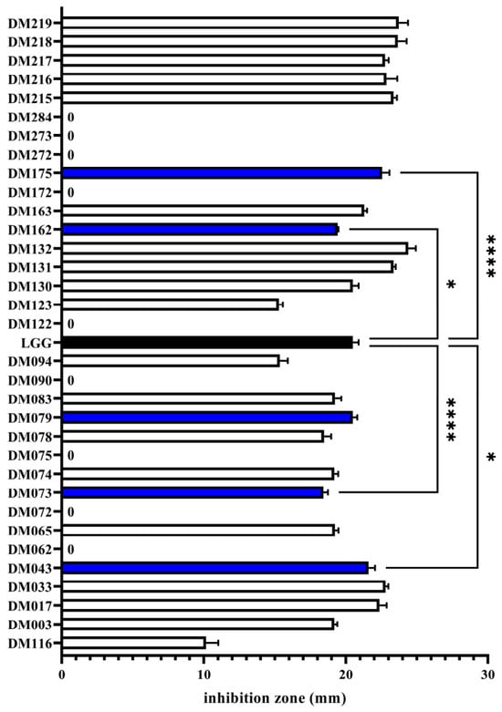
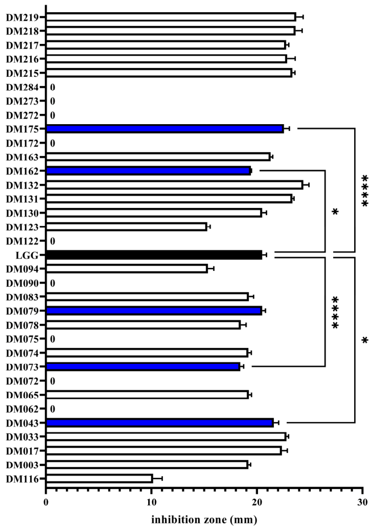
The antimicrobial activities of the selected probiotic strains against Cutibacterium acnes were evaluated using the agar diffusion assay (Figure 5). All selected strains exhibited inhibitory activity against C. acnes, with inhibition zone diameters ranging from approximately 18 to 23 mm.
Figure 5.
Antimicrobial activity of selected probiotic strains against Cutibacterium acnes. Antimicrobial activity was assessed using the agar diffusion assay and expressed as inhibition zone diameter (mm). Data are presented as mean ± SD. LGG (Lacticaseibacillus rhamnosus GG) was used as the reference strain. Statistical significance was determined relative to LGG (* p < 0.05, **** p < 0.0001). A value of 0 mm indicates that no detectable inhibition zone was observed. The reference strain, LGG (Lacticaseibacillus rhamnosus GG), is shown in black, the five selected candidate strains are highlighted in blue, and the other candidate strains are shown in white.
Among the tested strains, DM175 exhibited the strongest antimicrobial activity, showing the largest inhibition zone, followed by DM043 and DM079. In contrast, DM073 and DM162 displayed relatively smaller inhibition zones than the reference strain LGG. However, DM043 and DM175 showed significantly greater antimicrobial activity than LGG (* p < 0.05 and **** p < 0.0001, respectively), whereas no significant difference was observed between DM079 and LGG.
Although antimicrobial activities varied among the selected strains, all five candidates exhibited measurable inhibitory effects against C. acnes. These findings suggest that the selected probiotic strains possess anti-C. acnes activity and may contribute to the development of cosmetic ingredients for acne-prone skin.
Therefore, antimicrobial activity was considered together with antioxidant, anti-inflammatory, wound healing, and skin barrier-supportive properties for the final selection of probiotic candidates.
Representative agar diffusion images supporting the antimicrobial activity of the selected strains are provided in Supplementary Figure S1.
3.6. Anti-Inflammatory Effects of Selected Probiotic Strains in LPS-Stimulated RAW264.7 Macrophages
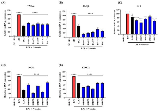
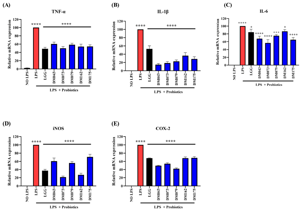
To further investigate the anti-inflammatory potential of the selected probiotic strains, the mRNA expression levels of pro-inflammatory mediators were analyzed in LPS-stimulated RAW264.7 macrophages using RT-qPCR (Figure 6). As expected, LPS stimulation markedly increased the expression of TNF-α, IL-1β, IL-6, iNOS, and COX-2 compared with the untreated control group.
Figure 6.
Anti-inflammatory effects of selected heat-killed probiotic strains in LPS-stimulated RAW264.7 macrophages. The expression levels of pro-inflammatory mediators, including (A) TNF-α, (B) IL-1β, (C) IL-6, (D) iNOS, and (E) COX-2, were analyzed by RT-qPCR following pretreatment with heat-killed probiotic strains and subsequent LPS stimulation. Relative mRNA expression levels were normalized to the housekeeping gene and expressed as percentages of the untreated control. Data are presented as mean ± SD (n = 3). Statistical significance was determined relative to the LPS-treated group (* p < 0.05, *** p < 0.001, **** p < 0.0001). The LPS-treated control group is shown in red, the reference strain (LGG) is shown in black, and the five selected probiotic strains are shown in blue.
Treatment with heat-killed probiotic strains significantly attenuated the expression of inflammatory mediators relative to the LPS-treated group. Among the tested strains, Lacticaseibacillus rhamnosus DM073 exhibited the strongest inhibitory effects, showing the lowest expression levels of IL-6, iNOS, and COX-2. DM043 also markedly suppressed IL-1β and COX-2 expression, whereas DM162 demonstrated a pronounced inhibitory effect on iNOS expression. In contrast, DM175 showed relatively weaker suppression of iNOS and COX-2 compared with the other selected strains.
Notably, several selected strains exhibited lower expression levels of pro-inflammatory genes than the reference strain LGG. In particular, DM073 consistently reduced IL-1β, IL-6, iNOS, and COX-2 expression to levels below those observed in the LGG-treated group, indicating superior anti-inflammatory activity. DM043 and DM079 also demonstrated comparable or improved suppression of selected inflammatory markers relative to LGG.
These findings were consistent with the NO inhibition assay results and suggest that selected heat-killed probiotic strains, particularly L. rhamnosus DM073, effectively suppress inflammatory responses in macrophages and may serve as promising candidates for cosmetic applications targeting inflammation-associated skin conditions.
3.7. Effects of Selected Probiotic Strains on Wound Healing Activity in HaCaT Keratinocytes
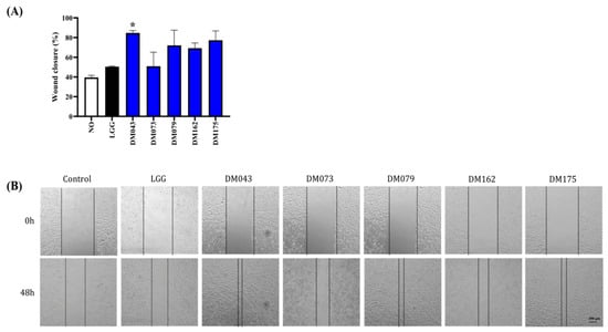
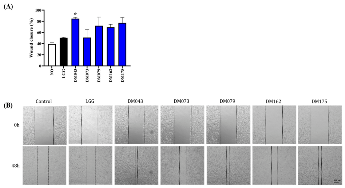
The wound healing activity of selected probiotic strains was evaluated using a scratch assay in HaCaT keratinocytes (Figure 7). All selected strains promoted wound closure compared with the untreated control group. Among the tested strains, DM043 exhibited the highest wound closure rate (83.4%) and significantly enhanced keratinocyte migration compared with the reference strain LGG (p < 0.05). DM079, DM162, and DM175 also showed enhanced wound closure activity, although the differences were not statistically significant. In contrast, DM073 demonstrated a relatively modest stimulatory effect on wound closure.
Figure 7.
Effects of selected heat-killed bacterial preparations on wound healing activity in HaCaT keratinocytes. (A) Quantitative analysis of wound closure after 48 h treatment with selected probiotic strains. Wound closure was calculated relative to the initial wound width and expressed as wound closure (%). (B) Representative microscopic images of scratch wounds at 0 h and 48 h. Dashed lines indicate wound boundaries. Data are presented as mean ± SD (n = 3). * p < 0.05 versus LGG. Scale bar = 200 μm. The reference strain (LGG) is shown in black, and the five selected probiotic strains are shown in blue.
Representative microscopic images further confirmed accelerated wound closure in probiotic-treated groups. In particular, DM043-treated cells exhibited greater wound closure after 48 h, indicating enhanced keratinocyte migration and regenerative potential. These results suggest that selected probiotic strains, especially DM043, may contribute to skin regeneration and wound repair.
Similar wound healing-promoting effects were also observed under TNF-α/IFN-γ-induced inflammatory conditions (Supplementary Figure S2).
3.8. Effects of Selected Probiotic Strains on Tight Junction and Skin Barrier-Related Gene Expression in HaCaT Keratinocytes
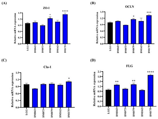
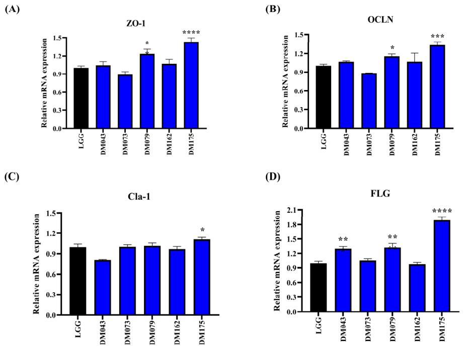
To investigate the skin barrier-supportive effects of the selected probiotic strains, the expression levels of tight junction- and skin barrier-related genes were analyzed in HaCaT keratinocytes by RT-qPCR (Figure 8).
Figure 8.
Effects of selected heat-killed bacterial preparations on tight junction and skin barrier-related gene expression in HaCaT keratinocytes. Relative mRNA expression levels of (A) ZO-1, (B) OCLN, (C) Claudin-1 (Cla-1), and (D) FLG were determined by RT-qPCR after treatment with selected heat-killed probiotic strains. Gene expression levels were normalized to GAPDH and expressed relative to the LGG-treated group. Data are presented as mean ± SD (n = 3). Statistical significance was determined using one-way ANOVA followed by Dunnett’s test. * p < 0.05, ** p < 0.01, *** p < 0.001, and **** p < 0.0001 versus LGG. The reference strain (LGG) is shown in black, and the five selected probiotic strains are shown in blue.
Among the tested strains, DM175 significantly increased the expression of ZO-1, OCLN, Claudin-1 (Cla-1), and FLG compared with the reference strain LGG, indicating a broad enhancement of skin barrier-associated gene expression. DM079 also significantly upregulated ZO-1, OCLN, and FLG expression, whereas DM043 markedly increased FLG expression. In contrast, DM073 and DM162 showed relatively limited effects on barrier-related gene expression.
Notably, FLG expression was strongly induced by DM175, reaching the highest level among all tested strains. Overall, these findings suggest that selected probiotic strains, particularly DM175 and DM079, may contribute to skin barrier reinforcement through the upregulation of tight junction and epidermal barrier-related genes.
3.9. Restoration of TNF-α/IFN-γ-Induced Skin Barrier Dysfunction by Selected Probiotic Strains
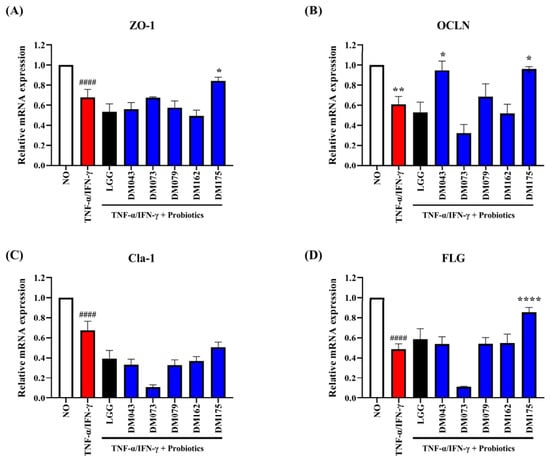
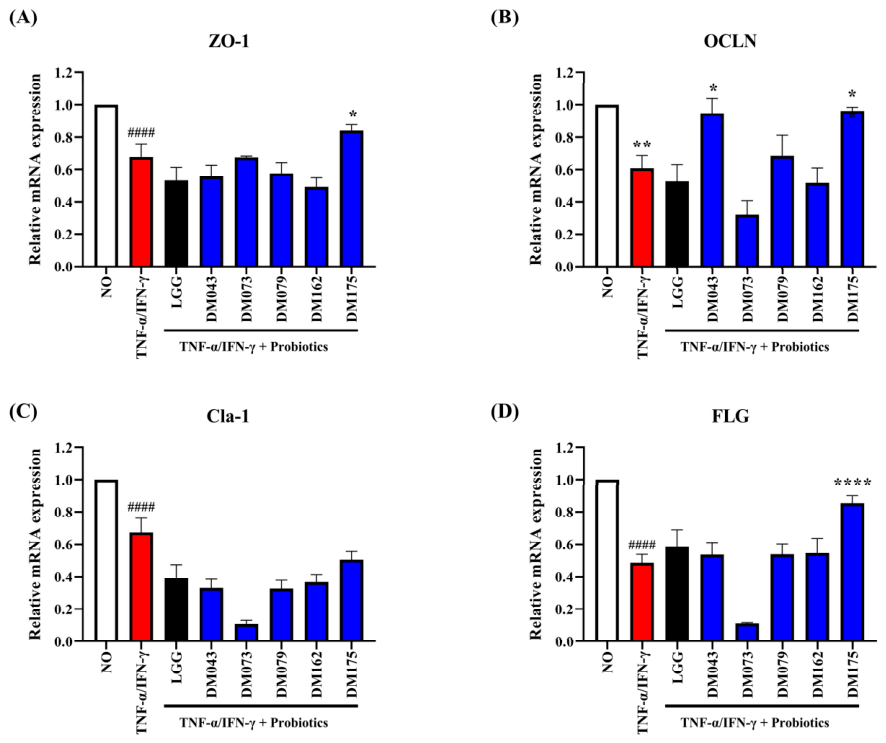
TNF-α/IFN-γ stimulation significantly reduced the expression levels of skin barrier-related genes, including ZO-1, OCLN, Claudin-1 (Cla-1), and FLG, compared with the untreated control group (Figure 9), indicating inflammatory barrier dysfunction in HaCaT keratinocytes.
Figure 9.
Effects of selected heat-killed bacterial preparations on TNF-α/IFN-γ-induced skin barrier dysfunction in HaCaT keratinocytes. Relative mRNA expression levels of (A) ZO-1, (B) OCLN, (C) Claudin-1 (Cla-1), and (D) FLG were analyzed by RT-qPCR following TNF-α/IFN-γ stimulation and heat-killed bacterial preparations. Gene expression levels were normalized to GAPDH and expressed relative to the untreated control group. Data are presented as mean ± SD (n = 3). Statistical significance was determined using one-way ANOVA followed by Dunnett’s test. #### p < 0.0001 versus untreated control group; * p < 0.05, ** p < 0.01, and **** p < 0.0001 versus TNF-α/IFN-γ-treated group. The TNF-α/IFN-γ-treated group is shown in red, the reference strain (LGG) is shown in black, and the five selected probiotic strains are shown in blue.
Treatment with selected probiotic strains restored the expression of barrier-associated genes to varying degrees. Among the tested strains, DM175 exhibited the most pronounced restorative effect, significantly upregulating the expression of ZO-1, OCLN, and FLG compared with the TNF-α/IFN-γ-treated group. DM043 also markedly increased OCLN expression, whereas DM079 partially restored the expression of ZO-1 and OCLN. In contrast, DM073 showed relatively limited protective effects under inflammatory conditions.
Notably, DM175 strongly restored FLG expression, reaching levels substantially higher than those observed in the inflammatory control group. These findings suggest that selected probiotic strains alleviate inflammatory skin barrier dysfunction and may contribute to the maintenance of epidermal integrity under pro-inflammatory conditions.
Under TNF-α/IFN-γ-induced inflammatory conditions, wound closure was significantly reduced compared with the untreated control. Treatment with LGG and all selected probiotic strains significantly improved wound closure, with DM043, DM079, and DM175 exhibiting the greatest recovery effects (Supplementary Figure S2).
In addition, selected probiotic strains reduced the expression of inflammatory chemokines, including RANTES, TARC, MDC, and IL-8, in TNF-α/IFN-γ-stimulated HaCaT keratinocytes (Supplementary Figure S3), further supporting their anti-inflammatory activity in keratinocytes.
These findings provide a basis for future studies investigating the underlying molecular mechanisms and the efficacy of these heat-killed bacterial preparations in more physiologically relevant skin models.
4. Discussion
The skin microbiome has emerged as an important regulator of epidermal homeostasis, immune balance, and barrier integrity. Accordingly, the identification of probiotic strains possessing multifunctional activities has gained increasing attention for cosmetic and dermatological applications. In the present study, an in-house bacterial library comprising 302 isolates was systematically screened to identify candidates with antioxidant, anti-inflammatory, antimicrobial, wound-healing, and skin barrier-supportive properties. Through a stepwise selection strategy, five strains (DM043, DM073, DM079, DM162, and DM175) were selected and further characterized using cellular and molecular assays.
Importantly, each selected strain exhibited distinct functional strengths. DM073 demonstrated the most potent anti-inflammatory activity, DM043 showed superior wound-healing capacity, and DM175 displayed the strongest skin barrier-supportive effects. These complementary properties suggest that individual strains may be strategically applied according to specific cosmetic objectives.
Oxidative stress is a major contributor to skin aging and inflammatory skin disorders. Excessive reactive oxygen species (ROS) promote lipid peroxidation, DNA damage, and inflammatory signaling, ultimately impairing epidermal barrier function. Therefore, radical scavenging activity is considered one of the fundamental characteristics of skin-beneficial probiotics. In the present study, several candidate strains exhibited strong DPPH radical scavenging activity comparable to or greater than that of the reference strain LGG. Previous studies have demonstrated that extracellular metabolites produced by Lactobacillus strains, including exopolysaccharides, bioactive peptides, and antioxidant compounds, contribute to ROS scavenging and cellular protection [21,22,23]. The high DPPH radical scavenging activities observed in the culture supernatants of the selected strains may therefore support their potential to alleviate oxidative stress-associated skin damage.
Inflammation represents another critical factor affecting skin health. Activated macrophages produce excessive nitric oxide (NO) and pro-inflammatory mediators that contribute to chronic inflammatory responses. Consistent with previous reports describing immunomodulatory properties of probiotic bacteria, the selected strains significantly reduced NO production in LPS-stimulated RAW264.7 macrophages. Furthermore, treatment with selected strains attenuated the expression of major inflammatory mediators, including TNF-α, IL-1β, IL-6, iNOS, and COX-2. These findings indicate that the selected strains possess potent anti-inflammatory activity and may regulate inflammatory signaling pathways involved in skin inflammation [9,18,24].
The antimicrobial activity against C. acnes was evaluated using viable probiotic cultures to assess strain-specific antagonistic potential. Although the present study primarily focused on heat-killed preparations and postbiotic-oriented cosmetic applications, future studies are warranted to determine whether heat-killed preparations or cell-free supernatants retain comparable anti-C. acnes activity. Such investigations would further support the development of postbiotic-based formulations for acne-prone skin [17,25].
Wound repair is an essential process for maintaining epidermal integrity following physical or inflammatory damage. In the present study, several selected strains enhanced HaCaT cell migration under normal conditions, indicating their potential to promote re-epithelialization. Among the tested strains, DM043 exhibited the greatest wound-healing activity [19,20]. In addition, under TNF-α/IFN-γ-induced inflammatory conditions, heat-killed bacterial preparations significantly improved wound closure compared with the inflammatory control group. These observations suggest that selected probiotic strains may facilitate tissue regeneration and recovery of damaged epidermal barriers under inflammatory environments.
An intact epidermal barrier is essential for preventing water loss and protecting the host from environmental stressors. Tight junction proteins, including ZO-1, occludin (OCLN), and claudin-1 (Cla-1), together with filaggrin (FLG), play central roles in maintaining barrier integrity and epidermal differentiation. Under normal conditions, several selected strains enhanced the expression of these barrier-associated genes, particularly DM175, which consistently increased ZO-1, OCLN, Cla-1, and FLG expression [15,26]. More importantly, heat-killed bacterial preparations partially restored barrier-related gene expression suppressed by TNF-α/IFN-γ stimulation. DM175 showed the strongest restorative effect, particularly for FLG expression, suggesting a robust barrier-protective function under inflammatory conditions. Filaggrin (FLG) is a key structural protein involved in epidermal differentiation and skin barrier maintenance, and reduced FLG expression has been closely associated with barrier dysfunction and inflammatory skin disorders such as atopic dermatitis [27,28]. Therefore, the marked restoration of FLG expression by DM175 may contribute to improved epidermal integrity and barrier function.
These findings are consistent with previous studies demonstrating that probiotic-derived metabolites can strengthen epithelial junctions and improve barrier function through modulation of host signaling pathways [15,26].
To further investigate the anti-inflammatory effects of the selected strains in keratinocytes, the expression of representative atopic dermatitis-associated chemokines was evaluated. TNF-α/IFN-γ stimulation markedly induced the expression of RANTES, TARC, MDC, and IL-8, whereas treatment with selected heat-killed bacterial preparations significantly reduced their expression. Notably, DM043 and DM079 strongly suppressed TARC and MDC expression, while DM175 exhibited broad inhibitory activity against multiple inflammatory chemokines. Since these mediators are involved in immune cell recruitment and chronic skin inflammation, their suppression further supports the potential application of these strains for inflammatory skin conditions [24,29,30].
Among the selected candidates, DM175 consistently demonstrated superior performance across multiple functional categories, including antimicrobial activity, barrier-supportive activity, anti-inflammatory activity, and inflammatory barrier recovery. DM043 exhibited particularly strong wound-healing activity, whereas DM079 showed notable effects on barrier-related gene expression and inflammatory chemokine suppression. These findings indicate that individual strains possess distinct functional strengths and that strain-specific characteristics should be considered when developing probiotic-based cosmetic formulations.
Several limitations should be acknowledged. First, all experiments were conducted using in vitro cell culture models and therefore may not fully reflect the complexity of human skin physiology. Second, antioxidant and antimicrobial activities were evaluated using viable bacterial cultures or their culture supernatants, whereas all other functional assays—including cytotoxicity, anti-inflammatory, wound-healing, and skin barrier-related gene expression—were performed using heat-killed bacterial preparations. Because antimicrobial and antioxidant activities are often dependent on metabolites generated during active bacterial growth, such as organic acids, bacteriocins, and exopolysaccharides, it remains to be determined whether the heat-killed forms of the selected strains retain comparable antioxidant and antimicrobial efficacy. Future studies directly comparing live, heat-killed, and cell-free supernatant preparations will be necessary to clarify which functional properties are preserved after heat inactivation, the form most relevant to postbiotic cosmetic applications. Third, the molecular mechanisms underlying the observed biological activities were not investigated in detail. Future studies should explore the involvement of signaling pathways and evaluate efficacy using reconstructed human skin models and clinical studies. In the present study, all functional evaluations, except for the initial antimicrobial screening against C. acnes, were performed using heat-killed bacterial preparations. Therefore, the observed DPPH radical scavenging, anti-inflammatory, and skin barrier-supportive activities are considered to reflect the effects of heat-killed bacterial cells and their associated components and support their potential application as postbiotic cosmetic ingredients rather than viable probiotics. Because heat-killed bacterial cells are non-viable, they are not expected to colonize the skin or displace the resident microbiota, suggesting a lower risk of disturbing the normal skin microbial community. The antimicrobial activity observed in this study was evaluated only against Cutibacterium acnes, a skin-associated opportunistic pathogen. In addition, the culture supernatants exhibiting DPPH radical scavenging activity may represent an additional source of bioactive components for future cosmetic formulations.
Importantly, the present study demonstrates that different probiotic strains exhibit distinct functional characteristics rather than uniformly exerting all skin-beneficial effects. Such strain-specific functionality may enable the development of targeted cosmetic formulations optimized for specific skin concerns, including inflammation, impaired barrier function, wound recovery, and microbiome imbalance.
5. Conclusions
A systematic screening approach was applied to identify multifunctional probiotic strains with skin-beneficial properties from a large probiotic strain library. Five strains (DM043, DM073, DM079, DM162, and DM175) were selected based on their antioxidant, anti-inflammatory, and antimicrobial activities and were further evaluated using in vitro skin-related assays. The selected strains exhibited favorable safety profiles and demonstrated wound-healing, skin barrier-supportive, and anti-inflammatory effects in HaCaT keratinocytes and RAW264.7 macrophages.
Among the selected candidates, Lacticaseibacillus rhamnosus DM073 exhibited superior anti-inflammatory activity, Lactiplantibacillus plantarum DM043 demonstrated the strongest wound-healing capacity, and Lactiplantibacillus plantarum DM175 showed remarkable skin barrier-supportive effects. These strain-specific functional characteristics highlight their potential as multifunctional cosmetic ingredients and provide a scientific basis for the development of probiotic-derived postbiotic formulations for skin soothing, barrier reinforcement, skin recovery, and microbiome-friendly skincare applications.
Furthermore, the distinct functional characteristics observed among the selected strains, rather than a uniform expression of all skin-beneficial effects, support their potential for developing targeted cosmetic formulations tailored to specific skin concerns, including inflammation, impaired barrier function, wound recovery, and microbiome imbalance.
These findings also provide a basis for future studies investigating the underlying molecular mechanisms and evaluating the efficacy of these heat-killed bacterial preparations in more physiologically relevant skin models and clinical cosmetic applications.
6. Patents
A patent application related to the findings presented in this study has been filed in Korea: “Probiotic Strains That Enhance Skin Barrier Function and Improve Inflammatory Skin, and Compositions Containing the Same”, Korean Patent Application No. 10-2026-0038736, filed on 4 March 2026 (Applicant: Docsmedi Co., Ltd.).
Supplementary Materials
The following supporting information can be downloaded at: https://www.mdpi.com/article/10.3390/cosmetics13040187/s1, Figure S1: Representative agar diffusion images showing the antimicrobial activity of selected probiotic strains against Cutibacterium acnes; Figure S2: Effects of selected probiotic strains on wound healing under TNF-α/IFN-γ-induced inflammatory conditions in HaCaT keratinocytes; Figure S3: Effects of selected probiotic strains on inflammatory chemokine expression (RANTES, TARC, MDC, and IL-8) in TNF-α/IFN-γ-stimulated HaCaT keratinocytes; Table S1: Normalized scores used for radar chart construction and functional comparison of selected probiotic strains.
Author Contributions
Conceptualization, J.-H.L. and H.-S.K.; methodology, J.-H.L.; validation, J.-H.L. and J.Y.; formal analysis, J.-H.L.; investigation, J.-H.L.; resources, Y.-Y.K. and H.-S.K.; data curation, J.-H.L.; writing—original draft preparation, J.-H.L.; writing—review and editing, J.-H.L. and H.-S.K.; visualization, J.-H.L.; supervision, H.-S.K.; project administration, H.-S.K.; funding acquisition, H.-S.K. All authors have read and agreed to the published version of the manuscript.
Funding
This research received no external funding. The article processing charge (APC) was funded by DOCSMEDI Co., Ltd.
Institutional Review Board Statement
Not applicable.
Informed Consent Statement
Not applicable.
Data Availability Statement
The data presented in this study are available within the article and its Supplementary Materials. The bacterial strains characterized in this study have been deposited at the Korean Collection for Type Cultures (KCTC) under accession numbers KCTC 16606BP (Lactiplantibacillus plantarum DM043), KCTC 16607BP (Lacticaseibacillus rhamnosus DM073), KCTC 15155BP (Ligilactobacillus salivarius DM079), KCTC 16609BP (Lacticaseibacillus paracasei DM162), and KCTC 16608BP (Lactiplantibacillus plantarum DM175). Additional data are available from the corresponding author upon reasonable request.
Conflicts of Interest
J.-H.L. and J.Y. are employed by DOCSMEDI Co., Ltd. The remaining authors declare no conflicts of interest. The funder had no role in the design of the present study; in the analysis or interpretation of data; in the writing of the manuscript; or in the decision to publish the results. The authors declare that the research was conducted in the absence of any commercial or financial relationships that could be construed as a potential conflict of interest.
Abbreviations
| C. acnes | Cutibacterium acnes |
| GAPDH | Glyceraldehyde-3-phosphate dehydrogenase |
| Cla-1 | Claudin-1 |
| COX-2 | Cyclooxygenase-2 |
| DPPH | 2,2-Diphenyl-1-picrylhydrazyl |
| FLG | Filaggrin |
| iNOS | Inducible nitric oxide synthase |
| IL-1β | Interleukin-1 beta |
| IL-6 | Interleukin-6 |
| IL-8 | Interleukin-8 (CXCL8) |
| ZO-1 | Zonula occludens-1 |
| OCLN | Occludin |
| RANTES | Regulated upon Activation, Normal T Cell Expressed and Secreted (CCL5) |
| MDC | Macrophage-derived chemokine (CCL22) |
| ROS | Reactive oxygen species |
| TARC | Thymus and activation-regulated chemokine (CCL17) |
| TNF-α | Tumor necrosis factor-alpha |
| IFN-γ | Interferon-gamma |
| NO | Nitric oxide |
| LPS | Lipopolysaccharide |
| LGG | Lacticaseibacillus rhamnosus GG |
References
- Carmona-Cruz, S.; Orozco-Covarrubias, L.; Sáez-de-Ocariz, M. The Human Skin Microbiome in Selected Cutaneous Diseases. Front. Cell. Infect. Microbiol. 2022, 12, 834135. [Google Scholar] [CrossRef] [Scilit] [PubMed]
- Ni, Q.; Zhang, P.; Li, Q.; Han, Z. Oxidative Stress and Gut Microbiome in Inflammatory Skin Diseases. Front. Cell Dev. Biol. 2022, 10, 849985. [Google Scholar] [CrossRef] [Scilit] [PubMed]
- Li, Z.; Zhang, J.; Zhang, Y.; Chen, H.; Bao, Y. Skin Microbiome in Health and Disease: Mechanisms and Emerging Therapeutic Strategies. Clin. Cosmet. Investig. Dermatol. 2025, 18, 3443–3455. [Google Scholar] [CrossRef] [Scilit] [PubMed]
- Gao, T.; Wang, X.; Li, Y.; Ren, F. The Role of Probiotics in Skin Health and Related Gut-Skin Axis: A Review. Nutrients 2023, 15, 3123. [Google Scholar] [CrossRef] [Scilit] [PubMed]
- De Almeida, C.V.; Antiga, E.; Lulli, M. Oral and Topical Probiotics and Postbiotics in Skincare and Dermatological Therapy: A Concise Review. Microorganisms 2023, 11, 1420. [Google Scholar] [CrossRef] [Scilit] [PubMed]
- Dou, J.; Feng, N.; Guo, F.; Chen, Z.; Liang, J.; Wang, T.; Guo, X.; Xu, Z. Applications of Probiotic Constituents in Cosmetics. Molecules 2023, 28, 6765. [Google Scholar] [CrossRef] [Scilit] [PubMed]
- Lim, H.W.; Huang, Y.H.; Kyeong, G.; Park, M.; Lim, C.J. Comparative Insights into the Skin Beneficial Properties of Probiotic Lactobacillus Isolates of Skin Origin. Biomed. Res. Int. 2022, 2022, 7728789. [Google Scholar] [CrossRef] [Scilit] [PubMed]
- Wang, J.; Zhang, J.; Guo, H.; Cheng, Q.; Abbas, Z.; Tong, Y.; Yang, T.; Zhou, Y.; Zhang, H.; Wei, X.; et al. Optimization of Exopolysaccharide Produced by Lactobacillus plantarum R301 and Its Antioxidant and Anti-Inflammatory Activities. Foods 2023, 12, 2481. [Google Scholar] [CrossRef] [Scilit] [PubMed]
- Hao, R.; Liu, Q.; Wang, L.; Jian, W.; Cheng, Y.; Zhang, Q.; Hayer, K.; Kamarudin Raja Idris, R.; Zhang, Y.; Lu, H.; et al. Anti-inflammatory effect of Lactiplantibacillus plantarum T1 cell-free supernatants through suppression of oxidative stress and NF-κB- and MAPK-signaling pathways. Appl. Environ. Microbiol. 2023, 89, e0060823. [Google Scholar] [CrossRef] [Scilit] [PubMed]
- Kim, G.; Choi, K.H.; Kim, H.; Chung, D.K. Alleviation of LPS-Induced Inflammation and Septic Shock by Lactiplantibacillus plantarum K8 Lysates. Int. J. Mol. Sci. 2021, 22, 5921. [Google Scholar] [CrossRef] [Scilit] [PubMed]
- Lee, K.S.; Kim, Y.; Lee, J.H.; Shon, S.; Kim, A.; Pham, A.V.Q.; Kim, C.; Kim, D.H.; Kim, Y.K.; Cho, E.G. Human Probiotic Lactobacillus paracasei-Derived Extracellular Vesicles Improve Tumor Necrosis Factor-α-Induced Inflammatory Phenotypes in Human Skin. Cells 2023, 12, 2789. [Google Scholar] [CrossRef] [Scilit] [PubMed]
- Brandner, J.M.; Kief, S.; Grund, C.; Rendl, M.; Houdek, P.; Kuhn, C.; Tschachler, E.; Franke, W.W.; Moll, I. Organization and formation of the tight junction system in human epidermis and cultured keratinocytes. Eur. J. Cell Biol. 2002, 81, 253–263. [Google Scholar] [CrossRef] [Scilit] [PubMed]
- Irvine, A.D.; McLean, W.H.; Leung, D.Y. Filaggrin mutations associated with skin and allergic diseases. N. Engl. J. Med. 2011, 365, 1315–1327. [Google Scholar] [CrossRef] [Scilit] [PubMed]
- Yuki, T.; Tobiishi, M.; Kusaka-Kikushima, A.; Ota, Y.; Tokura, Y. Impaired Tight Junctions in Atopic Dermatitis Skin and in a Skin-Equivalent Model Treated with Interleukin-17. PLoS ONE 2016, 11, e0161759. [Google Scholar] [CrossRef] [Scilit] [PubMed]
- Sultana, R.; McBain, A.J.; O’Neill, C.A. Strain-dependent augmentation of tight-junction barrier function in human primary epidermal keratinocytes by Lactobacillus and Bifidobacterium lysates. Appl. Environ. Microbiol. 2013, 79, 4887–4894. [Google Scholar] [CrossRef] [Scilit] [PubMed]
- Jung, Y.O.; Jeong, H.; Cho, Y.; Lee, E.O.; Jang, H.W.; Kim, J.; Nam, K.; Lim, K.M. Lysates of a Probiotic, Lactobacillus rhamnosus, Can Improve Skin Barrier Function in a Reconstructed Human Epidermis Model. Int. J. Mol. Sci. 2019, 20, 4289. [Google Scholar] [CrossRef] [Scilit] [PubMed]
- Biazzo, M.; Pinzauti, D.; Podrini, C. SkinDuoTM as a Targeted Probiotic Therapy: Shifts in Skin Microbiota and Clinical Outcomes in Acne Patients. Int. J. Mol. Sci. 2025, 26, 5000. [Google Scholar] [CrossRef] [Scilit] [PubMed]
- Kang, C.H.; Kim, J.S.; Kim, H.; Park, H.M.; Paek, N.S. Heat-Killed Lactic Acid Bacteria Inhibit Nitric Oxide Production via Inducible Nitric Oxide Synthase and Cyclooxygenase-2 in RAW 264.7 Cells. Probiotics Antimicrob. Proteins 2021, 13, 1530–1538. [Google Scholar] [CrossRef] [Scilit] [PubMed]
- Brandi, J.; Cheri, S.; Manfredi, M.; Di Carlo, C.; Vita Vanella, V.; Federici, F.; Bombiero, E.; Bazaj, A.; Rizzi, E.; Manna, L.; et al. Exploring the wound healing, anti-inflammatory, anti-pathogenic and proteomic effects of lactic acid bacteria on keratinocytes. Sci. Rep. 2020, 10, 11572. [Google Scholar] [CrossRef] [Scilit] [PubMed]
- Mohammedsaeed, W.; Cruickshank, S.; McBain, A.J.; O’Neill, C.A. Lactobacillus rhamnosus GG Lysate Increases Re-Epithelialization of Keratinocyte Scratch Assays by Promoting Migration. Sci. Rep. 2015, 5, 16147. [Google Scholar] [CrossRef] [Scilit] [PubMed]
- Lee, K.; Kim, H.J.; Kim, S.A.; Park, S.D.; Shim, J.J.; Lee, J.L. Exopolysaccharide from Lactobacillus plantarum HY7714 Protects against Skin Aging through Skin-Gut Axis Communication. Molecules 2021, 26, 1651. [Google Scholar] [CrossRef] [Scilit] [PubMed]
- Cha, S.Y.; Woo, I.K.; Cha, Y.J.; Lee, N.K.; Jang, H.J.; Paik, H.D. Anti-melanogenic and Antioxidant Activities of Lactiplantibacillus plantarum Strains in Skin Cells via the CREB/MITF and Nrf2/HO-1 Pathways. Probiotics Antimicrob. Proteins 2025, 17, 3795–3810. [Google Scholar] [CrossRef] [Scilit] [PubMed]
- Park, K.M.; Lee, Y.R.; Lee, N.K.; Paik, H.D. Lactiplantibacillus plantarum WB3801 and Lactiplantibacillus plantarum WB3808 Showed Antioxidant Effect and Anti-Apoptosis through Activation of the Keap1/Nrf2/HO-1 Pathway in H2O2-Induced HT-29 Cells. J. Microbiol. Biotechnol. 2025, 35, e2508014. [Google Scholar] [CrossRef] [Scilit] [PubMed]
- Lee, J.Y.; Park, J.Y.; Jeong, Y.; Kang, C.H. Anti-Inflammatory Response in TNFα/IFNγ-Induced HaCaT Keratinocytes and Probiotic Properties of Lacticaseibacillus rhamnosus MG4644, Lacticaseibacillus paracasei MG4693, and Lactococcus lactis MG5474. J. Microbiol. Biotechnol. 2023, 33, 1039–1049. [Google Scholar] [CrossRef] [Scilit] [PubMed]
- Lebeer, S.; Oerlemans, E.F.M.; Claes, I.; Henkens, T.; Delanghe, L.; Wuyts, S.; Spacova, I.; van den Broek, M.F.L.; Tuyaerts, I.; Wittouck, S.; et al. Selective targeting of skin pathobionts and inflammation with topically applied lactobacilli. Cell Rep. Med. 2022, 3, 100521. [Google Scholar] [CrossRef] [Scilit] [PubMed]
- Lee, C.; Pei, L.; Park, H.; Kim, H.; Huh, C.S. Skin Protection Effects of Lactobacillus paragasseri HN910 Lysate and the Role of Alanine. Probiotics Antimicrob. Proteins 2026, 18, 470–487. [Google Scholar] [CrossRef] [Scilit] [PubMed]
- McLean, W.H.; Irvine, A.D. Heritable filaggrin disorders: The paradigm of atopic dermatitis. J. Investig. Dermatol. 2012, 132, E20–E21. [Google Scholar] [CrossRef] [Scilit] [PubMed]
- Brown, S.J.; McLean, W.I. One remarkable molecule: Filaggrin. J. Investig. Dermatol. 2012, 132, 751–762. [Google Scholar] [CrossRef] [Scilit] [PubMed]
- Kaneki, M.; Ohira, C.; Magami, T.; Hamauzu, A.; Inaba, Y.; Togase, H.; Fukuyama, T. Topical Administration of a Mixed Microbial Culture of Lactobacillus paracasei, Pichia membranifaciens and Saccharomyces cerevisiae Significantly Inhibits the Development of Atopic Dermatitis in a Mouse Model Through IL-10 Overexpression by Dendritic Cells. Biomedicines 2025, 13, 2536. [Google Scholar] [CrossRef] [Scilit] [PubMed]
- Kim, Y.K.; Cho, M.; Kang, D.J. Anti-Inflammatory Response of New Postbiotics in TNF-α/IFN-γ-Induced Atopic Dermatitis-like HaCaT Keratinocytes. Curr. Issues Mol. Biol. 2024, 46, 6100–6111. [Google Scholar] [CrossRef] [Scilit] [PubMed]

Disclaimer/Publisher’s Note: The statements, opinions and data contained in all publications are solely those of the individual author(s) and contributor(s) and not of MDPI and/or the editor(s). MDPI and/or the editor(s) disclaim responsibility for any injury to people or property resulting from any ideas, methods, instructions or products referred to in the content. |
© 2026 by the authors. Licensee MDPI, Basel, Switzerland. This article is an open access article distributed under the terms and conditions of the Creative Commons Attribution (CC BY) license.