Next Issue
Volume 11, July
Previous Issue
Volume 11, May


Biosensors, Volume 11, Issue 6 (June 2021) – 38 articles

Cover Story (view full-size image): 
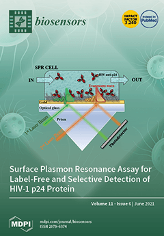
Multi-parameter SPR was used to develop a reliable and label-free detection method for HIV-1 p24 capsid protein. The study aimed at the assessment of a biosensing assay for the early detection of the human immunodeficiency virus (HIV). Remarkably, both physical and chemical immobilization of mouse monoclonal antibodies against HIV-1 p24 on the SPR gold detecting surface have been characterized for the first time. An equilibrium dissociation constant KD of 5.30 × 10⁻⁹ M was computed for the assay on the chemically modified surface. The system was also characterized in terms of sensitivity and selectivity, reaching a limit of detection of 4.1 ± 0.5 nM and an unprecedented selectivity ratio of 0.02. View this paper.
  • Issues are regarded as officially published after their release is announced to the table of contents alert mailing list.
  • You may sign up for e-mail alerts to receive table of contents of newly released issues.
  • PDF is the official format for papers published in both, html and pdf forms. To view the papers in pdf format, click on the "PDF Full-text" link, and use the free Adobe Reader to open them.
Order results
Result details
Section
Select all
Export citation of selected articles as:
13 pages, 1800 KiB  
Article
Electrochemical Immunosensing of ST2: A Checkpoint Target in Cancer Diseases
by Rebeca M. Torrente-Rodríguez, Cristina Muñoz-San Martín, Maria Gamella, María Pedrero, Neus Martínez-Bosch, Pilar Navarro, Pablo García de Frutos, José M. Pingarrón and Susana Campuzano
Biosensors 2021, 11(6), 202; https://doi.org/10.3390/bios11060202 - 21 Jun 2021
Cited by 11 | Viewed by 3026
Abstract
A magnetic beads (MB)-involved amperometric immunosensor for the determination of ST2, a member of the IL1 receptor family, is reported in this work. The method utilizes a sandwich immunoassay and disposable screen-printed carbon electrodes (SPCEs). Magnetic immunoconjugates built on the surface of carboxylic [...] Read more.
A magnetic beads (MB)-involved amperometric immunosensor for the determination of ST2, a member of the IL1 receptor family, is reported in this work. The method utilizes a sandwich immunoassay and disposable screen-printed carbon electrodes (SPCEs). Magnetic immunoconjugates built on the surface of carboxylic acid-microsized magnetic particles (HOOC-MBs) were used to selectively capture ST2. A biotinylated secondary antibody further conjugated with a streptavidin peroxidase conjugate (Strep-HRP) was used to accomplish the sandwiching of the target protein. The immune platform exhibits great selectivity and a low limit of detection (39.6 pg mL−1) for ST2, allowing the determination of soluble ST2 (sST2) in plasma samples from healthy individuals and patients diagnosed with pancreatic ductal adenocarcinoma (PDAC) in only 45 min once the immunoconjugates have been prepared. The good correlation of the obtained results with those provided by an ELISA kit performed using the same immunoreagents demonstrates the potential of the developed strategy for early diagnosis and/or prognosis of the fatal PDAC disease. Full article
Show Figures

Figure 1

10 pages, 2445 KiB  
Article
Tunable Goos-Hänchen Shift Surface Plasmon Resonance Sensor Based on Graphene-hBN Heterostructure
by Zihao Liu, Fangyuan Lu, Leyong Jiang, Wei Lin and Zhiwei Zheng
Biosensors 2021, 11(6), 201; https://doi.org/10.3390/bios11060201 - 21 Jun 2021
Cited by 16 | Viewed by 2411
Abstract
In this paper, a bimetallic sensor based on graphene-hexagonal boron nitride (hBN) heterostructure is theoretically studied. The sensitivity of the sensor can be improved by enhancing the Goos–Hänchen (GH) shift in the infrared band. The theoretical results show that adjusting the Fermi level, [...] Read more.
In this paper, a bimetallic sensor based on graphene-hexagonal boron nitride (hBN) heterostructure is theoretically studied. The sensitivity of the sensor can be improved by enhancing the Goos–Hänchen (GH) shift in the infrared band. The theoretical results show that adjusting the Fermi level, the number of graphene layers and the thickness of hBN, a GH shift of 182.09 λ can be obtained. Moreover, sensitivity of 2.02 × 10⁵ λ/RIU can be achieved with monolayer graphene, the thickness of gold layer is 20 nm, silver layer is 15 nm, and the hBN thickness of 492 nm. This heterogeneous infrared sensor has the advantages of high sensitivity and strong stability. The research results will provide a theoretical basis for the design of a new high-sensitivity infrared band sensor. Full article
(This article belongs to the Special Issue Optical Biosensor with 2D Materials and Metamaterials)
Show Figures

Figure 1

12 pages, 1427 KiB  
Article
Polystyrene Microsphere-Based Immunochromatographic Assay for Detection of Aflatoxin B1 in Maize
by Jin Wang, Xiangmei Li, Xing Shen, Ang Zhang, Jinxiu Liu and Hongtao Lei
Biosensors 2021, 11(6), 200; https://doi.org/10.3390/bios11060200 - 20 Jun 2021
Cited by 9 | Viewed by 2747
Abstract
Aflatoxin B1 (AFB1), a mycotoxin, is hepatotoxic, carcinogenic, and nephrotoxic in humans and animals, and contaminate a wide range of maize. In this study, an immunochromatographic assay (ICA) based on polystyrene microspheres (PMs) was developed for sensitive and quantitative detection [...] Read more.
Aflatoxin B1 (AFB1), a mycotoxin, is hepatotoxic, carcinogenic, and nephrotoxic in humans and animals, and contaminate a wide range of maize. In this study, an immunochromatographic assay (ICA) based on polystyrene microspheres (PMs) was developed for sensitive and quantitative detection of AFB1 in maize. The amounts of PMs, the condition for activating carboxyl groups of PMs, the amount of monoclonal antibody (mAb), and the volume of the immune probe were optimized to enhance the performance PMs-ICA for point-of-care testing of AFB1 in maize. The PMs-ICA showed the cut-off value of 1 ng/mL in phosphate buffer (PB) and 6 µg/kg in maize samples, respectively. The quantitative limit of detection (qLOD) was 0.27 and 1.43 µg/kg in PB and maize samples, respectively. The accuracy and precision of the PMs-ICA were evaluated by analysis of spiked maize samples with recoveries of 96.0% to 107.6% with coefficients of variation below 10%. In addition, the reliability of PMs-ICA was confirmed by the liquid chromatography-tandem mass spectrometry method. The results indicated that the PMs-ICA could be used as a sensitive, simple, rapid point-of-care testing of AFB1 in maize. Full article
(This article belongs to the Special Issue New Developments for Efficient Rapid Bioassays)
Show Figures

Figure 1

13 pages, 7052 KiB  
Article
Cranberry Proanthocyanidins-PANI Nanocomposite for the Detection of Bacteria Associated with Urinary Tract Infections
by Hilary Urena-Saborio, Anu Prathap M. Udayan, Emilia Alfaro-Viquez, Sergio Madrigal-Carballo, Jess D. Reed and Sundaram Gunasekaran
Biosensors 2021, 11(6), 199; https://doi.org/10.3390/bios11060199 - 19 Jun 2021
Cited by 4 | Viewed by 2730
Abstract
Consumption of cranberries is associated with the putative effects of preventing urinary tract infections (UTIs). Cranberry proanthocyanidins (PAC) contain unusual double A-type linkages, which are associated with strong interactions with surface virulence factors found on UTI-causing bacteria such as extra-intestinal pathogenic Escherichia coli [...] Read more.
Consumption of cranberries is associated with the putative effects of preventing urinary tract infections (UTIs). Cranberry proanthocyanidins (PAC) contain unusual double A-type linkages, which are associated with strong interactions with surface virulence factors found on UTI-causing bacteria such as extra-intestinal pathogenic Escherichia coli (ExPEC), depicting in bacterial agglutination processes. In this work, we demonstrated the efficacy of cranberry PAC (200 μg/mL) to agglutinate ExPEC (5.0 × 10⁸ CFU/mL) in vitro as a selective interaction for the design of functionalized biosensors for potential detection of UTIs. We fabricated functionalized screen-printed electrodes (SPEs) by modifying with PAC-polyaniline (PANI) nanocomposites and tested the effectiveness of the PAC-PANI/SPE biosensor for detecting the presence of ExPEC in aqueous suspensions. Results indicated that the PAC-PANI/SPE was highly sensitive (limit of quantification of 1 CFU/mL of ExPEC), and its response was linear over the concentration range of 1–70,000 CFU/mL, suggesting cranberry PAC-functionalized biosensors are an innovative alternative for the detection and diagnosis of ExPEC-associated UTIs. The biosensor was also highly selective, reproducible, and stable. Full article
(This article belongs to the Special Issue Biosensors for Food and Agricultural Research)
Show Figures

Figure 1

13 pages, 3700 KiB  
Article
Eye-Movement-Controlled Wheelchair Based on Flexible Hydrogel Biosensor and WT-SVM
by Xiaoming Wang, Yineng Xiao, Fangming Deng, Yugen Chen and Hailiang Zhang
Biosensors 2021, 11(6), 198; https://doi.org/10.3390/bios11060198 - 16 Jun 2021
Cited by 16 | Viewed by 3560
Abstract
To assist patients with restricted mobility to control wheelchair freely, this paper presents an eye-movement-controlled wheelchair prototype based on a flexible hydrogel biosensor and Wavelet Transform-Support Vector Machine (WT-SVM) algorithm. Considering the poor deformability and biocompatibility of rigid metal electrodes, we propose a [...] Read more.
To assist patients with restricted mobility to control wheelchair freely, this paper presents an eye-movement-controlled wheelchair prototype based on a flexible hydrogel biosensor and Wavelet Transform-Support Vector Machine (WT-SVM) algorithm. Considering the poor deformability and biocompatibility of rigid metal electrodes, we propose a flexible hydrogel biosensor made of conductive HPC/PVA (Hydroxypropyl cellulose/Polyvinyl alcohol) hydrogel and flexible PDMS (Polydimethylsiloxane) substrate. The proposed biosensor is affixed to the wheelchair user’s forehead to collect electrooculogram (EOG) and strain signals, which are the basis to recognize eye movements. The low Young’s modulus (286 KPa) and exceptional breathability (18 g m−2 h−1 of water vapor transmission rate) of the biosensor ensures a conformal and unobtrusive adhesion between it and the epidermis. To improve the recognition accuracy of eye movements (straight, upward, downward, left, and right), the WT-SVM algorithm is introduced to classify EOG and strain signals according to different features (amplitude, duration, interval). The average recognition accuracy reaches 96.3%, thus the wheelchair can be manipulated precisely. Full article
(This article belongs to the Section Intelligent Biosensors and Bio-Signal Processing)
Show Figures

Graphical abstract

33 pages, 4668 KiB  
Review
Trends in the Design of Intensity-Based Optical Fiber Biosensors (2010–2020)
by Nerea De Acha, Abián B. Socorro-Leránoz, César Elosúa and Ignacio R. Matías
Biosensors 2021, 11(6), 197; https://doi.org/10.3390/bios11060197 - 15 Jun 2021
Cited by 21 | Viewed by 3951
Abstract
There exists an increasing interest in monitoring low concentrations of biochemical species, as they allow the early-stage detection of illnesses or the monitoring of the environment quality. Thus, both companies and research groups are focused on the development of accurate, fast and highly [...] Read more.
There exists an increasing interest in monitoring low concentrations of biochemical species, as they allow the early-stage detection of illnesses or the monitoring of the environment quality. Thus, both companies and research groups are focused on the development of accurate, fast and highly sensitive biosensors. Optical fiber sensors have been widely employed for these purposes because they provide several advantages for their use in point-of-care and real-time applications. In particular, this review is focused on optical fiber biosensors based on luminescence and absorption. Apart from the key parameters that determine the performance of a sensor (limit of detection, sensibility, cross-sensibility, etc.), other features are analyzed, such as the optical fiber dimensions, the sensing set ups and the fiber functionalization. The aim of this review is to have a comprehensive insight of the different aspects that must be taken into account when working with this kind of sensors. Full article
(This article belongs to the Special Issue Optical Fiber Sensors for Environmental and Biomedical Applications)
Show Figures

Figure 1

16 pages, 2152 KiB  
Article
Development of a Diagnostic Biosensor Method of Hypersensitivity Pneumonitis towards a Point-of-Care Biosensor
by Tatiana Fiordelisio, Ivette Buendia-Roldan, Mathieu Hautefeuille, Diana Del-Rio, Diana G. Ríos-López, Diego Zamarrón-Hernández, Samuel Amat-Shapiro, Andrea Campa-Higareda, Edgar Jiménez-Díaz, Erika González-Villa, Janikua Nelson-Mora, Natllely García-Carreño, Jehú López-Aparicio, Eduardo Montes, Armando Santiago-Ruiz, Annie Pardo and Moisés Selman
Biosensors 2021, 11(6), 196; https://doi.org/10.3390/bios11060196 - 15 Jun 2021
Cited by 2 | Viewed by 3696
Abstract
In spite of a current increasing trend in the development of miniaturized, standalone point-of-care (PoC) biosensing platforms in the literature, the actual implementation of such systems in the field is far from being a reality although deeply needed. In the particular case of [...] Read more.
In spite of a current increasing trend in the development of miniaturized, standalone point-of-care (PoC) biosensing platforms in the literature, the actual implementation of such systems in the field is far from being a reality although deeply needed. In the particular case of the population screenings for local or regional diseases related to specific pathogens, the diagnosis of the presence of specific antibodies could drastically modify therapies and even the organization of public policies. The aim of this work was to develop a fast, cost-effective detection method based on the manipulation of functionalized magnetic beads for an efficient diagnosis of hypersensitivity pneumonitis (HP), looking for the presence of anti-pigeon antigen antibodies (APAA) in a patient’s serum. We presented a Diagnostic Biosensor Method (DBM) in detail, with validation by comparison with a traditional high-throughput platform (ELISA assay). We also demonstrated that it was compatible with a microfluidic chip that could be eventually incorporated into a PoC for easy and broad deployment using portable optical detectors. After standardization of the different reaction steps, we constructed and validated a plastic chip that could easily be scaled to high-volume manufacturing in the future. The solution proved comparable to conventional ELISA assays traditionally performed by the clinicians in their laboratory and should be compatible with other antibody detection directly from patient samples. Full article
(This article belongs to the Section Biosensors and Healthcare)
Show Figures

Figure 1

9 pages, 3035 KiB  
Communication
Study on a Plasmonic Tilted Fiber Grating-Based Biosensor for Calmodulin Detection
by Xiaoyong Chen, Jie Jiang, Nan Zhang, Wenwei Lin, Pin Xu and Jinghua Sun
Biosensors 2021, 11(6), 195; https://doi.org/10.3390/bios11060195 - 14 Jun 2021
Cited by 10 | Viewed by 3248
Abstract
Tilted fiber Bragg grating, which has the advantages of both fiber Bragg grating and long-period fiber grating, has been widely studied for sensing in many fields, especially in the field of biochemistry. Calmodulin, which has a wide distribution in eukaryotes, can regulate several [...] Read more.
Tilted fiber Bragg grating, which has the advantages of both fiber Bragg grating and long-period fiber grating, has been widely studied for sensing in many fields, especially in the field of biochemistry. Calmodulin, which has a wide distribution in eukaryotes, can regulate several enzymes such as adenylate cyclase and guanylate cyclase and mediates several cellular processes such as cell proliferation and cyclic nucleotide metabolism. The abnormal levels of calmodulin in the body will result in serious effects from metabolism to nerve growth and memory. Therefore, it is important to measure the calmodulin concentration in the body. In this work, we propose and experimentally demonstrate a plasmonic tilted fiber Bragg grating-based biosensor for calmodulin detection. The biosensor was made using an 18° tilted fiber Bragg grating with a 50 nm-thick gold nanofilm coating the surface of the fiber, and transient receptor potential channels were bonded onto the surface of the gold nanofilm to serve as bio-detectors for calmodulin detection. Experimental results showed that the limit of detection using our biosensor was 0.44 nM. Furthermore, we also demonstrated that the interaction between calmodulin and transient receptor potential channels was quite weak without calcium in the solution, which agrees with the biology. Our proposed biosensor has a simple structure, is easy to manufacture, and is of small size, making it a good choice for real-time, label-free, and microliter-volume biomolecule detection. Full article
(This article belongs to the Special Issue Recent Progress of Optical Fiber Based Biosensors)
Show Figures

Figure 1

19 pages, 3052 KiB  
Article
Stochastic Time Response and Ultimate Noise Performance of Adsorption-Based Microfluidic Biosensors
by Ivana Jokić, Zoran Djurić, Katarina Radulović, Miloš Frantlović, Gradimir V. Milovanović and Predrag M. Krstajić
Biosensors 2021, 11(6), 194; https://doi.org/10.3390/bios11060194 - 12 Jun 2021
Cited by 2 | Viewed by 2310
Abstract
In order to improve the interpretation of measurement results and to achieve the optimal performance of microfluidic biosensors, advanced mathematical models of their time response and noise are needed. The random nature of adsorption–desorption and mass transfer (MT) processes that generate the sensor [...] Read more.
In order to improve the interpretation of measurement results and to achieve the optimal performance of microfluidic biosensors, advanced mathematical models of their time response and noise are needed. The random nature of adsorption–desorption and mass transfer (MT) processes that generate the sensor response makes the sensor output signal inherently stochastic and necessitates the use of a stochastic approach in sensor response analysis. We present a stochastic model of the sensor time response, which takes into account the coupling of adsorption–desorption and MT processes. It is used for the analysis of response kinetics and ultimate noise performance of protein biosensors. We show that slow MT not only decelerates the response kinetics, but also increases the noise and decreases the sensor’s maximal achievable signal-to-noise ratio, thus degrading the ultimate sensor performance, including the minimal detectable/quantifiable analyte concentration. The results illustrate the significance of the presented model for the correct interpretation of measurement data, for the estimation of sensors’ noise performance metrics important for reliable analyte detection/quantification, as well as for sensor optimization in terms of the lower detection/quantification limit. They are also incentives for the further investigation of the MT influence in nanoscale sensors, as a possible cause of false-negative results in analyte detection experiments. Full article
(This article belongs to the Special Issue Microfluidics for Biosensing)
Show Figures

Figure 1

18 pages, 12349 KiB  
Article
“Green” Prussian Blue Analogues as Peroxidase Mimetics for Amperometric Sensing and Biosensing
by Galina Z. Gayda, Olha M. Demkiv, Yanna Gurianov, Roman Ya. Serkiz, Halyna M. Klepach, Mykhailo V. Gonchar and Marina Nisnevitch
Biosensors 2021, 11(6), 193; https://doi.org/10.3390/bios11060193 - 10 Jun 2021
Cited by 9 | Viewed by 3225
Abstract
Prussian blue analogs (PBAs) are well-known artificial enzymes with peroxidase (PO)-like activity. PBAs have a high potential for applications in scientific investigations, industry, ecology and medicine. Being stable and both catalytically and electrochemically active, PBAs are promising in the construction of biosensors and [...] Read more.
Prussian blue analogs (PBAs) are well-known artificial enzymes with peroxidase (PO)-like activity. PBAs have a high potential for applications in scientific investigations, industry, ecology and medicine. Being stable and both catalytically and electrochemically active, PBAs are promising in the construction of biosensors and biofuel cells. The “green” synthesis of PO-like PBAs using oxido-reductase flavocytochrome b2 is described in this study. When immobilized on graphite electrodes (GEs), the obtained green-synthesized PBAs or hexacyanoferrates (gHCFs) of transition and noble metals produced amperometric signals in response to H2O2. HCFs of copper, iron, palladium and other metals were synthesized and characterized by structure, size, catalytic properties and electro-mediator activities. The gCuHCF, as the most effective PO mimetic with a flower-like micro/nano superstructure, was used as an H2O2-sensitive platform for the development of a glucose oxidase (GO)-based biosensor. The GO/gCuHCF/GE biosensor exhibited high sensitivity (710 A M−1m−2), a broad linear range and good selectivity when tested on real samples of fruit juices. We propose that the gCuHCF and other gHCFs synthesized via enzymes may be used as artificial POs in amperometric oxidase-based (bio)sensors. Full article
Show Figures

Figure 1

13 pages, 1655 KiB  
Article
Computational Design of a Molecularly Imprinted Polymer for the Biomonitoring of the Organophosphorous Metabolite Chlorferron
by Bakhtiyar Qader, Issam Hussain, Mark Baron, Rebeca Jiménez-Pérez, Guzmán Gil-Ramírez and Jose Gonzalez-Rodriguez
Biosensors 2021, 11(6), 192; https://doi.org/10.3390/bios11060192 - 10 Jun 2021
Cited by 5 | Viewed by 2153
Abstract
Coumaphos is an organophosphorus compound used as insecticide and frequently used by beekeepers for the management of parasitic mites. The most important metabolite, chlorferron (CFN), has been identified in biological samples and foodstuff. The need to quickly identify the presence of typical metabolites, [...] Read more.
Coumaphos is an organophosphorus compound used as insecticide and frequently used by beekeepers for the management of parasitic mites. The most important metabolite, chlorferron (CFN), has been identified in biological samples and foodstuff. The need to quickly identify the presence of typical metabolites, as an indication of interaction with coumaphos has driven the need to produce a highly sensitive electrochemical method for chlorferron analysis, based on molecularly imprinting polymers (MIP) technology. It showed irreversible behaviour with mixed diffusion/adsorption-controlled reactions at the electrode surface. A monoelectronic mechanism of reaction for oxidation has also been suggested. The linear range observed was from 0.158 to 75 µM. Median precision in terms of %RSD around 3% was also observed. For DPV, the limit of detection (LOD) and the limit of quantitation (LOQ) for the CFN-MIP were 0.158 µM and 0.48 µM, respectively. The obtained median % recovery was around 98%. The results were also validated to reference values obtained using GC-MS. Urine and human synthetic plasma spiked with CFN were used to demonstrate the usability of the method in biological samples, showing the potential for biomonitoring. The developed imprinted sensor showed maximum signal change less than 16.8% when related metabolites or pesticide were added to the mix, suggesting high selectivity of the MIP sensor toward CFN molecules. The results from in vitro metabolism of CMP analysed also demonstrates the potential for detection and quantification of CFN in environmental samples. The newly developed CFN-MIP sensor offers similar LoDs than chromatographic methods with shorter analysis time. Full article
(This article belongs to the Special Issue Flexible Optical and Electrical Biosensors)
Show Figures

Figure 1

12 pages, 2652 KiB  
Article
Portable Chemiluminescence-Based Lateral Flow Assay Platform for the Detection of Cortisol in Human Serum
by Hyun Tae Kim, Enjian Jin and Min-Ho Lee
Biosensors 2021, 11(6), 191; https://doi.org/10.3390/bios11060191 - 10 Jun 2021
Cited by 18 | Viewed by 4399
Abstract
In this study, we developed the portable chemiluminescence (CL)-based lateral flow assay (LFA) platform for the detection of cortisol in human serum. Cortisol is well-known as a stress hormone due to its high relevancy for human mental and physical health, such as hypertension [...] Read more.
In this study, we developed the portable chemiluminescence (CL)-based lateral flow assay (LFA) platform for the detection of cortisol in human serum. Cortisol is well-known as a stress hormone due to its high relevancy for human mental and physical health, such as hypertension or depression. To date, a number of optical devices have provided the sensitive determination of levels of analytes. However, this modality type still requires costly optical modules. The developed CL platform is simply composed of two detection modules along with a loading part for the LFA strip. The LFA membrane contains gold nanoparticle probes conjugated with antibodies against cortisol and horseradish peroxidase (HRP), which can also efficiently increase the luminescent signal by providing many areas for anti-cortisol antibody and HRP. The measured voltage signals coming from the photodiode in a CL reader were compared with a standard microplate reader for the evaluation of accuracy. The linear range observed for cortisol was measured to be 0.78–12.5 μg/dL (R2 = 0.99) with a limit of detection (LOD) of 0.342 μg/dL. In addition, the CL-LFA reader showed a high correlation (R2 = 0.96) with the standard cortisol console (COBAS 8000, Roche), suggesting that our developed CL-based LFA platform can be usable in situ. Full article
(This article belongs to the Special Issue Fluorescent and Luminescent Biosensors: Development and Applications)
Show Figures

Figure 1

33 pages, 9111 KiB  
Review
Biosensors Coupled with Signal Amplification Technology for the Detection of Pathogenic Bacteria: A Review
by Fengchun Huang, Yingchao Zhang, Jianhan Lin and Yuanjie Liu
Biosensors 2021, 11(6), 190; https://doi.org/10.3390/bios11060190 - 09 Jun 2021
Cited by 34 | Viewed by 6607
Abstract
Foodborne disease caused by foodborne pathogens is a very important issue in food safety. Therefore, the rapid screening and sensitive detection of foodborne pathogens is of great significance for ensuring food safety. At present, many research works have reported the application of biosensors [...] Read more.
Foodborne disease caused by foodborne pathogens is a very important issue in food safety. Therefore, the rapid screening and sensitive detection of foodborne pathogens is of great significance for ensuring food safety. At present, many research works have reported the application of biosensors and signal amplification technologies to achieve the rapid and sensitive detection of pathogenic bacteria. Thus, this review summarized the use of biosensors coupled with signal amplification technology for the detection of pathogenic bacteria, including (1) the development, concept, and principle of biosensors; (2) types of biosensors, such as electrochemical biosensors, optical biosensors, microfluidic biosensors, and so on; and (3) different kinds of signal amplification technologies applied in biosensors, such as enzyme catalysis, nucleic acid chain reaction, biotin-streptavidin, click chemistry, cascade reaction, nanomaterials, and so on. In addition, the challenges and future trends for pathogenic bacteria based on biosensor and signal amplification technology were also discussed and summarized. Full article
(This article belongs to the Special Issue Microbial Toxins and Pathogen Biodetection)
Show Figures

Figure 1

15 pages, 1878 KiB  
Perspective
Continuous Non-Invasive Glucose Monitoring via Contact Lenses: Current Approaches and Future Perspectives
by David Bamgboje, Iasonas Christoulakis, Ioannis Smanis, Gaurav Chavan, Rinkal Shah, Masoud Malekzadeh, Ioannis Violaris, Nikolaos Giannakeas, Markos Tsipouras, Konstantinos Kalafatakis and Alexandros Tzallas
Biosensors 2021, 11(6), 189; https://doi.org/10.3390/bios11060189 - 09 Jun 2021
Cited by 31 | Viewed by 5337
Abstract
Diabetes mellitus (DM) is a chronic disease that must be carefully managed to prevent serious complications such as cardiovascular disease, retinopathy, nephropathy and neuropathy. Self-monitoring of blood glucose is a crucial tool for managing diabetes and, at present, all relevant procedures are invasive [...] Read more.
Diabetes mellitus (DM) is a chronic disease that must be carefully managed to prevent serious complications such as cardiovascular disease, retinopathy, nephropathy and neuropathy. Self-monitoring of blood glucose is a crucial tool for managing diabetes and, at present, all relevant procedures are invasive while they only provide periodic measurements. The pain and measurement intermittency associated with invasive techniques resulted in the exploration of painless, continuous, and non-invasive techniques of glucose measurement that would facilitate intensive management. The focus of this review paper is the existing solutions for continuous non-invasive glucose monitoring via contact lenses (CLs) and to carry out a detailed, qualitative, and comparative analysis to inform prospective researchers on viable pathways. Direct glucose monitoring via CLs is contingent on the detection of biomarkers present in the lacrimal fluid. In this review, emphasis is given on two types of sensors: a graphene-AgNW hybrid sensor and an amperometric sensor. Both sensors can detect the presence of glucose in the lacrimal fluid by using the enzyme, glucose oxidase. Additionally, this review covers fabrication procedures for CL biosensors. Ever since Google published the first glucose monitoring embedded system on a CL, CL biosensors have been considered state-of-the-art in the medical device research and development industry. The CL not only has to have a sensory system, it must also have an embedded integrated circuit (IC) for readout and wireless communication. Moreover, to retain mobility and ease of use of the CLs used for continuous glucose monitoring, the power supply to the solid-state IC on such CLs must be wireless. Currently, there are four methods of powering CLs: utilizing solar energy, via a biofuel cell, or by inductive or radiofrequency (RF) power. Although, there are many limitations associated with each method, the limitations common to all, are safety restrictions and CL size limitations. Bearing this in mind, RF power has received most of the attention in reported literature, whereas solar power has received the least attention in the literature. CLs seem a very promising target for cutting edge biotechnological applications of diagnostic, prognostic and therapeutic relevance. Full article
(This article belongs to the Section Biosensors and Healthcare)
Show Figures

Figure 1

12 pages, 13329 KiB  
Article
Integrating ECG Monitoring and Classification via IoT and Deep Neural Networks
by Li-Ren Yeh, Wei-Chin Chen, Hua-Yan Chan, Nan-Han Lu, Chi-Yuan Wang, Wen-Hung Twan, Wei-Chang Du, Yung-Hui Huang, Shih-Yen Hsu and Tai-Been Chen
Biosensors 2021, 11(6), 188; https://doi.org/10.3390/bios11060188 - 08 Jun 2021
Cited by 17 | Viewed by 3873
Abstract
Anesthesia assessment is most important during surgery. Anesthesiologists use electrocardiogram (ECG) signals to assess the patient’s condition and give appropriate medications. However, it is not easy to interpret the ECG signals. Even physicians with more than 10 years of clinical experience may still [...] Read more.
Anesthesia assessment is most important during surgery. Anesthesiologists use electrocardiogram (ECG) signals to assess the patient’s condition and give appropriate medications. However, it is not easy to interpret the ECG signals. Even physicians with more than 10 years of clinical experience may still misjudge. Therefore, this study uses convolutional neural networks to classify ECG image types to assist in anesthesia assessment. The research uses Internet of Things (IoT) technology to develop ECG signal measurement prototypes. At the same time, it classifies signal types through deep neural networks, divided into QRS widening, sinus rhythm, ST depression, and ST elevation. Three models, ResNet, AlexNet, and SqueezeNet, are developed with 50% of the training set and test set. Finally, the accuracy and kappa statistics of ResNet, AlexNet, and SqueezeNet in ECG waveform classification were (0.97, 0.96), (0.96, 0.95), and (0.75, 0.67), respectively. This research shows that it is feasible to measure ECG in real time through IoT and then distinguish four types through deep neural network models. In the future, more types of ECG images will be added, which can improve the real-time classification practicality of the deep model. Full article
(This article belongs to the Section Intelligent Biosensors and Bio-Signal Processing)
Show Figures

Figure 1

22 pages, 2866 KiB  
Review
Application of Raman Spectroscopic Methods in Food Safety: A Review
by Marlen Petersen, Zhilong Yu and Xiaonan Lu
Biosensors 2021, 11(6), 187; https://doi.org/10.3390/bios11060187 - 08 Jun 2021
Cited by 73 | Viewed by 9976
Abstract
Food detection technologies play a vital role in ensuring food safety in the supply chains. Conventional food detection methods for biological, chemical, and physical contaminants are labor-intensive, expensive, time-consuming, and often alter the food samples. These limitations drive the need of the food [...] Read more.
Food detection technologies play a vital role in ensuring food safety in the supply chains. Conventional food detection methods for biological, chemical, and physical contaminants are labor-intensive, expensive, time-consuming, and often alter the food samples. These limitations drive the need of the food industry for developing more practical food detection tools that can detect contaminants of all three classes. Raman spectroscopy can offer widespread food safety assessment in a non-destructive, ease-to-operate, sensitive, and rapid manner. Recent advances of Raman spectroscopic methods further improve the detection capabilities of food contaminants, which largely boosts its applications in food safety. In this review, we introduce the basic principles of Raman spectroscopy, surface-enhanced Raman spectroscopy (SERS), and micro-Raman spectroscopy and imaging; summarize the recent progress to detect biological, chemical, and physical hazards in foods; and discuss the limitations and future perspectives of Raman spectroscopic methods for food safety surveillance. This review is aimed to emphasize potential opportunities for applying Raman spectroscopic methods as a promising technique for food safety detection. Full article
(This article belongs to the Special Issue Sensors for Food Safety)
Show Figures

Figure 1

15 pages, 2534 KiB  
Article
NQS-Doped PDMS Solid Sensor: From Water Matrix to Urine Enzymatic Application
by Adrià Martínez-Aviñó, Lusine Hakobyan, Ana Ballester-Caudet, Yolanda Moliner-Martínez, Carmen Molins-Legua and Pilar Campíns-Falcó
Biosensors 2021, 11(6), 186; https://doi.org/10.3390/bios11060186 - 08 Jun 2021
Cited by 3 | Viewed by 2258
Abstract
The development of in situ analytical devices has gained outstanding scientific interest. A solid sensing membrane composed of 1,2-naphthoquinone-4-sulfonate (NQS) derivatizing reagent embedded into a polymeric polydimethylsiloxane (PDMS) composite was proposed for in situ ammonium (NH4+) and urea (NH2 [...] Read more.
The development of in situ analytical devices has gained outstanding scientific interest. A solid sensing membrane composed of 1,2-naphthoquinone-4-sulfonate (NQS) derivatizing reagent embedded into a polymeric polydimethylsiloxane (PDMS) composite was proposed for in situ ammonium (NH4+) and urea (NH2CONH2) analysis in water and urine samples, respectively. Satisfactory strategies were also applied for urease-catalyzed hydrolysis of urea, either in solution or glass-supported urease immobilization. Using diffuse reflectance measurements combined with digital image processing of color intensity (RGB coordinates), qualitative and quantitative analyte detection was assessed after the colorimetric reaction took place inside the sensing membrane. A suitable linear relationship was found between the sensor response and analyte concentration, and the results were validated by a thymol-PDMS-based sensor based on the Berthelot reaction. The suggested sensing device offers advantages such as rapidity, versatility, portability, and employment of non-toxic reagents that facilitate in situ analysis in an energy-efficient manner. Full article
(This article belongs to the Special Issue Biosensors for the Detection of Nitrogen-Based Compounds)
Show Figures

Figure 1

14 pages, 1368 KiB  
Article
A Validation Study of a Commercial Wearable Device to Automatically Detect and Estimate Sleep
by Dean J. Miller, Gregory D. Roach, Michele Lastella, Aaron T. Scanlan, Clint R. Bellenger, Shona L. Halson and Charli Sargent
Biosensors 2021, 11(6), 185; https://doi.org/10.3390/bios11060185 - 08 Jun 2021
Cited by 27 | Viewed by 5705
Abstract
The aims of this study were to: (1) compare actigraphy (ACTICAL) and a commercially available sleep wearable (i.e., WHOOP) under two functionalities (i.e., sleep auto-detection (WHOOP-AUTO) and manual adjustment of sleep (WHOOP-MANUAL)) for two-stage categorisation of sleep (sleep or wake) against polysomnography, and; [...] Read more.
The aims of this study were to: (1) compare actigraphy (ACTICAL) and a commercially available sleep wearable (i.e., WHOOP) under two functionalities (i.e., sleep auto-detection (WHOOP-AUTO) and manual adjustment of sleep (WHOOP-MANUAL)) for two-stage categorisation of sleep (sleep or wake) against polysomnography, and; (2) compare WHOOP-AUTO and WHOOP-MANUAL for four-stage categorisation of sleep (wake, light sleep, slow wave sleep (SWS), or rapid eye movement sleep (REM)) against polysomnography. Six healthy adults (male: n = 3; female: n = 3; age: 23.0 ± 2.2 yr) participated in the nine-night protocol. Fifty-four sleeps assessed by ACTICAL, WHOOP-AUTO and WHOOP-MANUAL were compared to polysomnography using difference testing, Bland–Altman comparisons, and 30-s epoch-by-epoch comparisons. Compared to polysomnography, ACTICAL overestimated total sleep time (37.6 min) and underestimated wake (−37.6 min); WHOOP-AUTO underestimated SWS (−15.5 min); and WHOOP-MANUAL underestimated wake (−16.7 min). For ACTICAL, sensitivity for sleep, specificity for wake and overall agreement were 98%, 60% and 89%, respectively. For WHOOP-AUTO, sensitivity for sleep, wake, and agreement for two-stage and four-stage categorisation of sleep were 90%, 60%, 86% and 63%, respectively. For WHOOP-MANUAL, sensitivity for sleep, wake, and agreement for two-stage and four-stage categorisation of sleep were 97%, 45%, 90% and 62%, respectively. WHOOP-AUTO and WHOOP-MANUAL have a similar sensitivity and specificity to actigraphy for two-stage categorisation of sleep and can be used as a practical alternative to polysomnography for two-stage categorisation of sleep and four-stage categorisation of sleep. Full article
(This article belongs to the Special Issue Frontiers of Wearable Biosensors for Human Health Monitoring)
Show Figures

Figure 1

11 pages, 2972 KiB  
Article
An Ultrasensitive Biosensor for Detection of Femtogram Levels of the Cancer Antigen AGR2 Using Monoclonal Antibody Modified Screen-Printed Gold Electrodes
by Wioleta Białobrzeska, Karolina Dziąbowska, Małgorzata Lisowska, M. Aiman Mohtar, Petr Muller, Borivoj Vojtesek, Radovan Krejcir, Robert O’Neill, Ted R. Hupp, Natalia Malinowska, Ewelina Bięga, Daniel Bigus, Zofia Cebula, Katarzyna Pala, Elżbieta Czaczyk, Sabina Żołędowska and Dawid Nidzworski
Biosensors 2021, 11(6), 184; https://doi.org/10.3390/bios11060184 - 07 Jun 2021
Cited by 9 | Viewed by 3630
Abstract
The detection of cancer antigens is a major aim of cancer research in order to develop better patient management through early disease detection. Many cancers including prostate, lung, and ovarian secrete a protein disulfide isomerase protein named AGR2 that has been previously detected [...] Read more.
The detection of cancer antigens is a major aim of cancer research in order to develop better patient management through early disease detection. Many cancers including prostate, lung, and ovarian secrete a protein disulfide isomerase protein named AGR2 that has been previously detected in urine and plasma using mass spectrometry. Here we determine whether a previously developed monoclonal antibody targeting AGR2 can be adapted from an indirect two-site ELISA format into a direct detector using solid-phase printed gold electrodes. The screen-printed gold electrode was surface functionalized with the anti-AGR2 specific monoclonal antibody. The interaction of the recombinant AGR2 protein and the anti-AGR2 monoclonal antibody functionalized electrode changed its electrochemical impedance spectra. Nyquist diagrams were obtained after incubation in an increasing concentration of purified AGR2 protein with a range of concentrations from 0.01 fg/mL to 10 fg/mL. In addition, detection of the AGR2 antigen can be achieved from cell lysates in medium or artificial buffer. These data highlight the utility of an AGR2-specific monoclonal antibody that can be functionalized onto a gold printed electrode for a one-step capture and quantitation of the target antigen. These platforms have the potential for supporting methodologies using more complex bodily fluids including plasma and urine for improved cancer diagnostics. Full article
(This article belongs to the Section Biosensor and Bioelectronic Devices)
Show Figures

Figure 1

9 pages, 2669 KiB  
Article
Inertial-Assisted Immunomagnetic Bioplatform towards Efficient Enrichment of Circulating Tumor Cells
by Yixing Gou, Jiawen Liu, Changku Sun, Peng Wang, Zheng You and Dahai Ren
Biosensors 2021, 11(6), 183; https://doi.org/10.3390/bios11060183 - 05 Jun 2021
Cited by 9 | Viewed by 3199
Abstract
Serving as an effective biomarker in liquid biopsy, circulating tumor cells (CTCs) can provide an accessible source for cancer biology study. For the in-depth evaluation of CTCs in cancer analysis, their efficient enrichment is essential, owing to their low abundance in peripheral blood. [...] Read more.
Serving as an effective biomarker in liquid biopsy, circulating tumor cells (CTCs) can provide an accessible source for cancer biology study. For the in-depth evaluation of CTCs in cancer analysis, their efficient enrichment is essential, owing to their low abundance in peripheral blood. In this paper, self-assembled immunomagnetic beads were developed to isolate CTCs from the ordered bundles of cells under the assistance of the spiral inertial effect. Parametric numerical simulations were performed to explore the velocity distribution in the cross section. Based on this chip, rare CTCs could be recovered under the throughput of 500 μL/min, making this device a valuable supplement in cancer analysis, diagnostics, and therapeutics. Full article
(This article belongs to the Special Issue Biosensors for MicroTAS)
Show Figures

Figure 1

9 pages, 1324 KiB  
Communication
Machine Learning-Guided Prediction of Central Anterior Chamber Depth Using Slit Lamp Images from a Portable Smartphone Device
by David Chen, Yvonne Ho, Yuki Sasa, Jieying Lee, Ching Chiuan Yen and Clement Tan
Biosensors 2021, 11(6), 182; https://doi.org/10.3390/bios11060182 - 05 Jun 2021
Cited by 7 | Viewed by 3330
Abstract
There is currently no objective portable screening modality for narrow angles in the community. In this prospective, single-centre image validation study, we used machine learning on slit lamp images taken with a portable smartphone device (MIDAS) to predict the central anterior chamber depth [...] Read more.
There is currently no objective portable screening modality for narrow angles in the community. In this prospective, single-centre image validation study, we used machine learning on slit lamp images taken with a portable smartphone device (MIDAS) to predict the central anterior chamber depth (ACD) of phakic patients with undilated pupils. Patients 60 years or older with no history of laser or intraocular surgery were recruited. Slit lamp images were taken with MIDAS, followed by anterior segment optical coherence tomography (ASOCT; Casia SS-1000, Tomey, Nagoya, Japan). After manual annotation of the anatomical landmarks of the slit lamp photos, machine learning was applied after image processing and feature extraction to predict the ACD. These values were then compared with those acquired from the ASOCT. Sixty-six eyes (right = 39, 59.1%) were included for analysis. The predicted ACD values formed a strong positive correlation with the measured ACD values from ASOCT (R2 = 0.91 for training data and R2 = 0.73 for test data). This study suggests the possibility of estimating central ACD using slit lamp images taken from portable devices. Full article
Show Figures

Figure 1

11 pages, 2850 KiB  
Article
Optimization of High-Density Fe-Au Nano-Arrays for Surface-Enhanced Raman Spectroscopy of Biological Samples
by Giovanni Marinaro, Maria Laura Coluccio and Francesco Gentile
Biosensors 2021, 11(6), 181; https://doi.org/10.3390/bios11060181 - 05 Jun 2021
Viewed by 2328
Abstract
The method of realizing nanostructures using porous alumina templates has attracted interest due to the precise geometry and cheap cost of nanofabrication. In this work, nanoporous alumina membranes were utilized to realize a forest of nanowires, providing a bottom-up nanofabrication method suitable for [...] Read more.
The method of realizing nanostructures using porous alumina templates has attracted interest due to the precise geometry and cheap cost of nanofabrication. In this work, nanoporous alumina membranes were utilized to realize a forest of nanowires, providing a bottom-up nanofabrication method suitable for surface-enhanced Raman spectroscopy (SERS). Gold and iron were electroplated through the straight channels of the membrane. The resulting nanowires are, indeed, made of an active element for plasmonic resonance and SERS as the hexagonal distribution of the nanowires and the extreme high density of the nanowires allows to excite the plasmon and detect the Raman signal. The method to reduce the distance between pores and, consequently, the distance of the nanowires after electrodeposition is optimized here. Indeed, it has been predicted that the light intensity enhancement factor is up to 1012 when the gap is small than 10 nm. Measurements of Raman signal of thiol groups drying on the gold nanowires show that the performance of the device is improved. As the thiol group can be linked to proteins, the device has the potential of a biosensor for the detection of a few biomolecules. To assess the performance of the device and demonstrate its ability to analyze biological solutions, we used it as SERS substrates to examine solutions of IgG in low abundance ranges. The results of the test indicate that the sensor can convincingly detect biomolecules in physiologically relevant ranges. Full article
(This article belongs to the Special Issue Surface Plasmon Resonance for Biosensing)
Show Figures

Figure 1

14 pages, 2334 KiB  
Article
Surface Plasmon Resonance Assay for Label-Free and Selective Detection of HIV-1 p24 Protein
by Lucia Sarcina, Giuseppe Felice Mangiatordi, Fabrizio Torricelli, Paolo Bollella, Zahra Gounani, Ronald Österbacka, Eleonora Macchia and Luisa Torsi
Biosensors 2021, 11(6), 180; https://doi.org/10.3390/bios11060180 - 03 Jun 2021
Cited by 15 | Viewed by 3872
Abstract
The early detection of the human immunodeficiency virus (HIV) is of paramount importance to achieve efficient therapeutic treatment and limit the disease spreading. In this perspective, the assessment of biosensing assay for the HIV-1 p24 capsid protein plays a pivotal role in the [...] Read more.
The early detection of the human immunodeficiency virus (HIV) is of paramount importance to achieve efficient therapeutic treatment and limit the disease spreading. In this perspective, the assessment of biosensing assay for the HIV-1 p24 capsid protein plays a pivotal role in the timely and selective detection of HIV infections. In this study, multi-parameter-SPR has been used to develop a reliable and label-free detection method for HIV-1 p24 protein. Remarkably, both physical and chemical immobilization of mouse monoclonal antibodies against HIV-1 p24 on the SPR gold detecting surface have been characterized for the first time. The two immobilization techniques returned a capturing antibody surface coverage as high as (7.5 ± 0.3) × 1011 molecule/cm2 and (2.4 ± 0.6) × 1011 molecule/cm2, respectively. However, the covalent binding of the capturing antibodies through a mixed self-assembled monolayer (SAM) of alkanethiols led to a doubling of the p24 binding signal. Moreover, from the modeling of the dose-response curve, an equilibrium dissociation constant KD of 5.30 × 10⁻⁹ M was computed for the assay performed on the SAM modified surface compared to a much larger KD of 7.46 × 10⁻⁵ M extracted for the physisorbed antibodies. The chemically modified system was also characterized in terms of sensitivity and selectivity, reaching a limit of detection of (4.1 ± 0.5) nM and an unprecedented selectivity ratio of 0.02. Full article
Show Figures

Figure 1

23 pages, 872 KiB  
Review
Electrochemical Biosensing of Dopamine Neurotransmitter: A Review
by Sophie Lakard, Ileana-Alexandra Pavel and Boris Lakard
Biosensors 2021, 11(6), 179; https://doi.org/10.3390/bios11060179 - 03 Jun 2021
Cited by 96 | Viewed by 8143
Abstract
Neurotransmitters are biochemical molecules that transmit a signal from a neuron across the synapse to a target cell, thus being essential to the function of the central and peripheral nervous system. Dopamine is one of the most important catecholamine neurotransmitters since it is [...] Read more.
Neurotransmitters are biochemical molecules that transmit a signal from a neuron across the synapse to a target cell, thus being essential to the function of the central and peripheral nervous system. Dopamine is one of the most important catecholamine neurotransmitters since it is involved in many functions of the human central nervous system, including motor control, reward, or reinforcement. It is of utmost importance to quantify the amount of dopamine since abnormal levels can cause a variety of medical and behavioral problems. For instance, Parkinson’s disease is partially caused by the death of dopamine-secreting neurons. To date, various methods have been developed to measure dopamine levels, and electrochemical biosensing seems to be the most viable due to its robustness, selectivity, sensitivity, and the possibility to achieve real-time measurements. Even if the electrochemical detection is not facile due to the presence of electroactive interfering species with similar redox potentials in real biological samples, numerous strategies have been employed to resolve this issue. The objective of this paper is to review the materials (metals and metal oxides, carbon materials, polymers) that are frequently used for the electrochemical biosensing of dopamine and point out their respective advantages and drawbacks. Different types of dopamine biosensors, including (micro)electrodes, biosensing platforms, or field-effect transistors, are also described. Full article
(This article belongs to the Special Issue Biosensors for Monitoring of Biologically Relevant Molecules)
Show Figures

Graphical abstract

15 pages, 2012 KiB  
Article
Silicene Quantum Capacitance Dependent Frequency Readout to a Label-Free Detection of DNA Hybridization— A Simulation Analysis
by Md. Sazzadur Rahman, Rokaia Laizu Naima, Khatuna Jannatun Shetu, Md. Mahabub Hossain, M. Shamim Kaiser, A. S. M. Sanwar Hosen, Md. Abdul Latif Sarker and Kelvin J. A. Ooi
Biosensors 2021, 11(6), 178; https://doi.org/10.3390/bios11060178 - 01 Jun 2021
Cited by 3 | Viewed by 2680
Abstract
The use of deoxyribonucleic acid (DNA) hybridization to detect disease-related gene expression is a valuable diagnostic tool. An ion-sensitive field-effect transistor (ISFET) with a graphene layer has been utilized for detecting DNA hybridization. Silicene is a two-dimensional silicon allotrope with structural properties similar [...] Read more.
The use of deoxyribonucleic acid (DNA) hybridization to detect disease-related gene expression is a valuable diagnostic tool. An ion-sensitive field-effect transistor (ISFET) with a graphene layer has been utilized for detecting DNA hybridization. Silicene is a two-dimensional silicon allotrope with structural properties similar to graphene. Thus, it has recently experienced intensive scientific research interest due to its unique electrical, mechanical, and sensing characteristics. In this paper, we proposed an ISFET structure with silicene and electrolyte layers for the label-free detection of DNA hybridization. When DNA hybridization occurs, it changes the ion concentration in the surface layer of the silicene and the pH level of the electrolyte solution. The process also changes the quantum capacitance of the silicene layer and the electrical properties of the ISFET device. The quantum capacitance and the corresponding resonant frequency readout of the silicene and graphene are compared. The performance evaluation found that the changes in quantum capacitance, resonant frequency, and tuning ratio indicate that the sensitivity of silicene is much more effective than graphene. Full article
(This article belongs to the Special Issue Bio-Chips and Biosensors)
Show Figures

Figure 1

28 pages, 2577 KiB  
Review
Advances in Electrochemical and Acoustic Aptamer-Based Biosensors and Immunosensors in Diagnostics of Leukemia
by Tibor Hianik
Biosensors 2021, 11(6), 177; https://doi.org/10.3390/bios11060177 - 31 May 2021
Cited by 14 | Viewed by 4265
Abstract
Early diagnostics of leukemia is crucial for successful therapy of this disease. Therefore, development of rapid, sensitive, and easy-to-use methods for detection of this disease is of increased interest. Biosensor technology is challenged for this purpose. This review includes a brief description of [...] Read more.
Early diagnostics of leukemia is crucial for successful therapy of this disease. Therefore, development of rapid, sensitive, and easy-to-use methods for detection of this disease is of increased interest. Biosensor technology is challenged for this purpose. This review includes a brief description of the methods used in current clinical diagnostics of leukemia and provides recent achievements in sensor technology based on immuno- and DNA aptamer-based electrochemical and acoustic biosensors. The comparative analysis of immuno- and aptamer-based sensors shows a significant advantage of DNA aptasensors over immunosensors in the detection of cancer cells. The acoustic technique is of comparable sensitivity with those based on electrochemical methods; moreover, it is label-free and provides straightforward evaluation of the signal. Several examples of sensor development are provided and discussed. Full article
(This article belongs to the Special Issue Aptamers to Replace Antibodies for in vitro Diagnostics)
Show Figures

Figure 1

16 pages, 8022 KiB  
Article
Simultaneous Imaging of Bio- and Non-Conductive Targets by Combining Frequency and Time Difference Imaging Methods in Electrical Impedance Tomography
by Xue Bai, Dun Liu, Jinzhao Wei, Xu Bai, Shijie Sun and Wenbin Tian
Biosensors 2021, 11(6), 176; https://doi.org/10.3390/bios11060176 - 31 May 2021
Cited by 4 | Viewed by 2475
Abstract
As a promising medical imaging modality, electrical impedance tomography (EIT) can image the electrical properties within a region of interest using electrical measurements applied at electrodes on the region boundary. This paper proposes to combine frequency and time difference imaging methods in EIT [...] Read more.
As a promising medical imaging modality, electrical impedance tomography (EIT) can image the electrical properties within a region of interest using electrical measurements applied at electrodes on the region boundary. This paper proposes to combine frequency and time difference imaging methods in EIT to simultaneously image bio- and non-conductive targets, where the image fusion is accomplished by applying a wavelet-based technique. To enable image fusion, both time and frequency difference imaging methods are investigated regarding the reconstruction of bio- or non-conductive inclusions in the target region at varied excitation frequencies, indicating that none of those two methods can tackle with the scenarios where both bio- and non-conductive inclusions exist. This dilemma can be resolved by fusing the time difference (td) and appropriate frequency difference (fd) EIT images since they are complementary to each other. Through simulation and in vitro experiment, it is demonstrated that the proposed fusion method can reasonably reconstruct both the bio- and non-conductive inclusions within the lung models established to simulate the ventilation process, which is expected to be beneficial for the diagnosis of lung-tissue related diseases by EIT. Full article
(This article belongs to the Special Issue Biomedical Sensing and Imaging)
Show Figures

Figure 1

12 pages, 1949 KiB  
Article
Plastic Antibody of Polypyrrole/Multiwall Carbon Nanotubes on Screen-Printed Electrodes for Cystatin C Detection
by Rui S. Gomes, Blanca Azucena Gomez-Rodríguez, Ruben Fernandes, M. Goreti F. Sales, Felismina T. C. Moreira and Rosa F. Dutra
Biosensors 2021, 11(6), 175; https://doi.org/10.3390/bios11060175 - 31 May 2021
Cited by 16 | Viewed by 3339
Abstract
This work reports the design of a novel plastic antibody for cystatin C (Cys-C), an acute kidney injury biomarker, and its application in point-of-care (PoC) testing. The synthetic antibody was obtained by tailoring a molecularly imprinted polymer (MIP) on a carbon screen-printed electrode [...] Read more.
This work reports the design of a novel plastic antibody for cystatin C (Cys-C), an acute kidney injury biomarker, and its application in point-of-care (PoC) testing. The synthetic antibody was obtained by tailoring a molecularly imprinted polymer (MIP) on a carbon screen-printed electrode (SPE). The MIP was obtained by electropolymerizing pyrrole (Py) with carboxylated Py (Py-COOH) in the presence of Cys-C and multiwall carbon nanotubes (MWCNTs). Cys-C was removed from the molecularly imprinted poly(Py) matrix (MPPy) by urea treatment. As a control, a non-imprinted poly(Py) matrix (NPPy) was obtained by the same procedure, but without Cys-C. The assembly of the MIP material was evaluated in situ by Raman spectroscopy and the binding ability of Cys-C was evaluated by the cyclic voltammetry (CV) and differential pulse voltammetry (DPV) electrochemical techniques. The MIP sensor responses were measured by the DPV anodic peaks obtained in the presence of ferro/ferricyanide. The peak currents decreased linearly from 0.5 to 20.0 ng/mL of Cys-C at each 20 min successive incubation and a limit of detection below 0.5 ng/mL was obtained at pH 6.0. The MPPy/SPE was used to analyze Cys-C in spiked serum samples, showing recoveries <3%. This device showed promising features in terms of simplicity, cost and sensitivity for acute kidney injury diagnosis at the point of care. Full article
(This article belongs to the Special Issue Biosensors for Diagnosis and Monitoring)
Show Figures

Figure 1

12 pages, 2390 KiB  
Article
Restoring the Oxidase-Like Activity of His@AuNCs for the Determination of Alkaline Phosphatase
by Fanfan Xiao, Yuting Yu, Yang Wu, Lili Tian, Guoyan Zhao, Hailong Pang and Jie Du
Biosensors 2021, 11(6), 174; https://doi.org/10.3390/bios11060174 - 30 May 2021
Cited by 7 | Viewed by 2459
Abstract
In this paper, we propose a simple colorimetric method for the sensitive and selective detection of alkaline phosphatase (ALP) activity based on the turn off/turn on oxidase mimic activity of His@AuNCs. His@AuNCs/graphene oxide hybrids (His@AuNCs/GO) were easily obtained using the self-assembly method with [...] Read more.
In this paper, we propose a simple colorimetric method for the sensitive and selective detection of alkaline phosphatase (ALP) activity based on the turn off/turn on oxidase mimic activity of His@AuNCs. His@AuNCs/graphene oxide hybrids (His@AuNCs/GO) were easily obtained using the self-assembly method with poly (diallyldimethylammonium chloride) (PDDA)-coated GO and showed high oxidase-like activity compared with His@AuNCs. We found that the pyrophosphate ion (P2O74−, PPi) could effectively inhibit the oxidase mimic activity of His@AuNCs/GO, and the hydrolysis of PPi by ALP restored the inhibited activity of His@AuNCs/GO, enabling them to efficiently catalyze the oxidation of 3,3′,5,5′-tetramethylbenzidine (TMB) to generate the blue oxidized product oxTMB. The intensity of the color showed a linear dependency with the ALP activity. ALP was detected in the linear range of 0–40 mU/mL with a low detection limit (LOD) of 0.26 mU/mL (S/N = 3). The proposed method is fast, easy, and can be applied to monitor the ALP activity in serum samples accurately and effectively, which suggests its practicability and reliability in the detection of ALP activity in clinical practice. Full article
(This article belongs to the Section Biosensor Materials)
Show Figures

Figure 1

11 pages, 1544 KiB  
Article
Evaluating the Performance of a Magnetic Nanoparticle-Based Detection Method Using Circle-to-Circle Amplification
by Darío Sánchez Martín, Reinier Oropesa-Nuñez and Teresa Zardán Gómez de la Torre
Biosensors 2021, 11(6), 173; https://doi.org/10.3390/bios11060173 - 28 May 2021
Cited by 2 | Viewed by 2750
Abstract
This work explores several issues of importance for the development of a diagnostic method based on circle-to-circle amplification (C2CA) and oligonucleotide-functionalized magnetic nanoparticles. Firstly, the performance of the detection method was evaluated in terms of sensitivity and speed. Synthetic target sequences for Newcastle [...] Read more.
This work explores several issues of importance for the development of a diagnostic method based on circle-to-circle amplification (C2CA) and oligonucleotide-functionalized magnetic nanoparticles. Firstly, the performance of the detection method was evaluated in terms of sensitivity and speed. Synthetic target sequences for Newcastle disease virus and Salmonella were used as model sequences. The sensitivity of the C2CA assay resulted in detection of 1 amol of starting DNA target with a total amplification time of 40 min for both target sequences. Secondly, the functionalization of the nanoparticles was evaluated in terms of robustness and stability. The functionalization was shown to be very robust, and the stability test showed that 92% of the oligos were still attached on the particle surface after three months of storage at 4 °C. Altogether, the results obtained in this study provide a strong foundation for the development of a quick and sensitive diagnostic assay. Full article
(This article belongs to the Section Biosensors and Healthcare)
Show Figures

Figure 1

Previous Issue
Next Issue
Back to TopTop