Next Issue
Volume 14, January
Previous Issue
Volume 13, September


J. Funct. Biomater., Volume 13, Issue 4 (December 2022) – 150 articles

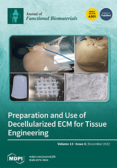
Cover Story (view full-size image): The extracellular matrix (ECM) is a crucial collection of proteins and other biomolecules which are highly conserved across evolution and, in the body, is approximately equal in mass to cells. The ECM is produced by cells and found in every tissue, where it provides cells with vital physical and biochemical cues that are crucial to the structure and function of tissues and organs. The ECM stimulates an anti-inflammatory state that promotes remodelling and repair. This makes the ECM an ideal biomaterial for tissue engineering, but first, it has to be decellularized through chemical and mechanical means. This review explores techniques to prepare and modify the ECM for tissue engineering applications, laboratory and clinical research with the ECM, and emerging trends and future directions for ECM research. View this paper
  • Issues are regarded as officially published after their release is announced to the table of contents alert mailing list.
  • You may sign up for e-mail alerts to receive table of contents of newly released issues.
  • PDF is the official format for papers published in both, html and pdf forms. To view the papers in pdf format, click on the "PDF Full-text" link, and use the free Adobe Reader to open them.
Order results
Result details
Section
Select all
Export citation of selected articles as:
4 pages, 203 KiB  
Editorial
Introduction to Problematics of the Special Issue “Green Biosynthesis of Nanomaterials for Biomedical Applications”
by Oleg V. Mikhailov and Ekaterina O. Mikhailova
J. Funct. Biomater. 2022, 13(4), 307; https://doi.org/10.3390/jfb13040307 - 19 Dec 2022
Cited by 2 | Viewed by 1142
Abstract
The concept of “green biosynthesis”, as well as the similar and more general concept of “green synthesis”, is collective and includes very diverse synthetic methods, using products vital for activity from various living organisms—of both plant and animal origin—for chemical compound production [...] [...] Read more.
The concept of “green biosynthesis”, as well as the similar and more general concept of “green synthesis”, is collective and includes very diverse synthetic methods, using products vital for activity from various living organisms—of both plant and animal origin—for chemical compound production [...] Full article
(This article belongs to the Special Issue Green Biosynthesis of Nanomaterials for Biomedical Applications)
19 pages, 7321 KiB  
Article
Nanohydroxyapatite/Titanate Nanotube Composites for Bone Tissue Regeneration
by Suziete B. S. Gusmão, Anupama Ghosh, Alan S. de Menezes, Antônio F. M. Pereira, Miriam T. P. Lopes, Madaline K. Souza, Dalton Dittz, Guilherme J. P. Abreu, Lucielma S. S. Pinto, Antônio L. M. Maia Filho, Gustavo O. M. Gusmão, Thomas J. Webster, Anderson O. Lobo and Bartolomeu C. Viana
J. Funct. Biomater. 2022, 13(4), 306; https://doi.org/10.3390/jfb13040306 - 17 Dec 2022
Cited by 2 | Viewed by 2130
Abstract
Strategies for the production of new nanocomposites that promote bone tissue regeneration are important, particularly those that enhance the osteoinduction of hydroxyapatite in situ. Here, we studied and report the synthesis of nanohydroxyapatite and titanate nanotube (nHAp/TiNT) composites formulated at different concentrations (1, [...] Read more.
Strategies for the production of new nanocomposites that promote bone tissue regeneration are important, particularly those that enhance the osteoinduction of hydroxyapatite in situ. Here, we studied and report the synthesis of nanohydroxyapatite and titanate nanotube (nHAp/TiNT) composites formulated at different concentrations (1, 2, 3, and 10 wt % TiNT) by means of a wet aqueous chemical reaction. The addition of TiNT affects the morphology of the nanocomposites, decreasing the average crystallite size from 54 nm (nHAp) to 34 nm (nHAp/TiNT10%), while confirming its interaction with the nanocomposite. The crystallinity index (CI) calculated by Raman spectroscopy and XRD showed that the values decreased according to the increase in TiNT concentration, which confirmed their addition to the structure of the nanocomposite. SEM images showed the presence of TiNTs in the nanocomposite. We further verified the potential cytotoxicity of murine fibroblast cell line L929, revealing that there was no remarkable cell death at any of the concentrations tested. In vivo regenerative activity was performed using oophorectomized animal (rat) models organized into seven groups containing five animals each over two experimental periods (15 and 30 days), with bone regeneration occurring in all groups tested within 30 days; however, the nHAp/TiNT10% group showed statistically greater tissue repair, compared to the untreated control group. Thus, the results of this study demonstrate that the presently formulated nHAp/TiNT nanocomposites are promising for numerous improved bone tissue regeneration applications. Full article
Show Figures

Graphical abstract

16 pages, 2807 KiB  
Article
Microbiological Properties and Cytotoxicity of PNVCL Hydrogels Containing Flavonoids as Intracanal Medication for Endodontic Therapy
by Gabriela Pacheco de Almeida Braga, Karina Sampaio Caiaffa, Jesse Augusto Pereira, Vanessa Rodrigues dos Santos, Amanda Caselato Andolfatto Souza, Lucas da Silva Ribeiro, Emerson Rodrigues Camargo, Anuradha Prakki and Cristiane Duque
J. Funct. Biomater. 2022, 13(4), 305; https://doi.org/10.3390/jfb13040305 - 17 Dec 2022
Cited by 1 | Viewed by 1559
Abstract
This study aimed to evaluate the cytotoxicity and microbiological properties of poly (N-vinylcaprolactam)—PNVCL hydrogels containing flavonoids as intracanal medication for endodontic therapy. Antimicrobial activity of ampelopsin (AMP), isoquercitrin and rutin was determined against Enterococcus faecalis, Actinomyces israelii, Lactobacillus casei, Streptococcus [...] Read more.
This study aimed to evaluate the cytotoxicity and microbiological properties of poly (N-vinylcaprolactam)—PNVCL hydrogels containing flavonoids as intracanal medication for endodontic therapy. Antimicrobial activity of ampelopsin (AMP), isoquercitrin and rutin was determined against Enterococcus faecalis, Actinomyces israelii, Lactobacillus casei, Streptococcus mutans, and Fusobacterium nucleatum by the microdilution method. After synthesis and characterization by rheology, PNVCL hydrogels were loaded with AMP and controls calcium hydroxide (CH) and chlorhexidine (CHX), and determined the compounds release profile. PNVCL+AMP, PNVCL+CH, PNVCL+CHX were evaluated on multi-species biofilms and analyzed by Scanning Electron Microscopy (SEM) and Confocal Laser Scanning Microscopy (CLSM). Cytotoxicity was determined after fibroblasts exposure to serial dilutions of AMP and PNVCL hydrogel extracts. AMP was effective against all of the bacteria tested, especially against S. mutans, A. israelli and F. nucleatum. SEM and CLSM analysis showed that PNVCL + AMP caused a significant decrease and disorganization of multi-species biofilms and reduction of intracanal viable cells, superior to the other groups. AMP affected fibroblast viability at concentrations above 0.125 mg/mL, and extracts of PNVCL+AMP showed low cytotoxicity. In conclusion, PNVCL containing AMP demonstrated cytocompatibility and potent effect against multi-species biofilms and could be potential intracanal medication for endodontic purposes. Full article
(This article belongs to the Special Issue Biomaterials in Restorative Dentistry and Endodontics)
Show Figures

Figure 1

13 pages, 893 KiB  
Review
Virus-like Particles for TEM Regulation and Antitumor Therapy
by Zhu Yang, Yongjie Chi, Jiaxin Bao, Xin Zhao, Jing Zhang and Lianyan Wang
J. Funct. Biomater. 2022, 13(4), 304; https://doi.org/10.3390/jfb13040304 - 17 Dec 2022
Cited by 2 | Viewed by 1806
Abstract
Tumor development and metastasis are intimately associated with the tumor microenvironment (TME), and it is difficult for vector-restricted drugs to act on the TME for long-term cancer immunotherapy. Virus-like particles (VLPs) are nanocage structures self-assembled from nucleic acid free viral proteins. Most VLPs [...] Read more.
Tumor development and metastasis are intimately associated with the tumor microenvironment (TME), and it is difficult for vector-restricted drugs to act on the TME for long-term cancer immunotherapy. Virus-like particles (VLPs) are nanocage structures self-assembled from nucleic acid free viral proteins. Most VLPs range from 20–200 nm in diameter and can naturally drain into lymph nodes to induce robust humoral immunity. As natural nucleic acid nanocarriers, their surfaces can also be genetically or chemically modified to achieve functions such as TME targeting. This review focuses on the design ideas of VLP as nanocarriers and the progress of their research in regulating TME. Full article
Show Figures

Graphical abstract

14 pages, 3349 KiB  
Article
Development and Characterization of Novel Orthodontic Adhesive Containing PCL–Gelatin–AgNPs Fibers
by Qihan Yuan, Qianqian Zhang, Xuecheng Xu, Yuqing Du, Jidong Xu, Yu Song and Yuanfei Wang
J. Funct. Biomater. 2022, 13(4), 303; https://doi.org/10.3390/jfb13040303 - 16 Dec 2022
Cited by 3 | Viewed by 1491
Abstract
Enamel demineralization around brackets is a relatively common complication of fixed orthodontic treatment, which seriously affects the aesthetics of teeth. In this study, a novel orthodontic adhesive containing polycaprolactone–gelatin–silver nanoparticles (PCL–gelatin–AgNPs) composite fibers was prepared to prevent enamel demineralization of orthodontic treatment. First, [...] Read more.
Enamel demineralization around brackets is a relatively common complication of fixed orthodontic treatment, which seriously affects the aesthetics of teeth. In this study, a novel orthodontic adhesive containing polycaprolactone–gelatin–silver nanoparticles (PCL–gelatin–AgNPs) composite fibers was prepared to prevent enamel demineralization of orthodontic treatment. First, PCL–gelatin–AgNPs fibers film prepared by electrospinning was made into short fibers and added to traditional orthodontic adhesives (Transbond XT, 3M Unitek) in three different ratios to design a series of composite adhesives containing antibacterial materials. The antimicrobial performance of the control product and the three samples were then evaluated by bacterial live/dead staining, colony-forming unit (CFU) counts, tensile bond strength (TBS), and adhesive residue index (ARI) scores. The composite adhesives’ antimicrobial properties increased with the increasing content of PCL–gelatin–AgNPs short fibers. The addition of complex antimicrobial fibers to 3M Transbond XT adhesive can significantly reduce the CFU of bacterial biofilms (p < 0.05). The bacterial survival rate on the surface of the specimen decreased with the increase of PCL–gelatin–AgNPs short fibers (p < 0.05). The TBS and ARI values (n = 10) indicated that adding PCL–gelatin–AgNPs short fibers had no significant adverse effect on adhesion. Therefore, adding PCL–gelatin–AgNPs short fibers makes it possible to fabricate orthodontic adhesives with strong antibacterial properties without compromising the bonding ability, which is essential for preventing enamel demineralization around the brackets. Full article
Show Figures

Graphical abstract

18 pages, 9219 KiB  
Article
Effect of Cell Geometry on the Mechanical Properties of 3D Voronoi Tessellation
by Zainab Alknery, Zhwan Dilshad Ibrahim Sktani and Ali Arab
J. Funct. Biomater. 2022, 13(4), 302; https://doi.org/10.3390/jfb13040302 - 16 Dec 2022
Cited by 1 | Viewed by 2749
Abstract
Irregular 3D biological scaffolds have been widely observed in nature. Therefore, in the current work, new designs are proposed for lightweight 3D scaffolds based on Voronoi tessellation with high porosity. The proposed designs are inspired by nature, which has undoubtedly proven to be [...] Read more.
Irregular 3D biological scaffolds have been widely observed in nature. Therefore, in the current work, new designs are proposed for lightweight 3D scaffolds based on Voronoi tessellation with high porosity. The proposed designs are inspired by nature, which has undoubtedly proven to be the best designer. Thus, the Rhinoceros 7/Grasshopper software was used to design three geometric models for both normal and elongated Voronoi structures: homogeneous, gradient I, and gradient II. Then, stereolithography (SLA) additive manufacturing was utilized to fabricate biopolymeric materials. Finally, a compression test was carried out to study and compare the mechanical properties of the designed samples. The gradient I cylinder show the highest Young’s modulus. For the Homogeneous and gradient II cylinders, elongated Voronoi structures show superior mechanical properties and energy absorption compared to normal Voronoi designs. Hence, these designs are promising topologies for future applications. Full article
Show Figures

Graphical abstract

12 pages, 2236 KiB  
Article
A High-Fidelity Artificial Urological System for the Quantitative Assessment of Endoscopic Skills
by Do Yeon Kim, Xiangzhou Tan, Moonkwang Jeong, Dandan Li, Arkadiusz Miernik and Tian Qiu
J. Funct. Biomater. 2022, 13(4), 301; https://doi.org/10.3390/jfb13040301 - 16 Dec 2022
Cited by 3 | Viewed by 1514
Abstract
Minimally-invasive surgery is rapidly growing and has become a standard approach for many operations. However, it requires intensive practice to achieve competency. The current training often relies on animal organ models or physical organ phantoms, which do not offer realistic surgical scenes or [...] Read more.
Minimally-invasive surgery is rapidly growing and has become a standard approach for many operations. However, it requires intensive practice to achieve competency. The current training often relies on animal organ models or physical organ phantoms, which do not offer realistic surgical scenes or useful real-time feedback for surgeons to improve their skills. Furthermore, the objective quantitative assessment of endoscopic skills is also lacking. Here, we report a high-fidelity artificial urological system that allows realistic simulation of endourological procedures and offers a quantitative assessment of the surgical performance. The physical organ model was fabricated by 3D printing and two-step polymer molding with the use of human CT data. The system resembles the human upper urinary tract with a high-resolution anatomical shape and vascular patterns. During surgical simulation, endoscopic videos are acquired and analyzed to quantitatively evaluate performance skills by a customized computer algorithm. Experimental results show significant differences in the performance between professional surgeons and trainees. The surgical simulator offers a unique chance to train endourological procedures in a realistic and safe environment, and it may also lead to a quantitative standard to evaluate endoscopic skills. Full article
(This article belongs to the Special Issue Advances and Challenges of Biomodels for Medical Applications)
Show Figures

Figure 1

25 pages, 10502 KiB  
Article
PVA-Based Nanofibers Containing Chitosan Modified with Graphene Oxide and Carbon Quantum Dot-Doped TiO2 Enhance Wound Healing in a Rat Model
by Fatemeh Norouzi, Mehrab Pourmadadi, Fatemeh Yazdian, Keyvan Khoshmaram, Javad Mohammadnejad, Mohammad Hossein Sanati, Faraz Chogan, Abbas Rahdar and Francesco Baino
J. Funct. Biomater. 2022, 13(4), 300; https://doi.org/10.3390/jfb13040300 - 15 Dec 2022
Cited by 32 | Viewed by 2820
Abstract
Electrospun nanofibrous constructs based on nanoparticles and biopolymers have recently been used in tissue engineering because of their similarity to the extracellular matrix in nature. In this study, electrospun chitosan-carbon quantum dot-titanium dioxide-graphene oxide (CS-CQD-TiO2-GO) nanofibrous mats were synthesized for use [...] Read more.
Electrospun nanofibrous constructs based on nanoparticles and biopolymers have recently been used in tissue engineering because of their similarity to the extracellular matrix in nature. In this study, electrospun chitosan-carbon quantum dot-titanium dioxide-graphene oxide (CS-CQD-TiO2-GO) nanofibrous mats were synthesized for use as wound dressings by the electrospinning method. To increase the biodegradation rate and water resistance, the fabricated nanofibrous mats were cross-linked. SEM images showed a uniform and coherent structure of CS-CQD-TiO2-GO nanocomposites and CS-CQD-TiO2-GO electrospun nanofibers mats. FTIR analysis, XRD pattern, SEM mapping, and EDS spectrum demonstrate the accuracy of the synthesis as well as the elemental and chemical structure of the nanofibrous mat. The water contact angle indicated that the nanofibrous mat had a hydrophilic property, which is essential for controlling wound exudates. The tensile strength and elongation tests showed that the nanofibrous mat has suitable mechanical properties for wound dressing, including significant flexibility and strength. Interestingly, antimicrobial testing illustrated that the fabricated nanofibrous mat had antibacterial activity against Gram-negative and Gram-positive bacteria. Appropriate cell viability and cytocompatibility of treated mouse fibroblast NIH3T3 cells with the nanofibrous mat were determined using an MTT assay. The animal study results confirmed the proper potential of the nanofibrous mat in wound dressing applications. Full article
(This article belongs to the Special Issue Fibrous Scaffolds for Tissue Engineering Application II)
Show Figures

Figure 1

15 pages, 6513 KiB  
Article
Antioxidant Stress of Transdermal Gene Delivery by Non-Viral Gene Vectors Based on Chitosan-Oligosaccharide
by Pengfei Cui, Ting Zhu, Pengju Jiang and Jianhao Wang
J. Funct. Biomater. 2022, 13(4), 299; https://doi.org/10.3390/jfb13040299 - 15 Dec 2022
Viewed by 1279
Abstract
Oxidative stress initiated by reactive oxygen species (ROS) is the cause of many acquired or congenital skin diseases. Oral antioxidants or using topical antioxidants preparations may bring the nonspecific distribution of drugs or anaphylaxis due to repeated use. In this study, a biocompatible [...] Read more.
Oxidative stress initiated by reactive oxygen species (ROS) is the cause of many acquired or congenital skin diseases. Oral antioxidants or using topical antioxidants preparations may bring the nonspecific distribution of drugs or anaphylaxis due to repeated use. In this study, a biocompatible gene vector by cross-linking of chitosan-oligosaccharide (CSO) and N,N’-cystamine-bis-acrylamide (CBA) was synthesized (CSO-CBA), which could carry therapeutic genes into the skin, express functional proteins in epidermal cells, and play an efficient antioxidant effect. Infrared and 1H NMR spectrum data showed that CSO-CBA was successfully synthesized. Gel electrophoresis results showed that the gene could be successfully compressed by the carrier and can be released in a reducing environment. Hemolysis experiments showed that the carrier had good biocompatibility. Transdermal gene delivery experiments proved that the vector can bring genes into the skin, express functional proteins, and protect the skin from reactive oxygen species damage after 7 days of administration. Skin compatibility experiments show that our therapy is biocompatible. Our study provides a minimally invasive and painless, high-biocompatibility, and long-term effective treatment for skin damage caused by reactive oxygen species, which has a potential application. Full article
(This article belongs to the Special Issue Biomedical Applications of Chitin and Chitosan-II)
Show Figures

Figure 1

16 pages, 3802 KiB  
Article
Degradable Pure Magnesium Used as a Barrier Film for Oral Bone Regeneration
by Xianfeng Shan, Yu Xu, Sharafadeen Kunle Kolawole, Ling Wen, Zhenwei Qi, Weiwei Xu and Junxiu Chen
J. Funct. Biomater. 2022, 13(4), 298; https://doi.org/10.3390/jfb13040298 - 15 Dec 2022
Cited by 7 | Viewed by 1536
Abstract
The barrier membrane plays an extremely critical role in guided bone regeneration (GBR), which determines the success or failure of GBR technology. In order to obtain barrier membranes with high mechanical strength and degradability, some researchers have focused on degradable magnesium alloys. However, [...] Read more.
The barrier membrane plays an extremely critical role in guided bone regeneration (GBR), which determines the success or failure of GBR technology. In order to obtain barrier membranes with high mechanical strength and degradability, some researchers have focused on degradable magnesium alloys. However, the degradation rate of pure Mg-based materials in body fluids is rather fast, thus posing an urgent problem to be solved in oral clinics. In this study, a novel micro-arc oxidation (MAO) surface-treated pure Mg membrane was prepared. Electrochemical tests, immersion experiments and in vivo experiments were carried out to investigate its potential use as a barrier membrane. The experimental results showed that the corrosion resistance of a pure Mg membrane treated by MAO is better than that of the uncoated pure Mg. The results of cell experiments showed no obvious cytotoxicity, which suggests the enhanced differentiation of osteoblasts. At the same time, the MAO-Mg membrane showed better biological activity than the pure Ti membrane in the early stage of implantation, exhibiting relatively good bone regeneration ability. Consequently, the MAO membrane has been proven to possess good application prospects for guided bone regeneration. Full article
(This article belongs to the Special Issue Advanced Biodegradable Biomaterials)
Show Figures

Figure 1

15 pages, 4490 KiB  
Article
Performance of Colombian Silk Fibroin Hydrogels for Hyaline Cartilage Tissue Engineering
by Augusto Zuluaga-Vélez, Carlos Andrés Toro-Acevedo, Adrián Quintero-Martinez, Jhon Jairo Melchor-Moncada, Francisco Pedraza-Ordoñez, Enrique Aguilar-Fernández and Juan Carlos Sepúlveda-Arias
J. Funct. Biomater. 2022, 13(4), 297; https://doi.org/10.3390/jfb13040297 - 14 Dec 2022
Viewed by 1484
Abstract
The development and evaluation of scaffolds play a crucial role in the engineering of hyaline cartilage tissue. This work aims to evaluate the performance of silk fibroin hydrogels fabricated from the cocoons of the Colombian hybrid in the in vitro regeneration of hyaline [...] Read more.
The development and evaluation of scaffolds play a crucial role in the engineering of hyaline cartilage tissue. This work aims to evaluate the performance of silk fibroin hydrogels fabricated from the cocoons of the Colombian hybrid in the in vitro regeneration of hyaline cartilage. The scaffolds were physicochemically characterized, and their performance was evaluated in a cellular model. The results showed that the scaffolds were rich in random coils and β-sheets in their structure and susceptible to various serine proteases with different degradation profiles. Furthermore, they showed a significant increase in ACAN, COL10A1, and COL2A1 expression compared to pellet culture alone and allowed GAG deposition. The soluble portion of the scaffold did not affect chondrogenesis. Furthermore, they promoted the increase in COL1A2, showing a slight tendency to differentiate towards fibrous cartilage. The results also showed that Colombian silk could be used as a source of biomedical devices, paving the way for sericulture to become a more diverse economic activity in emerging countries. Full article
Show Figures

Figure 1

15 pages, 4394 KiB  
Article
Surface Heparinization of a Magnesium-Based Alloy: A Comparison Study of Aminopropyltriethoxysilane (APTES) and Polyamidoamine (PAMAM) Dendrimers
by Masoumeh Ebrahimi, Atefeh Solouk, Ali Davoodi, Somaye Akbari, Masoumeh Haghbin Nazarpak and Alireza Nouri
J. Funct. Biomater. 2022, 13(4), 296; https://doi.org/10.3390/jfb13040296 - 13 Dec 2022
Cited by 2 | Viewed by 1183
Abstract
Magnesium (Mg)-based alloys are biodegradable metallic biomaterials that show promise in minimizing the risks of permanent metallic implants. However, their clinical applications are restricted due to their rapid in vivo degradation and low surface hemocompatibilities. Surface modifications are critically important for controlling the [...] Read more.
Magnesium (Mg)-based alloys are biodegradable metallic biomaterials that show promise in minimizing the risks of permanent metallic implants. However, their clinical applications are restricted due to their rapid in vivo degradation and low surface hemocompatibilities. Surface modifications are critically important for controlling the corrosion rates of Mg-based alloys and improving their hemocompatibilities. In the present study, two heparinization methods were developed to simultaneously increase the corrosion resistance and hemocompatibility of the AZ31 Mg alloy. In the first method, the surface of the AZ31 alloy was modified by alkali–heat treatment and then aminolyzed by 3-amino propyltriethoxy silane (APTES), a self-assembly molecule, and heparin was grafted onto the aminolyzed surface. In the second method, before heparinization, polyamidoamine dendrimers (PAMAM4-4) were grafted onto the aminolyzed surface with APTES to increase the number of surface functional groups, and heparinization was subsequently performed. The presence of a peak with a wavelength of about 1560 cm−1 in the FTIR spectrum for the sample modified with APTES and dendrimers indicated aminolysis of the surface. The results indicated that the corrosion resistance of the Mg alloy was significantly improved as a result of the formation of a passive layer following the alkali–heat treatment. The results obtained from a potentiodynamic polarization (PDP) test showed that the corrosion current in the uncoated sample decreased from 25 µA to 3.7 µA in the alkali–heat-treated sample. The corrosion current density was reduced by 14 and 50 times in samples treated with the self-assembly molecules, APTES and dendrimers, respectively. After heparinization, the clotting time for pristine Mg was greatly improved. Clotting time increased from 480 s for the pristine Mg sample to 630 s for the APTES- and heparin-modified samples and to 715 s for the PAMAM- and heparin-modified samples. Cell culture data showed a slight improvement in the cell-supporting behavior of the modified samples. Full article
Show Figures

Figure 1

21 pages, 6015 KiB  
Article
Carbopol Based Hydrogels for ITOPRIDE Hydrochloride Delivery; Synthesis, Characterization and Comparative Assessment with Various Monomers
by Muhammad Sarfraz, Rabia Iqbal, Kifayat Ullah Khan and Muhammad Usman Minhas
J. Funct. Biomater. 2022, 13(4), 295; https://doi.org/10.3390/jfb13040295 - 12 Dec 2022
Cited by 4 | Viewed by 1966
Abstract
The objective of the current study was to synthesize and characterize carbopol containing hydrogels with different monomers such as methacrylic acid (MAA), 2-acrylamido-2-methylpropane sulfonic acid (AMPS) and itaconic acid (ITA). Free radical polymerization method was optimized for the preparation of different formulations using [...] Read more.
The objective of the current study was to synthesize and characterize carbopol containing hydrogels with different monomers such as methacrylic acid (MAA), 2-acrylamido-2-methylpropane sulfonic acid (AMPS) and itaconic acid (ITA). Free radical polymerization method was optimized for the preparation of different formulations using N,N-methylene bis-acrylamide (MBA) as cross linking agent. Different studies were performed to evaluate the effect of different monomers on swelling, drug loading and drug release. Itopride Hydrochloride was used as model drug. FTIR, TGA, DSC and SEM were performed to probe the characteristics of fabricated hydrogels. Swelling studies of different fabricated hydrogels were performed in three pH conditions (1.2, 4.5 & 6.8). Higher swelling was observed at pH 6.8. An in-vitro release study was performed on pH 1.2 and 6.8. The synthesized hydrogels exhibited excellent mechanical strength, higher drug loading, pH sensitive and time dependent release up to 30 h. The excellent mechanical strength and extended drug release of Carbopol-co-poly-MAA-ITA hydrogels make them a potential candidate for controlled delivery of Itopride hydrochloride. Full article
(This article belongs to the Special Issue Biopolymer-Based Hydrogel Materials: Opportunities and Challenges)
Show Figures

Figure 1

16 pages, 3482 KiB  
Article
Corrosion Behavior and Biocompatibility of Hot-Extruded Mg–Zn–Ga–(Y) Biodegradable Alloys
by Viacheslav Bazhenov, Anna Li, Artem Iliasov, Vasily Bautin, Sofia Plegunova, Andrey Koltygin, Alexander Komissarov, Maxim Abakumov, Nikolay Redko and Kwang Seon Shin
J. Funct. Biomater. 2022, 13(4), 294; https://doi.org/10.3390/jfb13040294 - 12 Dec 2022
Cited by 6 | Viewed by 1581
Abstract
Fixation screws and other temporary magnesium alloy fixation devices are used in orthopedic practice because of their biodegradability, biocompatibility and acceptable biodegradation rates. The substitution of dissolving implant by tissues during the healing process is one of the main requirements for biodegradable implants. [...] Read more.
Fixation screws and other temporary magnesium alloy fixation devices are used in orthopedic practice because of their biodegradability, biocompatibility and acceptable biodegradation rates. The substitution of dissolving implant by tissues during the healing process is one of the main requirements for biodegradable implants. Previously, clinical tests showed the effectiveness of Ga ions on bone tissue regeneration. This work is the first systematic study on the corrosion rate and biocompatibility of Mg–Zn–Ga–(Y) alloys prepared by hot extrusion, where Ga is an additional major alloying element, efficient as a bone-resorption inhibitor. Most investigated alloys have a low corrosion rate in Hanks’ solution close to ~0.2 mm/year. No cytotoxic effects of Mg–2Zn–2Ga (wt.%) alloy on MG63 cells were observed. Thus, considering the high corrosion resistance and good biocompatibility, the Mg–2Zn–2Ga alloy is possible for applications in osteosynthesis implants with improved bone tissue regeneration ability. Full article
(This article belongs to the Special Issue Metallic Biomaterials for Medical Applications)
Show Figures

Figure 1

12 pages, 1962 KiB  
Article
Differential Foreign Body Reactions between Branched and Linear Glucomannan Scaffolds
by Yuwei Li, Yu Liu, Senio Campos de Souza, Tzuwei Chao, Lei Dong, Guoxing Sun, Chunming Wang and Yiming Niu
J. Funct. Biomater. 2022, 13(4), 293; https://doi.org/10.3390/jfb13040293 - 11 Dec 2022
Viewed by 1373
Abstract
The extent and patterns of foreign body reaction (FBR) influence the function and feasibility of biomaterials. Polysaccharides, as an important biomaterial category, have received increasing attention in diverse biomaterials design and biomedical applications due to their excellent polymeric and biocompatible characteristics. Their biological [...] Read more.
The extent and patterns of foreign body reaction (FBR) influence the function and feasibility of biomaterials. Polysaccharides, as an important biomaterial category, have received increasing attention in diverse biomaterials design and biomedical applications due to their excellent polymeric and biocompatible characteristics. Their biological effects are usually associated with their monosaccharide composition or functional groups, yet the contribution of their glycan structure is still unknown. Herein, two glucomannans, similar in composition and molecular weight with differences in glycan structure, linear-chain (Konjac glucomannan, KGM), and branched-chain (Bletilla striata polysaccharide, BSP), were adopted to explore the host–biomaterials interaction. After acetyl modification, these polysaccharides were fabricated into electrospun scaffolds to reduce the impacts derived from the physical properties and surface morphology. According to a systematic study of their biological effects on immune cells and host response in a subcutaneous implantation model in vivo, it was revealed that acetyl KGM (acKGM) scaffolds caused a stronger FBR than acetyl BSP materials. Additionally, acKGM could stimulate macrophages to release pro-inflammatory cytokines, suggesting the influence of sugar chain arrangement on FBR and providing clues for the fine regulation of immune response and novel biomaterials design. Full article
Show Figures

Figure 1

17 pages, 3863 KiB  
Article
Accuracy and Technical Predictability of Computer Guided Bone Harvesting from the Mandible: A Cone-Beam CT Analysis in 22 Consecutive Patients
by Luca De Stavola, Alessandro Cristoforetti, Andrea Fincato, Giandomenico Nollo, Paolo Ghensi, Anna Cantarutti and Francesco Tessarolo
J. Funct. Biomater. 2022, 13(4), 292; https://doi.org/10.3390/jfb13040292 - 10 Dec 2022
Cited by 2 | Viewed by 1559
Abstract
This study assesses the accuracy and technical predictability of a computer-guided procedure for harvesting bone from the external oblique ridge using a patient-specific cutting guide. Twenty-two patients needing bone augmentation for implant placement were subjected to mandibular osteotomy employing a case-specific stereolithographic surgical [...] Read more.
This study assesses the accuracy and technical predictability of a computer-guided procedure for harvesting bone from the external oblique ridge using a patient-specific cutting guide. Twenty-two patients needing bone augmentation for implant placement were subjected to mandibular osteotomy employing a case-specific stereolithographic surgical guide generated through computer aided design. Differences between planned and real cut planes were measured comparing pre- and post-operative Cone Beam Computed Tomography images of the donor site according to six validated angular and displacement indexes. Accuracy and technical predictability were assessed for 119 osteotomy planes over the study population. Three different guide fitting approaches were compared. An average root-mean-square discrepancy of 0.52 (0.30–0.97) mm was detected. The accuracy of apical and medial planes was higher than the mesial and distal planes due to occasional antero-posterior guide shift. Fitting the guide with an extra reference point on the closest tooth performed better than using only the bone surface, with two indexes significantly lower and less disperse. The study showed that the surgical plan was actualized with a 1 mm safety margin, allowing effective nerve preservation and reducing technical variability. When possible, surgical guide design should allow fitting on the closest tooth based on both radiological and/or intra-oral scan data. Full article
(This article belongs to the Special Issue Biomaterials and Bioengineering in Dentistry)
Show Figures

Figure 1

16 pages, 8180 KiB  
Review
Biomaterials and Clinical Applications of Customized Healing Abutment—A Narrative Review
by Parima Chokaree, Pongsakorn Poovarodom, Pisaisit Chaijareenont, Apichai Yavirach and Pimduen Rungsiyakull
J. Funct. Biomater. 2022, 13(4), 291; https://doi.org/10.3390/jfb13040291 - 10 Dec 2022
Cited by 15 | Viewed by 7806
Abstract
Customized healing abutments have been introduced in clinical practice along with implant surgery to preserve or create natural-appearing hard and soft tissue around the implant. This provides the benefits of reducing the overall treatment time by eliminating the second stage and reducing the [...] Read more.
Customized healing abutments have been introduced in clinical practice along with implant surgery to preserve or create natural-appearing hard and soft tissue around the implant. This provides the benefits of reducing the overall treatment time by eliminating the second stage and reducing the elapsed time of the fabrication of the final prostheses. This article aims to review the types and properties of materials used for the fabrication of customized healing abutments and their clinical applications. Articles published in English on customized healing abutments were searched in Google Scholar, PubMed/MEDLINE, ScienceDirect, and the Scopus databases up to August 2022. The relevant articles were selected and included in this literature review. Customized healing abutments can be fabricated from materials available for dental implants, including PEEK, PMMA, zirconia, resin composite, and titanium. All the materials can be used following both immediate and delayed implant placement. Each material provides different mechanical and biological properties that influence the peri-implant tissues. In conclusion, the studies have demonstrated promising outcomes for all the materials. However, further investigation comparing the effects of each material on peri-implant soft and hard tissues is required. Full article
(This article belongs to the Section Dental Biomaterials)
Show Figures

Figure 1

17 pages, 4180 KiB  
Article
Experimental Study of the Propagation Process of Dissection Using an Aortic Silicone Phantom
by Qing-Zhuo Chi, Yang-Yang Ge, Zhen Cao, Li-Li Long, Li-Zhong Mu, Ying He and Yong Luan
J. Funct. Biomater. 2022, 13(4), 290; https://doi.org/10.3390/jfb13040290 - 09 Dec 2022
Cited by 2 | Viewed by 1401
Abstract
Background: The mortality of acute aortic dissection (AD) can reach 65~70%. However, it is challenging to follow the progress of AD formation. The purpose of this work was to observe the process of dissection development using a novel tear-embedded silicone phantom. Methods: Silicone [...] Read more.
Background: The mortality of acute aortic dissection (AD) can reach 65~70%. However, it is challenging to follow the progress of AD formation. The purpose of this work was to observe the process of dissection development using a novel tear-embedded silicone phantom. Methods: Silicone phantoms were fabricated by embedding a torn area and primary tear feature on the inner layer. CT scanning and laser lightening were conducted to observe the variations in thickness and volume of the true lumen (TL) and false lumen (FL) during development. Results: The model with a larger interlayer adhesion damage required a lower pressure to trigger the development of dissection. At the initiation stage of dissection, the volume of TL increased by 25.5%, accompanied by a 19.5% enlargement of tear size. The force analysis based on the change of tear size verified the deduction of the process of interlaminar separation from the earlier studies. Conclusions: The primary tear and the weakening adhesion of the vessel layers are key factors in AD development, suggesting that some forms of primary damage to the arterial wall, in particular, the lumen morphology of vessels with straight inner lumen, should be considered as early risk predictors of AD. Full article
Show Figures

Figure 1

30 pages, 4059 KiB  
Review
Progress of Electrospun Nanofibrous Carriers for Modifications to Drug Release Profiles
by Ying Wang, Deng-Guang Yu, Yang Liu and Ya-Nan Liu
J. Funct. Biomater. 2022, 13(4), 289; https://doi.org/10.3390/jfb13040289 - 09 Dec 2022
Cited by 70 | Viewed by 4603
Abstract
Electrospinning is an advanced technology for the preparation of drug-carrying nanofibers that has demonstrated great advantages in the biomedical field. Electrospun nanofiber membranes are widely used in the field of drug administration due to their advantages such as their large specific surface area [...] Read more.
Electrospinning is an advanced technology for the preparation of drug-carrying nanofibers that has demonstrated great advantages in the biomedical field. Electrospun nanofiber membranes are widely used in the field of drug administration due to their advantages such as their large specific surface area and similarity to the extracellular matrix. Different electrospinning technologies can be used to prepare nanofibers of different structures, such as those with a monolithic structure, a core–shell structure, a Janus structure, or a porous structure. It is also possible to prepare nanofibers with different controlled-release functions, such as sustained release, delayed release, biphasic release, and targeted release. This paper elaborates on the preparation of drug-loaded nanofibers using various electrospinning technologies and concludes the mechanisms behind the controlled release of drugs. Full article
Show Figures

Figure 1

14 pages, 6788 KiB  
Article
Adhesive Property of 3D-Printed PEEK Abutments: Effects of Surface Treatment and Temporary Crown Material on Shear Bond Strength
by Dingjie Wang, Xingting Han, Feng Luo, Florian M. Thieringer, Yichen Xu, Guomin Ou and Sebastian Spintzyk
J. Funct. Biomater. 2022, 13(4), 288; https://doi.org/10.3390/jfb13040288 - 09 Dec 2022
Cited by 2 | Viewed by 1974
Abstract
Three-dimensionally printed polyetheretherketone (PEEK) materials are promising for fabricating customized dental abutments. This study aimed to investigate the adhesive property of a 3D-printed PEEK material. The effects of surface treatment and temporary crown materials on shear bond strength were evaluated. A total of [...] Read more.
Three-dimensionally printed polyetheretherketone (PEEK) materials are promising for fabricating customized dental abutments. This study aimed to investigate the adhesive property of a 3D-printed PEEK material. The effects of surface treatment and temporary crown materials on shear bond strength were evaluated. A total of 108 PEEK discs were 3D printed by fused-filament fabrication. Surface treatments, including sandblasting, abrasive paper grinding, and CO2 laser ablation, were applied to the PEEK discs, with the untreated specimens set as the control. Afterward, the surface topographies of each group were investigated by scanning electron microscopy (SEM, n = 1) and roughness measurements (n = 7). After preparing the bonding specimens with three temporary crown materials (Artificial teeth resin (ATR), 3M™ Filtek™ Supreme Flowable Restorative (FR), and Cool Temp NATURAL (CTN)), the shear bond strength was measured (n = 6), and the failure modes were analyzed by microscopy and SEM. The results showed that ATR exhibited a significantly higher shear bond strength compared to FR and CTN (p < 0.01), and the PEEK surfaces treated by sandblasting and abrasive paper grinding showed a statistically higher shear bond strength compared to the control (p < 0.05). For clinical application, the ATR material and subtractive surface treatments are recommended for 3D-printed PEEK abutments. Full article
Show Figures

Figure 1

20 pages, 978 KiB  
Review
Application of Tribology Concept in Dental Composites Field: A Scoping Review
by Giusy Rita Maria La Rosa, Luigi Generali, Calogero Bugea, Hani F. Ounsi, Gergely Benyőcs, Prasanna Neelakantan and Eugenio Pedullà
J. Funct. Biomater. 2022, 13(4), 287; https://doi.org/10.3390/jfb13040287 - 08 Dec 2022
Cited by 3 | Viewed by 1889
Abstract
Tribology is the discipline concerning the application of friction, lubrication, and wear concepts of interacting surfaces in relative motion. A growing interest has developed in tribology application in medical biomaterials, such as resin composites used in restorative dentistry. Yet, the keywords “tribology” and [...] Read more.
Tribology is the discipline concerning the application of friction, lubrication, and wear concepts of interacting surfaces in relative motion. A growing interest has developed in tribology application in medical biomaterials, such as resin composites used in restorative dentistry. Yet, the keywords “tribology” and “biotribology” are little applied in the pertinent publications. The aim of this scoping review was to offer an overview of tribology application in dental composites research and to identify knowledge gaps and address future research. A literature search was conducted on Pubmed and Scopus databases and the studies investigating the tribological behavior of resin composites were included for qualitative synthesis. The majority of studies on dental tribology were published in the research areas of mechanical engineering/nanotechnology and differed in several methodological aspects. The preponderant engineering approach and the lack of standardized testing make the laboratory findings poorly informative for clinicians. Future research should focus on the tribological behavior of dental materials composites by means of an integrated approach, i.e., engineering and clinical, for improving development and advancement in this field of research. Full article
(This article belongs to the Special Issue Tribology for Biomedical Applications)
Show Figures

Figure 1

13 pages, 2786 KiB  
Article
Biomechanical Testing of Two-Unit Bridges and a Comparison of Replacement Retention Depending on a Cementation Medium, Replacement Position, and Gap Size
by Alena Findrik Balogová, Viktória Rajťúková, Ľuboš Chromý, Andrej Somoš, Gabriela Ižaríková and Radovan Hudák
J. Funct. Biomater. 2022, 13(4), 286; https://doi.org/10.3390/jfb13040286 - 08 Dec 2022
Cited by 1 | Viewed by 1094
Abstract
Dental replacements are placed between the abutment teeth. The exceptions are two-unit bridges, as they are supported by a single tooth prepared only on one side of the missing tooth. The presented study deals with an analysis of a pressure force action on [...] Read more.
Dental replacements are placed between the abutment teeth. The exceptions are two-unit bridges, as they are supported by a single tooth prepared only on one side of the missing tooth. The presented study deals with an analysis of a pressure force action on two-unit bridges placed in the frontal part (20 samples), where the pressure action is lower, and in the distal part (20 samples), where the pressure action is higher. A CAD program by 3Shape was used for digital designing with two different gap settings, 10 μm (20 samples) and 30 μm (20 samples). Two-unit bridges were attached to the prepared tooth using two types of dental cement (20/20 samples), which were selected for their physical and bioactive properties. All two-unit bridges (a total of 80 samples) were fabricated from CoCr alloys on Mlab cusing R by applying the Selective Laser Melting (SLM) technology. Mechanical testing was performed using the Inspekt5 table blue. The obtained data were used to verify the hypotheses—a difference between both types of cement (A ≠ B), a difference between the frontal and distal two-unit bridges (F ≠ D) and a difference between the gap sizes (10 ≠ 30). To confirm the given theories, data were statistically evaluated using the F-test and subsequent t-tests. The resulting p-value was compared with the level of significance (α = 0.05). A statistical evaluation revealed a significant difference between the compared groups; however, no explicit correlation between the individual groups of specimens was identified. Full article
Show Figures

Graphical abstract

22 pages, 15639 KiB  
Article
Surface Modification of Additively Fabricated Titanium-Based Implants by Means of Bioactive Micro-Arc Oxidation Coatings for Bone Replacement
by Anna I. Kozelskaya, Sven Rutkowski, Johannes Frueh, Aleksey S. Gogolev, Sergei G. Chistyakov, Sergey V. Gnedenkov, Sergey L. Sinebryukhov, Andreas Frueh, Vladimir S. Egorkin, Evgeny L. Choynzonov, Mikhail Buldakov, Denis E. Kulbakin, Evgeny N. Bolbasov, Anton P. Gryaznov, Ksenia N. Verzunova, Margarita D. Apostolova and Sergei I. Tverdokhlebov
J. Funct. Biomater. 2022, 13(4), 285; https://doi.org/10.3390/jfb13040285 - 08 Dec 2022
Cited by 20 | Viewed by 2595
Abstract
In this work, the micro-arc oxidation method is used to fabricate surface-modified complex-structured titanium implant coatings to improve biocompatibility. Depending on the utilized electrolyte solution and micro-arc oxidation process parameters, three different types of coatings (one of them—oxide, another two—calcium phosphates) were obtained, [...] Read more.
In this work, the micro-arc oxidation method is used to fabricate surface-modified complex-structured titanium implant coatings to improve biocompatibility. Depending on the utilized electrolyte solution and micro-arc oxidation process parameters, three different types of coatings (one of them—oxide, another two—calcium phosphates) were obtained, differing in their coating thickness, crystallite phase composition and, thus, with a significantly different biocompatibility. An analytical approach based on X-ray computed tomography utilizing software-aided coating recognition is employed in this work to reveal their structural uniformity. Electrochemical studies prove that the coatings exhibit varying levels of corrosion protection. In vitro and in vivo experiments of the three different micro-arc oxidation coatings prove high biocompatibility towards adult stem cells (investigation of cell adhesion, proliferation and osteogenic differentiation), as well as in vivo biocompatibility (including histological analysis). These results demonstrate superior biological properties compared to unmodified titanium surfaces. The ratio of calcium and phosphorus in coatings, as well as their phase composition, have a great influence on the biological response of the coatings. Full article
(This article belongs to the Special Issue Biomaterials for Drug Release and Tissue Engineering)
Show Figures

Figure 1

12 pages, 4545 KiB  
Article
Characterization of Bioactive Compounds Having Antioxidant and Anti-Inflammatory Effects of Liliaceae Family Flower Petal Extracts
by Neha Kaushik, June-Hyun Kim, Linh Nhat Nguyen, Nagendra Kumar Kaushik and Kyung-A Choi
J. Funct. Biomater. 2022, 13(4), 284; https://doi.org/10.3390/jfb13040284 - 08 Dec 2022
Cited by 2 | Viewed by 3138
Abstract
Beneficial natural products utilized in cosmetics formulation and pharmaceutical applications are of enormous interest. Lily (Lilium) serves as an essential edible and medicinal plant species with wide classification. Here, we have performed the screening of various extracts that were prepared from flower petals [...] Read more.
Beneficial natural products utilized in cosmetics formulation and pharmaceutical applications are of enormous interest. Lily (Lilium) serves as an essential edible and medicinal plant species with wide classification. Here, we have performed the screening of various extracts that were prepared from flower petals grown from the bulbs of eight Lilium varieties, with a viewpoint to their applicability as a viable source of natural anti-inflammatory and antioxidants agent. Interestingly, our findings indicated that all ethanol and water extracts exhibited a substantially differential spectrum of antioxidant as well as anti-inflammatory properties. Specifically, Serrano showed a close similarity among ethanol and water extracts among all tested lily petal extracts. Therefore, to obtain a detailed analysis of chemical compounds, liquid chromatography–mass spectroscopy was performed in ethanolic and water extracts of Serrano petals. Together, our preliminary results indicated that lily petals extracts used in this study could serve as a basis to develop a potential new whitening agent with powerful antioxidant and anti-inflammatory properties for medicinal, functional food, and cosmetic applications. Full article
Show Figures

Figure 1

15 pages, 4470 KiB  
Article
Quasi-Static Mechanical Properties and Continuum Constitutive Model of the Thyroid Gland
by Peng Su, Chao Yue, Likun Cui, Qinjian Zhang, Baoguo Liu and Tian Liu
J. Funct. Biomater. 2022, 13(4), 283; https://doi.org/10.3390/jfb13040283 - 08 Dec 2022
Viewed by 1164
Abstract
The purpose of this study is to obtain the digital twin parameters of the thyroid gland and to build a constitutional model of the thyroid gland based on continuum mechanics, which will lay the foundation for the establishment of a surgical training system [...] Read more.
The purpose of this study is to obtain the digital twin parameters of the thyroid gland and to build a constitutional model of the thyroid gland based on continuum mechanics, which will lay the foundation for the establishment of a surgical training system for the thyroid surgery robot and the development of the digital twin of the thyroid gland. First, thyroid parenchyma was obtained from fresh porcine thyroid tissue and subjected to quasi-static unconfined uniaxial compression tests using a biomechanical test platform with two strain rates (0.005 s−1 and 0.05 s−1) and two loading orientations (perpendicular to the thyroid surface and parallel to the thyroid surface). Based on this, a tensile thyroid model was established to simulate the stretching process by using the finite element method. The thyroid stretching test was carried out under the same parameters to verify the validity of the hyperelastic constitutive model. The quasi-static mechanical property parameters of the thyroid tissue were obtained by a quasi-static unconstrained uniaxial compression test, and a constitutional model that can describe the quasi-static mechanical properties of thyroid tissue was proposed based on the principle of continuum media mechanics, which is of great value for the establishment of a surgical training system for the head and neck surgery robot and for the development of the thyroid digital twin. Full article
Show Figures

Figure 1

12 pages, 2710 KiB  
Article
Investigation of Cell Adhesion and Cell Viability of the Endothelial and Fibroblast Cells on Electrospun PCL, PLGA and Coaxial Scaffolds for Production of Tissue Engineered Blood Vessel
by Morteza Bazgir, Morvarid Saeinasab, Wei Zhang, Ximu Zhang, Ka Min Tsui, Abolfazl Maasoumi Sarvestani, Subhaan Nawaz, Phil Coates, Mansour Youseffi, Jacobo Elies and Farshid Sefat
J. Funct. Biomater. 2022, 13(4), 282; https://doi.org/10.3390/jfb13040282 - 08 Dec 2022
Cited by 7 | Viewed by 1924
Abstract
Endothelialization of artificial scaffolds is considered an effective strategy for increasing the efficiency of vascular transplantation. This study aimed to compare the biophysical/biocompatible properties of three different biodegradable fibrous scaffolds: Poly (ɛ-caprolactone) (PCL) alone, Poly Lactic-co-Glycolic Acid (PLGA) alone (both processed using Spraybase [...] Read more.
Endothelialization of artificial scaffolds is considered an effective strategy for increasing the efficiency of vascular transplantation. This study aimed to compare the biophysical/biocompatible properties of three different biodegradable fibrous scaffolds: Poly (ɛ-caprolactone) (PCL) alone, Poly Lactic-co-Glycolic Acid (PLGA) alone (both processed using Spraybase® electrospinning machine), and Coaxial scaffold where the fiber core and sheath was made of PCL and PLGA, respectively. Scaffold structural morphology was assessed by scanning electron microscope and tensile testing was used to investigate the scaffold tension resistance over time. Biocompatibility studies were carried out with human umbilical vein endothelial cells (HUVEC) and human vascular fibroblasts (HVF) for which cell viability (and cell proliferation over a 4-day period) and cell adhesion to the scaffolds were assessed by cytotoxicity assays and confocal microscopy, respectively. Our results showed that all biodegradable polymeric scaffolds are a reliable host to adhere and promote proliferation in HUVEC and HVF cells. In particular, PLGA membranes performed much better adhesion and enhanced cell proliferation compared to control in the absence of polymers. In addition, we demonstrate here that these biodegradable membranes present improved mechanical properties to construct potential tissue-engineered vascular graft. Full article
Show Figures

Figure 1

10 pages, 14129 KiB  
Article
Comparison of Regular and Speed Sintering on Low-Temperature Degradation and Fatigue Resistance of Translucent Zirconia Crowns for Implants: An In Vitro Study
by Suchada Kongkiatkamon and Chaimongkon Peampring
J. Funct. Biomater. 2022, 13(4), 281; https://doi.org/10.3390/jfb13040281 - 08 Dec 2022
Cited by 4 | Viewed by 1539
Abstract
Background: Although there are a few studies which compare fast and slow sintering in normal zirconia crowns, it is essential to compare the cracks and load-bearing capacity in zirconia screw-retained implant crowns between regular and speed sintering protocols. This research aimed to compare [...] Read more.
Background: Although there are a few studies which compare fast and slow sintering in normal zirconia crowns, it is essential to compare the cracks and load-bearing capacity in zirconia screw-retained implant crowns between regular and speed sintering protocols. This research aimed to compare the surface structure, cracks, and load-bearing capacity in zirconia screw-retained implant crowns between regular sintering (RS) and speed sintering (SS) protocol with and without cyclic loading (fatigue). Methods: A total of 60 screw-retained crowns were fabricated from zirconia (Katana STML Block) by the CAD/CAM system. Then, 30 crowns were subjected to the RS protocol and 30 crowns were subjected to the SS protocol. Cyclic loading was done in half zirconia crowns (15 crowns in each group) using a chewing simulator CS-4.8/CS-4.4 at room temperature. The loading force was applied on the middle of the crowns by a metal stylus underwater at room temperature with a chewing simulator at an axial 50 N load for 240,000 cycles and lateral movement at 2 mm. Scanning electron microscopy was done to study the surface of the crowns and the cracks in the crowns of the regular and speed sintering protocols, with and without fatigue. Results: For the speed sintering group, the surface looks more uniform, and the crack lines are present at a short distance compared to regular sintering. The sintering protocol with a larger Weibull module and durability increases the reliability. It showed that the Speed group showed the maximum fracture load, followed by the regular, speed fatigue, and regular fatigue groups. The fracture load in various groups showed significant differences. Conclusions: It was found that the speed group showed the maximum fracture load followed by the regular, speed fatigue, and regular fatigue. The crack lines ran from occlusal to bottoms (gingiva) and the arrest lines were perpendicular to the crack propagations. Full article
(This article belongs to the Special Issue Recent Advances in Dental Biomaterials)
Show Figures

Figure 1

15 pages, 3339 KiB  
Article
Polycaprolactone Electrospun Nanofiber Membrane with Sustained Chlorohexidine Release Capability against Oral Pathogens
by Zi-Jian Chen, Jia-Cheng Lv, Zhi-Guo Wang, Fei-Yu Wang, Ren-Huan Huang, Zi-Li Zheng, Jia-Zhuang Xu and Jing Wang
J. Funct. Biomater. 2022, 13(4), 280; https://doi.org/10.3390/jfb13040280 - 07 Dec 2022
Cited by 3 | Viewed by 1603
Abstract
Multiple-pathogen periodontal disease necessitates a local release and concentration of antibacterial medication to control inflammation in a particular location of the mouth cavity. Therefore, it is necessary to effectively load and deliver medicine/antibiotics to treat numerous complex bacterial infections. This study developed chlorhexidine [...] Read more.
Multiple-pathogen periodontal disease necessitates a local release and concentration of antibacterial medication to control inflammation in a particular location of the mouth cavity. Therefore, it is necessary to effectively load and deliver medicine/antibiotics to treat numerous complex bacterial infections. This study developed chlorhexidine (CHX)/polycaprolactone (PCL) nanofiber membranes with controlled release properties as periodontal dressings to prevent or treat oral disorders. Electrostatic spinning was adopted to endow the nanofiber membranes with a high porosity, hydrophilicity, and CHX loading capability. The release of CHX occurred in a concentration-dependent manner. The CHX/PCL nanofiber membranes exhibited good biocompatibility with human periodontal ligament stem cells, with cell viability over 85% in each group via CCK-8 assay and LIVE/DEAD staining; moreover, the good attachment of the membrane was illustrated by scanning electron microscopy imaging. Through the agar diffusion assay, the nanofiber membranes with only 0.075 wt% CHX exhibited high antibacterial activity against three typical oral infection-causing bacteria: Porphyromonas gingivalis, Enterococcus faecalis, and Prevotella intermedia. The results indicated that the CHX/PCL nanofiber holds great potential as a periodontal dressing for the prevention and treatment periodontal disorders associated with bacteria. Full article
(This article belongs to the Section Dental Biomaterials)
Show Figures

Figure 1

15 pages, 3580 KiB  
Article
Preparation and Characterization of Extracellular Matrix Hydrogels Derived from Acellular Cartilage Tissue
by Tsong-Hann Yu, Tsu-Te Yeh, Chen-Ying Su, Ni-Yin Yu, I-Cheng Chen and Hsu-Wei Fang
J. Funct. Biomater. 2022, 13(4), 279; https://doi.org/10.3390/jfb13040279 - 07 Dec 2022
Cited by 3 | Viewed by 2331
Abstract
Decellularized matrices can effectively reduce severe immune rejection with their cells and eliminated nucleic acid material and provide specific environments for tissue repair or tissue regeneration. In this study, we prepared acellular cartilage matrix (ACM) powder through the decellularization method and developed ACM [...] Read more.
Decellularized matrices can effectively reduce severe immune rejection with their cells and eliminated nucleic acid material and provide specific environments for tissue repair or tissue regeneration. In this study, we prepared acellular cartilage matrix (ACM) powder through the decellularization method and developed ACM hydrogels by physical, chemical, and enzymatic digestion methods. The results demonstrated that the small size group of ACM hydrogels exhibited better gel conditions when the concentration of ACM hydrogels was 30 and 20 mg/mL in 1N HCl through parameter adjustment. The data also confirmed that the ACM hydrogels retained the main components of cartilage: 61.18% of glycosaminoglycan (GAG) and 78.29% of collagen, with 99.61% of its DNA removed compared to samples without the decellularization procedure (set as 100%). Through turbidimetric gelation kinetics, hydrogel rheological property analysis, and hydrogel tissue physical property testing, this study also revealed that increasing hydrogel concentration is helpful for gelation. Besides, the ex vivo test confirmed that a higher concentration of ACM hydrogels had good adhesive properties and could fill in cartilage defects adequately. This study offers useful information for developing and manufacturing ACM hydrogels to serve as potential alternative scaffolds for future cartilage defect treatment. Full article
(This article belongs to the Special Issue Biomimetic Biomaterials-Based Scaffolds for Tissue Engineering)
Show Figures

Figure 1

23 pages, 5911 KiB  
Article
Delivery of Melittin as a Lytic Agent via Graphene Nanoparticles as Carriers to Breast Cancer Cells
by Karolina Daniluk, Agata Lange, Michał Pruchniewski, Artur Małolepszy, Ewa Sawosz and Sławomir Jaworski
J. Funct. Biomater. 2022, 13(4), 278; https://doi.org/10.3390/jfb13040278 - 07 Dec 2022
Cited by 3 | Viewed by 2206
Abstract
Melittin, as an agent to lyse biological membranes, may be a promising therapeutic agent in the treatment of cancer. However, because of its nonspecific actions, there is a need to use a delivery method. The conducted research determined whether carbon nanoparticles, such as [...] Read more.
Melittin, as an agent to lyse biological membranes, may be a promising therapeutic agent in the treatment of cancer. However, because of its nonspecific actions, there is a need to use a delivery method. The conducted research determined whether carbon nanoparticles, such as graphene and graphene oxide, could be carriers for melittin to breast cancer cells. The studies included the analysis of intracellular pH, the potential of cell membranes, the type of cellular transport, and the expression of receptor proteins. By measuring the particle size, zeta potential, and FT-IT analysis, we found that the investigated nanoparticles are connected by electrostatic interactions. The level of melittin encapsulation with graphene was 86%, while with graphene oxide it was 78%. A decrease in pHi was observed for all cell lines after administration of melittin and its complex with graphene. The decrease in membrane polarization was demonstrated for all lines treated with melittin and its complex with graphene and after exposure to the complex of melittin with graphene oxide for the MDA-MB-231 and HFFF2 lines. The results showed that the investigated melittin complexes and the melittin itself act differently on different cell lines (MDA-MB-231 and MCF-7). It has been shown that in MDA-MD-231 cells, melittin in a complex with graphene is transported to cells via caveolin-dependent endocytosis. On the other hand, the melittin–graphene oxide complex can reach breast cancer cells through various types of transport. Other differences in protein expression changes were also observed for tumor lines after exposure to melittin and complexes. Full article
Show Figures

Figure 1

Previous Issue
Next Issue
Back to TopTop