Previous Issue
Volume 14, May


Brain Sci., Volume 14, Issue 6 (June 2024) – 86 articles

  • Issues are regarded as officially published after their release is announced to the table of contents alert mailing list.
  • You may sign up for e-mail alerts to receive table of contents of newly released issues.
  • PDF is the official format for papers published in both, html and pdf forms. To view the papers in pdf format, click on the "PDF Full-text" link, and use the free Adobe Reader to open them.
Order results
Result details
Section
Select all
Export citation of selected articles as:
20 pages, 476 KiB  
Article
Neurocognitive and Neuropsychiatric Sequelae in Long COVID-19 Infection
by Marta Almeria, Juan Carlos Cejudo, Joan Deus and Jerzy Krupinski
Brain Sci. 2024, 14(6), 604; https://doi.org/10.3390/brainsci14060604 (registering DOI) - 14 Jun 2024
Abstract
Objective: To characterize the cognitive profile of long COVID-19 subjects and its possible association with clinical symptoms, emotional disturbance, biomarkers, and disease severity. Methods: We performed a single-center cross-sectional cohort study. Subjects between 20 and 60 years old with confirmed COVID-19 [...] Read more.
Objective: To characterize the cognitive profile of long COVID-19 subjects and its possible association with clinical symptoms, emotional disturbance, biomarkers, and disease severity. Methods: We performed a single-center cross-sectional cohort study. Subjects between 20 and 60 years old with confirmed COVID-19 infection were included. The assessment was performed 6 months following hospital or ambulatory discharge. Excluded were those with prior neurocognitive impairment and severe neurological/neuropsychiatric disorders. Demographic and laboratory data were extracted from medical records. Results: Altogether, 108 participants were included, 64 were male (59.25%), and the mean age was 49.10 years. The patients were classified into four groups: non-hospitalized (NH, n = 10), hospitalized without Intensive Care Unit (ICU) or oxygen therapy (HOSPI, n = 21), hospitalized without ICU but with oxygen therapy (OXY, n = 56), and ICU (ICU, n = 21) patients. In total, 38 (35.18%) reported Subjective Cognitive Complaints (SCC). No differences were found considering illness severity between groups. Females had more persistent clinical symptoms and SCC than males. Persistent dyspnea and headache were associated with higher scores in anxiety and depression. Persistent fatigue, anxiety, and depression were associated with worse overall cognition. Conclusions: No cognitive impairment was found regarding the severity of post-COVID-19 infection. SCC was not associated with a worse cognitive performance, but with higher anxiety and depression. Persistent clinical symptoms were frequent independent of illness severity. Fatigue, anxiety, and depression were linked to poorer cognitive function. Tests for attention, processing speed, and executive function were the most sensitive in detecting cognitive changes in these patients. Full article
(This article belongs to the Section Psychiatric Diseases)
8 pages, 1064 KiB  
Article
Inhibition of Acute mGluR5-Dependent Depression in Hippocampal CA1 by High-Frequency Magnetic Stimulation
by Norman Holl, Marco Heerdegen, Volker Zschorlich, Rüdiger Köhling and Timo Kirschstein
Brain Sci. 2024, 14(6), 603; https://doi.org/10.3390/brainsci14060603 (registering DOI) - 14 Jun 2024
Abstract
High-frequency magnetic stimulation (HFMS) applied directly to the hippocampal slice preparation in vitro induces activity-dependent synaptic plasticity and metaplasticity. In addition, changes in synaptic transmission following HFMS involve the activation of N-methyl-D-aspartate and metabotropic glutamate receptors (mGluR). Here, we asked whether a short [...] Read more.
High-frequency magnetic stimulation (HFMS) applied directly to the hippocampal slice preparation in vitro induces activity-dependent synaptic plasticity and metaplasticity. In addition, changes in synaptic transmission following HFMS involve the activation of N-methyl-D-aspartate and metabotropic glutamate receptors (mGluR). Here, we asked whether a short period of HFMS (5 × 10 delta-burst trains, duration of ~1 min) could alter mGluR5-mediated depression at Schaffer collateral–CA1 synapses in the acute brain slice preparation at 30 min after HFMS. To this end, we obtained field excitatory postsynaptic potential (fEPSP) slopes from Schaffer collateral–CA1 synapses after HFMS or control. First, we demonstrated that activity-dependent plasticity following HFMS depends on the slice orientation towards the magnetic coil indicating specific ion fluxes induced by magnetic fields. Second, we found that the mGluR5-specific agonist (RS)-2-chloro-5-hydroxyphenylglycine reduced the field excitatory postsynaptic potential (fEPSP) slopes in control slices but rather enhanced them in HFMS-treated slices. In contrast, the compound (S)-3,5-dihydroxyphenylglycine acting at both mGluR1 and mGluR5 reduced fEPSP slopes in both control and HFMS-treated slices. Importantly, the mGluR-dependent effects were independent from the slice-to-coil orientation indicating that asynchronous glutamate release could play a role. We conclude that a short period of HFMS inhibits subsequently evoked mGluR5-dependent depression at Schaffer collateral–CA1 synapses. This could be relevant for repetitive transcranial magnetic stimulation in psychiatric disorders such as major depression. Full article
Show Figures

Figure 1

9 pages, 247 KiB  
Brief Report
Neutrophil–Lymphocyte Ratio Values in Schizophrenia: A Comparison between Oral and Long-Acting Antipsychotic Therapies
by Antonino Messina, Fabrizio Bella, Giuliana Maccarone, Alessandro Rodolico and Maria Salvina Signorelli
Brain Sci. 2024, 14(6), 602; https://doi.org/10.3390/brainsci14060602 - 14 Jun 2024
Abstract
Background: Schizophrenia is a mental disorder affecting approximately 0.32% of the global population, according to the World Health Organization. Antipsychotic medications are used to treat this condition by inhibiting D2 dopamine and 5HT2 serotonin receptors. The selection of the appropriate mode of delivery [...] Read more.
Background: Schizophrenia is a mental disorder affecting approximately 0.32% of the global population, according to the World Health Organization. Antipsychotic medications are used to treat this condition by inhibiting D2 dopamine and 5HT2 serotonin receptors. The selection of the appropriate mode of delivery for these drugs is based on factors such as patient adherence, clinical presentation, and patient preferences. However, additional drivers of treatment selection are required in clinical practice. Mounting evidence suggests that neuroinflammation plays a crucial role in the pathogenesis of schizophrenia. NLR, a cost-effective biomarker of inflammation, has increased in several psychiatric conditions and may represent a valid method for studying the inflammatory stage in schizophrenia, relapse, and the first episode of psychosis. The aim of this study is to evaluate whether there are any variations in NLR values between patients given oral antipsychotics and those given long-acting antipsychotics. Methods: The study included 50 individuals with schizophrenia, either acute or in the follow-up phase. NLR was obtained by calculating the ratio of absolute neutrophil count (cells/μL) and absolute lymphocyte count (cells/μL). Results: Patients on long-acting antipsychotics exhibited significantly lower mean NLR scores (1.5 ± 0.7) compared to those on oral antipsychotics (2.2 ± 1.3) (p < 0.05). Conclusions: NLR appears promising as a neuroinflammatory biomarker. This study reveals significantly lower NLR values in patients on long-acting antipsychotics, which may signify reduced systemic inflammation and improved adherence. Full article
(This article belongs to the Section Psychiatric Diseases)
14 pages, 3714 KiB  
Brief Report
CD59 Protects Primary Human Cerebrovascular Smooth Muscle Cells from Cytolytic Membrane Attack Complex
by Carson D. Whinnery, Ying Nie, Danilo S. Boskovic, Salvador Soriano and Wolff M. Kirsch
Brain Sci. 2024, 14(6), 601; https://doi.org/10.3390/brainsci14060601 - 14 Jun 2024
Abstract
Cerebral amyloid angiopathy is characterized by a weakening of the small- and medium-sized cerebral arteries, as their smooth muscle cells are progressively replaced with acellular amyloid β, increasing vessel fragility and vulnerability to microhemorrhage. In this context, an aberrant overactivation of the complement [...] Read more.
Cerebral amyloid angiopathy is characterized by a weakening of the small- and medium-sized cerebral arteries, as their smooth muscle cells are progressively replaced with acellular amyloid β, increasing vessel fragility and vulnerability to microhemorrhage. In this context, an aberrant overactivation of the complement system would further aggravate this process. The surface protein CD59 protects most cells from complement-induced cytotoxicity, but expression levels can fluctuate due to disease and varying cell types. The degree to which CD59 protects human cerebral vascular smooth muscle (HCSM) cells from complement-induced cytotoxicity has not yet been determined. To address this shortcoming, we selectively blocked the activity of HCSM-expressed CD59 with an antibody, and challenged the cells with complement, then measured cellular viability. Unblocked HCSM cells proved resistant to all tested concentrations of complement, and this resistance decreased progressively with increasing concentrations of anti-CD59 antibody. Complete CD59 blockage, however, did not result in a total loss of cellular viability, suggesting that additional factors may have some protective functions. Taken together, this implies that CD59 plays a predominant role in HCSM cellular protection against complement-induced cytotoxicity. The overexpression of CD59 could be an effective means of protecting these cells from excessive complement system activity, with consequent reductions in the incidence of microhemorrhage. The precise extent to which cellular repair mechanisms and other complement repair proteins contribute to this resistance has yet to be fully elucidated. Full article
(This article belongs to the Section Molecular and Cellular Neuroscience)
Show Figures

Figure 1

16 pages, 259 KiB  
Review
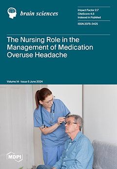
The Nursing Role in the Management of Medication Overuse Headache: Realities and Prospects
by Luigi Alberto Pini, Katiuscia Cottafavi and Paola Ferri
Brain Sci. 2024, 14(6), 600; https://doi.org/10.3390/brainsci14060600 - 14 Jun 2024
Abstract
This review aims to analyze the current literature to identify articles related to the role of nurses and, in general, the nursing management of patients suffering from medication overuse headache (MOH), a globally spread disease. We specifically argue for non-pharmacological approaches to pain [...] Read more.
This review aims to analyze the current literature to identify articles related to the role of nurses and, in general, the nursing management of patients suffering from medication overuse headache (MOH), a globally spread disease. We specifically argue for non-pharmacological approaches to pain management, such as multidisciplinary team approaches, holistic treatment, cognitive behavioral therapy and exercise. For this review, we investigated international scientific databases, including PubMed, CINAHL, Scopus and Embase, in the period between 2000 and 2024. We observed a wealth of scientific articles related to MOH, but a poverty of articles relating to the nursing management of headache. The research included the presence of academic-level training for nurses, whereas there are few institutions that train competent professionals in both pharmacological and non-pharmacological management of MOH patients. Nursing assessment and assistance strategies are indicated to plan tailored treatment paths related to the specific needs of these patients. Full article
(This article belongs to the Special Issue Medication Overuse Headache from Different Points of View)
12 pages, 250 KiB  
Article
Head Injury and Associated Sequelae in Individuals Seeking Asylum in the United States: A Retrospective Mixed-Methods Review of Medico-Legal Affidavits
by Altaf Saadi, Julia Asfour, Maria Vassimon De Assis, Tessa Wilson, Rohini J. Haar and Michele Heisler
Brain Sci. 2024, 14(6), 599; https://doi.org/10.3390/brainsci14060599 - 14 Jun 2024
Abstract
People seeking asylum are susceptible to head injury (HI) due to exposure to various forms of violence including war, torture, or interpersonal violence. Yet, the extents to which clinicians assess HI, and if so, what the associated characteristics are, are not well known. [...] Read more.
People seeking asylum are susceptible to head injury (HI) due to exposure to various forms of violence including war, torture, or interpersonal violence. Yet, the extents to which clinicians assess HI, and if so, what the associated characteristics are, are not well known. We analyzed 200 U.S.-based medico-legal affidavits using descriptive, multivariate regression, and thematic analysis. Head injury was documented in 38% of affidavits. Those who experienced physical violence were eight times likelier to experience HI than those who did not experience physical violence. Five themes emerged: (1) HI occurred commonly in the context of interpersonal violence (44%), followed by militarized violence (33%); (2) mechanisms of HI included direct blows to the head and asphyxiation, suggesting potential for both traumatic brain injury and brain injury from oxygen deprivation; (3) HI was often recurrent and concurrent with other physical injuries; (4) co-morbid psychiatric and post-concussive symptoms made it challenging to assess neurological and psychiatric etiologies; and (5) overall, there was a paucity of assessments and documentation of HI and sequelae. Among individuals assessed for asylum claims, HI is common, often recurrent, occurring in the context of interpersonal violence, and concurrent with psychological and other physical trauma. Physical violence is an important risk factor for HI, which should be assessed when physical violence is reported. Full article
(This article belongs to the Special Issue Shedding Light on the Hidden Epidemic of Violence and Brain Injury)
15 pages, 262 KiB  
Article
Wideband Tympanometry and Pressurized Otoacoustic Emissions in Children with Surgical Excision of Palatine and/or Pharyngeal Tonsils
by Aline Buratti Sanches, Milaine Dominici Sanfins, Piotr Henryk Skarzynski, Magdalena Beata Skarżyńska, Henrique Costa Penatti, Caroline Donadon, Ingrid Pereira de Souza, Ingridy Vitoria da Silva and Maria Francisca Colella-Santos
Brain Sci. 2024, 14(6), 598; https://doi.org/10.3390/brainsci14060598 - 14 Jun 2024
Abstract
Palatine and pharyngeal tonsil hypertrophy may lead to dysfunction of the auditory tube due to a propensity for infection, potentially giving rise to otitis media. This is a quantitative and longitudinal study, developed from 2019 to 2021, at the State University of Campinas [...] Read more.
Palatine and pharyngeal tonsil hypertrophy may lead to dysfunction of the auditory tube due to a propensity for infection, potentially giving rise to otitis media. This is a quantitative and longitudinal study, developed from 2019 to 2021, at the State University of Campinas (UNICAMP). The studied sample comprised 15 participants aged 5 to 12 years (mean 7.9 years), 12 male and 3 female, arranged into two groups: children diagnosed with pharyngeal and/or palatine tonsil hypertrophy who were candidates for surgery (G1), and children who were later evaluated after surgery (G2). As part of the test, an otoscopy and measurements of logoaudiometry, pure-tone threshold audiometry, wideband tympanometry (ambient and peak pressure), and otoacoustic emissions (TEOAEs and DPOAEs, both at ambient and peak pressure) were all performed. There were statistically significant differences between phases in pure-tone audiometry, in terms of 226 Hz tympanometry, wideband tympanometry in peak pressure conditions, in the amplitude measurement TEOAEs in both pressure conditions, in DPOAEs in ambient pressure conditions, and in the signal/noise measurement in both pressures in DPOAEs. Overall, it was found that hearing tests were different for subjects with palatine and pharyngeal tonsil hypertrophy compared to the post-surgical group. Full article
(This article belongs to the Special Issue Recent Advances in Hearing Impairment)
3 pages, 211 KiB  
Editorial
The Anxious Brain: The Influence of Stress on the Nervous System—Editorial
by Drozdstoy Stoyanov
Brain Sci. 2024, 14(6), 597; https://doi.org/10.3390/brainsci14060597 - 13 Jun 2024
Viewed by 35
Abstract
Anxiety disorders, including generalized anxiety, panic disorder, and post-traumatic stress, constitute the most frequent mental disorders and occur in about 14–18% of the overall population [...] Full article
(This article belongs to the Special Issue Anxious Brain: Stress Influence on the Nervous System)
17 pages, 2771 KiB  
Article
Neuroprotective Potentials of Berberine in Rotenone-Induced Parkinson’s Disease-like Motor Symptoms in Rats
by Hsiang-Chien Tseng, Mao-Hsien Wang, Chih-Hsiang Fang, Yi-Wen Lin and Hung-Sheng Soung
Brain Sci. 2024, 14(6), 596; https://doi.org/10.3390/brainsci14060596 - 13 Jun 2024
Viewed by 71
Abstract
Rotenone (RTN) induces neurotoxicity and motor dysfunction in rats, mirroring the pathophysiological traits of Parkinson’s disease (PD), including striatal oxidative stress, mitochondrial dysfunction, and changes in neural structure. This makes RTN a valuable model for PD research. Berberine (BBR), an isoquinoline alkaloid recognized [...] Read more.
Rotenone (RTN) induces neurotoxicity and motor dysfunction in rats, mirroring the pathophysiological traits of Parkinson’s disease (PD), including striatal oxidative stress, mitochondrial dysfunction, and changes in neural structure. This makes RTN a valuable model for PD research. Berberine (BBR), an isoquinoline alkaloid recognized for its antioxidative, anti-inflammatory, and neuroprotective properties, was evaluated for its ability to counteract RTN-induced impairments. Rats received subcutaneous RTN at 0.5 mg/kg for 21 days, resulting in weight loss and significant motor deficits assessed through open-field, bar catalepsy, beam-crossing, rotarod, and grip strength tests. BBR, administered orally at 30 or 100 mg/kg doses, one hour prior to RTN exposure for the same duration, effectively mitigated many of the RTN-induced motor impairments. Furthermore, BBR treatment reduced RTN-induced nitric oxide (NO) and lipid peroxidation (LPO) levels, bolstered antioxidative capacity, enhanced mitochondrial enzyme activities (e.g., succinate dehydrogenase (SDH), ATPase, and the electron transport chain (ETC)), and diminished striatal neuroinflammation and apoptosis markers. Notably, the co-administration of trigonelline (TGN), an inhibitor of the nuclear factor erythroid-2-related factor 2 (Nrf2) pathway, significantly attenuated BBR’s protective effects, indicating that BBR’s neuroprotective actions are mediated via the Nrf2 pathway. These results underscore BBR’s potential in ameliorating motor impairments akin to PD, suggesting its promise in potentially delaying or managing PD symptoms. Further research is warranted to translate these preclinical findings into clinical settings, enhancing our comprehension of BBR’s therapeutic prospects in PD. Full article
Show Figures

Figure 1

18 pages, 1240 KiB  
Article
A Hybrid EEG-Based Stress State Classification Model Using Multi-Domain Transfer Entropy and PCANet
by Yuefang Dong, Lin Xu, Jian Zheng, Dandan Wu, Huanli Li, Yongcong Shao, Guohua Shi and Weiwei Fu
Brain Sci. 2024, 14(6), 595; https://doi.org/10.3390/brainsci14060595 - 12 Jun 2024
Viewed by 112
Abstract
This paper proposes a new hybrid model for classifying stress states using EEG signals, combining multi-domain transfer entropy (TrEn) with a two-dimensional PCANet (2D-PCANet) approach. The aim is to create an automated system for identifying stress levels, which is crucial for early intervention [...] Read more.
This paper proposes a new hybrid model for classifying stress states using EEG signals, combining multi-domain transfer entropy (TrEn) with a two-dimensional PCANet (2D-PCANet) approach. The aim is to create an automated system for identifying stress levels, which is crucial for early intervention and mental health management. A major challenge in this field lies in extracting meaningful emotional information from the complex patterns observed in EEG. Our model addresses this by initially applying independent component analysis (ICA) to purify the EEG signals, enhancing the clarity for further analysis. We then leverage the adaptability of the fractional Fourier transform (FrFT) to represent the EEG data in time, frequency, and time–frequency domains. This multi-domain representation allows for a more nuanced understanding of the brain’s activity in response to stress. The subsequent stage involves the deployment of a two-layer 2D-PCANet network designed to autonomously distill EEG features associated with stress. These features are then classified by a support vector machine (SVM) to determine the stress state. Moreover, stress induction and data acquisition experiments are designed. We employed two distinct tasks known to trigger stress responses. Other stress-inducing elements that enhance the stress response were included in the experimental design, such as time limits and performance feedback. The EEG data collected from 15 participants were retained. The proposed algorithm achieves an average accuracy of over 92% on this self-collected dataset, enabling stress state detection under different task-induced conditions. Full article
(This article belongs to the Section Social Cognitive and Affective Neuroscience)
8 pages, 2887 KiB  
Case Report
Ictal Fear or Panic Attack, This Is the Question—A Video–EEG Study
by Francesco Castellana, Grazia D’Onofrio, Filomena Ciccone, Maria Teresa Di Claudio, Maura Pugliatti, Teresa Popolizio and Giuseppe d’Orsi
Brain Sci. 2024, 14(6), 594; https://doi.org/10.3390/brainsci14060594 - 12 Jun 2024
Viewed by 180
Abstract
Panic disorder (PD) and focal epilepsy, in particular, temporal lobe epilepsy, often present diagnostic challenges due to overlapping clinical manifestations. This article describes the case of a 25-year-old female, misdiagnosed with PD for 15 years, whose recurring episodes of sudden fear, palpitations, and [...] Read more.
Panic disorder (PD) and focal epilepsy, in particular, temporal lobe epilepsy, often present diagnostic challenges due to overlapping clinical manifestations. This article describes the case of a 25-year-old female, misdiagnosed with PD for 15 years, whose recurring episodes of sudden fear, palpitations, and nausea were later identified as manifestations of focal epilepsy. Initially unresponsive to conventional anti-anxiety medications, the patient’s correct diagnosis was only established through comprehensive electro-clinical, neuropsychological, and neuroimaging evaluations during her admission to our research hospital. Long-term video–EEG monitoring (LTVEM) played a pivotal role in identifying the epileptic nature of her episodes, which were characterized by paroxysmal activity in the right temporal and zygomatic regions, consistent with the location of a dysplastic lesion in the right amygdala, as revealed by high-resolution magnetic resonance imaging. These findings underline the importance of considering focal epilepsy in the differential diagnosis of PD, especially in cases refractory to standard psychiatric treatments. The misdiagnosis of epilepsy as PD can lead to significant delays in appropriate treatment, potentially exacerbating the patient’s condition and affecting their quality of life. This case emphasizes the necessity of a multidisciplinary approach and the utilization of advanced diagnostic tools like LTVEM in elucidating the underlying causes of paroxysmal psychiatric symptoms. Full article
(This article belongs to the Section Neurotechnology and Neuroimaging)
Show Figures

Figure 1

21 pages, 4125 KiB  
Article
Bidirectional Association between Sarcopenia and Depressive Symptoms among Chinese Middle- and Older-Aged Adults: Longitudinal Observational Study
by Na Zeng, Chao Li, Huan Mei, Shuilin Wu, Chang Liu, Xiaokun Wang, Jie Shi, Lin Lu and Yanping Bao
Brain Sci. 2024, 14(6), 593; https://doi.org/10.3390/brainsci14060593 - 11 Jun 2024
Viewed by 248
Abstract
Background: The study aimed to examine the bidirectional relationship between sarcopenia and depressive symptoms in a national, community-based cohort study, despite the unclear temporal sequence demonstrated previously. Methods: Data were derived from four waves (2011 baseline and 2013, 2015, and 2018 follow-ups) of [...] Read more.
Background: The study aimed to examine the bidirectional relationship between sarcopenia and depressive symptoms in a national, community-based cohort study, despite the unclear temporal sequence demonstrated previously. Methods: Data were derived from four waves (2011 baseline and 2013, 2015, and 2018 follow-ups) of the China Health and Retirement Longitudinal Study (CHARLS). A total of 17,708 participants aged 45 years or older who had baseline data on both sarcopenia status and depressive symptoms in 2011 were included in the study. For the two cohort analyses, a total of 8092 adults without depressive symptoms and 11,292 participants without sarcopenia in 2011 were included. Sarcopenia status was defined according to the Asian Working Group for Sarcopenia 2019 (AWGS 2019) criteria. Depressive symptoms were defined as a score of 20 or higher on the 10-item Center for Epidemiologic Studies Depressive Scale (CES-D-10). Cox proportional hazard regression models were conducted to examine the risk of depressive symptoms and sarcopenia risk, while cross-lagged panel models were used to examine the temporal sequence between depressive symptoms and sarcopenia over time. Results: During a total of 48,305.1 person-years follow-up, 1262 cases of incident depressive symptoms were identified. Sarcopenia exhibited a dose–response relationship with a higher risk of depressive symptoms (HR = 1.7, 95%CI: 1.2–2.3 for sarcopenia, and HR = 1.5, 95%CI: 1.2–1.8 for possible sarcopenia, p trend < 0.001). In the second cohort analysis, 240 incident sarcopenia cases were identified over 39,621.1 person-years. Depressive symptoms (HR = 1.5, 95%CI: 1.2–2.0) are significantly associated with a higher risk of developing sarcopenia after multivariable adjustment (p < 0.001, Cross-lagged panel analyses demonstrated that depressive symptoms were associated with subsequent sarcopenia (β = 0.003, p < 0.001). Simultaneously, baseline sarcopenia was also associated with subsequent depressive symptoms (β = 0.428, p < 0.001). Conclusion: This study identified a bidirectional relationship between depressive symptoms and sarcopenia. It seems more probable that baseline sarcopenia is associated with subsequent depressive symptoms in a stronger pattern than the reverse pathway. The interlinkage indicated that maintaining normal muscle mass and strength may serve as a crucial intervention strategy for alleviating mood disorders. Full article
(This article belongs to the Section Psychiatric Diseases)
Show Figures

Figure 1

23 pages, 551 KiB  
Review
Endocannabinoid System Changes throughout Life: Implications and Therapeutic Potential for Autism, ADHD, and Alzheimer’s Disease
by Kamila Gabrieli Dallabrida, Joyce Maria de Oliveira Bender, Ellen Schavarski Chade, Nathalia Rodrigues and Tuane Bazanella Sampaio
Brain Sci. 2024, 14(6), 592; https://doi.org/10.3390/brainsci14060592 - 10 Jun 2024
Viewed by 250
Abstract
The endocannabinoid system has been linked to various physiological and pathological processes, because it plays a neuromodulator role in the central nervous system. In this sense, cannabinoids have been used off-label for neurodevelopmental disorders, such as autism spectrum disorder (ASD) and attention-deficit/hyperactivity disorder [...] Read more.
The endocannabinoid system has been linked to various physiological and pathological processes, because it plays a neuromodulator role in the central nervous system. In this sense, cannabinoids have been used off-label for neurodevelopmental disorders, such as autism spectrum disorder (ASD) and attention-deficit/hyperactivity disorder (ADHA), as well as in Alzheimer’s disease (AD), a more prevalent neurodegenerative disease. Thus, this study aims, through a comprehensive literature review, to arrive at a better understanding of the impact of cannabinoids in the therapeutic treatment of patients with ASD, ADHD, and Alzheimer’s disease (AD). Overall, cannabis products rich in CBD displayed a higher therapeutic potential for ASD children, while cannabis products rich in THC have been tested more for AD therapy. For ADHD, the clinical studies are incipient and inconclusive, but promising. In general, the main limitations of the clinical studies are the lack of standardization of the cannabis-based products consumed by the participants, a lack of scientific rigor, and the small number of participants. Full article
(This article belongs to the Section Neuropharmacology and Neuropathology)
24 pages, 2901 KiB  
Technical Note
Modal Analysis of Cerebrovascular Effects for Digital Health Integration of Neurostimulation Therapies—A Review of Technology Concepts
by Marcel Stefanski, Yashika Arora, Mancheung Cheung and Anirban Dutta
Brain Sci. 2024, 14(6), 591; https://doi.org/10.3390/brainsci14060591 - 10 Jun 2024
Viewed by 381
Abstract
Transcranial electrical stimulation (tES) is increasingly recognized for its potential to modulate cerebral blood flow (CBF) and evoke cerebrovascular reactivity (CVR), which are crucial in conditions like mild cognitive impairment (MCI) and dementia. This study explores the impact of tES on the neurovascular [...] Read more.
Transcranial electrical stimulation (tES) is increasingly recognized for its potential to modulate cerebral blood flow (CBF) and evoke cerebrovascular reactivity (CVR), which are crucial in conditions like mild cognitive impairment (MCI) and dementia. This study explores the impact of tES on the neurovascular unit (NVU), employing a physiological modeling approach to simulate the vascular response to electric fields generated by tES. Utilizing the FitzHugh–Nagumo model for neuroelectrical activity, we demonstrate how tES can initiate vascular responses such as vasoconstriction followed by delayed vasodilation in cerebral arterioles, potentially modulated by a combination of local metabolic demands and autonomic regulation (pivotal locus coeruleus). Here, four distinct pathways within the NVU were modeled to reflect the complex interplay between synaptic activity, astrocytic influences, perivascular potassium dynamics, and smooth muscle cell responses. Modal analysis revealed characteristic dynamics of these pathways, suggesting that oscillatory tES may finely tune the vascular tone by modulating the stiffness and elasticity of blood vessel walls, possibly by also impacting endothelial glycocalyx function. The findings underscore the therapeutic potential vis-à-vis blood-brain barrier safety of tES in modulating neurovascular coupling and cognitive function needing the precise modulation of NVU dynamics. This technology review supports the human-in-the-loop integration of tES leveraging digital health technologies for the personalized management of cerebral blood flow, offering new avenues for treating vascular cognitive disorders. Future studies should aim to optimize tES parameters using computational modeling and validate these models in clinical settings, enhancing the understanding of tES in neurovascular health. Full article
20 pages, 2552 KiB  
Review
Research Progress on the Pathogenesis, Diagnosis, and Drug Therapy of Alzheimer’s Disease
by Yixuan Yang and Lina Qiu
Brain Sci. 2024, 14(6), 590; https://doi.org/10.3390/brainsci14060590 - 9 Jun 2024
Viewed by 466
Abstract
As the population ages worldwide, Alzheimer’s disease (AD), the most prevalent kind of neurodegenerative disorder among older people, has become a significant factor affecting quality of life, public health, and economies. However, the exact pathogenesis of Alzheimer’s remains elusive, and existing highly recognized [...] Read more.
As the population ages worldwide, Alzheimer’s disease (AD), the most prevalent kind of neurodegenerative disorder among older people, has become a significant factor affecting quality of life, public health, and economies. However, the exact pathogenesis of Alzheimer’s remains elusive, and existing highly recognized pathogenesis includes the amyloid cascade hypothesis, Tau neurofibrillary tangles hypothesis, and neuroinflammation hypothesis. The major diagnoses of Alzheimer’s disease include neuroimaging positron emission computed tomography, magnetic resonance imaging, and cerebrospinal fluid molecular diagnosis. The therapy of Alzheimer’s disease primarily relies on drugs, and the approved drugs on the market include acetylcholinesterase drugs, glutamate receptor antagonists, and amyloid-β monoclonal antibodies. Still, the existing drugs can only alleviate the symptoms of the disease and cannot completely reverse it. This review aims to summarize existing research results on Alzheimer’s disease pathogenesis, diagnosis, and drug therapy, with the objective of facilitating future research in this area. Full article
(This article belongs to the Section Neurodegenerative Diseases)
Show Figures

Figure 1

22 pages, 2403 KiB  
Review
A Narrative Review of the Dorsal Root Ganglia and Spinal Cord Mechanisms of Action of Neuromodulation Therapies in Neuropathic Pain
by Matheus Deroco Veloso da Silva, Geovana Martelossi-Cebinelli, Kelly Megumi Yaekashi, Thacyana T. Carvalho, Sergio M. Borghi, Rubia Casagrande and Waldiceu A. Verri
Brain Sci. 2024, 14(6), 589; https://doi.org/10.3390/brainsci14060589 - 9 Jun 2024
Viewed by 389
Abstract
Neuropathic pain arises from injuries to the nervous system in diseases such as diabetes, infections, toxicity, and traumas. The underlying mechanism of neuropathic pain involves peripheral and central pathological modifications. Peripheral mechanisms entail nerve damage, leading to neuronal hypersensitivity and ectopic action potentials. [...] Read more.
Neuropathic pain arises from injuries to the nervous system in diseases such as diabetes, infections, toxicity, and traumas. The underlying mechanism of neuropathic pain involves peripheral and central pathological modifications. Peripheral mechanisms entail nerve damage, leading to neuronal hypersensitivity and ectopic action potentials. Central sensitization involves a neuropathological process with increased responsiveness of the nociceptive neurons in the central nervous system (CNS) to their normal or subthreshold input due to persistent stimuli, leading to sustained electrical discharge, synaptic plasticity, and aberrant processing in the CNS. Current treatments, both pharmacological and non-pharmacological, aim to alleviate symptoms but often face challenges due to the complexity of neuropathic pain. Neuromodulation is emerging as an important therapeutic approach for the treatment of neuropathic pain in patients unresponsive to common therapies, by promoting the normalization of neuronal and/or glial activity and by targeting cerebral cortical regions, spinal cord, dorsal root ganglia, and nerve endings. Having a better understanding of the efficacy, adverse events and applicability of neuromodulation through pre-clinical studies is of great importance. Unveiling the mechanisms and characteristics of neuromodulation to manage neuropathic pain is essential to understand how to use it. In the present article, we review the current understanding supporting dorsal root ganglia and spinal cord neuromodulation as a therapeutic approach for neuropathic pain. Full article
(This article belongs to the Special Issue Neuromodulation and Pain)
Show Figures

Figure 1

4 pages, 182 KiB  
Editorial
Advances in Neuropsychology: Top Papers Published in Brain Sciences in 2022–2023
by Pierluigi Zoccolotti
Brain Sci. 2024, 14(6), 588; https://doi.org/10.3390/brainsci14060588 - 9 Jun 2024
Viewed by 348
Abstract
The spectrum of typical neuropsychology topics has gradually broadened in recent years thanks to advances in neuroimaging and electrophysiological techniques [...] Full article
(This article belongs to the Section Neuropsychology)
13 pages, 558 KiB  
Review
Long COVID in Brain Health Research: A Call to Action
by Thorsten Rudroff
Brain Sci. 2024, 14(6), 587; https://doi.org/10.3390/brainsci14060587 - 8 Jun 2024
Viewed by 416
Abstract
The COVID-19 pandemic has brought attention to the long-term consequences of the virus, particularly the persistent symptoms that characterize long COVID. This syndrome, which can last for months after the initial infection, includes a range of neurological and neuropsychiatric manifestations that have significant [...] Read more.
The COVID-19 pandemic has brought attention to the long-term consequences of the virus, particularly the persistent symptoms that characterize long COVID. This syndrome, which can last for months after the initial infection, includes a range of neurological and neuropsychiatric manifestations that have significant implications for brain health and dementia research. This review explores the current understanding of long COVID’s cognitive, neurological, and psychiatric symptoms and their potential impact on brain stimulation and neuroimaging studies. It argues that researchers must adapt their study designs and screening processes to account for the confounding effects of long COVID and ensure the accuracy and reliability of their findings. To advance the understanding of this condition and its long-term effects on brain health, the review proposes a series of strategies, including the development of standardized screening tools, the investigation of underlying mechanisms, and the identification of risk factors and protective factors. It also emphasizes the importance of collaborative research efforts and international data sharing platforms in accelerating the pace of discovery and developing targeted interventions for individuals with long COVID. As the prevalence of this condition continues to grow, it is imperative that the neuroscience community comes together to address this challenge and support those affected by long COVID. Full article
10 pages, 1125 KiB  
Article
Pork Liver Decomposition Product May Improve Frontal Lobe Function in Humans—Open Trial
by Miiru Suzuki, Ikuya Sato, Masatsugu Sato, Hideki Iwasaki, Takahiro Saito, Masahiko Kimura, Kenichi Sako, Tomoji Maeda, Hisao Haniu, Tamotsu Tsukahara and Yoshikazu Matsuda
Brain Sci. 2024, 14(6), 586; https://doi.org/10.3390/brainsci14060586 - 7 Jun 2024
Viewed by 274
Abstract
Porcine Liver Decomposition Product (PLDP) was obtained by treating pig liver homogenate with protease and filling it into capsules. We have already confirmed from three clinical trials that PLDP enhances visual memory and delays memory recall, and we believe that its activity is [...] Read more.
Porcine Liver Decomposition Product (PLDP) was obtained by treating pig liver homogenate with protease and filling it into capsules. We have already confirmed from three clinical trials that PLDP enhances visual memory and delays memory recall, and we believe that its activity is due to various phospholipids, including phosphatidylcholine (PC). In this study, we clinically evaluated PLDP for depressive symptoms caused by a decline in cognitive function. This clinical trial was conducted using the Revised Hasegawa Dementia Scale (HDS-R). The HDS-R (maximum score is 30 points) is a test similar to the Mini-Mental State Examination (MMSE), which is commonly used in Japan. Dementia is suspected if the score falls below 20 on the HDS-R. Additionally, in a previous clinical trial, there was no change in scores in the placebo group after three doses of the HDS-R. In order to clearly confirm the effectiveness of PLDP, this study was conducted under stricter conditions (HDS-R points of 15 to 23) than previous clinical trials (all participants had scores of 20 or higher). Therefore, from ethical considerations, a clinical trial was conducted using the scores before PLDP administration as a control. In this study, PLDP was administered orally at 4 capsules per day, and the HDS-R was confirmed 2 and 4 weeks after administration. A significant increase in HDS-R scores was observed at 2 and 4 weeks after PLDP administration. Additionally, regarding each item of the HDS-R, PLDP significantly increased 2 and 4 weeks after oral administration for the question items assessing delayed recall, and the question item assessing verbal fluency tasks was recognized. From the above results, we confirmed the reproducibility of the effect of PLDP in improving the delayed recall of verbal memories. Furthermore, increasing scores on verbal fluency tasks suggest that PLDP may enhance frontal lobe function and prevent or improve depressive symptoms. The effects observed in this study may differ from the mechanisms of action of existing antidepressants, and we believe that this may lead to the discovery of new antidepressants. Full article
(This article belongs to the Section Neuropharmacology and Neuropathology)
Show Figures

Figure 1

4 pages, 207 KiB  
Editorial
The Contribution of Internal and External Factors to Human Spatial Navigation
by Laura Piccardi, Raffaella Nori, Jose Manuel Cimadevilla and María Kozhevnikov
Brain Sci. 2024, 14(6), 585; https://doi.org/10.3390/brainsci14060585 - 7 Jun 2024
Viewed by 353
Abstract
Spatial navigation is a multifaceted cognitive function essential for planning and finding routes in one’s environment [...] Full article
14 pages, 526 KiB  
Article
Temporal–Posterior Alpha Power in Resting-State Electroencephalography as a Potential Marker of Complex Childhood Trauma in Institutionalized Adolescents
by Gabriela Mariana Marcu, Ciprian Ionuț Băcilă and Ana-Maria Zăgrean
Brain Sci. 2024, 14(6), 584; https://doi.org/10.3390/brainsci14060584 - 6 Jun 2024
Viewed by 322
Abstract
The present study explored whether, given the association of temporal alpha with fear circuitry (learning and conditioning), exposure to complex childhood trauma (CCT) is reflected in the temporal–posterior alpha power in resting-state electroencephalography (EEG) in complex trauma-exposed adolescents in a sample of 25 [...] Read more.
The present study explored whether, given the association of temporal alpha with fear circuitry (learning and conditioning), exposure to complex childhood trauma (CCT) is reflected in the temporal–posterior alpha power in resting-state electroencephalography (EEG) in complex trauma-exposed adolescents in a sample of 25 adolescents and similar controls aged 12–17 years. Both trauma and psychopathology were screened or assessed, and resting-state EEG was recorded following a preregistered protocol for data collection. Temporal–posterior alpha power, corresponding to the T5 and T6 electrode locations (international 10–20 system), was extracted from resting-state EEG in both eyes-open and eyes-closed conditions. We found that in the eyes-open condition, temporal–posterior alpha was significantly lower in adolescents exposed to CCT relative to healthy controls, suggesting that childhood trauma exposure may have a measurable impact on alpha oscillatory patterns. Our study highlights the importance of considering potential neural markers, such as temporal–posterior alpha power, to understanding the long-term consequences of CCT exposure in developmental samples, with possible important clinical implications in guiding neuroregulation interventions. Full article
(This article belongs to the Special Issue EEG and Event-Related Potentials)
16 pages, 931 KiB  
Systematic Review
Alterations in Neurotrophins in Alcohol-Addicted Patients during Alcohol Withdrawal
by Magda Malewska-Kasprzak, Maria Skibińska and Monika Dmitrzak-Węglarz
Brain Sci. 2024, 14(6), 583; https://doi.org/10.3390/brainsci14060583 - 6 Jun 2024
Viewed by 341
Abstract
Background: Alcohol use disorder (AUD) is related to mental and somatic disorders that result in alcohol withdrawal syndrome (AWS), with 30% of AWS cases leading to life-threatening delirium tremens (DTs). Currently, studies do not support using any one biomarker in DTs. Neurotrophins affect [...] Read more.
Background: Alcohol use disorder (AUD) is related to mental and somatic disorders that result in alcohol withdrawal syndrome (AWS), with 30% of AWS cases leading to life-threatening delirium tremens (DTs). Currently, studies do not support using any one biomarker in DTs. Neurotrophins affect neuromodulation, playing a role in the pathogenesis of AUD, AWS, and DTs. Methods: This review aims to summarize experimental and clinical data related to neurotrophins and S100B in neuroplasticity, as well as neurodegeneration in the context of AUD, AWS, and DTs. This work used publications that were selected based on the protocol consistent with the Preferred Reporting Items for Systematic Reviews and Meta-Analysis (PRISMA) statement. Results: The BDNF level could be a good candidate biomarker for relapse susceptibility, as it is significantly reduced during consumption and gradually increases during abstinence. GDNF influences AUD through its integral role in the function of dopaminergic neurons and ablates the return to alcohol-drinking behavior. NGF protects neurons from ethanol-induced cytotoxic damage and affects recovery from cognitive deficits after brain damage. The NT-3 level is decreased after alcohol exposure and is involved in compensatory mechanisms for cognitive decline in AUD. NT-4 affects oxidative stress, which is associated with chronic alcohol consumption. S100B is used as a biomarker of brain damage, with elevated levels in serum in AUD, and can protect 5-HT neurons from the damage caused by alcohol. Conclusions: BDNF, GDNF, NT-3, NT-4, NGF, and S100B may be valuable markers for withdrawal syndrome. In particular, the most relevant is their association with the development of delirium complications. However, there are few data concerning some neurotrophins in AWS and DTs, suggesting the need for further research. Full article
(This article belongs to the Special Issue Psychiatry and Addiction: A Multi-Faceted Issue)
Show Figures

Figure 1

3 pages, 160 KiB  
Editorial
The Role of Consciousness in Coupling Emotions, Motivations, and Behaviors
by Sergio Frumento and Danilo Menicucci
Brain Sci. 2024, 14(6), 582; https://doi.org/10.3390/brainsci14060582 - 5 Jun 2024
Viewed by 282
Abstract
A potential function of consciousness is to integrate emotions, motivations, and subsequent behaviors into a coherent narrative [...] Full article
10 pages, 973 KiB  
Brief Report
Transcriptomic Profile of Mouse Brain Ageing in Early Developmental Stages
by Karolina Kulis, Kevin Tabury, Mohammed Abderrafi Benotmane and Joanna Polanska
Brain Sci. 2024, 14(6), 581; https://doi.org/10.3390/brainsci14060581 - 5 Jun 2024
Viewed by 359
Abstract
Ageing is a continuous process that can cause neurodevelopmental changes in the body. Several studies have examined its effects, but few have focused on how time affects biological processes in the early stages of brain development. As studying the changes that occur in [...] Read more.
Ageing is a continuous process that can cause neurodevelopmental changes in the body. Several studies have examined its effects, but few have focused on how time affects biological processes in the early stages of brain development. As studying the changes that occur in the early stages of life is important to prevent age-related neurological and psychiatric disorders, we aim to focus on these changes. The transcriptomic markers of ageing that are common to the analysed brain regions of C57Bl/6J mice were identified after conducting two-way ANOVA tests and effect size analysis on the time courses of gene expression profiles in various mouse brain regions. A total of 16,374 genes (59.9%) significantly changed their expression level, among which 7600 (27.8%) demonstrated tissue-dependent differences only, and 1823 (6.7%) displayed time-dependent and tissue-independent responses. Focusing on genes with at least a large effect size gives the list of potential biomarkers 12,332 (45.1%) and 1670 (6.1%) genes, respectively. There were 305 genes that exhibited similar significant time response trends (independently of the brain region). Samples from an 11-day-old mouse embryo validated the identified early-stage brain ageing markers. The overall functional analysis revealed tRNA and rRNA processing in the mitochondrion and contact activation system (CAS), as well as the kallikrein/kinin system (KKS), together with clotting cascade and defective factor F9 activation being affected by ageing. Most ageing-related pathways were significantly enriched, especially those that are strongly connected to development processes and neurodegenerative diseases. Full article
(This article belongs to the Section Developmental Neuroscience)
20 pages, 739 KiB  
Article
Cognitive vs. Linguistic Training in Children with Developmental Language Disorder: Exploring Their Effectiveness on Verbal Short-Term Memory and Verbal Working Memory
by Theodora Bachourou, Stavroula Stavrakaki, Vasiliki Koukoulioti and Ioanna Talli
Brain Sci. 2024, 14(6), 580; https://doi.org/10.3390/brainsci14060580 - 5 Jun 2024
Viewed by 375
Abstract
The present study explores comparatively the effectiveness of a cognitive (verbal short-term memory (vSTM), verbal working memory (vWM)) and of a linguistic training (10-week duration each) in the diffusion of gains in cognitive abilities (vSTM and vWM) of in school-aged Greek-speaking children with [...] Read more.
The present study explores comparatively the effectiveness of a cognitive (verbal short-term memory (vSTM), verbal working memory (vWM)) and of a linguistic training (10-week duration each) in the diffusion of gains in cognitive abilities (vSTM and vWM) of in school-aged Greek-speaking children with developmental language disorder (DLD). To this purpose, two computerized training programs i.e., a linguistic and a cognitive one, were developed and applied to three groups (A, B, and C) of children with DLD (N = 49, in total). There were three assessments with two vSTM tasks (non-word repetition and forward digit span) and a vWM task (backward digit span): pre-therapeutically (time 1), where no significant between-group differences were found, post-therapeutically I (time 2), and post-therapeutically II (time 3) and two training phases. In phase Ι, group A received meta-syntactic training, whereas group B vSTM/vWM training and group C received no training. In phase ΙΙ, a reversal of treatment was performed for groups A and B: group A received vSTM/vWM while group B meta-syntactic training. Again, group C received no training. Overall, the results indicated a significant performance improvement for the treatment groups and revealed beneficial far-transfer effects as language therapy can affect vSTM and vWM in addition to direct and near transfer effects. In addition, the intervention type order affected performance as follows: first, better performance on the vSTM task (non-word repetition) was shown when the linguistic treatment was delivered first; second, better performance on the vWM in Time 2 and Time 3 was shown by group B, for which the cognitive treatment was delivered first. Concluding, not only intervention type but also intervention type order can affect performance in DLD. Full article
39 pages, 808 KiB  
Systematic Review
The Utility of Heartrate and Heartrate Variability Biofeedback for the Improvement of Interoception across Behavioural, Physiological and Neural Outcome Measures: A Systematic Review
by Lettie Wareing, Megan Rose Readman, Matthew R. Longo, Sally A. Linkenauger and Trevor J. Crawford
Brain Sci. 2024, 14(6), 579; https://doi.org/10.3390/brainsci14060579 - 5 Jun 2024
Viewed by 356
Abstract
Interoceptive dysfunctions are increasingly implicated in a number of physical and mental health conditions. Accordingly, there is a pertinent need for therapeutic interventions which target interoceptive deficits. Heartrate and heartrate variability biofeedback therapy (HR(V)-BF), interventions which train individuals to regulate their cardiovascular signals [...] Read more.
Interoceptive dysfunctions are increasingly implicated in a number of physical and mental health conditions. Accordingly, there is a pertinent need for therapeutic interventions which target interoceptive deficits. Heartrate and heartrate variability biofeedback therapy (HR(V)-BF), interventions which train individuals to regulate their cardiovascular signals and constrain these within optimal parameters through breathing, could enhance the functioning of interoceptive pathways via stimulation of the vagus nerve. Consequently, this narrative systematic review sought to synthesise the current state of the literature with regard to the potential of HR(V)-BF as an interoceptive intervention across behavioural, physiological and neural outcome measures related to interoception. In total, 77 papers were included in this review, with the majority using physiological outcome measures. Overall, findings were mixed with respect to improvements in the outcome measures after HR(V)-BF. However, trends suggested that effects on measures related to interoception were stronger when resonance frequency breathing and an intense treatment protocol were employed. Based on these findings, we propose a three-stage model by which HR(V)-BF may improve interoception which draws upon principles of interoceptive inference and predictive coding. Furthermore, we provide specific directions for future research, which will serve to advance the current knowledge state. Full article
(This article belongs to the Section Behavioral Neuroscience)
24 pages, 1988 KiB  
Article
Enhancing Working Memory and Reducing Anxiety in University Students: A Neurofeedback Approach
by Pierluigi Diotaiuti, Giuseppe Valente, Stefano Corrado, Beatrice Tosti, Chiara Carissimo, Tommaso Di Libero, Gianni Cerro, Angelo Rodio and Stefania Mancone
Brain Sci. 2024, 14(6), 578; https://doi.org/10.3390/brainsci14060578 - 5 Jun 2024
Viewed by 326
Abstract
(1) Background: Neurofeedback training (NFT) has emerged as a promising approach for enhancing cognitive functions and reducing anxiety, yet its specific impact on university student populations requires further investigation. This study aims to examine the effects of NFT on working memory improvement and [...] Read more.
(1) Background: Neurofeedback training (NFT) has emerged as a promising approach for enhancing cognitive functions and reducing anxiety, yet its specific impact on university student populations requires further investigation. This study aims to examine the effects of NFT on working memory improvement and anxiety reduction within this demographic. (2) Methods: A total of forty healthy university student volunteers were randomized into two groups: an experimental group that received NFT and a control group. The NFT protocol was administered using a 14-channel Emotiv Epoc X headset (EMOTIV, Inc., San Francisco, CA 94102, USA) and BrainViz software version Brain Visualizer 1.1 (EMOTIV, Inc., San Francisco, CA 94102, USA), focusing on the alpha frequency band to target improvements in working memory and reductions in anxiety. Assessment tools, including the Corsi Block and Memory Span tests for working memory and the State-Trait Anxiety Inventory-2 (STAI-2) for anxiety, were applied pre- and post-intervention. (3) Results: The findings indicated an increase in alpha wave amplitude in the experimental group from the second day of NFT, with statistically significant differences observed on days 2 (p < 0.05) and 8 (p < 0.01). Contrary to expectations based on the previous literature, the study did not observe a concurrent positive impact on working memory. Nonetheless, a significant reduction in state anxiety levels was recorded in the experimental group (p < 0.001), corroborating NFT’s potential for anxiety management. (4) Conclusions: While these results suggest some potential of the technique in enhancing neural efficiency, the variability across different days highlights the need for further investigation to fully ascertain its effectiveness. The study confirms the beneficial impact of NFT on reducing state anxiety among university students, underscoring its value in psychological and cognitive performance enhancement. Despite the lack of observed improvements in working memory, these results highlight the need for continued exploration of NFT applications across different populations and settings, emphasizing its potential utility in educational and therapeutic contexts. Full article
(This article belongs to the Section Social Cognitive and Affective Neuroscience)
Show Figures

Figure 1

16 pages, 2680 KiB  
Article
Correlation between DWI-ASPECTS Score, Ischemic Stroke Volume on DWI, Clinical Severity and Short-Term Prognosis: A Single-Center Study
by Oana Andreea Dogariu, Veronica Gheorman, Ioan Dogariu, Mihaela Corina Berceanu, Carmen Valeria Albu and Ioana Andreea Gheonea
Brain Sci. 2024, 14(6), 577; https://doi.org/10.3390/brainsci14060577 - 5 Jun 2024
Viewed by 275
Abstract
Ischemic stroke is a significant public health concern, with its incidence expected to double over the next 40 years, particularly among individuals over 75 years old. Previous studies, such as the DAWN trial, have highlighted the importance of correlating clinical severity with ischemic [...] Read more.
Ischemic stroke is a significant public health concern, with its incidence expected to double over the next 40 years, particularly among individuals over 75 years old. Previous studies, such as the DAWN trial, have highlighted the importance of correlating clinical severity with ischemic stroke volume to optimize patient management. Our study aimed to correlate the clinical severity of ischemic stroke, as assessed by the NIHSS score, with ischemic stroke volume measured using DWI, and short-term prognosis quantified by the mRS score at discharge. Conducted at the largest hospital in Gorj County from January 2023 to December 2023, this study enrolled 43 consecutive patients with acute ischemic stroke. In our patient cohort, we observed a strong positive correlation between NIHSS score and ischemic stroke volume (Spearman correlation coefficient = 0.982, p < 0.01), and a strong negative correlation between ASPECTS-DWI score and mRS score (Spearman correlation coefficient = −0.952, p < 0.01). Multiple linear regression analysis revealed a significant collective relationship between ASPECTS score, ischemic stroke volume, and NIHSS score (F(1, 41) = 600.28, p < 0.001, R2 = 0.94, R2adj = 0.93). These findings underscore the importance of DWI in assessing ischemic stroke severity and prognosis, warranting further investigation for its integration into clinical practice. Full article
(This article belongs to the Section Molecular and Cellular Neuroscience)
Show Figures

Figure 1

11 pages, 1038 KiB  
Article
Combining Anti-Mitochondrial Antibodies, Anti-Histone, and PLA2/COX Biomarkers to Increase Their Diagnostic Accuracy for Autism Spectrum Disorders
by Afaf El-Ansary, Hanan A. Alfawaz, Abir Ben Bacha and Laila Y. Al-Ayadhi
Brain Sci. 2024, 14(6), 576; https://doi.org/10.3390/brainsci14060576 - 5 Jun 2024
Viewed by 393
Abstract
Background: Autism spectrum disorder (ASD) is a neurodevelopmental disorder characterized by impairments in social interaction and restricted and repetitive behaviors. Oxidative stress may be a critical link between mitochondrial dysfunction and ASD as reactive oxygen species (ROS) generated from pro-oxidant environmental toxicants and [...] Read more.
Background: Autism spectrum disorder (ASD) is a neurodevelopmental disorder characterized by impairments in social interaction and restricted and repetitive behaviors. Oxidative stress may be a critical link between mitochondrial dysfunction and ASD as reactive oxygen species (ROS) generated from pro-oxidant environmental toxicants and activated immune cells can result in mitochondrial failure. Recently, mitochondrial dysfunction, autoimmunity, and abnormal lipid mediators have been identified in multiple investigations as an acknowledged etiological mechanism of ASD that can be targeted for therapeutic intervention. Methods: The relationship between lipid mediator markers linked to inflammation induction, such as phospholipase A2/cyclooxygenase-2 (PLA2/Cox-2), and the mitochondrial dysfunction marker anti-mitochondrial antibodies (AMA-M2), and anti-histone autoantibodies in the etiology of ASD was investigated in this study using combined receiver operating characteristic (ROC) curve analyses. This study also sought to identify the linear combination for a given set of markers that optimizes the partial area under ROC curves. This study included 40 age- and sex-matched controls and 40 ASD youngsters. The plasma of both groups was tested for PLA2/COX-2, AMA-M2, and anti-histone autoantibodies’ levels using ELISA kits. ROC curves and logistic regression models were used in the statistical analysis. Results: Using the integrated ROC curve analysis, a notable rise in the area under the curve was noticed. Additionally, the combined markers had markedly improved specificity and sensitivity. Conclusions: The current study suggested that measuring the predictive value of selected biomarkers related to mitochondrial dysfunction, autoimmunity, and lipid metabolism in children with ASD using a ROC curve analysis could lead to a better understanding of the etiological mechanism of ASD as well as its relationship with metabolism. Full article
Show Figures

Figure 1

12 pages, 2220 KiB  
Article
Tau Protein Accumulation Trajectory-Based Brain Age Prediction in the Alzheimer’s Disease Continuum
by Min Wang, Min Wei, Luyao Wang, Jun Song, Axel Rominger, Kuangyu Shi and Jiehui Jiang
Brain Sci. 2024, 14(6), 575; https://doi.org/10.3390/brainsci14060575 - 4 Jun 2024
Viewed by 325
Abstract
Clinical cognitive advancement within the Alzheimer’s disease (AD) continuum is intimately connected with sustained accumulation of tau protein pathology. The biological brain age and its gap show great potential for pathological risk and disease severity. In the present study, we applied multivariable linear [...] Read more.
Clinical cognitive advancement within the Alzheimer’s disease (AD) continuum is intimately connected with sustained accumulation of tau protein pathology. The biological brain age and its gap show great potential for pathological risk and disease severity. In the present study, we applied multivariable linear support vector regression to train a normative brain age prediction model using tau brain images. We further assessed the predicted biological brain age and its gap for patients within the AD continuum. In the AD continuum, evaluated pathologic tau binding was found in the inferior temporal, parietal-temporal junction, precuneus/posterior cingulate, dorsal frontal, occipital, and inferior-medial temporal cortices. The biological brain age gaps of patients within the AD continuum were notably higher than those of the normal controls (p < 0.0001). Significant positive correlations were observed between the brain age gap and global tau protein accumulation levels for mild cognitive impairment (r = 0.726, p < 0.001), AD (r = 0.845, p < 0.001), and AD continuum (r = 0.797, p < 0.001). The pathologic tau-based age gap was significantly linked to neuropsychological scores. The proposed pathologic tau-based biological brain age model could track the tau protein accumulation trajectory of cognitive impairment and further provide a comprehensive quantification index for the tau accumulation risk. Full article
(This article belongs to the Special Issue Challenges and Perspectives of Neurological Disorders: Series II)
Previous Issue
Back to TopTop