Next Issue
Volume 14, April
Previous Issue
Volume 14, February


Life, Volume 14, Issue 3 (March 2024) – 142 articles

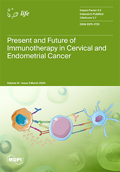
Cover Story (view full-size image): Immunotherapy has emerged as a promising strategy, harnessing the body’s immune system to combat gynecological cancer. This literature review provides a comprehensive overview of the current landscape and future directions of immunotherapy in cervical and endometrial cancer. It presents the most critical methods and treatments employed in immunotherapy for cervical and endometrial cancer, including immune checkpoint inhibitors, cancer vaccines, and adoptive cell therapies. Immune checkpoint inhibitors, such as anti-PD-1/PD-L1 antibodies, have shown remarkable clinical efficacy in certain gynecological malignancies, particularly in advanced or recurrent cases. Ongoing research on cancer vaccines and adoptive cell therapies holds promise for personalized and targeted treatment options. View this paper
  • Issues are regarded as officially published after their release is announced to the table of contents alert mailing list.
  • You may sign up for e-mail alerts to receive table of contents of newly released issues.
  • PDF is the official format for papers published in both, html and pdf forms. To view the papers in pdf format, click on the "PDF Full-text" link, and use the free Adobe Reader to open them.
Order results
Result details
Section
Select all
Export citation of selected articles as:
10 pages, 2042 KiB  
Article
Extra-Anogenital Giant Cutaneous Squamous Cell Carcinomas
by Mateusz K. Mateuszczyk, Iwona Chlebicka, Magdalena Łyko, Joanna Maj and Jacek C. Szepietowski
Life 2024, 14(3), 421; https://doi.org/10.3390/life14030421 - 21 Mar 2024
Viewed by 803
Abstract
Extra-anogenital giant cSCCs are rare but have worse outcomes compared to smaller tumors. Prompted by limited data, the authors conducted a retrospective study to gather more information about giant cSCCs to optimize clinical care. We identified seven cases of giant cSCCs from a [...] Read more.
Extra-anogenital giant cSCCs are rare but have worse outcomes compared to smaller tumors. Prompted by limited data, the authors conducted a retrospective study to gather more information about giant cSCCs to optimize clinical care. We identified seven cases of giant cSCCs from a review of cSCC cases treated in the Unit of Dermatosurgery between 2016 and 2022. Most patients were male (85.71%) with a mean age of 80.29 ± 12.22 years. UV radiation was the most common risk factor (five cases) followed by smoking (three cases) and hidradenitis suppurativa (one case). Most giant cases were located in the head area (71.4%) and the diameter of lesions ranged from 6 to 18 cm. All patients corresponded to tumor stage T3, and 42.86% of patients had lymph node metastases. Surgical excision was the treatment of choice in most cases (85.71%), while a combination of cemiplimab and RP1 was used in some cases due to the ineffectiveness of treatment or contraindications to other therapies. The authors emphasize the importance of early detection and prevention of modifiable risk factors, such as UV radiation, and a multidisciplinary approach to treatment. Other therapies, including immunotherapy, may become increasingly important. Full article
(This article belongs to the Special Issue New Insights in Skin Oncology: From Pathogenesis to Treatment)
Show Figures

Figure 1

25 pages, 900 KiB  
Review
Antitumor Mechanisms of Lycium barbarum Fruit: An Overview of In Vitro and In Vivo Potential
by Maria Rosaria Miranda, Vincenzo Vestuto, Giuseppina Amodio, Michele Manfra, Giacomo Pepe and Pietro Campiglia
Life 2024, 14(3), 420; https://doi.org/10.3390/life14030420 - 21 Mar 2024
Viewed by 842
Abstract
Lycium barbarum, known as goji berry or wolfberry, is a fruit long associated with health benefits, showing a plethora of effects ranging from antioxidant, anticancer, anti-inflammatory, and immunomodulatory effects. Its potential is attributed to the significant presence of polysaccharides, glycopeptides, polyphenols, flavonoids, [...] Read more.
Lycium barbarum, known as goji berry or wolfberry, is a fruit long associated with health benefits, showing a plethora of effects ranging from antioxidant, anticancer, anti-inflammatory, and immunomodulatory effects. Its potential is attributed to the significant presence of polysaccharides, glycopeptides, polyphenols, flavonoids, carotenoids, and their derivatives. These compounds effectively counteract the action of free radicals, positively influencing cellular balance and intracellular signaling, contributing to overall cell health and function acting on multiple molecular pathways. Several fractions extracted from goji berries demonstrate antitumor properties, particularly effective against breast cancer, without showing cytotoxic effects on normal human cells. Hence, the review explored the fundamental traits of bioactive elements in Lycium barbarum and their potential in cancer treatment and, specifically, breast cancer. It focused on elucidating wolfberry’s influenced biochemical pathways, its synergism with anticancer drugs, and its potential to alleviate the side effects associated with existing cancer treatments. Full article
Show Figures

Figure 1

20 pages, 10775 KiB  
Article
Generative-Adversarial-Network-Based Image Reconstruction for the Capacitively Coupled Electrical Impedance Tomography of Stroke
by Mikhail Ivanenko, Damian Wanta, Waldemar T. Smolik, Przemysław Wróblewski and Mateusz Midura
Life 2024, 14(3), 419; https://doi.org/10.3390/life14030419 - 21 Mar 2024
Viewed by 714
Abstract
This study investigated the potential of machine-learning-based stroke image reconstruction in capacitively coupled electrical impedance tomography. The quality of brain images reconstructed using the adversarial neural network (cGAN) was examined. The big data required for supervised network training were generated using a two-dimensional [...] Read more.
This study investigated the potential of machine-learning-based stroke image reconstruction in capacitively coupled electrical impedance tomography. The quality of brain images reconstructed using the adversarial neural network (cGAN) was examined. The big data required for supervised network training were generated using a two-dimensional numerical simulation. The phantom of an axial cross-section of the head without and with impact lesions was an average of a three-centimeter-thick layer corresponding to the height of the sensing electrodes. Stroke was modeled using regions with characteristic electrical parameters for tissues with reduced perfusion. The head phantom included skin, skull bone, white matter, gray matter, and cerebrospinal fluid. The coupling capacitance was taken into account in the 16-electrode capacitive sensor model. A dedicated ECTsim toolkit for Matlab was used to solve the forward problem and simulate measurements. A conditional generative adversarial network (cGAN) was trained using a numerically generated dataset containing samples corresponding to healthy patients and patients affected by either hemorrhagic or ischemic stroke. The validation showed that the quality of images obtained using supervised learning and cGAN was promising. It is possible to visually distinguish when the image corresponds to the patient affected by stroke, and changes caused by hemorrhagic stroke are the most visible. The continuation of work towards image reconstruction for measurements of physical phantoms is justified. Full article
(This article belongs to the Special Issue Advances in Artificial Intelligence for Medical Image Analysis)
Show Figures

Figure 1

19 pages, 702 KiB  
Review
Chronic Kidney Disease with Mineral Bone Disorder and Vascular Calcification: An Overview
by Carmine Izzo, Carmine Secondulfo, Giancarlo Bilancio, Valeria Visco, Nicola Virtuoso, Serena Migliarino, Michele Ciccarelli, Paola Di Pietro, Lucia La Mura, Antonio Damato, Albino Carrizzo and Carmine Vecchione
Life 2024, 14(3), 418; https://doi.org/10.3390/life14030418 - 21 Mar 2024
Viewed by 1291
Abstract
Chronic kidney disease (CKD) is a global health issue with a rising prevalence, affecting 697.5 million people worldwide. It imposes a substantial burden, contributing to 35.8 million disability-adjusted life years (DALYs) and 1.2 million deaths in 2017. The mortality rate for CKD has [...] Read more.
Chronic kidney disease (CKD) is a global health issue with a rising prevalence, affecting 697.5 million people worldwide. It imposes a substantial burden, contributing to 35.8 million disability-adjusted life years (DALYs) and 1.2 million deaths in 2017. The mortality rate for CKD has increased by 41.5% between 1990 and 2017, positioning it as a significant cause of global mortality. CKD is associated with diverse health complications, impacting cardiovascular, neurological, nutritional, and endocrine aspects. One prominent complication is CKD–mineral and bone disorder (MBD), a complex condition involving dysregulation of bone turnover, mineralization, and strength, accompanied by soft tissue and vascular calcification. Alterations in mineral metabolism, including calcium, phosphate, parathyroid hormone (PTH), vitamin D, fibroblast growth factor-23 (FGF-23), and Klotho, play pivotal roles in CKD-MBD. These disturbances, observed early in CKD, contribute to the progression of bone disorders and renal osteodystrophy (ROD). Vascular calcification (VC) is a key component of CKD-MBD, accelerated by CKD. The pathophysiology involves complex processes in vascular smooth muscle cells and the formation of calciprotein particles (CPP). VC is closely linked to cardiovascular events and mortality, emphasizing its prognostic significance. Various serum markers and imaging techniques, including lateral plain X-ray, Kauppila Score, Adragao Score, and pulse wave velocity, aid in VC detection. Additionally, pQCT provides valuable information on arterial calcifications, offering an advantage over traditional scoring systems. CKD poses a substantial global health burden, and its complications, including CKD-MBD and VC, significantly contribute to morbidity and mortality. Understanding the intricate relationships between mineral metabolism, bone disorders, and vascular calcification is crucial for effective diagnosis and therapeutic interventions. Full article
Show Figures

Figure 1

22 pages, 1397 KiB  
Review
Cell Immortality: In Vitro Effective Techniques to Achieve and Investigate Its Applications and Challenges
by Mahla Chalak, Mahdi Hesaraki, Seyedeh Nasim Mirbahari, Meghdad Yeganeh, Shaghayegh Abdi, Sarah Rajabi and Farhid Hemmatzadeh
Life 2024, 14(3), 417; https://doi.org/10.3390/life14030417 - 21 Mar 2024
Viewed by 868
Abstract
Cells are very important to researchers due to their use in various biological studies in in vitro and in vivo settings. This importance stems from the short lifespan of most cells under laboratory conditions, which can pose significant challenges, such as the difficulties [...] Read more.
Cells are very important to researchers due to their use in various biological studies in in vitro and in vivo settings. This importance stems from the short lifespan of most cells under laboratory conditions, which can pose significant challenges, such as the difficulties associated with extraction from the source tissue, ethical concerns about separating cells from human or animal models, limited cell passage ability, and variation in results due to differences in the source of the obtained cells, among other issues. In general, cells in laboratory conditions can divide into a limited number, known as the Hayflick limit, due to telomere erosion at the end of each cellular cycle. Given this problem, researchers require cell lines that do not enter the senescence phase after a limited number of divisions. This can allow for more stable studies over time, prevent the laborious work associated with cell separation and repeated cultivation, and save time and money in research projects. The aim of this review is to summarize the function and effect of immortalization techniques, various methods, their advantages and disadvantages, and ultimately the application of immortalization and cell line production in various research fields. Full article
(This article belongs to the Special Issue Advances in Cell Biology and Drug Discovery)
Show Figures

Figure 1

12 pages, 2035 KiB  
Article
Rapid Detection of Tannin Content in Wine Grapes Using Hyperspectral Technology
by Peng Zhang, Qiang Wu, Yanhan Wang, Yun Huang, Min Xie and Li Fan
Life 2024, 14(3), 416; https://doi.org/10.3390/life14030416 - 21 Mar 2024
Viewed by 691
Abstract
Wine grape quality is influenced by the variety and growing environment, and the quality of the grapes has a significant impact on the quality of the wine. Tannins are a crucial indicator of wine grape quality, and, therefore, rapid and non-destructive methods for [...] Read more.
Wine grape quality is influenced by the variety and growing environment, and the quality of the grapes has a significant impact on the quality of the wine. Tannins are a crucial indicator of wine grape quality, and, therefore, rapid and non-destructive methods for detecting tannin content are necessary. This study collected spectral data of Pinot Noir and Chardonnay using a geophysical spectrometer, with a focus on the 500–1800 nm spectrum. The spectra were preprocessed using Savitzky–Golay (SG), first-order differential (1D), standard normal transform (SNV), and their respective combinations. Characteristic bands were extracted through correlation analysis (PCC). Models such as partial least squares (PLS), support vector machine (SVM), random forest (RF), and one-dimensional neural network (1DCNN) were used to model tannin content. The study found that preprocessing the raw spectra improved the models’ predictive capacity. The SVM–RF model was the most effective in predicting grape tannin content, with a test set R2 of 0.78, an RMSE of 0.31, and an RE of 10.71%. These results provide a theoretical basis for non-destructive testing of wine grape tannin content. Full article
(This article belongs to the Special Issue Crop Phenotyping Based on Artificial Intelligence Methods)
Show Figures

Figure 1

28 pages, 17007 KiB  
Article
Could Life Have Started on Mars? Planetary Conditions That Assemble and Destroy Protocells
by Francesca C. A. Cary, David W. Deamer, Bruce F. Damer, Sarah A. Fagents, Kathleen C. Ruttenberg and Stuart P. Donachie
Life 2024, 14(3), 415; https://doi.org/10.3390/life14030415 - 20 Mar 2024
Viewed by 1594
Abstract
Early Mars was likely habitable, but could life actually have started there? While cellular life emerged from prebiotic chemistry through a pre-Darwinian selection process relevant to both Earth and Mars, each planet posed unique selection ‘hurdles’ to this process. We focus on drivers [...] Read more.
Early Mars was likely habitable, but could life actually have started there? While cellular life emerged from prebiotic chemistry through a pre-Darwinian selection process relevant to both Earth and Mars, each planet posed unique selection ‘hurdles’ to this process. We focus on drivers of selection in prebiotic chemistry generic to Earth-like worlds and specific to Mars, such as an iron-rich surface. Iron, calcium, and magnesium cations are abundant in hydrothermal settings on Earth and Mars, a promising environment for an origin of life. We investigated the impact of cations on the stability and disruption of different primitive cell membranes under different pH conditions. The relative destabilizing effect of cations on membranes observed in this study is Ca2+ > Fe2+ > Mg2+. Cation concentrations in Earth systems today are too low to disrupt primitive membranes, but on Mars concentrations could have been elevated enough to disrupt membranes during surface dehydration. Membranes and RNA interact during dehydration–rehydration cycles to mutually stabilize each other in cation-rich solutions, and optimal membrane composition can be ‘selected’ by environmental factors such as pH and cation concentrations. We introduce an approach that considers how life may have evolved differently under the Martian planetary conditions and selective pressures. Full article
Show Figures

Figure 1

22 pages, 911 KiB  
Review
Gut Microbiota Modulators Based on Polyphenols Extracted from Winery By-Products and Their Applications in the Nutraceutical Industry
by Laura-Dorina Dinu and Emanuel Vamanu
Life 2024, 14(3), 414; https://doi.org/10.3390/life14030414 - 20 Mar 2024
Viewed by 815
Abstract
Vine-growing for the production of wine is one of the oldest and most important agricultural activities worldwide, but the winemaking process leads to vast amounts of waste. Viticulture and vinification by-products have many bioactive molecules, including polyphenols, prebiotic fibers, organic acids, and minerals. [...] Read more.
Vine-growing for the production of wine is one of the oldest and most important agricultural activities worldwide, but the winemaking process leads to vast amounts of waste. Viticulture and vinification by-products have many bioactive molecules, including polyphenols, prebiotic fibers, organic acids, and minerals. While research on the specific human health effects of grapevine residues (pomace, seeds, barks, stalks, canes, and leaves) is still ongoing, the available data suggest the potential to positively modulate the normal and dysbiotic gut microbiota (GM) using polyphenol-rich extracts obtained from winery by-products. This review provides an updated summary of the in vitro and in vivo evidence in animal models and humans concerning the ability of polyphenol-rich winery residue to be used as a GM modulator that supports their nutraceutical applications as a functional ingredient. Additionally, this review aims to enhance interest in viticulture waste (grapevine stems and leaves), as the levels of polyphenols are similar to those found in red grapes or seeds. However, more research is still needed to obtain innovative products. The valorization of winery residues is not only environmentally friendly; it can also be economically beneficial, creating added-value nutraceuticals that modulate microbiota and a new revenue stream for wine producers. Full article
(This article belongs to the Special Issue Advances in Food Bioactive and Gut Microbiota Interactions)
Show Figures

Figure 1

10 pages, 1525 KiB  
Communication
Supplementation of Nicotinic Acid and Its Derivatives Up-Regulates Cellular NAD+ Level Rather than Nicotinamide Derivatives in Cultured Normal Human Epidermal Keratinocytes
by Takahiro Oyama, Takumi Yamamoto, Takeshi Kameda, Takanori Kamiya, Hideaki Abe, Takehiko Abe and Sei-ichi Tanuma
Life 2024, 14(3), 413; https://doi.org/10.3390/life14030413 - 20 Mar 2024
Viewed by 1175
Abstract
Nicotinamide adenine dinucleotide (NAD+) plays a pivotal role in various physiological processes within mammalian cells, including energy metabolism, redox homeostasis, and genetic regulation. In the majority of mammalian cellular contexts, NAD+ biosynthesis primarily relies on vitamin B3, including nicotinamide (NAM) [...] Read more.
Nicotinamide adenine dinucleotide (NAD+) plays a pivotal role in various physiological processes within mammalian cells, including energy metabolism, redox homeostasis, and genetic regulation. In the majority of mammalian cellular contexts, NAD+ biosynthesis primarily relies on vitamin B3, including nicotinamide (NAM) and nicotinic acid (NA). The concept of NAD+ augmentation therapy has recently emerged as a promising strategy to mitigate aging-associated phenomena, termed rejuvenation. Despite the involvement of diverse enzymatic cascades in NAD+ biosynthesis, certain cellular environments exhibit deficiencies in specific enzymes, suggesting cell type-dependent variability in optimal NAD+ precursor selection. However, the optimization of NAD+ precursors for topical formulations has received scant attention thus far. In the present investigation, we sought to delineate the most efficacious precursor for augmenting NAD+ levels in human skin keratinocytes. Remarkably, NA supplementation led to a significant 1.3-fold elevation in intracellular NAD+ levels, even in the presence of nicotinamide phosphoribosyltransferase inhibition by FK866. Additionally, NA mononucleotide demonstrated a 1.5-fold increase (but not significant) in NAD+ levels following 100 μM application. Conversely, NAM and its derivatives failed to elicit a NAD+ response in keratinocytes. Notably, NA supplementation elicited up-regulation of mitochondrial superoxide dismutase (SOD2) and sirtuin 3 (SIRT3), indicative of its beneficial impact on mitochondrial function. Furthermore, NA mitigated rotenone-induced mitochondrial reactive oxygen species (ROS) accumulation. Collectively, these findings advocate for the potential utility of NA in topical applications aimed at skin rejuvenation. Full article
Show Figures

Figure 1

17 pages, 5197 KiB  
Article
Aerobic Exercise Modulates Proteomic Profiles in Gastrocnemius Muscle of db/db Mice, Ameliorating Sarcopenia
by Yen-Chun Huang, Monika Renuka Sanotra, Chi-Chang Huang, Yi-Ju Hsu and Chen-Chung Liao
Life 2024, 14(3), 412; https://doi.org/10.3390/life14030412 - 20 Mar 2024
Viewed by 909
Abstract
Type-2 diabetes mellitus (T2DM)-induced sarcopenia is intertwined with diminished insulin sensitivity and extracellular matrix (ECM) remodeling in skeletal muscle and other organs. Physical activities such as aerobic exercise play a crucial role in regulating blood glucose levels, insulin sensitivity, metabolic pathways, oxidative stress, [...] Read more.
Type-2 diabetes mellitus (T2DM)-induced sarcopenia is intertwined with diminished insulin sensitivity and extracellular matrix (ECM) remodeling in skeletal muscle and other organs. Physical activities such as aerobic exercise play a crucial role in regulating blood glucose levels, insulin sensitivity, metabolic pathways, oxidative stress, fibrosis, ECM remodeling, and muscle regeneration by modulating differentially expressed protein (DEP) levels. The objectives of our research were to investigate the effect of six weeks of aerobic exercise on the gastrocnemius and soleus muscle of db/db mice’s DEP levels compared to those of sedentary db/db mice. A total of eight db/db mice were divided into two groups (n = 4 per group), namely sedentary mice (SED) and exercise-trained mice (ET), of which the latter were subjected to six weeks of a moderate-intensity aerobic exercise intervention for five days per week. After the exercise intervention, biochemical tests, including analyses of blood glucose and HbA1c levels, were performed. Histological analysis using H & E staining on tissue was performed to compare morphological characters. Gastrocnemius and soleus muscles were dissected and processed for proteomic analysis. Data were provided and analyzed based on the DEPs using the label-free quantification (LFQ) algorithm. Functional enrichment analysis and Ingenuity Pathway Analysis (IPA) were employed as bioinformatics tools to elucidate the molecular mechanisms involved in the DEPs and disease progression. Significantly reduced blood glucose and HbA1c levels and an increased cross-sectional area (CSA) of gastrocnemius muscle fibers were seen in the ET group after the exercise interventions due to upregulations of metabolic pathways. Using proteomics data analysis, we found a significant decrease in COL1A1, COL4A2, ENG, and LAMA4 protein levels in the ET gastrocnemius, showing a significant improvement in fibrosis recovery, ECM remodeling, and muscle regeneration via the downregulation of the TGF-β signaling pathway. Upregulated metabolic pathways due to ET-regulated DEPs in the gastrocnemius indicated increased glucose metabolism, lipid metabolism, muscle regeneration, and insulin sensitivity, which play a crucial role in muscle regeneration and maintaining blood glucose and lipid levels. No significant changes were observed in the soleus muscle due to the type of exercise and muscle fiber composition. Our research suggests that engaging in six weeks of aerobic exercise may have a positive impact on the recovery of T2DM-induced sarcopenia, which might be a potential candidate for mitigation, prevention, and therapeutic treatment in the future. Full article
Show Figures

Figure 1

18 pages, 1240 KiB  
Systematic Review
Effects of Low-Load Blood Flow Restriction Training on Muscle Anabolism Biomarkers and Thrombotic Biomarkers Compared with Traditional Training in Healthy Adults Older Than 60 Years: Systematic Review and Meta-Analysis
by Raúl Fabero-Garrido, Miguel Gragera-Vela, Tamara del Corral, Marta Hernández-Martín, Gustavo Plaza-Manzano and Ibai López-de-Uralde-Villanueva
Life 2024, 14(3), 411; https://doi.org/10.3390/life14030411 - 20 Mar 2024
Viewed by 769
Abstract
The aim of this meta-analysis was to determine the effects of low-load blood flow restriction training (LL-BFRT) on muscle anabolism and thrombotic biomarkers compared with the effects of traditional LL training and to analyse the changes in these biomarkers in the short and [...] Read more.
The aim of this meta-analysis was to determine the effects of low-load blood flow restriction training (LL-BFRT) on muscle anabolism and thrombotic biomarkers compared with the effects of traditional LL training and to analyse the changes in these biomarkers in the short and medium term (acute/immediate and after at least 4 weeks of the training programme, respectively). A search was conducted in the following electronic databases from inception to 1 March 2024: MEDLINE, CENTRAL, Web of Science, PEDro, Science Direct, CINHAL, and Scopus. A total of 13 randomized controlled trials were included, with a total of 256 healthy older adults (mean (min–max) age 68 (62–71) years, 44.53% female). The outcome measures were muscle anabolism biomarkers and thrombosis biomarkers. The standardized mean difference (SMD) was calculated to compare the outcomes reported by the studies. The overall meta-analysis showed that LL-BFRT produces a large increase in muscle anabolism biomarkers compared with traditional LL training (eight studies; SMD = 0.88 [0.39; 1.37]) and compared with a passive control (four studies; SMD = 0.91 [0.54; 1.29]). LL-BFRT does not produce an increase in thrombotic biomarkers compared with traditional LL training (four studies; SMD = −0.02 [−0.41; 0.36]) or compared with a passive control (two studies; SMD = 0.20 [−0.41; 0.80]). The increase in muscle anabolism biomarkers was large after applying a single session (four studies; SMD = 1.29 [0.18; 2.41]) and moderate after applying a training programme (four studies; SMD = 0.58 [0.09; 1.06]). In conclusion, LL-BFRT increases muscle anabolism biomarkers to a greater extent than traditional LL training (low-quality evidence) or a passive control (moderate-quality evidence) in healthy older adults. This superior anabolic potential of LL-BFRT compared with LL training is sustained in the short to medium term. LL-BFRT is a safe training methodology for older adults, showing moderate-quality evidence of no increase in thrombotic biomarkers compared with traditional LL training. Full article
(This article belongs to the Section Medical Research)
Show Figures

Figure 1

11 pages, 234 KiB  
Article
Treatment with Remdesivir of Children with SARS-CoV-2 Infection: Experience from a Clinical Hospital in Romania
by Maria-Elena Cocuz, Iuliu Gabriel Cocuz, Ligia Rodina, Elena Tataranu, Olga Adriana Caliman-Sturdza and Florin Filip
Life 2024, 14(3), 410; https://doi.org/10.3390/life14030410 - 20 Mar 2024
Viewed by 646
Abstract
Background: The COVID-19 pandemic was characterized by mild-to-moderate disease in children and adolescents, with low incidences of severe cases and mortality. Most of the information on drug therapy in COVID-19-positive children was derived from research in adult patients. Remdesivir, an inhibitor of viral [...] Read more.
Background: The COVID-19 pandemic was characterized by mild-to-moderate disease in children and adolescents, with low incidences of severe cases and mortality. Most of the information on drug therapy in COVID-19-positive children was derived from research in adult patients. Remdesivir, an inhibitor of viral RNA polymerase, was shown to be effective in COVID-19 patients with moderate-to-severe disease. In this study, we present our experience of the use of remdesivir in pediatric patients hospitalized with COVID-19. Materials and methods: This retrospective study was based on the early use of remdesivir in 14 children with mild, moderate, and severe clinical forms of COVID-19, who were hospitalized between 1 January 2022, and 30 September 2023. Results: The patients included eight infants and six children older than 1 day (the age range was 2 months to 17 years). Most of them (92.85%) had documented pneumonia. Four patients had associated acute laryngitis, and another had bronchiolitis. Coinfections with Streptococcus pneumoniae were diagnosed in two patients. The clinical course was favorable in 12/14 (85.71%) children. Two patients were transferred to the pediatric intensive care unit because of aggravation of associated acute diseases (acute laryngitis and bronchiolitis, respectively). Mild increases in alanine aminotransferase levels occurred in two patients, with no increase in serum creatinine, during treatment with remdesivir. Conclusion: The appropriate use of remdesivir proved safe and efficient in our group of patients. However, further studies are required to support the efficiency, tolerability, and safety of remdesivir in children. Full article
(This article belongs to the Special Issue COVID-19 Prevention and Treatment: 2nd Edition)
16 pages, 3288 KiB  
Article
Biodistribution Assessment of a Novel 68Ga-Labeled Radiopharmaceutical in a Cancer Overexpressing CCK2R Mouse Model: Conventional and Radiomics Methods for Analysis
by Anna Maria Pavone, Viviana Benfante, Paolo Giaccone, Alessandro Stefano, Filippo Torrisi, Vincenzo Russo, Davide Serafini, Selene Richiusa, Marco Pometti, Fabrizio Scopelliti, Massimo Ippolito, Antonino Giulio Giannone, Daniela Cabibi, Mattia Asti, Elisa Vettorato, Luca Morselli, Mario Merone, Marcello Lunardon, Alberto Andrighetto, Antonino Tuttolomondo, Francesco Paolo Cammarata, Marco Verona, Giovanni Marzaro, Francesca Mastrotto, Rosalba Parenti, Giorgio Russo and Albert Comelliadd Show full author list remove Hide full author list
Life 2024, 14(3), 409; https://doi.org/10.3390/life14030409 - 20 Mar 2024
Viewed by 810
Abstract
The aim of the present study consists of the evaluation of the biodistribution of a novel 68Ga-labeled radiopharmaceutical, [68Ga]Ga-NODAGA-Z360, injected into Balb/c nude mice through histopathological analysis on bioptic samples and radiomics analysis of positron emission tomography/computed tomography (PET/CT) images. [...] Read more.
The aim of the present study consists of the evaluation of the biodistribution of a novel 68Ga-labeled radiopharmaceutical, [68Ga]Ga-NODAGA-Z360, injected into Balb/c nude mice through histopathological analysis on bioptic samples and radiomics analysis of positron emission tomography/computed tomography (PET/CT) images. The 68Ga-labeled radiopharmaceutical was designed to specifically bind to the cholecystokinin receptor (CCK2R). This receptor, naturally present in healthy tissues such as the stomach, is a biomarker for numerous tumors when overexpressed. In this experiment, Balb/c nude mice were xenografted with a human epidermoid carcinoma A431 cell line (A431 WT) and overexpressing CCK2R (A431 CCK2R+), while controls received a wild-type cell line. PET images were processed, segmented after atlas-based co-registration and, consequently, 112 radiomics features were extracted for each investigated organ / tissue. To confirm the histopathology at the tissue level and correlate it with the degree of PET uptake, the studies were supported by digital pathology. As a result of the analyses, the differences in radiomics features in different body districts confirmed the correct targeting of the radiopharmaceutical. In preclinical imaging, the methodology confirms the importance of a decision-support system based on artificial intelligence algorithms for the assessment of radiopharmaceutical biodistribution. Full article
(This article belongs to the Section Radiobiology and Nuclear Medicine)
Show Figures

Figure 1

9 pages, 33583 KiB  
Case Report
Extracellular Vesicles as Next-Generation Biomarkers in Lung Cancer Patients: A Case Report on Adenocarcinoma and Squamous Cell Carcinoma
by Monika Ruzycka-Ayoush, Monika Prochorec-Sobieszek, Andrzej Cieszanowski, Maciej Glogowski, Anna Szumera-Cieckiewicz, Joanna Podgorska, Alicja Targonska, Kamil Sobczak, Grazyna Mosieniak and Ireneusz P. Grudzinski
Life 2024, 14(3), 408; https://doi.org/10.3390/life14030408 - 20 Mar 2024
Viewed by 746
Abstract
Extracellular vesicles (EVs) released from primary cell lines, originating from resected tissues during biopsies in patients with non-small cell lung cancer (NSCLC) revealing adenocarcinoma and squamous cell carcinoma subtypes, were examined for membrane proteomic fingerprints using a proximity barcoding assay. All the collected [...] Read more.
Extracellular vesicles (EVs) released from primary cell lines, originating from resected tissues during biopsies in patients with non-small cell lung cancer (NSCLC) revealing adenocarcinoma and squamous cell carcinoma subtypes, were examined for membrane proteomic fingerprints using a proximity barcoding assay. All the collected EVs expressed canonical tetraspanins (CD9, CD63, and CD81) highly coexpressed with molecules such as lysosome-associated membrane protein-1 (LAMP1–CD107a), sialomucin core protein 24 (CD164), Raph blood group (CD151), and integrins (ITGB1 and ITGA2). This representation of the protein molecules on the EV surface may provide valuable information on NSCLC subtypes and offer new diagnostic opportunities as next-generation biomarkers in personalized oncology. Full article
(This article belongs to the Section Medical Research)
Show Figures

Figure 1

12 pages, 1334 KiB  
Article
The Repeatability and Agreement of Ocular Parameters Measured with the MYAH and Myopia Master Devices between Expert and Non-Expert Practitioners
by Sara Ortiz-Toquero, Irene Sanchez, Carmen Gurrea, Alba Recio, David Balsa and Raul Martin
Life 2024, 14(3), 407; https://doi.org/10.3390/life14030407 - 19 Mar 2024
Viewed by 664
Abstract
In myopia control, it is essential to measure the axial length (AL) and corneal parameters, and to monitor whether changes in these parameters have occurred over time. The aim of this study was to analyse the repeatability and agreement between expert and non-expert [...] Read more.
In myopia control, it is essential to measure the axial length (AL) and corneal parameters, and to monitor whether changes in these parameters have occurred over time. The aim of this study was to analyse the repeatability and agreement between expert and non-expert practitioners in ocular parameters measured by the MYAH and Myopia Master. Three repeated measurements (n = 42) were recorded with the MYAH and Myopia Master by two (an expert and a non-expert) observers in a randomized order. The AL, K1, K2, and white-to-white (WTW) distance were collected. The intraobserver repeatability was excellent in all parameters measured with both devices in both observers. The AL outcome presented the best repeatability with the MYAH and Myopia Master (intraclass correlation coefficient, ICC = 1.0; coefficient of variation, CV ≤ 0.06% for both observers), while the WTW presented poorer results (ICC ≤ 0.991; CV ≤ 0.52%). The Myopia Master provides a significantly (p ≤ 0.01) flatter K1 and K2 as well as a lower WTW (p ≤ 0.01) than the MYAH. No statistically significant difference in AL measurements was found with either device (p ≥ 0.10; ICC = 1.0). None of the parameters showed differences (p ≥ 0.12) between the expert and non-expert observer. The MYAH and Myopia Master provide consistent measurements in a healthy adult population regardless of the previous clinical experience of the observer. AL measurements should be used interchangeably but K1, K2, and WTW should be used interchangeably with caution. Full article
(This article belongs to the Special Issue Cornea and Anterior Eye Diseases)
Show Figures

Figure 1

21 pages, 6319 KiB  
Article
Sensitivity Analysis and Uncertainty Quantification of Nanoparticle Deposition from Tongue Morphological Variations
by Tiancheng Yang, Xiuhua Si and Jinxiang Xi
Life 2024, 14(3), 406; https://doi.org/10.3390/life14030406 - 19 Mar 2024
Viewed by 720
Abstract
The human tongue has highly variable morphology. Its role in regulating respiratory flows and deposition of inhaled aerosols remains unclear. The objective of this study was to quantify the uncertainty of nanoparticle deposition from the variability in tongue shapes and positions and to [...] Read more.
The human tongue has highly variable morphology. Its role in regulating respiratory flows and deposition of inhaled aerosols remains unclear. The objective of this study was to quantify the uncertainty of nanoparticle deposition from the variability in tongue shapes and positions and to rank the importance of these morphological factors. Oropharyngeal models with different tongue postures were reconstructed by modifying an existent anatomically accurate upper airway geometry. An LRN k-ω model was applied to solve the multiregime flows, and the Lagrangian tracking approach with near-wall treatment was used to simulate the behavior and fate of inhaled aerosols. Once the database of deposition rates was completed, a surrogate model was trained using Gaussian process regression with polynomial kernels and was validated by comparing its predictions to new CFD simulations. Input sensitivity analysis and output updateability quantification were then performed using the surrogate model. Results show that particle size is the most significant parameter in determining nanoparticle deposition in the upper airway. Among the morphological factors, the shape variations in the central tongue had a higher impact on the total deposition than those in the back tongue and glottal aperture. When considering subregional deposition, mixed sensitivity levels were observed among morphological factors, with the back tongue being the major factor for throat deposition and the central tongue for oral deposition. Interaction effects between flow rate and morphological factors were much higher than the effects from individual parameters and were most significant in the throat (pharyngolaryngeal region). Given input normal variances, the nanoparticle deposition exhibits logarithmical normal distributions, with much lower uncertainty in 100-nm than 2-nm aerosols. Full article
(This article belongs to the Section Physiology and Pathology)
Show Figures

Figure 1

16 pages, 748 KiB  
Article
Effects of Dietary l-Glutamine Supplementation on the Intestinal Function and Muscle Growth of Piglets
by Lei Wang, Meng Shen, Jiale Liu, Yanyan Zhang, Zhekun Zhu, Baocheng Li, Shuangshuang Guo, Dan Yi, Binying Ding, Tao Wu, Di Zhao, Kang Yao and Yongqing Hou
Life 2024, 14(3), 405; https://doi.org/10.3390/life14030405 - 19 Mar 2024
Viewed by 882
Abstract
The aim of this study was to investigate the effects of dietary l-glutamine (Gln) supplementation on the morphology and function of the intestine and the growth of muscle in piglets. In this study, sixteen 21-day-old piglets were randomly divided into two groups: [...] Read more.
The aim of this study was to investigate the effects of dietary l-glutamine (Gln) supplementation on the morphology and function of the intestine and the growth of muscle in piglets. In this study, sixteen 21-day-old piglets were randomly divided into two groups: the Control group (fed a basal diet) and the Gln group (fed a basal diet supplemented with 0.81% Gln). Blood, gut, and muscle samples were collected from all piglets on Day 20 of the trial. Compared with the Control group, the supplementation of Gln increased (p < 0.05) the villus height, villus width, villus surface area, and villus height/crypt depth ratio of the small intestine. Furthermore, the supplementation of Gln increased (p < 0.05) total protein, total protein/DNA, and RNA/DNA in both the jejunum and ileum. It also increased (p < 0.05) the concentrations of carnosine and citrulline in the jejunal mucosa, as well as citrulline and cysteine concentrations in the ileum. Conversely, Gln supplementation decreased (p < 0.05) Gln concentrations in both the jejunum and ileum, along with β-aminoisobutyric acid and 1-Methylhistidine concentrations, specifically in the ileum. Subsequent research revealed that Gln supplementation increased (p < 0.05) the mRNA levels for glutathione-S-transferase omega 2 and interferon-β in the duodenum. In addition, Gln supplementation led to an increase (p < 0.05) in the number of Lactobacillus genus in the colon, but a decrease (p < 0.05) in the level of HSP70 in the jejunum and the activity of diamine oxidase in plasma. Also, Gln supplementation reduced (p < 0.05) the mRNA levels of glutathione-S-transferase omega 2 and interferon stimulated genes, such as MX1, OAS1, IFIT1, IFIT2, IFIT3, and IFIT5 in both the jejunum and ileum, and the numbers of Clostridium coccoides, Enterococcus genus, and Enterobacterium family in the colon. Moreover, Gln supplementation enhanced (p < 0.05) the concentrations of total protein, RNA/DNA, and total protein/DNA ratio in the longissimus dorsi muscle, the concentrations of citrulline, ornithine, arginine, and hydroxyproline, and the mRNA level of peptide transporter 1, while reducing the contents of hydrogen peroxide and malondialdehyde and the mRNA level of glutathione-S-transferase omega 2 in the longissimus dorsi muscle. In conclusion, dietary Gln supplementation can improve the intestinal function of piglets and promote the growth of the longissimus dorsi muscle. Full article
(This article belongs to the Section Animal Science)
Show Figures

Figure 1

17 pages, 2889 KiB  
Article
Effects of Chemical and Biological Fungicide Applications on Sexual Sporulation of Rhizoctonia solani AG-3 TB on Tobacco
by Yingmei Yang, Jie Zhang, Jiduo Yan, Lianjin Zhao, Li Luo, Chengyun Li and Genhua Yang
Life 2024, 14(3), 404; https://doi.org/10.3390/life14030404 - 18 Mar 2024
Viewed by 726
Abstract
Rhizoctonia solani AG-3 TB primarily causes tobacco target spot disease by producing a large number of sexual spores. However, inducing sexual spore formation under in vitro conditions has been challenging, impeding further research on its control. In this study, field experiments were conducted [...] Read more.
Rhizoctonia solani AG-3 TB primarily causes tobacco target spot disease by producing a large number of sexual spores. However, inducing sexual spore formation under in vitro conditions has been challenging, impeding further research on its control. In this study, field experiments were conducted to assess the effects of different concentrations of chemical and biological fungicides on the production of sexual spores of R. solani AG-3 TB on tobacco plants. The results demonstrated that four chemical fungicides (propiconazole-morpholine guanidine, bordeaux mixture, thiophanate-methyl, and mancozeb) significantly induced sexual spore formation. Among them, increasing the concentrations of the first three fungicides resulted in an increase in the number of sexual spores, while increasing the concentration of mancozeb led to a decrease in spore count. The pathogenic fungus produced more sexual spores during the night than during the day. Temperature, humidity, and light conditions influenced spore production. Additionally, the infection rate of sexual spores was directly proportional to their concentration and inoculation time, but their survival time did not exceed 6 h in vitro. Importantly, Streptomyces rectiolaceus A8 significantly suppressed sexual spore formation, achieving an 83.63% control efficacy in the field and producing antimicrobial substances against R. solani AG-3 TB. In conclusion, appropriate concentrations of chemical fungicides can induce sexual spore formation, while A8 can inhibit their production, showing potential value for controlling tobacco target spot disease. Full article
Show Figures

Figure 1

14 pages, 1220 KiB  
Article
A Comparison of Prognostic Factors in a Large Cohort of In-Hospital and Out-of-Hospital Cardiac Arrest Patients
by Rossana Soloperto, Federica Magni, Anita Farinella, Elisa Gouvea Bogossian, Lorenzo Peluso, Nicola De Luca, Fabio Silvio Taccone and Filippo Annoni
Life 2024, 14(3), 403; https://doi.org/10.3390/life14030403 - 18 Mar 2024
Viewed by 823
Abstract
We investigated independent factors predicting neurological outcome and death, comparing in-hospital (IHCA) and out-of-hospital cardiac arrest (OHCA) patients. The study was conducted in the mixed 34-bed Intensive Care Department at the Hôpital Universitaire de Bruxelles (HUB), Belgium. All adult consecutive cardiac arrest (CA) [...] Read more.
We investigated independent factors predicting neurological outcome and death, comparing in-hospital (IHCA) and out-of-hospital cardiac arrest (OHCA) patients. The study was conducted in the mixed 34-bed Intensive Care Department at the Hôpital Universitaire de Bruxelles (HUB), Belgium. All adult consecutive cardiac arrest (CA) survivors were included between 2004 and 2022. For all patients, demographic data, medical comorbidities, CA baseline characteristics, treatments received during Intensive Care Unit stay, in-hospital major complications, and neurological outcome at three months after CA, using the Cerebral Performance Category (CPC) scale, were collected. In the multivariable analysis, in the IHCA group (n = 540), time to return of spontaneous circulation (ROSC), older age, unwitnessed CA, higher lactate on admission, asystole as initial rhythm, a non-cardiac cause of CA, the occurrence of shock, the occurrence of acute kidney injury (AKI), and the presence of previous neurological disease and of liver cirrhosis were independent predictors of an unfavorable neurological outcome. Among patients with OHCA (n = 567), time to ROSC, older age, higher lactate level on admission, unwitnessed CA, asystole or pulseless electrical activity (PEA) as initial rhythm, the occurrence of shock, a non-cardiac cause of CA, and a previous neurological disease were independent predictors of an unfavorable neurological outcome. To conclude, in our large cohort of mixed IHCA and OHCA patients, we observed numerous factors independently associated with a poor neurological outcome, with minimal differences between the two groups, reflecting the greater vulnerability of hospitalized patients. Full article
Show Figures

Figure 1

19 pages, 375 KiB  
Review
Breast Cancer Exposomics
by Anca-Narcisa Neagu, Taniya Jayaweera, Lilian Corrice, Kaya Johnson and Costel C. Darie
Life 2024, 14(3), 402; https://doi.org/10.3390/life14030402 - 18 Mar 2024
Viewed by 1174
Abstract
We are exposed to a mixture of environmental man-made and natural xenobiotics. We experience a wide spectrum of environmental exposure in our lifetime, including the effects of xenobiotics on gametogenesis and gametes that undergo fertilization as the starting point of individual development and, [...] Read more.
We are exposed to a mixture of environmental man-made and natural xenobiotics. We experience a wide spectrum of environmental exposure in our lifetime, including the effects of xenobiotics on gametogenesis and gametes that undergo fertilization as the starting point of individual development and, moreover, in utero exposure, which can itself cause the first somatic or germline mutation necessary for breast cancer (BC) initiation. Most xenobiotics are metabolized or/and bioaccumulate and biomagnify in our tissues and cells, including breast tissues, so the xenobiotic metabolism plays an important role in BC initiation and progression. Many considerations necessitate a more valuable explanation regarding the molecular mechanisms of action of xenobiotics which act as genotoxic and epigenetic carcinogens. Thus, exposomics and the exposome concept are based on the diversity and range of exposures to physical factors, synthetic chemicals, dietary components, and psychosocial stressors, as well as their associated biologic processes and molecular pathways. Existing evidence for BC risk (BCR) suggests that food-borne chemical carcinogens, air pollution, ionizing radiation, and socioeconomic status are closely related to breast carcinogenesis. The aim of this review was to depict the dynamics and kinetics of several xenobiotics involved in BC development, emphasizing the role of new omics fields related to BC exposomics, such as environmental toxicogenomics, epigenomics and interactomics, metagenomics, nutrigenomics, nutriproteomics, and nutrimiRomics. We are mainly focused on food and nutrition, as well as endocrine-disrupting chemicals (EDCs), involved in BC development. Overall, cell and tissue accumulation and xenobiotic metabolism or biotransformation can lead to modifications in breast tissue composition and breast cell morphology, DNA damage and genomic instability, epimutations, RNA-mediated and extracellular vesicle effects, aberrant blood methylation, stimulation of epithelial–mesenchymal transition (EMT), disruption of cell–cell junctions, reorganization of the actin cytoskeleton, metabolic reprogramming, and overexpression of mesenchymal genes. Moreover, the metabolism of xenobiotics into BC cells impacts almost all known carcinogenic pathways. Conversely, in our food, there are many bioactive compounds with anti-cancer potential, exerting pro-apoptotic roles, inhibiting cell cycle progression and proliferation, migration, invasion, DNA damage, and cell stress conditions. We can conclude that exposomics has a high potential to demonstrate how environmental exposure to xenobiotics acts as a double-edged sword, promoting or suppressing tumorigenesis in BC. Full article
(This article belongs to the Special Issue Advances in Breast Cancer Research and Treatment)
10 pages, 581 KiB  
Hypothesis
Hypothesis: Cancer Hormesis and Its Potential for Cancer Therapeutics
by Michael Bordonaro and Darina Lazarova
Life 2024, 14(3), 401; https://doi.org/10.3390/life14030401 - 18 Mar 2024
Viewed by 610
Abstract
Primary tumors can inhibit the growth of secondary lesions, particularly metastases, in a phenomenon termed “concomitant resistance”. Several mechanisms have been proposed for this effect, each supported by experimental data. In this paper, we hypothesize that concomitant resistance is a form of hormesis, [...] Read more.
Primary tumors can inhibit the growth of secondary lesions, particularly metastases, in a phenomenon termed “concomitant resistance”. Several mechanisms have been proposed for this effect, each supported by experimental data. In this paper, we hypothesize that concomitant resistance is a form of hormesis, a biphasic dose response in which a stimulus has a positive and/or stimulatory effect at low dosages and a negative, inhibitory, and/or toxic effect at higher dosages. When this paradigm applies to tumorigenesis, it is referred to as “cancer hormesis”. Thus, low numbers of benign neoplastic cells or less tumorigenic malignant cells may result in resistance to the development of malignant neoplasms, including metastases. A host containing a number of (less tumorigenic) neoplastic cells may exhibit greater protection against more tumorigenic malignant neoplasms than a host who lacks neoplastic cells, or who has too few neoplastic cells to stimulate a protective response. As a theoretical endeavor, this paper also proposes that cancer hormesis can be leveraged for therapeutic purposes, by the implantation of safely controlled, benign artificial tumors in high-risk patients. These tumors would prevent the development of endogenous malignant neoplasms by creating an inhibitory environment for such growth. Strategies for testing the hypothesis are proposed. Full article
(This article belongs to the Section Medical Research)
Show Figures

Figure 1

13 pages, 1085 KiB  
Article
Cardio-Oncoimmunology: Cardiac Toxicity, Cardiovascular Hypersensitivity, and Kounis Syndrome
by Nicholas G. Kounis, Ming-Yow Hung, Cesare de Gregorio, Virginia Mplani, Christos Gogos, Stelios F. Assimakopoulos, Panagiotis Plotas, Periklis Dousdampanis, Sophia N. Kouni, Anastasopoulou Maria, Grigorios Tsigkas and Ioanna Koniari
Life 2024, 14(3), 400; https://doi.org/10.3390/life14030400 - 18 Mar 2024
Viewed by 1034
Abstract
Cancer therapy can result in acute cardiac events, such as coronary artery spasm, acute myocardial infarction, thromboembolism, myocarditis, bradycardia, tachyarrhythmias, atrio-ventricular blocks, QT prolongation, torsades de pointes, pericardial effusion, and hypotension, as well as chronic conditions, such as hypertension, and systolic and diastolic [...] Read more.
Cancer therapy can result in acute cardiac events, such as coronary artery spasm, acute myocardial infarction, thromboembolism, myocarditis, bradycardia, tachyarrhythmias, atrio-ventricular blocks, QT prolongation, torsades de pointes, pericardial effusion, and hypotension, as well as chronic conditions, such as hypertension, and systolic and diastolic left ventricular dysfunction presenting clinically as heart failure or cardiomyopathy. In cardio-oncology, when referring to cardiac toxicity and cardiovascular hypersensitivity, there is a great deal of misunderstanding. When a dose-related cardiovascular side effect continues even after the causative medication is stopped, it is referred to as a cardiotoxicity. A fibrotic response is the ultimate outcome of cardiac toxicity, which is defined as a dose-related cardiovascular adverse impact that lasts even after the causative treatment is stopped. Cardiotoxicity can occur after a single or brief exposure. On the other hand, the term cardiac or cardiovascular hypersensitivity describes an inflammatory reaction that is not dose-dependent, can occur at any point during therapy, even at very low medication dosages, and can present as Kounis syndrome. It may also be accompanied by anti-drug antibodies and tryptase levels. In this comprehensive review, we present the current views on cardiac toxicity and cardiovascular hypersensitivity, together with the reviewed cardiac literature on the chemotherapeutic agents inducing hypersensitivity reactions. Cardiac hypersensitivity seems to be the pathophysiologic basis of coronary artery spasm, acute coronary syndromes such as Kounis syndrome, and myocarditis caused by cancer therapy. Full article
(This article belongs to the Section Physiology and Pathology)
Show Figures

Figure 1

12 pages, 1322 KiB  
Article
Dermoscopy of Chronic Radiation-Induced Dermatitis in Patients with Head and Neck Cancers Treated with Radiotherapy
by Aleksandra Pilśniak, Anastazja Szlauer-Stefańska, Andrzej Tukiendorf, Tomasz Rutkowski, Krzysztof Składowski and Grażyna Kamińska-Winciorek
Life 2024, 14(3), 399; https://doi.org/10.3390/life14030399 - 18 Mar 2024
Viewed by 712
Abstract
Radiotherapy (RT) is an integral part of many cancer treatment protocols. Chronic radiation-induced dermatitis (CRD) is a cutaneous toxicity that occurs in one-third of all patients treated with this method. CRD is usually observed several months after completion of treatment. Typical symptoms of [...] Read more.
Radiotherapy (RT) is an integral part of many cancer treatment protocols. Chronic radiation-induced dermatitis (CRD) is a cutaneous toxicity that occurs in one-third of all patients treated with this method. CRD is usually observed several months after completion of treatment. Typical symptoms of CRD are telangiectasia, skin discoloration, atrophy, thickening, and cutaneous fibrosis. There are currently no data in the literature on the evaluation of the dermoscopic features of CRD. The aim of this prospective study was the identification of clinical and dermoscopic features in a group of 32 patients with head and neck cancer (HNC) in whom CRD developed after RT. CRD was assessed at 3, 6, and 12 months after RT in 16, 10, and 10 patients, respectively. CRD was assessed at one time point and two time points in 28 and 4 patients, respectively. The control included skin areas of the same patient not exposed to RT. The dataset consisted of 36 clinical and 216 dermoscopic photos. Clinical evaluation was performed according to the RTOG/EORTC radiation-induced dermatitis scale. The highest score was grade 2 observed in 21 patients. Clinical observations revealed the presence of slight and patchy atrophy, pigmentation change, moderate telangiectasias, and some and total hair loss. Dotted vessels, clustered vessel distribution, white patchy scale, perifollicular white color, white structureless areas, brown dots and globules, and white lines were the most frequently noted features in dermoscopy. Three independent risk factors for chronic toxicity, such as age, gender, and surgery before RT, were identified. The dermoscopic features that had been shown in our study reflect the biological reaction of the skin towards radiation and may be used for the parametrization of CRD regarding its intensity and any other clinical consequences in the future. Full article
(This article belongs to the Special Issue Applications of Dermatoscopy in Skin Diseases)
Show Figures

Figure 1

13 pages, 8415 KiB  
Article
Presenting Eco-Anatomical Data for Saponaria jagelii, a Species on the Edge of the Blade
by Aikaterina L. Stefi, Konstantina Mitsigiorgi and Nikolaos S. Christodoulakis
Life 2024, 14(3), 398; https://doi.org/10.3390/life14030398 - 17 Mar 2024
Viewed by 751
Abstract
The seeds, roots, leaves, flowers and fruits of the critically endangered (CR) species Saponaria jagelii Phitos & Greuter (Caryophyllaceae) were studied. The morphology of the seeds was investigated with scanning electron microscopy. The seeds were imbibed, germinated and developed into young plants. These [...] Read more.
The seeds, roots, leaves, flowers and fruits of the critically endangered (CR) species Saponaria jagelii Phitos & Greuter (Caryophyllaceae) were studied. The morphology of the seeds was investigated with scanning electron microscopy. The seeds were imbibed, germinated and developed into young plants. These plants, along with strictly selected wild-growing plants, were used for optical microscopic observations. The leaves and flowers were observed with scanning electron microscopy as well. At least two types of active glandular trichomes were detected on both the leaves and the calyxes of the flowers. The structures of the primary and secondary roots were also investigated. The roots turned into secondary structures very quickly and very close to the root tip. Light microscopy and histochemical reagents were employed to detect secondary metabolites of interest in the leaves. All the metabolites detected were already reported to be synthesized in stressed plants. Distribution data are presented. Conservation actions based on the habitat morphology and the human activities within it, such as the limitation of beach access during the seed-dispersing period and the prohibition of vehicle usage, are recommended in order to protect this tolerant yet severely stressed plant species. Full article
(This article belongs to the Section Plant Science)
Show Figures

Figure 1

11 pages, 552 KiB  
Article
Humanizing Birth in a Third-Level Hospital: Revealing the Benefits of Natural Cesarean Sections
by Paula Recacha-Ponce, Pablo Baliño Remiro, Laura García-Rayo-Reolid, Violeta Dominguez-Gomez, María Pilar Suárez-Alcázar, Ana Folch-Ayora, Pablo Salas-Medina and Eladio Joaquin Collado-Boira
Life 2024, 14(3), 397; https://doi.org/10.3390/life14030397 - 17 Mar 2024
Viewed by 728
Abstract
Background and Aims: Efforts to humanize childbirth focus on promoting skin-to-skin contact, labor accompaniment, and breastfeeding. Despite these advancements, cesarean sections often lack a consideration of immediate mother–child contact, early breastfeeding initiation, and follow-up. This underscores the need for a ‘natural’ approach to [...] Read more.
Background and Aims: Efforts to humanize childbirth focus on promoting skin-to-skin contact, labor accompaniment, and breastfeeding. Despite these advancements, cesarean sections often lack a consideration of immediate mother–child contact, early breastfeeding initiation, and follow-up. This underscores the need for a ‘natural’ approach to cesarean sections, aiming to ‘humanize’ the procedure and emulate some aspects of vaginal birth. Materials and Methods: An observational longitudinal cohort study was conducted, involving pregnant women scheduled for a cesarean section. Two comparison groups were established: one undergoing conventional cesarean sections and the other receiving a humanization intervention. While in “conventional cesarean sections,” newborns are separated from mothers at birth, preventing actions such as early breastfeeding or skin-to-skin contact, and maternal companionship is lacking in the operating room, the intervention of cesarean section humanization was based on avoiding the separation of the mother and newborn, promoting skin-to-skin contact, early breastfeeding, and maternal accompaniment during surgery. Descriptive data on maternal and neonatal variables, including breastfeeding initiation, maintenance, and baby weight trends, were collected. Additionally, a validated survey assessed the pain, satisfaction, and anxiety among the 73 participating women. Results: Women undergoing natural cesarean sections reported higher satisfaction, lower anxiety, and reduced postoperative pain, requiring less analgesia. Although their exclusive breastfeeding rates at 10 days postpartum showed no significant difference, statistically significant differences favored natural cesarean sections at 3 months (67.5% vs. 25%) and 6 months (50% vs. 4.5%). Neonates in the natural cesarean group exhibited greater weight gain at 10 days postpartum compared to those delivered conventionally (+49.90 g vs. −39.52 g). No significant differences in blood counts were observed between the groups. Conclusions: This study underscores the manifold advantages offered by the natural cesarean procedure compared to the conventional cesarean approach. Notably, a NC demonstrates superior outcomes in terms of heightened maternal satisfaction with the obstetric process, the enhanced sustainability of exclusive breastfeeding, and augmented neonatal weight gain. Full article
(This article belongs to the Section Reproductive and Developmental Biology)
Show Figures

Figure 1

22 pages, 2571 KiB  
Article
Activation of a Rhythmic Lower Limb Movement Pattern during the Use of a Multimodal Brain–Computer Interface: A Case Study of a Clinically Complete Spinal Cord Injury
by Carla Pais-Vieira, José Gabriel Figueiredo, André Perrotta, Demétrio Matos, Mafalda Aguiar, Júlia Ramos, Márcia Gato, Tânia Poleri and Miguel Pais-Vieira
Life 2024, 14(3), 396; https://doi.org/10.3390/life14030396 - 16 Mar 2024
Viewed by 1051
Abstract
Brain–computer interfaces (BCIs) that integrate virtual reality with tactile feedback are increasingly relevant for neurorehabilitation in spinal cord injury (SCI). In our previous case study employing a BCI-based virtual reality neurorehabilitation protocol, a patient with complete T4 SCI experienced reduced pain and emergence [...] Read more.
Brain–computer interfaces (BCIs) that integrate virtual reality with tactile feedback are increasingly relevant for neurorehabilitation in spinal cord injury (SCI). In our previous case study employing a BCI-based virtual reality neurorehabilitation protocol, a patient with complete T4 SCI experienced reduced pain and emergence of non-spastic lower limb movements after 10 sessions. However, it is still unclear whether these effects can be sustained, enhanced, and replicated, as well as the neural mechanisms that underlie them. The present report outlines the outcomes of extending the previous protocol with 24 more sessions (14 months, in total). Clinical, behavioral, and neurophysiological data were analyzed. The protocol maintained or reduced pain levels, increased self-reported quality of life, and was frequently associated with the appearance of non-spastic lower limb movements when the patient was engaged and not experiencing stressful events. Neural activity analysis revealed that changes in pain were encoded in the theta frequency band by the left frontal electrode F3. Examination of the lower limbs revealed alternating movements resembling a gait pattern. These results suggest that sustained use of this BCI protocol leads to enhanced quality of life, reduced and stable pain levels, and may result in the emergence of rhythmic patterns of lower limb muscle activity reminiscent of gait. Full article
(This article belongs to the Special Issue The Clinical Application of Brain–Machine Interfaces (BMIs))
Show Figures

Figure 1

12 pages, 590 KiB  
Article
Effects on Lipid Profile after One Year of Apremilast Therapy in Patients with Psoriasis: A Monocentric Experience
by Prisca Guerra, Antonella Di Cesare, Elia Rosi, Ilaria Scandagli, Gianmarco Silvi, Giulia Nunziati and Francesca Prignano
Life 2024, 14(3), 395; https://doi.org/10.3390/life14030395 - 16 Mar 2024
Viewed by 721
Abstract
Apremilast, a phosphodiesterase-4 inhibitor, has shown promise to have a potential beneficial metabolic effect. We conducted a single-centre retrospective study on adult patients with moderate-to-severe psoriasis who underwent apremilast treatment over at least 12 and 52 weeks, respectively. Baseline characteristics, weight, lipid profile, [...] Read more.
Apremilast, a phosphodiesterase-4 inhibitor, has shown promise to have a potential beneficial metabolic effect. We conducted a single-centre retrospective study on adult patients with moderate-to-severe psoriasis who underwent apremilast treatment over at least 12 and 52 weeks, respectively. Baseline characteristics, weight, lipid profile, and fasting glucose levels were collected at baseline and at 12, 24, and 52 weeks. Furthermore, we conducted a narrative review of the current scientific knowledge on the metabolic effects of apremilast in patients with psoriasis and psoriatic arthritis. We observed a significant reduction in average weight and body mass index (BMI) in patients treated with apremilast in both the initial and the subgroup analysis, a significant reduction in triglycerides levels at 24 and 52 weeks, and a significant increase in high-density lipoprotein (HDL) levels at 52 weeks, whereas there were no significant changes in total cholesterol or low-density lipoprotein (LDL) concentrations over the 52-week treatment period. These findings suggest a potential positive impact of apremilast on both weight management and lipid profile in individuals with moderate-to-severe psoriasis in the medium–long term. Full article
Show Figures

Figure 1

9 pages, 1156 KiB  
Case Report
Non-Infectious Pneumonitis and Acute Respiratory Distress Syndrome in a Patient on Ustekinumab Treatment: Case Report and Literature Review
by Valentina Cioffi, Giulia Di Napoli, Pierfrancesco Tozzi, Sabina Martelli, Katia Bruno, Andrea Longo, Helena Buso, Francesco Pugliese and Cinzia Milito
Life 2024, 14(3), 394; https://doi.org/10.3390/life14030394 - 16 Mar 2024
Viewed by 780
Abstract
Ustekinumab is a monoclonal antibody targeting the p40 subunit of IL-12 and IL-23, approved for treating psoriasis, psoriatic arthritis, and inflammatory bowel disease. Despite a remarkable success in treating chronic inflammatory conditions and a generally favorable safety profile, its role in inducing rare [...] Read more.
Ustekinumab is a monoclonal antibody targeting the p40 subunit of IL-12 and IL-23, approved for treating psoriasis, psoriatic arthritis, and inflammatory bowel disease. Despite a remarkable success in treating chronic inflammatory conditions and a generally favorable safety profile, its role in inducing rare adverse events, such as interstitial pneumonia and acute respiratory distress syndrome (ARDS), remains largely uncharted. We report a case of a 66-year-old male patient treated with Ustekinumab for severe psoriasis who, after almost two years of treatment, developed dyspnea, asthenia, and fever progressing to non-infectious pneumonia and ARDS leading to ICU admission. Moreover, we conducted a literature review on Ustekinumab-associated pulmonary complications. Our case underscores the importance of appropriate and long-term clinical monitoring in patients on Ustekinumab treatment, particularly considering the potential lung complications. The possibility of non-infectious pneumonitis should be considered alongside infectious causes, facilitating prompt management in the case of negative infectious screening. Additionally, the severity of ARDS underscores the importance of timely recognition and proper management. Further investigations are recommended to investigate the immunological basis of Ustekinumab-induced ARDS for designing appropriate monitoring strategies. Full article
(This article belongs to the Section Medical Research)
Show Figures

Figure 1

11 pages, 929 KiB  
Systematic Review
Ovarian Tissue Cryopreservation versus Other Fertility Techniques for Chemoradiation-Induced Premature Ovarian Insufficiency in Women: A Systematic Review and Future Directions
by Eman N. Chaudhri, Ayman Salman, Khalid Awartani, Zaraq Khan and Shahrukh K. Hashmi
Life 2024, 14(3), 393; https://doi.org/10.3390/life14030393 - 15 Mar 2024
Viewed by 876
Abstract
Current advances in cancer therapy have increased survival, emphasizing the need for life quality improvement. Fertility loss is common post-chemotherapy. Current guidelines establish embryo and oocyte cryopreservation to address premature ovarian insufficiency (POI). Ovarian tissue cryopreservation has also recently become an acceptable option [...] Read more.
Current advances in cancer therapy have increased survival, emphasizing the need for life quality improvement. Fertility loss is common post-chemotherapy. Current guidelines establish embryo and oocyte cryopreservation to address premature ovarian insufficiency (POI). Ovarian tissue cryopreservation has also recently become an acceptable option for fertility preservation, particularly as it is the only option for pre-pubertal patients. Few definitions for optimum fertility outcomes, and few systematic reviews comparing embryo, oocyte, and ovarian tissue cryopreservation as a means of fertility preservation (FP) in pre- and post-pubertal female cancer patients exist. This systematic review aims to improve understanding of gonadotoxic effects of chemoradiation therapy in cancer patients, to analyze the different fertility preservation techniques and procedures available to women with chemoradiation induced ovarian insufficiency, and to compare and recognize the benefits of each technique in restoring fertility, sexual hormone function, and quality of life. Searches were conducted electronically on PubMed, Cochrane, and EBSCOHost, including clinical trials, prospective, and retrospective studies of female cancer patients undergoing anti-cancer therapy, with predefined MeSH terminology. Data were collected, analyzed, and compared. Non-randomized clinical studies were evaluated for risk bias through the Newcastle–Ottawa Scale. In total, 23 studies were included. From there, 647 patients opted for oocyte cryopreservation, 267 for embryo cryopreservation, and 1382 for ovarian tissue cryopreservation (OTC). A total of 175, 18, and 121 live births resulted respectively from oocyte, embryo, and OTC, respectively. Studies without live births discussed other fertility markers as indicators of improvement in sexual hormone function and fertility. The gonadotoxic effects of chemotherapy call for FP intervention. Oocyte and embryo cryopreservation/implantation are well-established procedures. With changing trends and life quality consideration, OTC is a promising interventional method for pre-pubertal patients facing the prospect of fertility loss. Full article
Show Figures

Figure 1

10 pages, 242 KiB  
Article
Effects of the COVID-19 Pandemic on Brief Resolved Unexplained Events (BRUEs) in Children: A Comparative Analysis of Pre-Pandemic and Pandemic Periods
by Luana Nosetti, Marco Zaffanello, Giorgio Piacentini, Francesca De Bernardi, Cristina Cappelluti, Camilla Sangiorgio and Massimo Agosti
Life 2024, 14(3), 392; https://doi.org/10.3390/life14030392 - 15 Mar 2024
Viewed by 609
Abstract
Background: Brief Resolved Unexplained Events (BRUEs), formerly known as Apparent Life-Threatening Events (ALTEs), are concerning episodes of short duration (typically <1 min) characterized by a change in breathing, consciousness, muscle tone, and/or skin color. In some cases, SARS-CoV-2 infection has been associated with [...] Read more.
Background: Brief Resolved Unexplained Events (BRUEs), formerly known as Apparent Life-Threatening Events (ALTEs), are concerning episodes of short duration (typically <1 min) characterized by a change in breathing, consciousness, muscle tone, and/or skin color. In some cases, SARS-CoV-2 infection has been associated with episodes of BRUEs in previously healthy children. This study aimed to compare the demographic, respiratory, perinatal, and infectious characteristics in children affected by BRUEs before the COVID-19 pandemic and after the spread of SARS-CoV-2. Methods: We conducted a retrospective observational study covering January 2018 to March 2020 (pre-COVID-19) and April 2023 (during the ongoing COVID-19 pandemic). Collected variables included clinical information during pregnancy and neonatal details of children with BRUEs. Results: The number of children in the pre-COVID-19 period was 186 (41%); after the emergence and spread of SARS-CoV-2 this number was 268 (59%). The risk of infection at birth for children developing BRUEs was higher during the pandemic. Children were less likely to have ongoing symptomatic infection during BRUEs during the pandemic (coefficient B = 0.783; p = 0.009). Respiratory symptoms during BRUEs were more frequent during the pandemic (coefficient B = 0.654; p = 0.052). Fever during BRUEs was less likely during the pandemic (coefficient B = −0.465, p = 0.046). Conclusions: These findings could have significant clinical implications for managing children with BRUEs during the COVID-19 pandemic. Full article
(This article belongs to the Section Epidemiology)
Previous Issue
Next Issue
Back to TopTop