Next Issue
Volume 9, December
Previous Issue
Volume 9, October


Toxins, Volume 9, Issue 11 (November 2017) – 42 articles

Cover Story (view full-size image): 
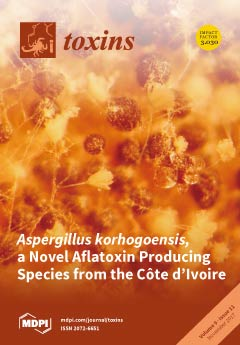
Aspergillus korhogoensis is a new aflatoxinogenic species of the Flavi section isolated from peanuts and peanut paste in Côte d'Ivoire. This view under stereomicroscope of a culture on MEA shows few conidial heads on a field of numerous hard brown sclerotia. These sparse heads are born by small conidiophores located on a thin aerial mycelium. (photo S. Bailly) View this paper
  • Issues are regarded as officially published after their release is announced to the table of contents alert mailing list.
  • You may sign up for e-mail alerts to receive table of contents of newly released issues.
  • PDF is the official format for papers published in both, html and pdf forms. To view the papers in pdf format, click on the "PDF Full-text" link, and use the free Adobe Reader to open them.
Order results
Result details
Section
Select all
Export citation of selected articles as:
1524 KiB  
Correction
Correction: S. Vogelgsang et al. Fusarium Mycotoxins in Swiss Wheat: A Survey of Growers’ Samples between 2007 and 2014 Shows Strong Year and Minor Geographic Effects. Toxins 2017, 9, 246
by Susanne Vogelgsang, Tomke Musa, Irene Bänziger, Andreas Kägi, Thomas D. Bucheli, Felix E. Wettstein, Matias Pasquali and Hans-Rudolf Forrer
Toxins 2017, 9(11), 377; https://doi.org/10.3390/toxins9110377 - 21 Nov 2017
Cited by 6 | Viewed by 3661
Abstract
The authors wish to correct Figures 3–5 in this paper [1]. In all three figures, the y axis stated as a unit “milligram/kg” (mg/kg) instead of “microgram/kg” (µg/kg). Full article
(This article belongs to the Collection Understanding Mycotoxin Occurrence in Food and Feed Chains)
Show Figures

Figure 3

2176 KiB  
Review
Microvesicle Involvement in Shiga Toxin-Associated Infection
by Annie Villysson, Ashmita Tontanahal and Diana Karpman
Toxins 2017, 9(11), 376; https://doi.org/10.3390/toxins9110376 - 19 Nov 2017
Cited by 29 | Viewed by 10450
Abstract
Shiga toxin is the main virulence factor of enterohemorrhagic Escherichia coli, a non-invasive pathogen that releases virulence factors in the intestine, causing hemorrhagic colitis and, in severe cases, hemolytic uremic syndrome (HUS). HUS manifests with acute renal failure, hemolytic anemia and thrombocytopenia. [...] Read more.
Shiga toxin is the main virulence factor of enterohemorrhagic Escherichia coli, a non-invasive pathogen that releases virulence factors in the intestine, causing hemorrhagic colitis and, in severe cases, hemolytic uremic syndrome (HUS). HUS manifests with acute renal failure, hemolytic anemia and thrombocytopenia. Shiga toxin induces endothelial cell damage leading to platelet deposition in thrombi within the microvasculature and the development of thrombotic microangiopathy, mostly affecting the kidney. Red blood cells are destroyed in the occlusive capillary lesions. This review focuses on the importance of microvesicles shed from blood cells and their participation in the prothrombotic lesion, in hemolysis and in the transfer of toxin from the circulation into the kidney. Shiga toxin binds to blood cells and may undergo endocytosis and be released within microvesicles. Microvesicles normally contribute to intracellular communication and remove unwanted components from cells. Many microvesicles are prothrombotic as they are tissue factor- and phosphatidylserine-positive. Shiga toxin induces complement-mediated hemolysis and the release of complement-coated red blood cell-derived microvesicles. Toxin was demonstrated within blood cell-derived microvesicles that transported it to renal cells, where microvesicles were taken up and released their contents. Microvesicles are thereby involved in all cardinal aspects of Shiga toxin-associated HUS, thrombosis, hemolysis and renal failure. Full article
(This article belongs to the Special Issue Ribosome Inactivating Toxins)
Show Figures

Figure 1

239 KiB  
Article
Ultrasonographic Evaluation of Botulinum Toxin Injection Site for the Medial Approach to Tibialis Posterior Muscle in Chronic Stroke Patients with Spastic Equinovarus Foot: An Observational Study
by Alessandro Picelli, Alessio Baricich, Elena Chemello, Nicola Smania, Carlo Cisari, Marialuisa Gandolfi, Nicoletta Cinone, Maurizio Ranieri and Andrea Santamato
Toxins 2017, 9(11), 375; https://doi.org/10.3390/toxins9110375 - 18 Nov 2017
Cited by 9 | Viewed by 5095
Abstract
The tibialis posterior muscle is a frequent target for injection of botulinum toxin during the management of spastic equinovarus foot in adults with post-stroke spasticity. Although it is deep-seated, the needle insertion into the tibialis posterior muscle is usually performed using anatomical landmarks [...] Read more.
The tibialis posterior muscle is a frequent target for injection of botulinum toxin during the management of spastic equinovarus foot in adults with post-stroke spasticity. Although it is deep-seated, the needle insertion into the tibialis posterior muscle is usually performed using anatomical landmarks and safety information obtained from healthy subjects and cadavers. Our aim was to evaluate the botulinum toxin injection site for the medial approach to the tibialis posterior muscle in chronic stroke patients with spastic equinovarus foot. Forty-six patients were evaluated at the affected middle lower leg medial surface with ultrasonography according to the following parameters: tibialis posterior muscle depth, thickness, and echo intensity. As to the spastic tibialis posterior, we found a mean muscle depth of 26.5 mm and a mean muscle thickness of 10.1 mm. Furthermore we observed a median tibialis posterior muscle echo intensity of 3.00 on the Heckmatt scale. The tibialis posterior muscle thickness was found to be inversely associated with its depth (p < 0.001) and echo intensity (p = 0.006). Furthermore, tibialis posterior muscle depth was found to be directly associated with its echo intensity (p = 0.004). Our findings may usefully inform manual needle placement into the tibialis posterior for the botulinum toxin treatment of spastic equinovarus foot in chronic stroke patients. Full article
(This article belongs to the Special Issue Muscle Selection for BoNT)
2471 KiB  
Article
Impact of a Single Oral Acute Dose of Aflatoxin B1 on Liver Function/Cytokines and the Lymphoproliferative Response in C57Bl/6 Mice
by Angélica Tieme Ishikawa, Elisa Yoko Hirooka, Paula Leonello Alvares e Silva, Ana Paula Frederico Rodrigues Loureiro Bracarense, Karina Keller Marques da Costa Flaiban, Claudia Yuri Akagi, Osamu Kawamura, Marcio Carvalho da Costa and Eiko Nakagawa Itano
Toxins 2017, 9(11), 374; https://doi.org/10.3390/toxins9110374 - 17 Nov 2017
Cited by 40 | Viewed by 5801
Abstract
Aflatoxin B1 (AFB1), a mycotoxin found in food and feed, exerts harmful effects on humans and animals. The liver is the earliest target of AFB1, and its effects have been evaluated in animal models exposed to acute or [...] Read more.
Aflatoxin B1 (AFB1), a mycotoxin found in food and feed, exerts harmful effects on humans and animals. The liver is the earliest target of AFB1, and its effects have been evaluated in animal models exposed to acute or chronic doses. Considering the possibility of sporadic ingestion of AFB1-contaminated food, this study investigated the impact of a single oral dose of AFB1 on liver function/cytokines and the lymphoproliferative response in mice. C57BL/6 mice were treated with a single oral AFB1 dose (44, 442 or 663 μg AFB1/kg of body weight) on the first day. Liver function (ALT, γ-GT, and total protein), cytokines (IL-4, IFN-γ, and IL-17), histopathology, and the spleen lymphoproliferative response to mitogens were evaluated on the 5th day. Although AFB1 did not produce any significant changes in the biochemical parameters, 663 μg AFB1/kg-induced hepatic upregulation of IL-4 and IFN-γ, along with liver tissue injury and suppression of the lymphoproliferative response to ConA (p < 0.05). In conclusion, a single oral dose of AFB1 exposure can induce liver tissue lesions, liver cytokine modulation, and immune suppression in C57BL/6 mice. Full article
(This article belongs to the Special Issue Impact of Human Metabolism on the Toxicological Effects of Mycotoxins)
Show Figures

Figure 1

1222 KiB  
Article
Comparative Study of Biological Activities of Venom from Colubrid Snakes Rhabdophis tigrinus (Yamakagashi) and Rhabdophis lateralis
by Yumiko Komori, Toru Hifumi, Akihiko Yamamoto, Atsushi Sakai, Manabu Ato, Kyoko Sawabe and Toshiaki Nikai
Toxins 2017, 9(11), 373; https://doi.org/10.3390/toxins9110373 - 17 Nov 2017
Cited by 4 | Viewed by 4035
Abstract
Rhabdophis lateralis, a colubrid snake distributed throughout the continent of Asia, has recently undergone taxonomic revisions. Previously, Rhabdophis lateralis was classified as a subspecies of R. tigrinus (Yamakagashi) until 2012, when several genetic differences were discovered which classified this snake as its [...] Read more.
Rhabdophis lateralis, a colubrid snake distributed throughout the continent of Asia, has recently undergone taxonomic revisions. Previously, Rhabdophis lateralis was classified as a subspecies of R. tigrinus (Yamakagashi) until 2012, when several genetic differences were discovered which classified this snake as its own species. To elucidate the toxicity of venom from this poorly studied colubrid, various biological activities were compared between the venom from the two snake species. The components of their venom were compared by the elution profiles of reversed-phase HPLC and SDS-PAGE, and gel filtrated fractions were tested for effects on blood coagulation. Proteolytic activities of these fractions were also assayed by using synthetic substrates, fibrinogen, and matrix proteins. Similar to the R. tigrinus venom, the higher molecular weight fraction of R. lateralis venom contained a prothrombin activator. Both prothrombin time (PT) and activated partial thromboplastin time (APTT) of human plasma were shortened by the addition of R. lateralis and R. tigrinus venom. The thrombin formation was estimated by the uses of SDS-PAGE and chromogenic substrates. These venom fractions also possessed very specific proteinase activity on human fibrinogen, but the substrates for matrix metalloproteinase, such as collagen and laminin, were not hydrolyzed. However, there were some notable differences in reactivity to synthetic substrates for matrix metalloproteinase, and R. tigrinus venom possessed relatively higher activity. Our chemical investigation indicates that the components included in both venoms resemble each other closely. However, the ratio of components and proteolytic activity of some ingredients are slightly different, indicating differences between two closely-related snakes. Full article
(This article belongs to the Section Animal Venoms)
Show Figures

Figure 1

893 KiB  
Review
G-Protein Coupled Receptors Targeted by Analgesic Venom Peptides
by James T. Daniel and Richard J. Clark
Toxins 2017, 9(11), 372; https://doi.org/10.3390/toxins9110372 - 16 Nov 2017
Cited by 14 | Viewed by 7217
Abstract
Chronic pain is a complex and debilitating condition associated with a large personal and socioeconomic burden. Current pharmacological approaches to treating chronic pain such as opioids, antidepressants and anticonvulsants exhibit limited efficacy in many patients and are associated with dose-limiting side effects that [...] Read more.
Chronic pain is a complex and debilitating condition associated with a large personal and socioeconomic burden. Current pharmacological approaches to treating chronic pain such as opioids, antidepressants and anticonvulsants exhibit limited efficacy in many patients and are associated with dose-limiting side effects that hinder their clinical use. Therefore, improved strategies for the pharmacological treatment of pathological pain are urgently needed. G-protein coupled receptors (GPCRs) are ubiquitously expressed on the surface of cells and act to transduce extracellular signals and regulate physiological processes. In the context of pain, numerous and diverse families of GPCRs expressed in pain pathways regulate most aspects of physiological and pathological pain and are thus implicated as potential targets for therapy of chronic pain. In the search for novel compounds that produce analgesia via GPCR modulation, animal venoms offer an enormous and virtually untapped source of potent and selective peptide molecules. While many venom peptides target voltage-gated and ligand-gated ion channels to inhibit neuronal excitability and blunt synaptic transmission of pain signals, only a small proportion are known to interact with GPCRs. Of these, only a few have shown analgesic potential in vivo. Here we review the current state of knowledge regarding venom peptides that target GPCRs to produce analgesia, and their development as therapeutic compounds. Full article
(This article belongs to the Special Issue Animal Venoms and Pain)
Show Figures

Figure 1

2372 KiB  
Article
Ameliorative Effects of Grape Seed Proanthocyanidin Extract on Growth Performance, Immune Function, Antioxidant Capacity, Biochemical Constituents, Liver Histopathology and Aflatoxin Residues in Broilers Exposed to Aflatoxin B1
by Shahid Ali Rajput, Lvhui Sun, Niya Zhang, Mahmoud Mohamed Khalil, Xin Gao, Zhao Ling, Luoyi Zhu, Farhan Anwar Khan, Jiacai Zhang and Desheng Qi
Toxins 2017, 9(11), 371; https://doi.org/10.3390/toxins9110371 - 15 Nov 2017
Cited by 87 | Viewed by 7603 | Correction
Abstract
Aflatoxicosis is a grave threat to the poultry industry. Dietary supplementation with antioxidants showed a great potential in enhancing the immune system; hence, protecting animals against aflatoxin B1-induced toxicity. Grape seed proanthocyanidin extract (GSPE) one of the most well-known and powerful [...] Read more.
Aflatoxicosis is a grave threat to the poultry industry. Dietary supplementation with antioxidants showed a great potential in enhancing the immune system; hence, protecting animals against aflatoxin B1-induced toxicity. Grape seed proanthocyanidin extract (GSPE) one of the most well-known and powerful antioxidants. Therefore, the purpose of this research was to investigate the effectiveness of GSPE in the detoxification of AFB1 in broilers. A total of 300 one-day-old Cobb chicks were randomly allocated into five treatments of six replicates (10 birds per replicate), fed ad libitum for four weeks with the following dietary treatments: 1. Basal diet (control); 2. Basal diet + 1 mg/kg AFB1 contaminated corn (AFB1); 3. Basal diet + GSPE 250 mg/kg; (GSPE 250 mg/kg) 4. Basal diet + AFB1 (1 mg/kg) + GSPE 250 mg/kg; (AFB1 + GSPE 250 mg/kg) 5. Basal diet + AFB1 (1mg/kg) + GSPE 500 mg/kg, (AFB1 + GSPE 500 mg/kg). When compared with the control group, feeding broilers with AFB1 alone significantly reduced growth performance, serum immunoglobulin contents, negatively altered serum biochemical contents, and enzyme activities, and induced histopathological lesion in the liver. In addition, AFB1 significantly increased malondialdehyde content and decreased total superoxide dismutase, catalase, glutathione peroxide, glutathione-S transferase, glutathione reductase activities, and glutathione concentration within the liver and serum. The supplementation of GSPE (250 and 500 mg/kg) to AFB1 contaminated diet reduced AFB1 residue in the liver and significantly mitigated AFB1 negative effects. From these results, it can be concluded that dietary supplementation of GSPE has protective effects against aflatoxicosis caused by AFB1 in broiler chickens. Full article
(This article belongs to the Section Mycotoxins)
Show Figures

Figure 1

454 KiB  
Review
Evidence to Use Botulinum Toxin Injections in Tension-Type Headache Management: A Systematic Review
by Mieszko Wieckiewicz, Natalia Grychowska, Marek Zietek, Gniewko Wieckiewicz and Joanna Smardz
Toxins 2017, 9(11), 370; https://doi.org/10.3390/toxins9110370 - 15 Nov 2017
Cited by 19 | Viewed by 5486
Abstract
Tension-type headache (TTH) is the most common type of chronic recurring head pain. It can occur twice as often in women as in men. It is the most common type of headache. Its lifetime prevalence is 30% to 78% in the general population. [...] Read more.
Tension-type headache (TTH) is the most common type of chronic recurring head pain. It can occur twice as often in women as in men. It is the most common type of headache. Its lifetime prevalence is 30% to 78% in the general population. TTH treatment should be multilevel. It often consists of taking pain medication, muscle relaxants, antidepressants, using biofeedback therapy, acupuncture, and attending behavioral therapy. Several clinical trials also suggest that botulinum toxin (BTX) may be an effective treatment option for such patients. The aim of this study was to evaluate if BTX can be used as a treatment method in TTH in the light of current medical literature. The authors searched the PubMed, EBSCOhost, OVID, Web of Knowledge, Cochrane Library and CINAHL databases to identify relevant publications. The authors finally included 11 papers—prospective and retrospective cohort studies. Among most of the selected studies, there was a significant correlation between using BTX and reduction of TTH pain intensity and severity. By analyzing qualified studies, it can be concluded that botulinum toxin seems to be effective in TTH management. Full article
(This article belongs to the Special Issue Muscle Selection for BoNT)
Show Figures

Figure 1

2020 KiB  
Article
Membrane-Active Properties of an Amphitropic Peptide from the CyaA Toxin Translocation Region
by Alexis Voegele, Orso Subrini, Nicolas Sapay, Daniel Ladant and Alexandre Chenal
Toxins 2017, 9(11), 369; https://doi.org/10.3390/toxins9110369 - 14 Nov 2017
Cited by 21 | Viewed by 5470
Abstract
The adenylate cyclase toxin CyaA is involved in the early stages of infection by Bordetella pertussis, the causative agent of whooping cough. CyaA intoxicates target cells by a direct translocation of its catalytic domain (AC) across the plasma membrane and produces supraphysiological [...] Read more.
The adenylate cyclase toxin CyaA is involved in the early stages of infection by Bordetella pertussis, the causative agent of whooping cough. CyaA intoxicates target cells by a direct translocation of its catalytic domain (AC) across the plasma membrane and produces supraphysiological levels of cAMP, leading to cell death. The molecular process of AC translocation remains largely unknown, however. We have previously shown that deletion of residues 375–485 of CyaA selectively abrogates AC translocation into eukaryotic cells. We further identified within this “translocation region” (TR), P454 (residues 454–484), a peptide that exhibits membrane-active properties, i.e., is able to bind and permeabilize lipid vesicles. Here, we analyze various sequences from CyaA predicted to be amphipatic and show that although several of these peptides can bind membranes and adopt a helical conformation, only the P454 peptide is able to permeabilize membranes. We further characterize the contributions of the two arginine residues of P454 to membrane partitioning and permeabilization by analyzing the peptide variants in which these residues are substituted by different amino acids (e.g., A, K, Q, and E). Our data shows that both arginine residues significantly contribute, although diversely, to the membrane-active properties of P454, i.e., interactions with both neutral and anionic lipids, helix formation in membranes, and disruption of lipid bilayer integrity. These results are discussed in the context of the translocation process of the full-length CyaA toxin. Full article
(This article belongs to the Special Issue Adenylate Cyclase (CyaA) Toxin)
Show Figures

Figure 1

1370 KiB  
Communication
The Apoptogenic Toxin AIP56 Is Secreted by the Type II Secretion System of Photobacterium damselae subsp. piscicida
by Ana Do Vale, Cassilda Pereira, Carlos R. Osorio and Nuno M. S. dos Santos
Toxins 2017, 9(11), 368; https://doi.org/10.3390/toxins9110368 - 14 Nov 2017
Cited by 13 | Viewed by 4723
Abstract
AIP56 (apoptosis-inducing protein of 56 kDa) is a key virulence factor of Photobacterium damselae subsp. piscicida (Phdp), the causative agent of a septicaemia affecting warm water marine fish species. Phdp-associated pathology is triggered by AIP56, a short trip AB toxin with a [...] Read more.
AIP56 (apoptosis-inducing protein of 56 kDa) is a key virulence factor of Photobacterium damselae subsp. piscicida (Phdp), the causative agent of a septicaemia affecting warm water marine fish species. Phdp-associated pathology is triggered by AIP56, a short trip AB toxin with a metalloprotease A domain that cleaves the p65 subunit of NF-κB, an evolutionarily conserved transcription factor that regulates the expression of inflammatory and anti-apoptotic genes and plays a central role in host responses to infection. During infection by Phdp, AIP56 is systemically disseminated and induces apoptosis of macrophages and neutrophils, compromising the host phagocytic defence and contributing to the genesis of pathology. Although it is well established that the secretion of AIP56 is crucial for Phdp pathogenicity, the protein secretion systems operating in Phdp and the mechanism responsible for the extracellular release of the toxin remain unknown. Here, we report that Phdp encodes a type II secretion system (T2SS) and show that mutation of the EpsL component of this system impairs AIP56 secretion. This work demonstrates that Phdp has a functional T2SS that mediates secretion of its key virulence factor AIP56. Full article
(This article belongs to the Section Bacterial Toxins)
Show Figures

Figure 1

260 KiB  
Article
Management of Ciguatoxin Risk in Eastern Australia
by Hazel Farrell, Shauna A. Murray, Anthony Zammit and Alan W. Edwards
Toxins 2017, 9(11), 367; https://doi.org/10.3390/toxins9110367 - 14 Nov 2017
Cited by 22 | Viewed by 6378
Abstract
Between 2014 and 2016, five cases of ciguatera fish poisoning (CFP), involving twenty four individuals, were linked to Spanish Mackerel (Scomberomorus commerson) caught in the coastal waters of the state of New South Wales (NSW) on the east coast of Australia. [...] Read more.
Between 2014 and 2016, five cases of ciguatera fish poisoning (CFP), involving twenty four individuals, were linked to Spanish Mackerel (Scomberomorus commerson) caught in the coastal waters of the state of New South Wales (NSW) on the east coast of Australia. Previously, documented cases of CFP in NSW were few, and primarily linked to fish imported from other regions. Since 2015, thirteen individuals were affected across four additional CFP cases in NSW, linked to fish imported from tropical locations. The apparent increase in CFP in NSW from locally sourced catch, combined with the risk of CFP from imported fish, has highlighted several considerations that should be incorporated into risk management strategies to minimize CFP exposure for seafood consumers. Full article
(This article belongs to the Special Issue Public Health Outreach to Prevention of Aquatic Toxin Exposure)
1010 KiB  
Article
Differential Gene Expression Analysis of Bovine Macrophages after Exposure to the Penicillium Mycotoxins Citrinin and/or Ochratoxin A
by Kristen M. Brennan, Se-Young Oh, Alexandros Yiannikouris, Daniel E. Graugnard and Niel A. Karrow
Toxins 2017, 9(11), 366; https://doi.org/10.3390/toxins9110366 - 13 Nov 2017
Cited by 20 | Viewed by 5070
Abstract
Mycotoxins produced by fungal species commonly contaminate livestock feedstuffs, jeopardizing their health and diminishing production. Citrinin (CIT) and ochratoxin A (OTA) are mycotoxins produced by Penicillium spp. and commonly co-occur. Both CIT and OTA can modulate immune response by inhibiting cell proliferation and [...] Read more.
Mycotoxins produced by fungal species commonly contaminate livestock feedstuffs, jeopardizing their health and diminishing production. Citrinin (CIT) and ochratoxin A (OTA) are mycotoxins produced by Penicillium spp. and commonly co-occur. Both CIT and OTA can modulate immune response by inhibiting cell proliferation and differentiation, altering cell metabolism, and triggering programmed cell death. The objective of this study was to determine the effects of sublethal exposure (i.e., the concentration that inhibited cell proliferation by 25% (IC25)) to CIT, OTA or CIT + OTA on the bovine macrophage transcriptome. Gene expression was determined using the Affymetrix Bovine Genome Array. After 6 h of exposure to CIT, OTA or CIT + OTA, the number of differentially expressed genes (DEG), respectively, was as follows: 1471 genes (822 up-regulated, 649 down-regulated), 5094 genes (2611 up-regulated, 2483 down-regulated) and 7624 genes (3984 up-regulated, 3640 down-regulated). Of these, 179 genes (88 up-regulated, 91 down-regulated) were commonly expressed between treatments. After 24 h of exposure to CIT, OTA or CIT + OTA the number of DEG, respectively, was as follows: 3230 genes (1631 up-regulated, 1599 down-regulated), 8558 genes (4167 up-regulated, 4391 down-regulated), and 10,927 genes (6284 up-regulated, 4643 down-regulated). Of these, 770 genes (247 up-regulated, 523 down-regulated) were commonly expressed between treatments. The categorization of common biological functions and pathway analysis suggests that the IC25 of both CIT and OTA, or their combination, induces cellular oxidative stress, a slowing of cell cycle progression, and apoptosis. Collectively, these effects contribute to inhibiting bovine macrophage proliferation. Full article
(This article belongs to the Collection Ochratoxins-Collection)
Show Figures

Figure 1

839 KiB  
Article
Proteomic Characterization of the Venom of Five Bombus (Thoracobombus) Species
by Nezahat Pınar Barkan, Mustafa Bilal Bayazit and Duygu Ozel Demiralp
Toxins 2017, 9(11), 362; https://doi.org/10.3390/toxins9110362 - 11 Nov 2017
Cited by 12 | Viewed by 4546
Abstract
Venomous animals use venom, a complex biofluid composed of unique mixtures of proteins and peptides, to act on vital systems of the prey or predator. In bees, venom is solely used for defense against predators. However, the venom composition of bumble bees ( [...] Read more.
Venomous animals use venom, a complex biofluid composed of unique mixtures of proteins and peptides, to act on vital systems of the prey or predator. In bees, venom is solely used for defense against predators. However, the venom composition of bumble bees (Bombus sp.) is largely unknown. The Thoracobombus subgenus of Bombus sp. is a diverse subgenus represented by 14 members across Turkey. In this study, we sought out to proteomically characterize the venom of five Thoracobombus species by using bottom-up proteomic techniques. We have obtained two-dimensional polyacrylamide gel (2D-PAGE) images of each species’ venom sample. We have subsequently identified the protein spots by using matrix assisted laser desorption ionization/time-of-flight mass spectrometry (MALDI-TOF MS). We have identified 47 proteins for Bombus humilis, 32 for B. pascuorum, 60 for B. ruderarius, 39 for B. sylvarum, and 35 for B. zonatus. Moreover, we illustrated that intensities of 2DE protein spots corresponding to putative venom toxins vary in a species-specific manner. Our analyses provide the primary proteomic characterization of five bumble bee species’ venom composition. Full article
(This article belongs to the Section Animal Venoms)
Show Figures

Figure 1

214 KiB  
Review
Botulinum Toxin in Management of Limb Tremor
by Elina Zakin and David Simpson
Toxins 2017, 9(11), 365; https://doi.org/10.3390/toxins9110365 - 10 Nov 2017
Cited by 15 | Viewed by 4763
Abstract
Essential tremor is characterized by persistent, usually bilateral and symmetric, postural or kinetic activation of agonist and antagonist muscles involving either the distal or proximal upper extremity. Quality of life is often affected and one’s ability to perform daily tasks becomes impaired. Oral [...] Read more.
Essential tremor is characterized by persistent, usually bilateral and symmetric, postural or kinetic activation of agonist and antagonist muscles involving either the distal or proximal upper extremity. Quality of life is often affected and one’s ability to perform daily tasks becomes impaired. Oral therapies, including propranolol and primidone, can be effective in the management of essential tremor, although adverse effects can limit their use and about 50% of individuals lack response to oral pharmacotherapy. Locally administered botulinum toxin injection has become increasingly useful in the management of essential tremor. Targeting of select muscles with botulinum toxin is an area of active research, and muscle selection has important implications for toxin dosing and functional outcomes. The use of anatomical landmarks with palpation, EMG guidance, electrical stimulation, and ultrasound has been studied as a technique for muscle localization in toxin injection. Earlier studies implemented a standard protocol for the injection of (predominantly) wrist flexors and extensors using palpation and EMG guidance. Targeting of muscles by selection of specific activators of tremor (tailored to each patient) using kinematic analysis might allow for improvement in efficacy, including functional outcomes. It is this individualized muscle selection and toxin dosing (requiring injection within various sites of a single muscle) that has allowed for success in the management of tremors. Full article
1520 KiB  
Review
Exolysin Shapes the Virulence of Pseudomonas aeruginosa Clonal Outliers
by Emeline Reboud, Pauline Basso, Antoine P. Maillard, Philippe Huber and Ina Attrée
Toxins 2017, 9(11), 364; https://doi.org/10.3390/toxins9110364 - 09 Nov 2017
Cited by 21 | Viewed by 6298
Abstract
Bacterial toxins are important weapons of toxicogenic pathogens. Depending on their origin, structure and targets, they show diverse mechanisms of action and effects on eukaryotic cells. Exolysin is a secreted 170 kDa pore-forming toxin employed by clonal outliers of Pseudomonas aeruginosa providing to [...] Read more.
Bacterial toxins are important weapons of toxicogenic pathogens. Depending on their origin, structure and targets, they show diverse mechanisms of action and effects on eukaryotic cells. Exolysin is a secreted 170 kDa pore-forming toxin employed by clonal outliers of Pseudomonas aeruginosa providing to some strains a hyper-virulent behaviour. This group of strains lacks the major virulence factor used by classical strains, the Type III secretion system. Here, we review the structural features of the toxin, the mechanism of its secretion and the effects of the pore formation on eukaryotic cells. Full article
(This article belongs to the Section Bacterial Toxins)
Show Figures

Graphical abstract

614 KiB  
Article
Awareness and Prevalence of Mycotoxin Contamination in Selected Nigerian Fermented Foods
by Ifeoluwa Adekoya, Patrick Njobeh, Adewale Obadina, Cynthia Chilaka, Sheila Okoth, Marthe De Boevre and Sarah De Saeger
Toxins 2017, 9(11), 363; https://doi.org/10.3390/toxins9110363 - 08 Nov 2017
Cited by 35 | Viewed by 8080
Abstract
Fermented food samples (n = 191) including maize gruel (ogi), sorghum gruel (ogi-baba), melon seed (ogiri), locust bean (iru) and African oil bean seed (ugba) from Southwest Nigeria were quantified for 23 [...] Read more.
Fermented food samples (n = 191) including maize gruel (ogi), sorghum gruel (ogi-baba), melon seed (ogiri), locust bean (iru) and African oil bean seed (ugba) from Southwest Nigeria were quantified for 23 mycotoxins, including aflatoxin B1 (AFB1), fumonisin B1 (FB1), and sterigmatocystin (STE) using liquid chromatography-tandem mass spectrometry. The practices, perceived understanding and health risks related to fungal and mycotoxin contamination amongst fermented food sellers was also established. Data obtained revealed that 82% of the samples had mycotoxins occurring singly or in combination. FB1 was present in 83% of ogi-baba samples, whereas 20% of ugba samples contained AFB1 (range: 3 to 36 µg/kg) and STE was present in 29% of the ogi samples. In terms of multi-mycotoxin contamination, FB1 + FB2 + FB3 + STE + AFB1 + alternariol + HT-2 co-occurred within one sample. The awareness study revealed that 98% of respondents were unaware of mycotoxin contamination, and their education level slightly correlated with their level of awareness (p < 0.01, r = 0.308). The extent to which the analyzed mycotoxins contaminated these food commodities, coupled with the poor perception of the population under study on fungi and mycotoxins, justifies the need to enact fungal and mycotoxin mitigation strategies along the food chain. Full article
Show Figures

Figure 1

1654 KiB  
Article
Efficacy of Bee Venom Acupuncture for Chronic Low Back Pain: A Randomized, Double-Blinded, Sham-Controlled Trial
by Byung-Kwan Seo, Kyungsun Han, Ojin Kwon, Dae-Jean Jo and Jun-Hwan Lee
Toxins 2017, 9(11), 361; https://doi.org/10.3390/toxins9110361 - 07 Nov 2017
Cited by 34 | Viewed by 10459
Abstract
Bee venom acupuncture (BVA) is an effective treatment for chronic low back pain (CLBP) through the pharmacological effects of bee venom and the simultaneous stimulation of acupoints. However, evidence of its efficacy and safety in humans remains unclear. Using a double-blind, randomized study, [...] Read more.
Bee venom acupuncture (BVA) is an effective treatment for chronic low back pain (CLBP) through the pharmacological effects of bee venom and the simultaneous stimulation of acupoints. However, evidence of its efficacy and safety in humans remains unclear. Using a double-blind, randomized study, 54 patients with non-specific CLBP were assigned to the BVA and sham groups. All participants underwent six sessions of real or sham BVA for 3 weeks, in addition to administration of 180 mg of loxonin per day. The primary outcome, that is, “bothersomeness” derived from back pain, was assessed using the visual analog scale. Secondary outcomes included pain intensity, dysfunction related to back pain (Oswestry Disability Index), quality of life (EuroQol 5-Dimension), and depressive mood (Beck’s depression inventory). Outcomes were evaluated every week during the treatment period and followed up at weeks 4, 8, and 12. After 3 weeks of the treatment, significant improvements were observed in the bothersomeness, pain intensity, and functional status in the BVA group compared with the sham group. Although minimal adverse events were observed in both groups, subsequent recovery was achieved without treatment. Consequently, our results suggest that it can be used along with conventional pharmacological therapies for the treatment of CLBP. Full article
(This article belongs to the Special Issue Animal Venoms and Pain)
Show Figures

Figure 1

2303 KiB  
Article
Effects of Lonomia obliqua Venom on Vascular Smooth Muscle Cells: Contribution of NADPH Oxidase-Derived Reactive Oxygen Species
by João Alfredo Moraes, Genilson Rodrigues, Vany Nascimento-Silva, Mariana Renovato-Martins, Markus Berger, Jorge Almeida Guimarães and Christina Barja-Fidalgo
Toxins 2017, 9(11), 360; https://doi.org/10.3390/toxins9110360 - 07 Nov 2017
Cited by 15 | Viewed by 5371
Abstract
Envenomation caused by human contact with the caterpillar Lonomia is characterized by deleterious effects on coagulation and patency of blood vessels. The cellular effects induced by Lonomia obliqua venom highlights its capacity to activate endothelial cells, leading to a proinflammatory phenotype. Having more [...] Read more.
Envenomation caused by human contact with the caterpillar Lonomia is characterized by deleterious effects on coagulation and patency of blood vessels. The cellular effects induced by Lonomia obliqua venom highlights its capacity to activate endothelial cells, leading to a proinflammatory phenotype. Having more knowledge about the mechanisms involved in envenomation may contribute to better treatment. We aimed to evaluate the effects of Lonomia obliqua caterpillar bristle extract (LOCBE) on vascular smooth muscle cells (VSMC). We observed that LOCBE induced VSMC migration, which was preceded by alterations in actin cytoskeleton dynamics and Focal Adhesion Kinase activation. LOCBE also induced Extracellular Signal-Regulated Kinase (ERK) phosphorylation in VSMC, and the inhibition of this pathway impaired cell proliferation. Stimulation of VSMC with LOCBE triggered reactive oxygen species (ROS) production through the activation of NADPH oxidase. The rapid increase in these ROS further induced mitochondrial ROS production, however only NADPH oxidase-derived ROS were involved in ERK activation in VSMC. We that demonstrated the chemotactic and proliferative effects of LOCBE on VSMC were dependent on ROS production, mainly through NADPH oxidase. Together, the data show that Lonomia obliqua venom can interact with and activate VSMC. These effects rely on ROS production, suggesting new potential targets for treatment against vascular damage during envenomation. Full article
(This article belongs to the Section Animal Venoms)
Show Figures

Figure 1

2958 KiB  
Article
TGF-β1/Smad3 Signaling Pathway Mediates T-2 Toxin-Induced Decrease of Type II Collagen in Cultured Rat Chondrocytes
by Yang Li, Ning Zou, Jing Wang, Ke-Wei Wang, Fu-Yuan Li, Fu-Xun Chen, Bing-Yu Sun and Dian-Jun Sun
Toxins 2017, 9(11), 359; https://doi.org/10.3390/toxins9110359 - 05 Nov 2017
Cited by 18 | Viewed by 4563
Abstract
T-2 toxin can cause damage to the articular cartilage, but the molecular mechanism remains unclear. By employing the culture of rat chondrocytes, we investigated the effect of the TGF-β1/Smad3 signaling pathway on the damage to chondrocytes induced by T-2 toxin. It was found [...] Read more.
T-2 toxin can cause damage to the articular cartilage, but the molecular mechanism remains unclear. By employing the culture of rat chondrocytes, we investigated the effect of the TGF-β1/Smad3 signaling pathway on the damage to chondrocytes induced by T-2 toxin. It was found that T-2 toxin could reduce cell viability and increased the number of apoptotic cells when compared with the control group. After the addition of the T-2 toxin, the production of type II collagen was reduced at mRNA and protein levels, while the levels of TGF-β1, Smad3, ALK5, and MMP13 were upregulated. The production of the P-Smad3 protein was also increased. Inhibitors of TGF-β1 and Smad3 were able to reverse the effect of the T-2 toxin on the protein level of above-mentioned signaling molecules. The T-2 toxin could promote the level of MMP13 via the stimulation of TGF-β1 signaling in chondrocytes, resulting in the downregulation of type II collagen and chondrocyte damage. Smad3 may be involved in the degradation of type II collagen, but the Smad3 has no connection with the regulation of MMP13 level. This study provides a new clue to elucidate the mechanism of T-2 toxin-induced chondrocyte damage. Full article
(This article belongs to the Section Mycotoxins)
Show Figures

Figure 1

11256 KiB  
Article
The First Cry2Ac-Type Protein Toxic to Helicoverpa armigera: Cloning and Overexpression of Cry2ac7 Gene from SBS-BT1 Strain of Bacillus thuringiensis
by Faiza Saleem and Abdul Rauf Shakoori
Toxins 2017, 9(11), 358; https://doi.org/10.3390/toxins9110358 - 03 Nov 2017
Cited by 7 | Viewed by 4957
Abstract
The Cry (crystal) proteins from Bacillus thuringiensis are known to have toxicity against a variety of insects and have been exploited to control insect pests through transgenic plants and biopesticides. B. thuringiensis SBS BT-1 carrying the cry2 genes was isolated from soil samples [...] Read more.
The Cry (crystal) proteins from Bacillus thuringiensis are known to have toxicity against a variety of insects and have been exploited to control insect pests through transgenic plants and biopesticides. B. thuringiensis SBS BT-1 carrying the cry2 genes was isolated from soil samples in Pakistan. The 2-kb full length cry2Ac gene was cloned, sequenced, and submitted to the EMBL DNA database (Accession No. AM292031). For expression analysis, Escherichia coli DH5α was transformed with the fragment sub-cloned in pET22b expression vector using NdeI and HindIII restriction sites, and later confirmed by restriction endonuclease analysis. To assess the toxicity of Cry2Ac7 protein against lepidopteran and dipteran insects, BL21 (codon plus) strain of E. coli was further transformed with the recombinant plasmid. The 65-kDa protein was expressed in the form of inclusion bodies up to 180 OD units per liter of the medium. Inclusions were washed with a buffer containing 1.5% Triton-X 100 and >90% pure Cry2Ac7 was obtained. The inclusion bodies were dissolved in 50 mM K2CO3 (pH 11.5), dialyzed, and freeze-dried. This freeze-dried protein as well as inclusion bodies were used in bioassays against larvae of Helicoverpa armigera and Musca domestica. The freeze-dried protein was toxic to H. armigera larvae with an LC50 value of 131 ng/mL. However, Cry2Ac7 produced in E. coli did not show any mortality to M. domestica larvae. This is the first report of Cry2Ac protein toxic to H. armigera. Full article
(This article belongs to the Section Bacterial Toxins)
Show Figures

Figure 1

643 KiB  
Perspective
The Unexpected Tuners: Are LncRNAs Regulating Host Translation during Infections?
by Primoz Knap, Toma Tebaldi, Francesca Di Leva, Marta Biagioli, Mauro Dalla Serra and Gabriella Viero
Toxins 2017, 9(11), 357; https://doi.org/10.3390/toxins9110357 - 03 Nov 2017
Cited by 5 | Viewed by 4659
Abstract
Pathogenic bacteria produce powerful virulent factors, such as pore-forming toxins, that promote their survival and cause serious damage to the host. Host cells reply to membrane stresses and ionic imbalance by modifying gene expression at the epigenetic, transcriptional and translational level, to recover [...] Read more.
Pathogenic bacteria produce powerful virulent factors, such as pore-forming toxins, that promote their survival and cause serious damage to the host. Host cells reply to membrane stresses and ionic imbalance by modifying gene expression at the epigenetic, transcriptional and translational level, to recover from the toxin attack. The fact that the majority of the human transcriptome encodes for non-coding RNAs (ncRNAs) raises the question: do host cells deploy non-coding transcripts to rapidly control the most energy-consuming process in cells—i.e., host translation—to counteract the infection? Here, we discuss the intriguing possibility that membrane-damaging toxins induce, in the host, the expression of toxin-specific long non-coding RNAs (lncRNAs), which act as sponges for other molecules, encoding small peptides or binding target mRNAs to depress their translation efficiency. Unravelling the function of host-produced lncRNAs upon bacterial infection or membrane damage requires an improved understanding of host lncRNA expression patterns, their association with polysomes and their function during this stress. This field of investigation holds a unique opportunity to reveal unpredicted scenarios and novel approaches to counteract antibiotic-resistant infections. Full article
(This article belongs to the Special Issue Cellular Entry of Binary and Pore-Forming Bacterial Toxins)
Show Figures

Graphical abstract

1418 KiB  
Review
Chemodenervation of the Larynx
by Rachel Kaye and Andrew Blitzer
Toxins 2017, 9(11), 356; https://doi.org/10.3390/toxins9110356 - 02 Nov 2017
Cited by 9 | Viewed by 12952
Abstract
Botulinum neurotoxin (BoNT) has existed for thousands of years; however, it was not medically utilized until investigations into its therapeutic use began in sincerity during the late 1970s and 1980s. This, coupled with the reclassification of spasmodic dysphonia as a focal dystonia, led [...] Read more.
Botulinum neurotoxin (BoNT) has existed for thousands of years; however, it was not medically utilized until investigations into its therapeutic use began in sincerity during the late 1970s and 1980s. This, coupled with the reclassification of spasmodic dysphonia as a focal dystonia, led to the use of chemodenervation for this disorder, which has since become a refined technique. Indeed, due to its safety and efficacy, BoNT has been investigated in multiple neurolaryngology disorders, including spasmodic dysphonia, vocal tremor, and muscle tension dysphonia. BoNT has been shown to be a useful and safe adjunct in the treatment for these disorders and may reduce or eliminate oral pharmacotherapy and/or prevent the need for a surgical intervention. We present the historical background, development, proposed mechanisms of action, uses, and techniques for administering BoNT for laryngeal disorders, with a particular focus on spasmodic dysphonia. Full article
(This article belongs to the Special Issue Muscle Selection for BoNT)
Show Figures

Figure 1

4349 KiB  
Article
Influence of Glycine and Arginine on Cylindrospermopsin Production and aoa Gene Expression in Aphanizomenon ovalisporum
by Ángel Barón-Sola, Francisca Fernández del Campo and Soledad Sanz-Alférez
Toxins 2017, 9(11), 355; https://doi.org/10.3390/toxins9110355 - 01 Nov 2017
Cited by 4 | Viewed by 4474
Abstract
Arginine (Arg) and glycine (Gly) seem to be the only substrates accepted by the amidinotransferase that catalyze the first step of the synthesis pathway of the cyanotoxin cylindrospermopsin (CYN), leading to guanidinoacetate (GAA). Here, the effect of these amino acids on the production [...] Read more.
Arginine (Arg) and glycine (Gly) seem to be the only substrates accepted by the amidinotransferase that catalyze the first step of the synthesis pathway of the cyanotoxin cylindrospermopsin (CYN), leading to guanidinoacetate (GAA). Here, the effect of these amino acids on the production of CYN in cultures of the cylindrospermopsin-producing strain, Aphanizomenon ovalisporum UAM-MAO, has been studied. Arg clearly increased CYN content, the increment appearing triphasic along the culture. On the contrary, Gly caused a decrease of CYN, observable from the first day on. Interestingly, the transcript of the gene ntcA, key in nitrogen metabolism control, was also enhanced in the presence of Arg and/or Gly, the trend of the transcript oscillations being like that of aoa/cyr. The inhibitory effect of Gly in CYN production seems not to result from diminishing the activity of genes considered involved in CYN synthesis, since Gly, as Arg, enhance the transcription of genes aoaA-C and cyrJ. On the other hand, culture growth is affected by Arg and Gly in a similar way to CYN production, with Arg stimulating and Gly impairing it. Taken together, our data show that the influence of both Arg and Gly on CYN changes seems not to be due to a specific effect on the first step of CYN synthesis; it rather appears to be the result of changes in the physiological cell status. Full article
(This article belongs to the Special Issue Selected Papers from the 5th Iberoamerican Cyanotoxins Meeting)
Show Figures

Figure 1

15138 KiB  
Article
Molecular Dynamics Simulation Reveals Specific Interaction Sites between Scorpion Toxins and Kv1.2 Channel: Implications for Design of Highly Selective Drugs
by Shouli Yuan, Bin Gao and Shunyi Zhu
Toxins 2017, 9(11), 354; https://doi.org/10.3390/toxins9110354 - 01 Nov 2017
Cited by 6 | Viewed by 4772
Abstract
The Kv1.2 channel plays an important role in the maintenance of resting membrane potential and the regulation of the cellular excitability of neurons, whose silencing or mutations can elicit neuropathic pain or neurological diseases (e.g., epilepsy and ataxia). Scorpion venom contains [...] Read more.
The Kv1.2 channel plays an important role in the maintenance of resting membrane potential and the regulation of the cellular excitability of neurons, whose silencing or mutations can elicit neuropathic pain or neurological diseases (e.g., epilepsy and ataxia). Scorpion venom contains a variety of peptide toxins targeting the pore region of this channel. Despite a large amount of structural and functional data currently available, their detailed interaction modes are poorly understood. In this work, we choose four Kv1.2-targeted scorpion toxins (Margatoxin, Agitoxin-2, OsK-1, and Mesomartoxin) to construct their complexes with Kv1.2 based on the experimental structure of ChTx-Kv1.2. Molecular dynamics simulation of these complexes lead to the identification of hydrophobic patches, hydrogen-bonds, and salt bridges as three essential forces mediating the interactions between this channel and the toxins, in which four Kv1.2-specific interacting amino acids (D353, Q358, V381, and T383) are identified for the first time. This discovery might help design highly selective Kv1.2-channel inhibitors by altering amino acids of these toxins binding to the four channel residues. Finally, our results provide new evidence in favor of an induced fit model between scorpion toxins and K+ channel interactions. Full article
(This article belongs to the Special Issue Toxins in Drug Discovery and Pharmacology)
Show Figures

Figure 1

3858 KiB  
Article
Aspergillus korhogoensis, a Novel Aflatoxin Producing Species from the Côte d’Ivoire
by Amaranta Carvajal-Campos, Ama Lethicia Manizan, Souria Tadrist, David Koffi Akaki, Rose Koffi-Nevry, Geromy G. Moore, Stephen O. Fapohunda, Sylviane Bailly, Didier Montet, Isabelle P. Oswald, Sophie Lorber, Catherine Brabet and Olivier Puel
Toxins 2017, 9(11), 353; https://doi.org/10.3390/toxins9110353 - 31 Oct 2017
Cited by 32 | Viewed by 8570
Abstract
Several strains of a new aflatoxigenic species of Aspergillus, A. korhogoensis, were isolated in the course of a screening study involving species from section Flavi found contaminating peanuts (Arachis hypogaea) and peanut paste in the Côte d’Ivoire. Based on [...] Read more.
Several strains of a new aflatoxigenic species of Aspergillus, A. korhogoensis, were isolated in the course of a screening study involving species from section Flavi found contaminating peanuts (Arachis hypogaea) and peanut paste in the Côte d’Ivoire. Based on examination of four isolates, this new species is described using a polyphasic approach. A concatenated alignment comprised of nine genes (ITS, benA, cmdA, mcm7, amdS, rpb1, preB, ppgA, and preA) was subjected to phylogenetic analysis, and resulted in all four strains being inferred as a distinct clade. Characterization of mating type for each strain revealed A. korhogoensis as a heterothallic species, since three isolates exhibited a singular MAT1-1 locus and one isolate exhibited a singular MAT1-2 locus. Morphological and physiological characterizations were also performed based on their growth on various types of media. Their respective extrolite profiles were characterized using LC/HRMS, and showed that this new species is capable of producing B- and G-aflatoxins, aspergillic acid, cyclopiazonic acid, aflavarins, and asparasones, as well as other metabolites. Altogether, our results confirm the monophyly of A. korhogoensis, and strengthen its position in the A. flavus clade, as the sister taxon of A. parvisclerotigenus. Full article
(This article belongs to the Collection Aflatoxins)
Show Figures

Graphical abstract

2470 KiB  
Article
Suppressive Effects of Bee Venom Acupuncture on Paclitaxel-Induced Neuropathic Pain in Rats: Mediation by Spinal α2-Adrenergic Receptor
by Jiho Choi, Changhoon Jeon, Ji Hwan Lee, Jo Ung Jang, Fu Shi Quan, Kyungjin Lee, Woojin Kim and Sun Kwang Kim
Toxins 2017, 9(11), 351; https://doi.org/10.3390/toxins9110351 - 31 Oct 2017
Cited by 32 | Viewed by 6205
Abstract
Paclitaxel, a chemotherapy drug for solid tumors, induces peripheral painful neuropathy. Bee venom acupuncture (BVA) has been reported to have potent analgesic effects, which are known to be mediated by activation of spinal α-adrenergic receptor. Here, we investigated the effect of BVA on [...] Read more.
Paclitaxel, a chemotherapy drug for solid tumors, induces peripheral painful neuropathy. Bee venom acupuncture (BVA) has been reported to have potent analgesic effects, which are known to be mediated by activation of spinal α-adrenergic receptor. Here, we investigated the effect of BVA on mechanical hyperalgesia and spinal neuronal hyperexcitation induced by paclitaxel. The role of spinal α-adrenergic receptor subtypes in the analgesic effect of BVA was also observed. Administration of paclitaxel (total 8 mg/kg, intraperitoneal) on four alternate days (days 0, 2, 4, and 6) induced significant mechanical hyperalgesic signs, measured using a von Frey filament. BVA (1 mg/kg, ST36) relieved this mechanical hyperalgesia for at least two hours, and suppressed the hyperexcitation in spinal wide dynamic range neurons evoked by press or pinch stimulation. Both melittin (0.5 mg/kg, ST36) and phospholipase A2 (0.12 mg/kg, ST36) were shown to play an important part in this analgesic effect of the BVA, as they significantly attenuated the pain. Intrathecal pretreatment with the α2-adrenergic receptor antagonist (idazoxan, 50 µg), but not α1-adrenergic receptor antagonist (prazosin, 30 µg), blocked the analgesic effect of BVA. These results suggest that BVA has potent suppressive effects against paclitaxel-induced neuropathic pain, which were mediated by spinal α2-adrenergic receptor. Full article
(This article belongs to the Special Issue Toxins in Drug Discovery and Pharmacology)
Show Figures

Figure 1

787 KiB  
Review
Neurophysiological Measures of Efficacy and Safety for Botulinum Toxin Injection in Facial and Bulbar Muscles: Special Considerations
by Mohammad Alimohammadi and Anna Rostedt Punga
Toxins 2017, 9(11), 352; https://doi.org/10.3390/toxins9110352 - 30 Oct 2017
Cited by 11 | Viewed by 19638
Abstract
Botulinum toxin (BoNT) injections into facial and bulbar muscles are widely and increasingly used as medical treatments for cervical and facial dystonia, facial hemispasm, correction of facial palsy, hyperhidrosis, as well as cosmetic treatment of glabellar lines associated with grief and anger. Although [...] Read more.
Botulinum toxin (BoNT) injections into facial and bulbar muscles are widely and increasingly used as medical treatments for cervical and facial dystonia, facial hemispasm, correction of facial palsy, hyperhidrosis, as well as cosmetic treatment of glabellar lines associated with grief and anger. Although BoNT treatment is generally considered safe, the diffusion of the toxin to surrounding muscles may result in complications, including difficulties swallowing, in a dose-dependent manner. The sensitivity of clinical examination for detecting adverse events after BoNT treatment is limited. Few reports have highlighted the potential effects on other muscles in the facial area due to the spreading of the toxin. The possibilities of spreading and thus unknown pharmacological BoNT effects in non-targeted muscles emphasise the importance of correct administration of BoNT in terms of dose selection, injection points, and appropriate effect surveillance. In this review article, we will focus on novel objective measures of efficacy and safety regarding BoNT treatment of facial muscles and the reasons why this is important. Full article
(This article belongs to the Special Issue Muscle Selection for BoNT)
Show Figures

Figure 1

3056 KiB  
Article
Overlooked Short Toxin-Like Proteins: A Shortcut to Drug Design
by Michal Linial, Nadav Rappoport and Dan Ofer
Toxins 2017, 9(11), 350; https://doi.org/10.3390/toxins9110350 - 29 Oct 2017
Cited by 7 | Viewed by 5305
Abstract
Short stable peptides have huge potential for novel therapies and biosimilars. Cysteine-rich short proteins are characterized by multiple disulfide bridges in a compact structure. Many of these metazoan proteins are processed, folded, and secreted as soluble stable folds. These properties are shared by [...] Read more.
Short stable peptides have huge potential for novel therapies and biosimilars. Cysteine-rich short proteins are characterized by multiple disulfide bridges in a compact structure. Many of these metazoan proteins are processed, folded, and secreted as soluble stable folds. These properties are shared by both marine and terrestrial animal toxins. These stable short proteins are promising sources for new drug development. We developed ClanTox (classifier of animal toxins) to identify toxin-like proteins (TOLIPs) using machine learning models trained on a large-scale proteomic database. Insects proteomes provide a rich source for protein innovations. Therefore, we seek overlooked toxin-like proteins from insects (coined iTOLIPs). Out of 4180 short (<75 amino acids) secreted proteins, 379 were predicted as iTOLIPs with high confidence, with as many as 30% of the genes marked as uncharacterized. Based on bioinformatics, structure modeling, and data-mining methods, we found that the most significant group of predicted iTOLIPs carry antimicrobial activity. Among the top predicted sequences were 120 termicin genes from termites with antifungal properties. Structural variations of insect antimicrobial peptides illustrate the similarity to a short version of the defensin fold with antifungal specificity. We also identified 9 proteins that strongly resemble ion channel inhibitors from scorpion and conus toxins. Furthermore, we assigned functional fold to numerous uncharacterized iTOLIPs. We conclude that a systematic approach for finding iTOLIPs provides a rich source of peptides for drug design and innovative therapeutic discoveries. Full article
(This article belongs to the Special Issue Toxins in Drug Discovery and Pharmacology)
Show Figures

Figure 1

1824 KiB  
Review
Nano-Aptasensing in Mycotoxin Analysis: Recent Updates and Progress
by Amina Rhouati, Gonca Bulbul, Usman Latif, Akhtar Hayat, Zhan-Hong Li and Jean Louis Marty
Toxins 2017, 9(11), 349; https://doi.org/10.3390/toxins9110349 - 28 Oct 2017
Cited by 50 | Viewed by 6892
Abstract
Recent years have witnessed an overwhelming integration of nanomaterials in the fabrication of biosensors. Nanomaterials have been incorporated with the objective to achieve better analytical figures of merit in terms of limit of detection, linear range, assays stability, low production cost, etc. Nanomaterials [...] Read more.
Recent years have witnessed an overwhelming integration of nanomaterials in the fabrication of biosensors. Nanomaterials have been incorporated with the objective to achieve better analytical figures of merit in terms of limit of detection, linear range, assays stability, low production cost, etc. Nanomaterials can act as immobilization support, signal amplifier, mediator and artificial enzyme label in the construction of aptasensors. We aim in this work to review the recent progress in mycotoxin analysis. This review emphasizes on the function of the different nanomaterials in aptasensors architecture. We subsequently relate their features to the analytical performance of the given aptasensor towards mycotoxins monitoring. In the same context, a critically analysis and level of success for each nano-aptasensing design will be discussed. Finally, current challenges in nano-aptasensing design for mycotoxin analysis will be highlighted. Full article
(This article belongs to the Collection Biorecognition Assays for Mycotoxins)
Show Figures

Figure 1

1981 KiB  
Article
The Effects of Melittin and Apamin on Airborne Fungi-Induced Chemical Mediator and Extracellular Matrix Production from Nasal Polyp Fibroblasts
by Seung-Heon Shin, Mi-Kyung Ye, Sung-Yong Choi and Kwan-Kyu Park
Toxins 2017, 9(11), 348; https://doi.org/10.3390/toxins9110348 - 27 Oct 2017
Cited by 17 | Viewed by 4770
Abstract
Melittin and apamin are the main components of bee venom and they have been known to have anti-inflammatory and anti-fibrotic properties. The aim of this study was to evaluate the effect of melittin and apamin on airborne fungi-induced chemical mediator and extracellular matrix [...] Read more.
Melittin and apamin are the main components of bee venom and they have been known to have anti-inflammatory and anti-fibrotic properties. The aim of this study was to evaluate the effect of melittin and apamin on airborne fungi-induced chemical mediator and extracellular matrix (ECM) production in nasal fibroblasts. Primary nasal fibroblasts were isolated from nasal polyps, which were collected during endoscopic sinus surgery. Nasal fibroblasts were treated with Alternaria and Aspergillus. The effects of melittin and apamin on the production of interleukin (IL)-6 and IL-8 were determined with enzyme linked immunosorbent assay. ECM mRNA and protein expressions were determined with the use of quantitative RT-PCR and Western blot. Alternaria-induced IL-6 and IL-8 production was significantly inhibited by apamin. However, melittin did not influence the production of IL-6 and IL-8 from nasal fibroblasts. Melittin or apamin significantly inhibited collagen type I, TIMP-1, and MMP-9 mRNA expression and protein production from nasal fibroblasts. Melittin and apamin inhibited Alternaria-induced phosphorylation of Smad 2/3 and p38 MAPK. Melittin and apamin can inhibit the fungi-induced production of chemical mediators and ECM from nasal fibroblasts. These results suggest the possible role of melittin and apamin in the treatment of fungi induced airway inflammatory diseases. Full article
(This article belongs to the Special Issue Toxins in Drug Discovery and Pharmacology)
Show Figures

Figure 1

Previous Issue
Next Issue
Back to TopTop