Next Issue
Volume 18, February
Previous Issue
Volume 17, December


Neurol. Int., Volume 18, Issue 1 (January 2026) – 20 articles

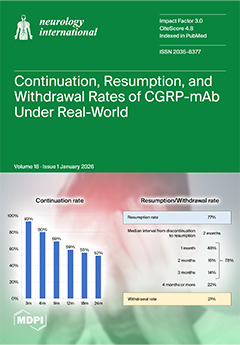
Cover Story (view full-size image): Anti-calcitonin gene-related peptide monoclonal antibodies (CGRP-mAbs) have dramatically improved migraine treatment, with guidelines recommending continued treatment for 12 to 18 months. However, CGRP-mAb treatment is expensive and requires monthly injections, placing a financial and physical burden on patients. This study was conducted under real-world clinical conditions in which patients are free to choose their own treatment. The continuation rate of CGRP-mAb treatment at 12 months after initiation was 59%, and more than half of patients remained on treatment at 24 months. Among patients who discontinued CGRP-mAb treatment due to its effectiveness, the resumption rate was 77%, and 78% of those patients resumed treatment within 3 months of discontinuation. The withdrawal rate was 21%. View this paper
  • Issues are regarded as officially published after their release is announced to the table of contents alert mailing list.
  • You may sign up for e-mail alerts to receive table of contents of newly released issues.
  • PDF is the official format for papers published in both, html and pdf forms. To view the papers in pdf format, click on the "PDF Full-text" link, and use the free Adobe Reader to open them.
Order results
Result details
Section
Select all
Export citation of selected articles as:
12 pages, 756 KB  
Article
Pre-Hospital Rate-Pressure Product Is Not Positively Associated with Hematoma Expansion or Initial Hematoma Volume in Spontaneous Intracranial Hemorrhage
by Stephanie Q. Liang, Daniel M. Oh, Fawaz Philip Tarzi, Nerses Sanossian, David S. Liebeskind, Jeffery L. Saver, Melissa Wilson and Roy A. Poblete
Neurol. Int. 2026, 18(1), 20; https://doi.org/10.3390/neurolint18010020 - 20 Jan 2026
Viewed by 851
Abstract
Background: The management of spontaneous intracerebral hemorrhage (ICH) has centered around controlling blood pressure in order to prevent hematoma expansion (HE). Rate-pressure product (RPP) has emerged as a hemodynamic marker that accounts for heart rate (HR) and systolic blood pressure (SBP), both [...] Read more.
Background: The management of spontaneous intracerebral hemorrhage (ICH) has centered around controlling blood pressure in order to prevent hematoma expansion (HE). Rate-pressure product (RPP) has emerged as a hemodynamic marker that accounts for heart rate (HR) and systolic blood pressure (SBP), both of which are crucial in modifying shear stress to the vasculature. We hypothesized that RPP in the pre-hospital hyperacute phase is positively associated with initial hematoma volume and HE. Methods: We analyzed 263 patients with primary ICH from the Field Administration of Stroke Therapy-Magnesium (FAST-MAG) study with initial and interval neuroimaging. RPP was calculated as the product of HR and SBP in pre-hospital and pre-treatment phases, stratified into quintiles. HE was defined by volume expansion of >6 mL or >33% from baseline volume on repeat neuroimaging performed within 48 h of the first scan. The primary outcome was the initial hematoma volume by quintiles of hyperacute RPP. The secondary outcome was the occurrence of HE across RPP quintiles. Multivariable logistic regression was used to assess the degree to which RPP affects HE. Results: Of the 263 patients analyzed, 116 (44%) had HE. The proportion of patients with HE or the initial hematoma volume was not statistically significant across RPP quintiles overall. HE was significantly more common in female patients or patients on anticoagulation. Conclusions: Elevated RPP was not associated with increased initial hematoma volume or subsequent HE in the hyperacute period after spontaneous ICH. Future research is necessary to determine the clinical importance of RPP as a biomarker in the clinical outcome of ICH. Full article
(This article belongs to the Topic Neurological Updates in Neurocritical Care)
Show Figures

Figure 1

24 pages, 2782 KB  
Systematic Review
Global Prevalence of Sleep-Disordered Breathing in Intracerebral Hemorrhage Survivors: A Meta-Analysis and Systematic Review
by Farhan Ishaq
Neurol. Int. 2026, 18(1), 19; https://doi.org/10.3390/neurolint18010019 - 20 Jan 2026
Viewed by 1950
Abstract
Background: Sleep-disordered breathing (SDB) and intracerebral hemorrhage (ICH) share a bidirectional relationship: SDB may increase ICH risk, while ICH can induce or exacerbate SDB. However, the prevalence and characteristics of post-ICH SDB remain poorly defined. Objective: To estimate the prevalence of SDB among [...] Read more.
Background: Sleep-disordered breathing (SDB) and intracerebral hemorrhage (ICH) share a bidirectional relationship: SDB may increase ICH risk, while ICH can induce or exacerbate SDB. However, the prevalence and characteristics of post-ICH SDB remain poorly defined. Objective: To estimate the prevalence of SDB among ICH survivors and examine associated clinical factors, including the relative burden of obstructive (OSA) versus central sleep apnea (CSA). Methods: A systematic review and meta-analysis were performed across PubMed, Scopus, CINAHL, and ClinicalTrials.gov. Studies assessing SDB in adults with ICH using American Academy of Sleep Medicine (AASM) category 1–4 diagnostic devices were included. Random-effects models estimated pooled prevalence at varying apnea–hypopnea index (AHI) thresholds, with subgroup analyses by setting, timing, geography, and diagnostic factors. Results: Seventeen studies met inclusion criteria. Pooled SDB prevalence was 85% (95% CI: 80–91%) at AHI > 5, with 49% (95% CI: 42–57%) experiencing moderate SDB (AHI > 15), and 21% (95% CI: 15–27%) experiencing severe SDB (AHI > 30). The prevalence of OSA predominated 73% (95% CI: 64% to 82%),while CSA occurred in 5% (95% CI: 2–9%), corresponding to a pooled RR of 7.44 and OR of 53.08 for OSA versus CSA. Conclusions: SDB—primarily OSA—is highly prevalent following ICH, underscoring the need for early, routine screening and intervention to improve neurological and cardiovascular outcomes. Full article
Show Figures

Graphical abstract

28 pages, 1252 KB  
Review
Reframing Dementia Prevention Strategies Aligned with the WHO Global Action Plan: A Structured Narrative Review Focusing on Mild Behavioral Impairment
by Efthalia Angelopoulou, Sokratis Papageorgiou and John Papatriantafyllou
Neurol. Int. 2026, 18(1), 18; https://doi.org/10.3390/neurolint18010018 - 16 Jan 2026
Cited by 2 | Viewed by 3008
Abstract
Background/Objectives: Dementia represents a growing public health challenge. The WHO Global Action Plan on the Public Health Response to Dementia emphasizes early detection, risk reduction, and innovation as key priorities. Mild Behavioral Impairment (MBI), defined as the emergence of persistent neuropsychiatric symptoms [...] Read more.
Background/Objectives: Dementia represents a growing public health challenge. The WHO Global Action Plan on the Public Health Response to Dementia emphasizes early detection, risk reduction, and innovation as key priorities. Mild Behavioral Impairment (MBI), defined as the emergence of persistent neuropsychiatric symptoms in older individuals, represents a potential marker of early neurodegeneration and possible window for early intervention. This review explores the role of MBI in dementia prevention, mapping current evidence within the WHO Global Action Plan framework. Methods: A comprehensive search was performed in PubMed, Scopus, and the official WHO website, during 1 September 2025–10 November 2025, without time restrictions. Eligible sources included original clinical studies, reviews, and policy documents addressing MBI, dementia prevention, and public health. Data were thematically synthesized according to the seven objectives of WHO: (1) dementia as a public health priority, (2) dementia awareness and friendliness, (3) dementia risk reduction, (4) dementia diagnosis, treatment, care and support, (5) support for dementia carers, (6) information systems for dementia, and (7) dementia research and innovation. Results: Accumulating evidence indicates that MBI assessment can capture early behavioral manifestations of neurodegenerative and other forms of dementia, correlating with fluid, neuroimaging and genetic biomarkers. Integrating MBI screening through the easy-to-administer MBI Checklist (MBI-C) into clinical and community-based care, including telemedicine pathways and research, may enhance early identification and personalized interventions, enrich the pool for clinical trials, and facilitate research in biomarker and therapy. MBI-related research further supports its integration in remote digital monitoring and population-based prevention. Conclusions: Embedding MBI-informed screening and interventions into national dementia strategies aligns with WHO objectives for early, equitable and scalable prevention and brain health. Full article
(This article belongs to the Section Aging Neuroscience)
Show Figures

Graphical abstract

16 pages, 2407 KB  
Review
Modeling Late-Onset Sporadic Alzheimer’s Disease Using Patient-Derived Cells: A Review
by Alisar Katbe, Ismaïla Diagne and Gilbert Bernier
Neurol. Int. 2026, 18(1), 17; https://doi.org/10.3390/neurolint18010017 - 14 Jan 2026
Viewed by 1414
Abstract
Late-onset sporadic Alzheimer’s disease (LOAD) is the most common form of dementia. The disease is characterized by progressive loss of memory and behavioral changes followed by neurodegeneration of all cortical areas. While the contribution of genetic and environmental factors is important, advanced aging [...] Read more.
Late-onset sporadic Alzheimer’s disease (LOAD) is the most common form of dementia. The disease is characterized by progressive loss of memory and behavioral changes followed by neurodegeneration of all cortical areas. While the contribution of genetic and environmental factors is important, advanced aging remains the most important disease risk factor. Because LOAD does not naturally occur in most animal species, except humans, studies have traditionally relied on the use of transgenic mouse models recapitulating early-onset familial Alzheimer’s disease (EOAD). Hence, the development of more representative LOAD models through reprograming of patient-derived cells into neuronal, glial, and immune cells became a necessity to better understand the disease’s origin and pathophysiology. Herein, and focusing on neurons, we review current work in the field and compare results obtained with two different reprograming methods to generate LOAD patient’s neuronal cells: the induced pluripotent stem cell and induced neuron technologies. We also evaluate if these models can faithfully mimic cellular and molecular pathologies observed in LOAD patients’ brains. Full article
(This article belongs to the Special Issue Advances in Molecular Mechanisms of Neurodegenerative Diseases)
Show Figures

Graphical abstract

14 pages, 4400 KB  
Article
Simulator Training on Neurointerventional Skill Acquisition in Novices: A Pilot Study
by Alexander von Hessling, Tim von Wyl, Dirk Lehnick, Chloé Sieber, Justus E. Roos and Grzegorz M. Karwacki
Neurol. Int. 2026, 18(1), 16; https://doi.org/10.3390/neurolint18010016 - 14 Jan 2026
Viewed by 850
Abstract
Background: Simulation-based training may offer a useful approach to support skill acquisition in neurointerventional stroke treatment without exposing patients to procedural risks. As the global demand for thrombectomy rises, training strategies that ensure procedural competence while addressing workforce constraints are increasingly important. With [...] Read more.
Background: Simulation-based training may offer a useful approach to support skill acquisition in neurointerventional stroke treatment without exposing patients to procedural risks. As the global demand for thrombectomy rises, training strategies that ensure procedural competence while addressing workforce constraints are increasingly important. With this pilot study, we aim to generate a hypothesis as to whether additional exposure of trainees to mechanical thrombectomy could benefit from simulator training on top of the standard training carried out on flow models. This study was designed as an exploratory pilot investigation and was not able to provide inferential or confirmatory statistical conclusions. Methods: Six novice participants (advanced clinical-year medical students with completed anatomical and preclinical training, but without previous exposure to catheter-based interventions) performed two neurointerventional tasks, vascular access and mechanical thrombectomy (MTE), on flow models. After a baseline assessment, three participants received standard model-based training (control group), and three received additional simulator training using a high-fidelity angiography simulator (Mentice VIST G5). Performance was reassessed after four weeks using technical and clinical surrogate metrics, which were ranked and descriptively analyzed. Results: No relevant differences were observed between groups for the vascular access task. In contrast, the simulator group demonstrated a trend toward improved performance in the MTE task, with greater gains in efficiency, autonomy, and procedural safety. Conclusions: Our findings indicate a possible benefit of even brief simulator exposure for skill acquisition for complex endovascular procedures such as MTE. While conventional training may suffice for basic skills, simulation may be particularly helpful in supporting learning in more advanced tasks. Full article
Show Figures

Graphical abstract

15 pages, 760 KB  
Systematic Review
The Multifaceted Role of Irisin in Neurological Disorders: A Systematic Review Integrating Preclinical Evidence with Clinical Observations
by Foad Alzoughool, Loai Alanagreh, Yousef Aljawarneh, Haitham Zraigat and Mohammad Alzghool
Neurol. Int. 2026, 18(1), 15; https://doi.org/10.3390/neurolint18010015 - 9 Jan 2026
Cited by 3 | Viewed by 2134
Abstract
Background: Irisin, an exercise-induced myokine, has emerged as a potent neuroprotective factor, though a systematic synthesis of its role across neurological disorders is lacking. This review systematically evaluates clinical and preclinical evidence on irisin’s association with neurological diseases and its underlying mechanisms. Methods: [...] Read more.
Background: Irisin, an exercise-induced myokine, has emerged as a potent neuroprotective factor, though a systematic synthesis of its role across neurological disorders is lacking. This review systematically evaluates clinical and preclinical evidence on irisin’s association with neurological diseases and its underlying mechanisms. Methods: Following PRISMA 2020 guidelines, a systematic search of PubMed/MEDLINE, Scopus, Web of Science, Embase, and Cochrane Library was conducted. The review protocol was prospectively registered in PROSPERO. Twenty-one studies were included, comprising predominantly preclinical evidence (n = 14), alongside clinical observational studies (n = 6), and a single randomized controlled trial (RCT) investigating irisin in cerebrovascular diseases, Parkinson’s disease (PD), Alzheimer’s disease (AD), and other neurological conditions. Eligible studies were original English-language research on irisin or FNDC5 and their neuroprotective effects, excluding reviews and studies without direct neuronal outcomes. Risk of bias was independently assessed using SYRCLE, the Newcastle–Ottawa Scale, and RoB 2, where disagreements between reviewers were resolved through discussion and consensus. Results were synthesized narratively, integrating mechanistic, pre-clinical, and clinical evidence to highlight consistent neuroprotective patterns of irisin across disease categories. Results: Clinical studies consistently demonstrated that reduced circulating irisin levels predict poorer outcomes. Lower serum irisin was associated with worse functional recovery and post-stroke depression after ischemic stroke, while decreased plasma irisin in PD correlated with greater motor severity, higher α-synuclein, and reduced dopamine uptake. In AD, cerebrospinal fluid irisin levels were significantly correlated with global cognitive efficiency and specific domain performance, and correlation analyses within studies suggested a closer association with amyloid-β pathology than with markers of general neurodegeneration. However, diagnostic accuracy metrics (e.g., AUC, sensitivity, specificity) for irisin as a standalone biomarker are not yet established. Preclinical findings revealed that irisin exerts neuroprotection through multiple mechanisms: modulating microglial polarization from pro-inflammatory M1 to anti-inflammatory M2 phenotype, suppressing NLRP3 inflammasome activation, enhancing autophagy, activating integrin αVβ5/AMPK/SIRT1 signaling, improving mitochondrial function, and reducing neuronal apoptosis. Irisin administration improved outcomes across models of stroke, PD, AD, postoperative cognitive dysfunction, and epilepsy. Conclusions: Irisin represents a critical mediator linking exercise to brain health, with consistent neuroprotective effects across diverse neurological conditions. Its dual ability to combat neuroinflammation and directly protect neurons, demonstrated in preclinical models, positions it as a promising therapeutic candidate for future investigation. Future research must prioritize the resolution of fundamental methodological challenges in irisin measurement, alongside investigating pharmacokinetics and sex-specific effects, to advance irisin toward rigorous clinical evaluation. Full article

Show Figures

Figure 1

19 pages, 474 KB  
Case Report
Rehabilitation After Severe Traumatic Brain Injury with Acute Symptomatic Seizure: Neurofeedback and Motor Therapy in a 6-Month Follow-Up Case Study
by Annamaria Leone, Luna Digioia, Rosita Paulangelo, Nicole Brugnera, Luciana Lorenzon, Fabiana Montenegro, Pietro Fiore, Petronilla Battista, Stefania De Trane and Gianvito Lagravinese
Neurol. Int. 2026, 18(1), 14; https://doi.org/10.3390/neurolint18010014 - 8 Jan 2026
Viewed by 2018
Abstract
Background/Objectives: Post-traumatic epileptogenesis is a frequent and clinically relevant consequence of traumatic brain injury (TBI), often contributing to worsened neurological and functional outcomes. In patients experiencing early post-injury seizures, rehabilitative strategies that support recovery while considering increased epileptogenic risk are needed. This case [...] Read more.
Background/Objectives: Post-traumatic epileptogenesis is a frequent and clinically relevant consequence of traumatic brain injury (TBI), often contributing to worsened neurological and functional outcomes. In patients experiencing early post-injury seizures, rehabilitative strategies that support recovery while considering increased epileptogenic risk are needed. This case study explores the potential benefits of combining neurofeedback (NFB) with motor therapy on cognitive and motor recovery. Methods: A patient hospitalized for severe TBI who experienced an acute symptomatic seizure in the early post-injury phase underwent baseline quantitative EEG (qEEG), neuromotor, functional, and neuropsychological assessments. The patient then completed a three-week rehabilitation program (five days/week) including 30 sensorimotor rhythm (SMR) NFB sessions (35 min each) combined with daily one-hour motor therapy. qEEG and clinical assessments were repeated post-intervention and at 6-month follow-up. Results: Post-intervention qEEG showed significant reductions in Delta and Theta power, reflecting decreased cortical slowing and enhanced neural activation. Relative power analysis indicated reduced Theta activity and Alpha normalization, suggesting improved cortical stability. Increases were observed in Beta and High-beta activity, alongside significant reductions in the Theta/Beta ratio, consistent with improved attentional regulation. Neuropsychological outcomes revealed reliable improvements in global cognition, memory, and visuospatial abilities, mostly maintained or enhanced at follow-up. Depressive and anxiety symptoms decreased markedly. Motor and functional assessments demonstrated meaningful improvements in motor performance, coordination, and functional independence. Conclusions: Findings suggest that integrating NFB with motor therapy may support recovery processes and be associated with sustained neuroplastic changes in the early post-injury phase after TBI, a condition associated with elevated risk for post-traumatic epilepsy. Full article
(This article belongs to the Section Brain Tumor and Brain Injury)
Show Figures

Graphical abstract

10 pages, 1665 KB  
Case Report
Targeted and Sequential Cryoneurolysis Improves Gait After Botulinum-Toxin Unresponsiveness in Post-Stroke Spasticity: A Laboratory-Verified Case
by Frédéric Chantraine, José Alexandre Pereira, Céline Schreiber, Tanja Classen, Gilles Areno and Frédéric Dierick
Neurol. Int. 2026, 18(1), 13; https://doi.org/10.3390/neurolint18010013 - 7 Jan 2026
Cited by 3 | Viewed by 1631
Abstract
Background: Chronic post-stroke spasticity often limits gait despite best-practice botulinum-toxin intramuscular injections (BTIs), whose benefit is constrained by short duration, dose ceilings, and tachyphylaxis. Cryoneurolysis (CNL) induces a reversible axonotmesis with preserved endoneurium, potentially providing longer tone reduction with fewer adverse effects, but [...] Read more.
Background: Chronic post-stroke spasticity often limits gait despite best-practice botulinum-toxin intramuscular injections (BTIs), whose benefit is constrained by short duration, dose ceilings, and tachyphylaxis. Cryoneurolysis (CNL) induces a reversible axonotmesis with preserved endoneurium, potentially providing longer tone reduction with fewer adverse effects, but its impact on whole-gait quality and its compatibility with implanted functional electrical stimulation (FES) remain poorly documented. Case presentation: A 43-year-old man, 12 years after right middle cerebral artery stroke, walked independently with an implanted common peroneal FES system but complained of effortful gait with left-knee “locking” and drop foot without FES. Multiple BTI series to triceps surae and quadriceps yielded only transient benefit. Two ultrasound-guided CNL sessions targeted tibial (soleus, medial gastrocnemius) and femoral (rectus femoris, vastus intermedius) motor branches. Quantitative gait analysis and fine-wire electromyography (EMG) were performed at baseline, 6 weeks after each CNL, and at 6 months, with and without FES. CNL produced immediate and sustained reductions in triceps surae and quadriceps overactivity, resolution of genu recurvatum, normalization of stiff-knee gait, improved ankle dorsiflexion, and increased swing phase knee flexion (>50°). Gait Deviation Index rose from 69 to 80 and Gillette Gait Index decreased by more than 50%, with preserved strength and without adverse events. Conclusions: Targeted, sequential CNL of tibial and femoral motor branches can safely deliver durable, clinically meaningful gait improvements when BTI has reached its ceiling and can act synergistically with implanted FES. Quantitative gait analysis and EMG sharpen clinical decision-making in spasticity management. Full article
(This article belongs to the Section Movement Disorders and Neurodegenerative Diseases)
Show Figures

Figure 1

14 pages, 525 KB  
Article
The Impact of Successful Transurethral Indwelling Catheter Removal on Health-Related Quality of Life in Patients Undergoing Neurological Rehabilitation
by Anke K. Jaekel, Manuel Pickermann, Ann Katrin Walter, Anna-Lena Butscher, John Bitter, Franziska I. Winterhagen, Ruth Kirschner-Hermanns and Stephanie C. Knüpfer
Neurol. Int. 2026, 18(1), 12; https://doi.org/10.3390/neurolint18010012 - 6 Jan 2026
Viewed by 965
Abstract
Background/Objectives: Patients with acute severe neurological disorders often receive a transurethral indwelling catheter (TUIC) during their initial treatment. These TUICs often remain in place until the transfer to a rehabilitation or a long-term care facility. There are no systematic concepts for bladder management [...] Read more.
Background/Objectives: Patients with acute severe neurological disorders often receive a transurethral indwelling catheter (TUIC) during their initial treatment. These TUICs often remain in place until the transfer to a rehabilitation or a long-term care facility. There are no systematic concepts for bladder management and no data regarding the impact on the catheter associated, health-related quality of life (HRQoL) in this patient group. The aim of this study was to investigate the impact of successful TUIC removal on the HRQoL of those affected and to contribute to the development of systematic bladder management. Methods: A prospective longitudinal study was conducted on 33 patients treated at a neurological rehabilitation centre due to acute severe neurological disorders. The HRQoL was assessed using the SF-36 Health Survey prior to and following the TUIC removal. The influence of urinary incontinence was analysed. The mean differences were determined using a one-sample t-test adjusted for age and gender. Results: TUIC removal was successful in 61.8% (21/33). The SF-36 Health Survey showed the following improvements (adj. mean diff., 95% CI, p-value): Mental Component Summary measure (4.36, 0.34; 8.38, p = 0.035), Role-Emotional (20.89, 0.54; 41.24, p = 0.045), Physical Functioning (10.03, 3.18; 16.88, p = 0.007). The comparison between incontinent and continent patients showed a poorer HRQoL for the incontinent group. Conclusions: Successful TUIC removal has a positive influence on psychological/emotional aspects and physical functioning. Structured bladder management that considers the physical and psychological aspects of patients and nursing staff, as well as medical and economic aspects, should be pursued with vigour. Full article
(This article belongs to the Topic Advances in Neurorehabilitation)
Show Figures

Figure 1

12 pages, 288 KB  
Review
Understanding the Failure of Medical Therapy in PFO-Associated Stroke and the Benefits of Closure: A Narrative Review
by Riwaj Bhagat
Neurol. Int. 2026, 18(1), 11; https://doi.org/10.3390/neurolint18010011 - 5 Jan 2026
Viewed by 1975
Abstract
Patent foramen ovale (PFO) is present in roughly one quarter of adults and is over-represented among younger patients with cryptogenic ischemic stroke. The past decade has produced compelling evidence from randomized trials showing that PFO closure is beneficial than medical therapy in preventing [...] Read more.
Patent foramen ovale (PFO) is present in roughly one quarter of adults and is over-represented among younger patients with cryptogenic ischemic stroke. The past decade has produced compelling evidence from randomized trials showing that PFO closure is beneficial than medical therapy in preventing recurrent ischemic stroke in appropriately selected patients. Despite this, anticoagulation continues to be used when closure is not feasible, declined, contraindicated, or considered after recurrent events. The observation that some patients experience “breakthrough” stroke or transient ischemic attack (TIA) despite therapeutic anticoagulation raises a critical question: why does medical therapy fail in PFO-associated stroke, and why does closure appear superior? This narrative review synthesizes the latest evidence on the pathophysiology of PFO-associated stroke, with attention to mechanisms that remain incompletely addressed by anticoagulation. It analyzes randomized trial data comparing antiplatelet therapy, anticoagulation, and transcatheter closure. It examines the role of high-risk PFO anatomical characteristics, the Risk of Paradoxical Embolism (RoPE) score, and the PFO-Associated Stroke Causal Likelihood (PASCAL) classification in understanding medical therapy failure. Additionally, the review explores whether PFO “type” predicts anticoagulation failure and highlights future research directions needed to further optimize therapy. In conclusion, in appropriately selected patients with high-risk PFO features, closure provides greater stroke risk reduction than medical therapy alone, albeit with small absolute risk differences and a procedural risk of atrial fibrillation. Full article
17 pages, 868 KB  
Review
Neuromarkers of Adaptive Neuroplasticity and Cognitive Resilience Across Aging: A Multimodal Integrative Review
by Jordana Mariane Neyra Chauca, Manuel de Jesús Ornelas Sánchez, Nancy García Quintana, Karen Lizeth Martín del Campo Márquez, Brenda Areli Carvajal Juarez, Nancy Rojas Mendoza and Martha Ayline Aguilar Díaz
Neurol. Int. 2026, 18(1), 10; https://doi.org/10.3390/neurolint18010010 - 5 Jan 2026
Cited by 3 | Viewed by 4397
Abstract
Background: Aging is traditionally characterized by progressive structural and cognitive decline; however, increasing evidence shows that the aging brain retains a remarkable capacity for reorganization. This adaptive neuroplasticity supports cognitive resilience—defined as the ability to maintain efficient cognitive performance despite age-related neural vulnerability. [...] Read more.
Background: Aging is traditionally characterized by progressive structural and cognitive decline; however, increasing evidence shows that the aging brain retains a remarkable capacity for reorganization. This adaptive neuroplasticity supports cognitive resilience—defined as the ability to maintain efficient cognitive performance despite age-related neural vulnerability. Objective: To synthesize current molecular, cellular, neuroimaging, and electrophysiological neuromarkers that characterize adaptive neuroplasticity and to examine how these mechanisms contribute to cognitive resilience across aging. Methods: This narrative review integrates findings from molecular neuroscience, multimodal neuroimaging (fMRI, DTI, PET), electrophysiology (EEG, MEG, TMS), and behavioral research to outline multiscale biomarkers associated with compensatory and efficient neural reorganization in older adults. Results: Adaptive neuroplasticity emerges from the coordinated interaction of neurotrophic signaling (BDNF, CREB, IGF-1), glial modulation (astrocytic lactate metabolism, regulated microglial activity), synaptic remodeling, and neurovascular support (VEGF, nitric oxide). Multimodal neuromarkers—including preserved frontoparietal connectivity, DMN–FPCN coupling, synaptic density (SV2A-PET), theta–gamma coherence, and LTP-like excitability—consistently correlate with resilience in executive functions, memory, and processing speed. Behavioral enrichment, physical activity, and cognitive training further enhance these biomarkers, creating a bidirectional loop between experience and neural adaptability. Conclusions: Adaptive neuroplasticity represents a fundamental mechanism through which older adults maintain cognitive function despite biological aging. Integrating molecular, imaging, electrophysiological, and behavioral neuromarkers provides a comprehensive framework to identify resilience trajectories and to guide personalized interventions aimed at preserving cognition. Understanding these multilevel adaptive mechanisms reframes aging not as passive decline but as a dynamic continuum of biological compensation and cognitive preservation. Full article
(This article belongs to the Section Aging Neuroscience)
Show Figures

Figure 1

10 pages, 611 KB  
Systematic Review
Exploratory Dietary Approaches for Drug-Resistant Epilepsy Beyond Standard Ketogenic Diet and Fish Oil: A Systematic Review of Preliminary Clinical Evidence
by Xianghong Meng and Kequan Zhou
Neurol. Int. 2026, 18(1), 9; https://doi.org/10.3390/neurolint18010009 - 4 Jan 2026
Viewed by 1734
Abstract
Background: Standard ketogenic diets (KD) and fish oil have established efficacy for drug-resistant epilepsy (DRE), but adherence and variability remain challenging. Objective: The objective of this study is to provide the first systematic evaluation of clinical evidence for emerging dietary interventions for epilepsy—specifically [...] Read more.
Background: Standard ketogenic diets (KD) and fish oil have established efficacy for drug-resistant epilepsy (DRE), but adherence and variability remain challenging. Objective: The objective of this study is to provide the first systematic evaluation of clinical evidence for emerging dietary interventions for epilepsy—specifically those other than standard KD and fish oil—and to rigorously evaluate their effectiveness and certainty of evidence to address the current gap in dietary management literature. Unlike prior reviews focused on standard KD or carbohydrate-modified versions, this study is the first to synthesize evidence for “non-standard” interventions—including olive oil-based KDs, probiotics, and restrictive gluten/glutamate-free diets—which are typically excluded from traditional dietary meta-analyses. Methods: Following PRISMA 2020 guidelines, we searched PubMed, Web of Science, Cochrane, and Google Scholar up to March 2025. Randomized Controlled Trials (RCTs) and Non-Randomized Studies of Interventions (NRSIs) were included, with quality assessed using RoB 2 and ROBINS-I tools. Results: Eight studies (total n = 675) were identified, comprising 2 RCTs and 6 NRSIs. These included olive oil-based KDs (n = 1), probiotic/synbiotic supplementation (n = 2), medium-chain triglyceride (MCT) additions (n = 2), and gluten-free (n = 1) or glutamate-free (n = 1) diets. Evidence quality is generally low, with 75% of studies at high risk of bias. Preliminary responder rates reached 83.1% in uncontrolled olive oil-based KD studies, whereas the only RCT evaluating a low-glutamate diet showed no significant seizure reduction (p = 0.57). Conclusion: Evidence for emerging dietary interventions beyond standard KD is nascent and of low certainty. Interpretation: While preliminary signals exist for olive oil-based KDs and probiotics, current data are insufficient for clinical recommendation; this review identifies these as promising exploratory targets requiring validation through rigorous, blinded RCTs. Full article
(This article belongs to the Section Movement Disorders and Neurodegenerative Diseases)
Show Figures

Figure 1

7 pages, 213 KB  
Case Report
Adult Botulism of Unknown Source with Post-Toxin Anti-GQ1b Antibodies: Implications for Molecular Mimicry—A Case Report
by Regev Cohen, Adi Hersalis Eldar, Yaron River, Ofir Schuster, Zina Baider, Shelly Lipman-Arens, Yael Galnoor Tene, Linor Ishay, Lamis Mahamid, Olga Feld Simon, Nina Avshovitch, Alvira Zbiger, Eran Diamant, Amram Torgeman, Elad Milrot, Ofir Israeli, Shlomo Shmaya, Itzhak Braverman and Shlomo E. Blum
Neurol. Int. 2026, 18(1), 8; https://doi.org/10.3390/neurolint18010008 - 29 Dec 2025
Viewed by 1049
Abstract
Background: Botulism is a rare but potentially fatal neuroparalytic illness caused by Clostridium botulinum neurotoxins (BoNTs). While adult cases usually result from foodborne exposure or wound infection, intestinal colonization is exceedingly uncommon. Diagnosis can be delayed by overlap with other neuromuscular syndromes, [...] Read more.
Background: Botulism is a rare but potentially fatal neuroparalytic illness caused by Clostridium botulinum neurotoxins (BoNTs). While adult cases usually result from foodborne exposure or wound infection, intestinal colonization is exceedingly uncommon. Diagnosis can be delayed by overlap with other neuromuscular syndromes, and confirmation requires specialized assays. Anti-GQ1b antibodies, classically associated with Miller–Fisher syndrome (MFS), have rarely been reported in confirmed botulism, raising questions about shared pathophysiology. Case Presentation: We describe an adult patient with acute dyspnea, xerostomia, and cranial neuropathies. No foodborne source was identified, but intestinal colonization of BoNT/B toxigenic Clostridium botulinum was confirmed by stool enrichment and mouse lethality bioassay. The patient improved promptly following heptavalent antitoxin. Unexpectedly, anti-GQ1b antibodies were detected during recovery, a finding typically linked to MFS rather than botulism. Discussion: This case illustrates the diagnostic challenges of sporadic cases of botulism, especially when respiratory compromise and autonomic dysfunction dominate the initial presentation. The autoantibodies finding raises the possibility of molecular mimicry, whereby toxin–ganglioside interactions expose neuronal epitopes and trigger an immune response. While causality cannot be proven, the overlap between botulism and GQ1b-positive neuropathies merits further investigation. Conclusions: Clinicians should maintain high suspicion for botulism in adults with acute dyspnea, especially when associated with cranial neuropathies, even in the absence of foodborne exposure. Anti-ganglioside antibodies in this context should be interpreted with caution, as they do not exclude botulism but may highlight immunological overlap with autoimmune neuropathies. Full article
(This article belongs to the Section Movement Disorders and Neurodegenerative Diseases)
24 pages, 1026 KB  
Review
Emerging Therapeutic Approaches for Tic Alleviation in Tourette Syndrome: The Role of Micronutrients
by Samskruthi Madireddy and Sahithi Madireddy
Neurol. Int. 2026, 18(1), 7; https://doi.org/10.3390/neurolint18010007 - 26 Dec 2025
Viewed by 5048
Abstract
Tourette syndrome (TS), or Tourette’s, is a tic disorder (TD) belonging to a group of neuropsychiatric conditions marked by recurrent motor movements or vocalizations known as tics. TD, including TS, typically begins in childhood between 4 and 18 years of age and affects [...] Read more.
Tourette syndrome (TS), or Tourette’s, is a tic disorder (TD) belonging to a group of neuropsychiatric conditions marked by recurrent motor movements or vocalizations known as tics. TD, including TS, typically begins in childhood between 4 and 18 years of age and affects approximately 3% of children and adolescents. The etiology and pathogenesis of TD are multifactorial, involving genetic, immunologic, psychological, and environmental factors. Evidence suggests that neurotransmitter dysregulation, particularly within the cortical dopaminergic networks of the basal ganglia and limbic system, which support motor control and cognition, may be involved in the development of TD. Nutritional factors may modulate TD through various mechanisms, including effects on neurotransmitter synthesis and metabolism, neurodevelopment, neural architecture, and neuroimmune activity. This review integrates current evidence on the roles of vitamins D, B6, and A, as well as iron, magnesium, zinc, and copper, in TD. For each micronutrient, its physiological and neurobiological functions are discussed, along with possible mechanistic links to TD pathophysiology. Additionally, we summarize the impact of nutrient deficiencies and assess available evidence regarding their potential therapeutic potential role in TD management. Overall, this synthesis highlights how nutritional status may influence TD onset and symptom severity, suggesting that nutrient-based interventions could potentially serve as valuable adjunctive strategies in treatment. Full article
(This article belongs to the Section Movement Disorders and Neurodegenerative Diseases)
Show Figures

Figure 1

13 pages, 1013 KB  
Article
Long-Term Health Consequences of SARS-CoV-2: Reaction Time and Brain Fog
by Ana Lesac Brizić, Branislava Popović, Tina Zavidić, Nevena Todorović, Verica Petrović, Nataša Pilipović-Broćeta, Ana R. Miljković, Aleksandar Ljubotina and Ema Dejhalla
Neurol. Int. 2026, 18(1), 6; https://doi.org/10.3390/neurolint18010006 - 26 Dec 2025
Viewed by 2587
Abstract
Background/Objectives: Beyond respiratory problems, COVID-19 can cause a variety of symptoms, such as neurological disorders caused by biological and psychological factors. Brain fog (BF), a post-illness cognitive impairment that many patients report, can be evaluated with reaction time (RT) testing. Response latency [...] Read more.
Background/Objectives: Beyond respiratory problems, COVID-19 can cause a variety of symptoms, such as neurological disorders caused by biological and psychological factors. Brain fog (BF), a post-illness cognitive impairment that many patients report, can be evaluated with reaction time (RT) testing. Response latency is measured by RT, which can be either simple (sRT) or complex (cRT). This study focuses on how COVID-19 affects cognitive function, with particular attention on RT changes, BF prevalence, and implications for daily life. Methods: The study included 599 participants from Bosnia and Herzegovina, Croatia and Serbia. RT was measured using PsyToolkit and participants completed a COVID-19-associated BF questionnaire. Participants who experienced BF after their latest COVID-19 infection rated its severity using a visual analogue scale (VAS). Additional clinical data were obtained from medical records. Results: BF was reported by 40% of participants post-COVID-19. Men reported it less frequently but found it more disruptive. RT progressively declined post-infection, reaching peak impairment at 15 weeks, following recovery, with RT normalizing by six months. Conclusions: COVID-19 is linked to temporary RT impairment, peaking at 15 weeks post-infection and resolving by six months, independent of BF presence. This study emphasizes the need for a biopsychosocial approach to BF management. Easily available RT assessments should be incorporated into routine clinical practice. Full article
(This article belongs to the Section Aging Neuroscience)
Show Figures

Graphical abstract

19 pages, 5043 KB  
Article
Functional Suppression of CLOCK Activity in Ventromedial Hypothalamic Prodynorphin Neurons Alters Locomotor Activity and Rapid Eye Movement Sleep
by Ting He and Xu Wang
Neurol. Int. 2026, 18(1), 5; https://doi.org/10.3390/neurolint18010005 - 25 Dec 2025
Viewed by 1650
Abstract
Background/Objectives: The circadian regulator, circadian locomotor output cycles kaput (CLOCK), is well-established in maintaining sleep–wake rhythms, yet its cell-type-specific functions in sleep regulation remain largely unexplored. While ventromedial hypothalamic (VMH) prodynorphin (PDYN)-expressing (VMHPDYN+) neurons are known to modulate homeostatic and [...] Read more.
Background/Objectives: The circadian regulator, circadian locomotor output cycles kaput (CLOCK), is well-established in maintaining sleep–wake rhythms, yet its cell-type-specific functions in sleep regulation remain largely unexplored. While ventromedial hypothalamic (VMH) prodynorphin (PDYN)-expressing (VMHPDYN+) neurons are known to modulate homeostatic and motivational processes, their potential role in circadian sleep regulation has not been investigated. Methods: To address this, we developed mice with PDYN neuron-specific functional suppression of CLOCK activity (mClkΔ19) by interfering with their internal clock through Adeno-Associated Virus (AAV)-mediated overexpression of dominant-negative CLOCKΔ19 in PDYN-Cre mice. Results: We found that mClkΔ19 mice exhibited reduced locomotor activity during the dark phase, earlier activity peaks, and impaired rhythmicity of rapid eye movement (REM) and non-REM (NREM) sleep. Sleep analysis in mClkΔ19 mice showed selective reductions and fragmentation of light-phase REM sleep, more frequent sleep–wake transitions, and shorter REM cycles during the dark phase, indicating disrupted REM sleep timing. EEG spectral analysis in mClkΔ19 mice revealed decreased gamma activity during REM sleep in the light phase and an increase in delta activity coupled with decreased gamma during wakefulness in the dark phase. Conclusions: These findings suggest that the CLOCK activity in VMHPDYN+ neurons is vital for circadian accuracy, REM sleep stability, and brain oscillations during sleep–wake cycles. Full article
Show Figures

Figure 1

14 pages, 594 KB  
Review
Experimental Primary Brain Calcification Model and Its Application to Pathogenesis Mechanism Analysis and Therapeutic Research
by Hisaka Kurita, Junya Murata, Kazuki Ohuchi, Yuichi Hayashi and Masatoshi Inden
Neurol. Int. 2026, 18(1), 4; https://doi.org/10.3390/neurolint18010004 - 24 Dec 2025
Viewed by 1413
Abstract
Primary Brain Calcification (PBC) is a neurodegenerative disorder of unknown etiology that results in bilateral calcifications within the brain. PBC symptoms vary, including Parkinsonian symptoms and psychiatric symptoms. Abnormalities in phosphate metabolism within the brain are hypothesized to be a mechanism underlying the [...] Read more.
Primary Brain Calcification (PBC) is a neurodegenerative disorder of unknown etiology that results in bilateral calcifications within the brain. PBC symptoms vary, including Parkinsonian symptoms and psychiatric symptoms. Abnormalities in phosphate metabolism within the brain are hypothesized to be a mechanism underlying the onset of PBC, but the precise pathophysiological mechanism remains unclear. Furthermore, no fundamental treatment or therapeutic agent for PBC has been established. Previous studies have reported SLC20A2, PDGFB, PDGFRB, XPR1, MYORG, JAM2, CMPK2, and NAA60 as causative genes for familial PBC. Elucidating the pathophysiological mechanisms of PBC and developing treatments and therapeutic agents requires appropriate experimental disease models. Knockout mice and mutant mice targeting familial causative genes have been reported to be useful as in vivo models of PBC. Furthermore, several disease-specific iPS cells for PBC have been reported, suggesting their potential utility as PBC models. This paper reviews each familial causative gene and current PBC models, including genetically modified animals and disease-specific iPS cells, and examines their usefulness for understanding disease mechanisms and advancing therapeutic research. Full article
(This article belongs to the Special Issue Advances in Molecular Mechanisms of Neurodegenerative Diseases)
Show Figures

Graphical abstract

15 pages, 506 KB  
Article
Continuation, Resumption, and Withdrawal Rates of CGRP-mAb Treatment for Migraine Under Real-World Clinical Conditions in Which Patients Are Free to Choose Own Treatment
by Takafumi Tanei, Satoshi Yamashita, Satoshi Maesawa, Yusuke Nishimura, Tomotaka Ishizaki, Yoshitaka Nagashima, Takahiro Suzuki, Hajime Hamasaki, Shun Yamamoto, Toshihiko Wakabayashi and Ryuta Saito
Neurol. Int. 2026, 18(1), 3; https://doi.org/10.3390/neurolint18010003 - 24 Dec 2025
Cited by 1 | Viewed by 2162
Abstract
Background/Objectives: Anti-calcitonin gene-related peptide monoclonal antibodies (CGRP-mAbs) are effective injectable medications for the treatment of migraine. This study aimed to evaluate continuation, resumption, and withdrawal rates of CGRP-mAb treatment under real-world clinical conditions. Methods: Treatment-naïve patients with at least 3 months of follow-up [...] Read more.
Background/Objectives: Anti-calcitonin gene-related peptide monoclonal antibodies (CGRP-mAbs) are effective injectable medications for the treatment of migraine. This study aimed to evaluate continuation, resumption, and withdrawal rates of CGRP-mAb treatment under real-world clinical conditions. Methods: Treatment-naïve patients with at least 3 months of follow-up after starting CGRP-mAb treatment were included. The decision to continue, discontinue, or resume CGRP-mAb treatment was made freely by the patients. Headache Impact Test-6 (HIT-6) and the Migraine-Specific Quality of Life Questionnaire (MSQ) were administered before starting treatment and one month after each CGRP-mAb injection. The endpoints were as follows: continuation rates of CGRP-mAb treatment after treatment initiation; resumption rate; withdrawal rate; changes in HIT-6 and MSQ scores; and differences in background factors between the resumption and withdrawal groups. Results: Of the 1162 migraine patients, 146 were included in the analysis. Continuation rates of CGRP-mAb treatment at 3, 6, 9, 12, 18, and 24 months were 93.2%, 80.2%, 68.9%, 58.8%, 55.4%, and 51.7%, respectively. For the patients who discontinued, the resumption rate was 76.8%, and the withdrawal rate was 20.7%. HIT-6 and MSQ scores were significantly decreased at all assessment points compared with before CGRP-mAb treatment. There were no significant differences in factors between the resumption and withdrawal groups. Conclusions: Under real-world clinical conditions in which patients were free to choose their own treatment, the continuation rate of CGRP-mAb treatment 12 months after treatment initiation was 58.8%, and more than half of patients remained on treatment after 24 months. The resumption rate was 76.8% and the withdrawal rate was 20.7%. Full article
(This article belongs to the Section Pain Research)
Show Figures

Figure 1

18 pages, 713 KB  
Opinion
Multiple Sclerosis: An Ethnically Diverse Disease with Worldwide Equity Challenges Accessing Care
by Victor M. Rivera
Neurol. Int. 2026, 18(1), 2; https://doi.org/10.3390/neurolint18010002 - 22 Dec 2025
Cited by 4 | Viewed by 2748
Abstract
Multiple sclerosis (MS) affects approximately 2.9 million people in the world, exerting a significant economic and societal burden. The disease is increasingly identified among populations considered as uncommonly affected. MS is reported in all regions of the World Health Organization (WHO) member states [...] Read more.
Multiple sclerosis (MS) affects approximately 2.9 million people in the world, exerting a significant economic and societal burden. The disease is increasingly identified among populations considered as uncommonly affected. MS is reported in all regions of the World Health Organization (WHO) member states in Africa, the Americas, South-East Asia, Europe, the Eastern Mediterranean and the Western Pacific, affecting all ethnicities while exhibiting substantially variable prevalences. Countries with high MS prevalence and some with moderate frequencies generally have economically better structured healthcare systems. Nevertheless, health disparities in these countries are accentuated by suboptimal accessibility of care for their minorities, immigrants and other underserved populations. Social Determinants of Health (SDOH) might have an impact on morbidity and higher rates of disability. Large segments of the world population (i.e., African, Latin American, people from the Middle East and Southeast Asia) do not have access to adequate MS diagnostic procedures, compounded by reduced availability of neurologists. Healthcare disparities exist practically in every country of the world. Active wars and a large number of refugees resulting from conflict augments the challenges to healthcare systems. These global factors constitute obstacles to the adequate management of MS. A collective international path is required to facilitate access to highly effective, albeit onerous treatments, some already approved and being utilized, i.e., monoclonal antibodies and B-lymphocyte depletory agents, and others foreseen in the future as advanced therapeutic molecules continue to develop. Full article
(This article belongs to the Special Issue Advances in Multiple Sclerosis, Third Edition)
Show Figures

Figure 1

31 pages, 1046 KB  
Review
The Role of Blood–Brain Barrier Disruption in Epilepsy: Mechanisms and Consequences
by Elena Suleymanova and Anna Karan
Neurol. Int. 2026, 18(1), 1; https://doi.org/10.3390/neurolint18010001 - 22 Dec 2025
Cited by 12 | Viewed by 2849
Abstract
The blood–brain barrier (BBB) is essential for maintaining cerebral homeostasis, and its dysfunction is increasingly recognized as an active driver of epilepsy. This review explores the mechanisms by which BBB disruption contributes to seizures and the development of chronic epilepsy. Potentially epileptogenic insults, [...] Read more.
The blood–brain barrier (BBB) is essential for maintaining cerebral homeostasis, and its dysfunction is increasingly recognized as an active driver of epilepsy. This review explores the mechanisms by which BBB disruption contributes to seizures and the development of chronic epilepsy. Potentially epileptogenic insults, including traumatic brain injury, stroke, and status epilepticus, induce acute and often persistent BBB leakage. This breach permits the extravasation of serum albumin, which activates transforming growth factor-beta (TGF-β) signaling in astrocytes. This cascade leads to astrocytic dysfunction, impaired potassium buffering, neuroinflammation, and synaptic remodeling, collectively fostering neuronal hyperexcitability. Furthermore, BBB disruption facilitates the infiltration of peripheral immune cells, amplifying neuroinflammation and propagating a pathologic cycle of BBB damage and seizure activity. BBB damage is mediated by multiple processes, including the activation of the plasminogen activation (PA) system. Furthermore, these processes of BBB disruption and neuroinflammation provide a shared pathological basis for neuropsychiatric disorders like depression and anxiety, which are common comorbidities of epilepsy, through shared mechanisms of neuroinflammation and neurovascular unit (NVU) dysregulation. BBB dysfunction can also contribute to the resistance to antiepileptic drugs. Finally, we discuss the therapeutic potential of stabilizing the BBB as a viable strategy for developing disease-modifying therapies for epilepsy. Full article
(This article belongs to the Section Movement Disorders and Neurodegenerative Diseases)
Show Figures

Graphical abstract

Previous Issue
Next Issue
Back to TopTop