Review

Die Another Day: Inhibition of Cell Death Pathways by Cytomegalovirus

by
Wolfram Brune
1,* and
Christopher E. Andoniou
2,3,*
1
Heinrich Pette Institute, Leibniz Institute for Experimental Virology, Hamburg 20251, Germany
2
Immunology and Virology Program, Centre for Ophthalmology and Visual Science, the University of Western Australia, Crawley 6009, WA, Australia
3
Centre for Experimental Immunology, Lions Eye Institute, Nedlands 6009, WA, Australia
*
Authors to whom correspondence should be addressed.
Viruses 2017, 9(9), 249; https://doi.org/10.3390/v9090249
Submission received: 26 July 2017 / Revised: 27 August 2017 / Accepted: 28 August 2017 / Published: 2 September 2017
(This article belongs to the Special Issue Viral Infection and Apoptosis)

Abstract

:
Multicellular organisms have evolved multiple genetically programmed cell death pathways that are essential for homeostasis. The finding that many viruses encode cell death inhibitors suggested that cellular suicide also functions as a first line of defence against invading pathogens. This theory was confirmed by studying viral mutants that lack certain cell death inhibitors. Cytomegaloviruses, a family of species-specific viruses, have proved particularly useful in this respect. Cytomegaloviruses are known to encode multiple death inhibitors that are required for efficient viral replication. Here, we outline the mechanisms used by the host cell to detect cytomegalovirus infection and discuss the methods employed by the cytomegalovirus family to prevent death of the host cell. In addition to enhancing our understanding of cytomegalovirus pathogenesis we detail how this research has provided significant insights into the cross-talk that exists between the various cell death pathways.

1. Introduction

The capacity to eliminate damaged or unwanted cells via regulated cell death programmes is essential for the wellbeing of multicellular organisms. Indeed, defective regulation of cell death pathways can result in the development of pathological conditions such as cancer, autoimmunity and inflammatory disease [1,2]. In addition to a role in embryonic development and tissue homeostasis [3], programmed cell death has the capacity to act as defence mechanism against invading pathogens [4,5]. The finding that many pathogens encode cell death inhibitors suggests that the ability to circumvent this process is essential for optimal replication and/or transmission of many pathogens.
Human cytomegalovirus (HCMV) is a large DNA virus that is common in the human population. After the initial acute infection, that is typically subclinical, HCMV establishes a latent infection that lasts for the lifetime of the host [6,7]. HCMV is known to employ an array of strategies designed to interfere with the host immune response and thereby prevent viral clearance. Since HCMV infection is strictly species-specific, animal models are often employed to study how virally-encoded genes contribute to viral pathogenesis in vivo. Murine CMV (MCMV), a natural mouse pathogen, shares a high degree of sequence homology and biology with HCMV making it an excellent model for HCMV infection. Targeted disruption of selected viral open reading frames has established that efficient viral replication depends on the capacity to inhibit the host cell death pathways. In this review we describe how CMV infection triggers a death response in the host cell, outline the countermeasures employed by CMV to prevent death, and detail how these contribute to in vivo viral pathogenesis.

2. Multiple Pathways to Death

Several genetically controlled cell death pathways are now recognised. Apoptosis is characterised by cell shrinkage, nuclear condensation, DNA fragmentation, and the formation of apoptotic bodies. The process is generally non-inflammatory since phagocytic cells rapidly engulf and digest the apoptotic bodies. Additional pathways to death such as necroptosis and pyroptosis have also been described. Unlike apoptosis, these necrotic forms of death are accompanied by cellular swelling, and ultimately rupture of the cell membrane, resulting in the release of cellular components into the extracellular space. Significant cross-talk between the different cell death pathways exist, allowing the distinct pathways to operate in a coordinated manner in order to promote immune responses to invading pathogens.

2.1. Apoptosis

The process of apoptosis may be initiated by either extrinsic or intrinsic signals. The intrinsic pathway is triggered in response to stimuli such as DNA damage, growth factor deprivation, or endoplasmic reticulum (ER) stress. Bcl-2 family proteins are the principle regulators of the intrinsic pathway that function by regulating the integrity of the mitochondrial outer membrane (MOM) [8]. MOM permeabilization (MOMP) results in the release of proteins such as cytochrome c, Smac/DIABLO, and Htr2/Omi that promotes the activation of caspases, a family of cysteine proteases responsible for mediating cellular destruction [9,10,11,12]. The Bcl-2 family is composed of three functional subgroups, BH3-only proteins that act as stress sensors and initiate apoptosis, the effector proteins Bax and Bak that mediate MOMP, and pro-survival proteins that maintain mitochondrial membrane integrity. Pro-survival Bcl-2 proteins have the capacity to bind to Bax and Bak and thus prevent their activation [8]. All pro-survival proteins appear capable of inhibiting Bax, while only Mcl-1, Bcl-xL and A1 seem capable of holding Bak in check [13,14,15]. BH3-only proteins initiate apoptosis by binding to pro-survival Bcl-2 proteins and thereby releasing Bax and Bak [13,14]. Alternatively, some BH3-only proteins have the capacity to interact with Bax and Bak and directly catalyse their activation [16,17,18,19,20]. In healthy cells Bax and Bak exist as inert monomers, but as apoptosis proceeds the proteins undergo conformational changes resulting in the formation of large homo-oligomers that permeabilize the MOM. The Bcl-2 pathway therefore determines cell fate by regulating the activity of Bax and Bak. The importance of Bax and Bak to the apoptotic cascade was confirmed by the finding that cells isolated from Bax/Bak−/− mice are highly resistant to many forms of apoptosis [21,22].
Death receptors (DR) may promote cell survival or death depending on the composition of the signalling complexes formed after receptor activation. DR are a subset of the tumour necrosis factor (TNF) receptor family characterised by a cytoplasmic domain of approximately 80 amino acids termed the death domain (DD). Fas-associated DD protein (FADD) is an adaptor protein that is critical for apoptotic signalling downstream of DR. Following activation of the DR Fas, DR4 or DR5, FADD is recruited to the receptor, via homotypic DD interactions, and FADD in turn, recruits initiator caspase-8 or caspase-10 [23,24]. Recruitment of the initiator caspases promotes the formation of dimers resulting in autocatalytic caspase activation [25]. Once activated, the initiator caspases promote apoptosis by cleaving effector caspases, such as caspase-3 and caspase-7, that then degrade critical cellular components. This process can be inhibited by the cellular FLICE inhibitor protein (cFLIP), a non-catalytic paralogue of caspase-8 (FLICE), that forms a heterodimer with caspase-8, thus preventing autocatalytic activation [26]. Initiator caspase activation, and hence apoptosis, may also result after TNF receptor 1 (TNFR1) activation. Ligand binding by TNFR1 results in the recruitment of the adaptors TNFR1-associated DD protein (TRADD) and TNFR-associated factor 2 (TRAF2), the receptor-interacting protein 1 (RIP1) kinase, and the cellular inhibitor of apoptosis protein (cIAP) into a signalling structure termed complex I. Formation of complex I promotes cell survival by activating the NF-κB pathway and inducing the production of pro-survival proteins [27]. Following internalization of complex I, complex II, a cytoplasmic signalling unit that includes the FADD adaptor and caspase-8 is formed. Several forms of complex II have been described with the stability and constituents of the complex regulated by an intricate series of ubiquitination event and phosphorylation events. Formation of complex II results in apoptosis only if autocatalytic processing of caspase-8 is able to take place, for example when cFLIP levels are low [28].

2.2. Necroptosis

Receptor interacting protein kinase-1 and -3 (RIPK1 and RIPK3) are key mediators of necroptosis with the study of signalling events downstream of TNFR1 being critical in understanding how these kinases promote necrotic death [29]. As outlined above, complex II is assembled in response to TNFR1 activation. Complex II, also called the “necrosome”, is composed of a heterodimer of caspase-8 and cFLIP along with FADD, RIPK1 and RIPK3. The presence of cFLIP in the complex prevents the autocatalytic activation of caspase-8, thus sparing the cell from apoptotic death. While caspase-8 is unable to undergo full catalytic activation when bound to cFLIP, a basal protease activity is present, and inhibition of caspase-8 activity has long been recognised as a method to promote necroptosis [30,31]. The finding that RIPK1 and RIPK3 are substrates of caspase-8 suggests that caspase-8 acts as a negative regulator of necroptosis by degrading and hence silencing the pro-necroptotic kinases [32,33]. Under circumstances where caspase-8 activity is silenced, RIPK1 recruits and activates RIPK3 through a RIP homotypic interaction motif (RHIM) [34,35,36]. Once activated, RIPK3 phosphorylates the mixed lineage kinase domain-like (MLKL) protein causing a conformational change within the MLKL that promotes its oligomerization. The oligomeric form of MLKL translocates to the plasma membrane where it interacts with the cell membrane and causes membrane rupture [37,38,39,40]. RIPK1-independent mechanisms for the activation of RIPK3 have also been described: the intracellular nucleotide sensor DNA-dependent activator of IFN-regulatory factors (DAI), also known as Z-DNA-binding protein 1(ZBP1), and the TIR-domain-containing adaptor inducing interferon-β (TRIF), both of which carry an RHIM, are capable of engaging RIPK3 and activating the necroptotic pathway [41,42].

2.3. Pyroptosis

The inflammatory caspases, that includes caspase-1, are critical for mediating innate defence. In response to cellular stress, inflammasomes are assembled that act as platforms for the activation of caspase-1. Activated caspase-1 promotes inflammation by cleaving pro-interleukin (IL)-1β and pro-IL-18 into their active forms, and initiates pyroptotic death by cleaving the gasdermin D protein. Once cleaved, the N-terminus of gasdermin D forms pores within the cell membrane causing the cell to swell and eventually results in membrane rupture [43,44,45]. The inflammasome is formed when pattern recognition receptors (PRRs), such as members of the Nod-like receptor family (NLR) or the absent in melanoma 2 (AIM2) protein, oligomerize in response to the detection of danger- or pathogen-associated molecules [46]. Clustering of the PRR results in the recruitment of the apoptosis-associated speck-like adaptor protein containing a CARD (ASC) that binds pro-caspase-1. The recruitment of pro-caspase-1 into the multi-protein assembly triggers proteolytic cleavage of pro-caspase-1 into its active form allowing the processing of downstream substrates. It should be noted that while caspase-1 is essential for the processing of pro-IL-1β and pro-IL-18, activation of caspase-11 can induce pyoptosis in some settings [47]. The inflammatory caspases therefore link the processes of inflammation and cell death induced following infection.

2.4. Cross-Talk between Death Pathways

The preceding description of the cell death pathways may give the impression that cell death results from the activation of linear signalling cascades. In fact, significant cross-talk between the various pathways exist that determines if a cell will die, and if so, by what mechanism. The interconnection of the pathways is exemplified by caspase-8 whose activation initiates apoptosis, while basal activity of uncleaved casapse-8 is essential to prevent necroptosis. Similarly, instances where activation of RIPK1 causes apoptosis, or promotes cell survival, rather than activating necroptosis have been reported. Antagonism of the ubiquitin ligases cIAP1 and cIAP2 is sufficient to result in the formation of the “Ripoptosome” a complex composed of FADD, caspase-8 and RIPK1 that assembles independently of DR signalling [48,49]. The kinase activity of RIPK1 is required for formation of the Ripoptosome with the complex mediating caspase-8 dependent apoptosis. The finding that RIPK1-deficent mice die shortly after birth suggested that in addition to its role in promoting death RIPK1 has a pro-survival function [50]. Inactivation of both caspase-8 and RIPK3 (or FADD and RIPK1) was required to prevent the lethality associated with RIPK1, indicating that FADD and caspase-8 promote survival by suppressing RIPK1 and RIPK3-mediated necroptosis during development [51,52,53]. Moreover, RIPK1 also has a kinase-independent pro-survival function under certain conditions [54,55,56].
The processes of apoptosis and pyroptosis are also linked. Procasapse-8 can be recruited to the inflammasome by interacting with the ASC adaptor protein leading to apoptotic death [57]. These observations demonstrate some of the ways in which the cell death pathways are linked. A feature of the interlocking regulation of these pathways is that inhibiting one cell death mechanism often results in the activation of an alternative pathway. An extension of this observation then is that ability of pathogens to inhibit the innate death response will require multiple inhibitory proteins.

3. Activation of Cell Death Pathways by CMV

In order for cell death to function as a defence mechanism, the host cell must have an effective means for detecting the presence of an invading pathogen. Cell death can be triggered when the host cell detects pathogen-associated molecular patterns (PAMPs), or can occur in response to cellular stress that is induced by the replication of intracellular pathogens. The following section describes some of the known mechanisms used by the host to detect CMV and how these can activate a cell death response.

3.1. Direct Recognition of CMV Infection

PRRs are a class of receptors that recognise molecules expressed, or produced by pathogens. Toll-like receptors (TLRs) were the first members of the PRR family to be identified with several implicated in the detection of CMV. A heterodimer of TLR1 and TRL2 was found to bind glycoproteins B and H produced by HCMV [58]. Whether MCMV can be detected by this mechanism has not been determined, but TLR2 knockout mice were found to be more susceptible to MCMV infection suggesting this pathway could be relevant during in vivo infection [59]. Increased titres of MCMV were also detected in mice lacking TLR3, which recognises double-stranded RNA (dsRNA), or TLR9, a receptor for unmethylated DNA [60,61]. TLR signalling typically results in the production of pro-inflammatory cytokines and chemokines. More recently, though, activation of many TLRs was found to result in the formation of signalling complexes capable of inducing RIPK-dependent necroptosis or caspase-8-dependent apoptosis [42,49,62].
In addition to TLRs, the DAI/ZBP1 and AIM2 sensors detect MCMV infection. Both of these PRR recognise cytoplasmic dsDNA, and both are activated during MCMV infection. Upon activation, DAI/ZBP1 can interact with RIPK3, promoting its activation that ultimately results in necroptosis [41]. More recent studies have shown that DAI/ZBP1 activation during MCMV infection requires viral gene transcription, but not viral DNA replication, and that DAI/ZBP1 is activated by the dsRNA in the Z-conformation (Z-RNA) rather than by the Z-DNA [63,64]. By contrast, the DNA sensor AIM2 suppresses MCMV replication via activation of the inflammasome [65]. While AIM2 was found to be required for the production of anti-viral cytokines in this context, the potential contribution of pyroptosis to the control of MCMV was not addressed. Activation of dsRNA-dependent protein kinase (PKR) is an additional mechanism by which CMV infection is detected [66,67]. PKR forms a homodimer and undergoes autophosphorylation after binding an RNA target resulting in catalytic activation of PKR. Once activated, PKR inhibits viral replication via several mechanisms, the best characterised of which is the inhibition of cellular protein translation by phosphorylation of the eukaryotic translation initiation factor 2α (eIF2α) [68]. How PKR activation mediates cell death has not been completely elucidated, but activation of NF-κB and/or FADD-dependent caspase-8 activation have been implicated [69,70]. PKR induces cell death in response to infection by influenza or poxviruses, but whether this process occurs following CMV infection has not been determined.

3.2. Cellular Stress Responses

Viral replication places a significant burden on the host cell, which responds by activating stress response pathways. CMV replication requires large amounts of protein to be synthesised. As a consequence, unfolded proteins accumulate within the ER resulting in the activation of a stress response after CMV infection [71,72]. The unfolded protein response is designed to restore ER homeostasis, however, if ER stress is not resolved cell death can result [73]. In response to ER stress the PKR-like ER kinase (PERK) phosphorylates eIF2α, resulting in the attenuation of mRNA translation, and in doing so, preventing the influx of additional protein into the stressed ER. Activation of PERK also allows for the preferential translation of proteins that contribute to the resolution of ER stress, or the induction of apoptosis, including the transcription factor C/EBP homologous protein (CHOP). A second mediator of ER stress responses is inositol-requiring enzyme 1 (IRE1), which undergoes oligomerisation in response to the accumulation of unfolded proteins within the ER. Once activated, IRE1 stimulates expression of ER-associated degradation factors and chaperones in order to alleviate ER stress. However, IRE1 can also promote apoptosis by activating a TRAF2-ASK1-JNK signalling cascade [73], by promoting caspase-12 or caspase-2 activation [74,75], or by interacting with Bax and Bak [76].
A second form of stress response triggered by CMV infection is the DNA damage response. Following detection of DNA damage, pathways that inhibit cell cycle progression are activated allowing for repair of the damaged DNA, or if the damage is too severe, apoptosis ensues [77]. Replication of the HCMV genome is achieved by a rolling-circle mechanism, and the resulting concatameric genomes are then cleaved to unit-length genomes [7]. This mechanism produces multiple exposed ends that can be recognised as dsDNA breaks. The ataxia-telangiectasia mutated (ATM) kinase pathway that responds to dsDNA breaks is activated following HCMV infection [78]. ATM has the capacity to activate p53 that can then induce pro-apoptotic proteins including Bax, Fas and the p53-induced protein with a DD (PIDD). HCMV replication also triggers a second type of DNA damage response activated in response to stalled replication forks [79]. The ATM-Rad3-related kinase (ATR) is the main transducer of this process, with p53 again acting as one of the downstream effectors for this pathway. Hence, replication of the viral genome, a process that is essential for viral propagation, activates several host cell defence mechanisms.
In summary, the host cell has the capacity to activate multiple cell death pathways either by directly detecting CMV constituents or indirectly by stress pathways activated by viral replication.

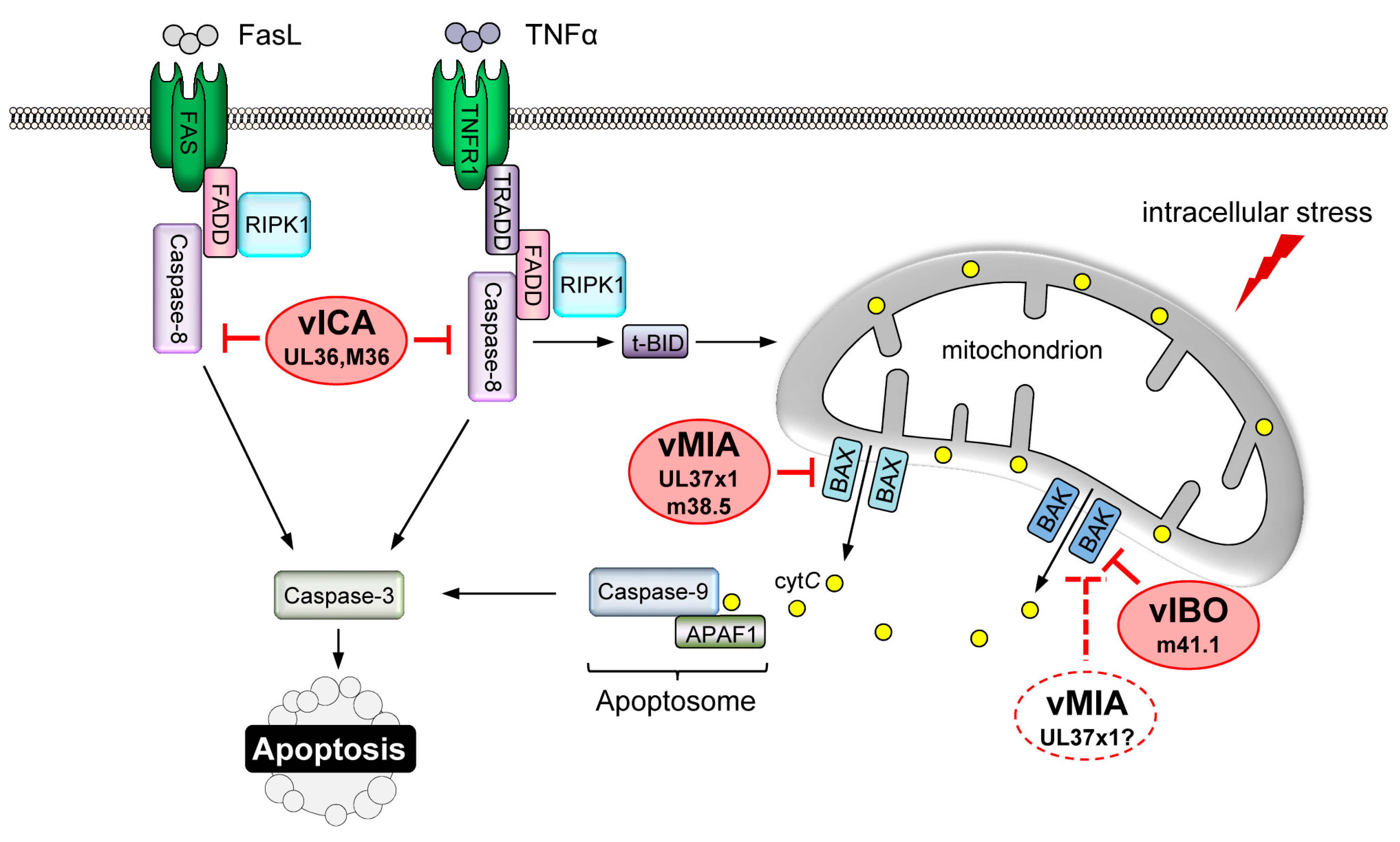
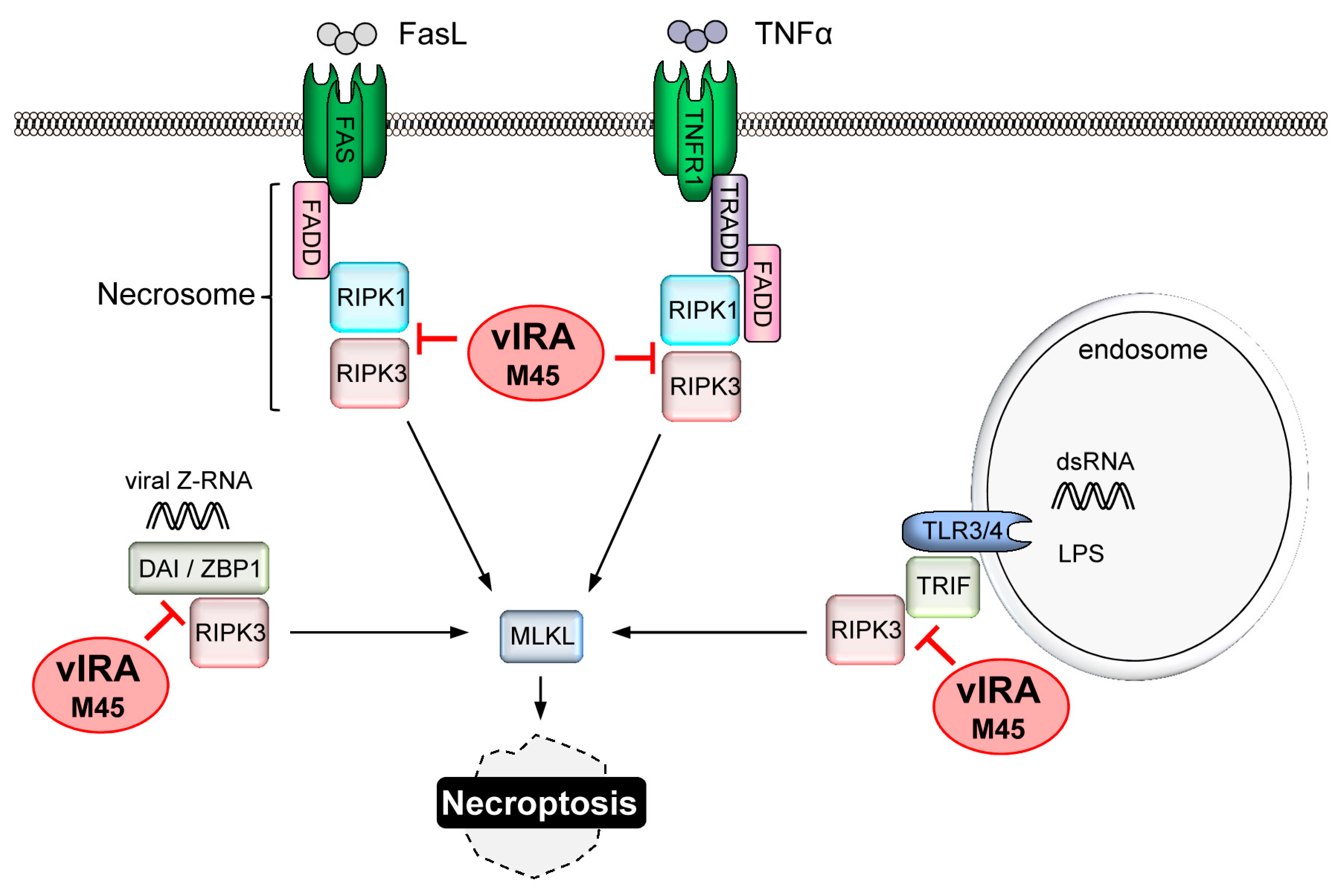
4. CMV-Encoded Death Inhibitors

Given the array of mechanisms that can lead to cell death in response to viral infection, a slow growing virus such as CMV would be expected to encode multiple death inhibitors. The ability to rapidly generate viral mutants lacking specific viral open reading frames has resulted in the identification of numerous CMV-encoded death inhibitors and aided in understanding how these proteins contribute to viral replication.

4.1. Inhibition of MOMP

A key step in the execution of intrinsic apoptosis is the permeabilization of the MOM by Bax and/or Bak. HCMV prevents MOMP by targeting Bax, and possibly Bak. The protein product of UL37 exon 1 (UL37x1) of HCMV, termed viral mitochondria-localized inhibitor of apoptosis (vMIA), binds and sequesters Bax at the mitochondrial membrane [80,81,82] (Figure 1). These early reports defined vMIA as a Bax-specific inhibitor, and suggested that activation of Bak requires the action of Bax in some cell types [81]. Thus, the inhibition of Bax by vMIA may be sufficient to prevent MOMP during HCMV infection. Several later studies have demonstrated an association between vMIA and Bak, implying that vMIA is capable of inhibiting both pro-apoptotic effector proteins [83,84]. If vMIA inhibits Bak, in addition to Bax, during viral infection remains to be confirmed. Several additional functions have been ascribed to vMIA. Expression of vMIA is sufficient to induce the release of calcium stores from the ER, a process that may modulate the apoptotic response [85]. In addition to its anti-apoptotic role vMIA inhibits anti-viral signalling downstream of the mitochondrial antiviral-signaling protein (MAVS) at mitochondria and peroxisomes [86,87]. vMIA therefore has the potential to promote viral replication via several mechanisms.
Unlike HCMV, MCMV encodes distinct inhibitors of Bax and Bak. The m38.5 protein of MCMV localises to mitochondria where it binds Bax and prevents its activation [84,88,89,90]. Although the MCMV m38.5 and HCMV UL37x1 proteins share little sequence similarity, they are very similar in their functions and their genes are located at analogous positions within the viral genomes. Therefore, m38.5 is also referred to as the vMIA of MCMV (Figure 1). A second MCMV-derived inhibitor, m41.1, associates with Bak at the mitochondrial membrane and acts as a viral inhibitor of Bak oligomerisation (vIBO) [91] (Figure 1). Cells infected in vitro with MCMV mutants lacking either m38.5 or m41.1 are sensitive to apoptosis induced by a range of stimuli [88,89,91,92]. Since activation of either Bax or Bak is sufficient to induce apoptosis, it is surprising that the in vivo growth characteristics of an Δm38.5 mutant differed from that observed when m41.1 was absent. Replication of a Δm41.1 mutant was attenuated in the liver and lungs, while deletion of m38.5 had no impact on viral replication at these sites [89,92,93,94]. By contrast, MCMV replication in leukocytes was reduced to a similar extent when either m38.5 or m41.1 was absent [89,92,93]. Optimal replication of MCMV therefore depends upon m38.5 and m41.1, whose combined activities maintain mitochondrial integrity. Overall the data suggest that inhibition of the intrinsic apoptotic pathway is an important requirement for CMV replication.
The perturbation of mitochondrial metabolism that occurs during viral infection can induce apoptosis. HCMV prevents cell death induced by oxidative stress by producing large amounts of a 2.7-kilobase non-coding RNA. During infection the β2.7 RNA interacts with complex I of the respiratory transport chain, resulting in maintenance of mitochondrial membrane potential [95]. Genes associated with retinoid/interferon-induced mortality (GRIM)-19 is an essential component of complex I that relocalises to a perinuclear region in response to oxidative stress [96]. β2.7 interacts with GRIM-19 and prevents its relocalisation from the mitochondria, allowing oxidative phosphorylation to continue and preventing oxidative stress-induced death [95].

4.2. Suppression of the ER Stress Response

The survival of CMV-infected cells depends on the ability to modulate the ER stress response. HCMV counteracts this process, in part, via the production of UL38. Cells infected with a HCMV mutant lacking UL38 die prematurely with cells displaying morphological changes consistent with the induction of apoptosis [97]. UL38 is a multifunctional protein with expression of the N-terminal 239 amino acids sufficient to suppress apoptosis [98,99]. Expression of UL38 is associated with accumulation of the activating transcription factor 4 (ATF4) and suppression of JNK activity [98]. The ATF4 transcription factor helps to resolve ER stress by inducing the production of proteins that facilitate protein folding within the ER. The inhibition of JNK activation prevents phosphorylation of Bcl-2 and Bim and so maintains the integrity of the mitochondrial membrane. Importantly, overexpression of ATF4 or inhibition of JNK activity reduced the death of cells infected with a pUL38-deficient virus demonstrating the functional relevance of these changes to the suppression of apoptosis [98]. The M38 gene of MCMV shares significant homology with UL38, suggesting that MCMV might have conserved this mechanism for inhibiting ER stress-induced death, however, this has not yet been formally tested.
A second mechanism for manipulating the ER stress response by CMV is downregulation of IRE1 protein levels. The M50 protein of MCMV interacts with IRE1, causing its degradation at late times post infection [100]. UL50, the HCMV homologue of M50, was found to have a similar impact on IRE1 expression [100]. By inducing the degradation of IRE1 the M50/UL50 proteins should restrict all IRE1 signalling events, including the activation of apoptosis-inducing pathways. However, the impact of M50/UL50 on ER stress-induced apoptosis has not yet been investigated.

4.3. Inhibition of DR-Mediated Apoptosis

Multiple proteins involved in immune recognition including DR are downregulated following infection with CMV. Cell surface expression of the Fas and TNF receptors are reduced following infection with HCMV or MCMV, respectively [101,102]. Surprisingly, the UL138 protein encoded by HCMV increases TNFR1 levels at the cell surface [103,104]. While these changes have been noted following in vitro infection how they impact on viral replication in vivo is unclear. The impact of CMV infection on the expression of the TNF-related apoptosis-inducing ligand (TRAIL) receptors has been more extensively characterised. The MCMV m166 open reading frame inhibits the cell surface expression of the TRAIL receptor [105]. Importantly, the in vivo replication of an m166 deletion virus was compromised, an effect that could be overcome by depleting NK cells. Direct targeting of the TRAIL receptor therefore allows MCMV-infected cells to avoid elimination by innate immune effector cells. A similar pathway is likely to exist in humans since the UL141 glycoprotein of HCMV is capable of binding to TRAIL receptors and promoting receptor retention within the cell [106].
CMV-encoded proteins that interfere with DR signalling also contribute to viral pathogenesis. HCMV encodes the viral inhibitor of caspase-8 activation (vICA), the protein product of the UL36 gene. HCMV vICA inhibits Fas-induced cell death by binding to pro-caspase-8 and preventing its activation [107] (Figure 1). Homologues of vICA have been identified in genomes of CMVs from different species implying that the capacity to inhibit DR signalling is an important requirement for CMV pathogenesis [108]. M36 is the vICA gene of MCMV. It is dispensable for viral replication in fibroblasts, but required for optimal replication in macrophages in vitro [109]. The replication defect of an MCMV M36 deletion mutant in macrophages was fully rescued by expression of HCMV UL36 or overexpression of a dominant-negative FADD [110,111] and partially rescued by expression of MC159, a viral FLIP of the molluscum contagiosum virus [112]. Remarkably, the in vivo growth defect of MCMV mutants lacking M36 was rescued by depletion of macrophages [113]. This finding is consistent with in vitro experiments where TNFα produced by macrophages induced apoptosis in cells infected with a ΔM36 virus. Thus, M36 allows MCMV-infected cells to resist the action of DR ligands produced by the host in response to infection. Similar to what has been observed with MCMV M36, HCMV UL36 was also required for cell death inhibition and viral replication in monocyte-derived macrophages [114]. However, cell death induced by a UL36-deficient virus could be inhibited by the broad-spectrum caspase inhibitor zVAD-fmk only at early but not at late times of monocyte-to-macrophage differentiation. This finding was interpreted as an indication that UL36 inhibits caspase-dependent as well as caspase-independent cell death programs in monocyte-derived macrophages [114].

4.4. Replication of CMV Requires Suppression of Necroptosis

As mentioned previously, the necroptotic death pathway can be activated under circumstances where caspase-8 activity is suppressed. The action of vICA, while offering protection from DR-mediated apoptosis, could conceivably sensitise cells to necroptosis. Detection of MCMV infection by host PRRs constitutes an additional mechanism by which the necroptotic pathway could be activated. Analysis of MCMV mutants has established that the capacity to inhibit necroptosis is essential for viral replication in vivo. Screening of a random transposon library identified the M45 protein as being important for preventing the death of infected endothelial cells and macrophages in vitro, and that the activity of M45 is essential for in vivo replication [115,116]. M45 interacts with both RIPK1 and RIPK3 and prevents necroptosis initiated by Fas or TNFR activation in vitro [117,118]. Therefore, M45 has been termed viral inhibitor of RIP activation (vIRA) (Figure 2). Construction of an MCMV mutant bearing an inactivating mutation within M45 clarified the physiological relevance of these findings [119]. Knockdown of RIPK1 expression or pharmacological inhibition of RIPK1 activity was unable to prevent necroptosis induced by the M45 mutant virus in vitro establishing that MCMV infection activates necroptosis via a RIPK1-independent mechanism [119]. By contrast, growth of the M45 mutant virus was equivalent to that of wild type (WT) virus in RIPK3 knockout mice [119]. The growth defect of the M45 mutant could also be rescued by infecting mice lacking DAI/ZBP1 [41] or expressing a Z-RNA binding-deficient DAI/ZBP1 protein [63]. This elegant series of observations established that the DAI/ZBP1 PRR senses Z-RNA produced during MCMV infection resulting in activation of RIPK3-dependent necroptosis (Figure 2), and that successful replication of MCMV depends upon suppression of this process by M45. Similarly, M45 can block TLR3 and TLR4-induced necroptosis by inhibiting the RHIM-dependent activation of RIP3 by the adaptor protein TRIF [42] (Figure 2).
Besides inhibiting necroptosis, M45 also modulates TNFR-dependent activation of transcription factor NF-κB and p38 mitogen-activated protein kinase by interacting with RIPK1 [117,120]. Moreover, M45 also interacts with the NF-κB essential modulator (NEMO) and redirects it to autophagosomes for degradation [121]. By blocking all canonical NF-κB activating pathways, M45 inhibits NF-κB-dependent expression of proinflammatory cytokines and survival factors. Thus, M45 has multiple functions that affect cell death, survival, and inflammation.
How the processes of apoptosis and necroptosis interact during viral infection was recently investigated using an MCMV mutant lacking functional M36 and M45. Macrophages infected with the double mutant activated caspase-8 to drive apoptosis early, with cells then progressing to secondary RIPK3-dependant necroptosis [122]. This form of cell death was highly inflammatory, resulting in an improved T cell response after in vivo infection. This data suggests that the simultaneous suppression of apoptosis and necroptosis not only prevents the premature death of infected cells, but also serves as a means of restricting the inflammatory response.
HCMV infection is also known to block the induction of necroptosis [123]. In contrast to MCMV, inhibition of necroptosis by HCMV occurs after the activation of RIPK3 and phosphorylation of MLKL. The viral protein responsible for conferring protection is regulated by IE1, but its identity has not been determined [123]. Interestingly, HCMV UL45 differs from its MCMV homologue, M45, in that it does not contain a RHIM. In contrast, the homologous proteins in herpes simplex virus (HSV) type 1 and 2 carry a RHIM [124]. The HSV1 ICP6 protein inhibits necroptosis in human cells [125], but surprisingly activates necroptosis in murine cells [126,127]. These results have established that necroptosis is an evolutionarily conserved response to herpesvirus infection. RHIM-mediated inhibition of necroptosis is a conserved strategy of both MCMV and HSV (at least in cells of their natural host), but MCMV and HCMV have developed distinct methods for antagonizing this process.

4.5. Interference with Pyroptosis?

CMV infection is known to activate the inflammasome [65]. If CMV can suppress this pathway is unclear, but some evidence from in vitro experiments suggests this may be the case. Expression of HCMV UL83 was sufficient to cause a reduction in the expression of AIM2 and inhibit the processing of IL-1β [128]. A reduction in the expression level of pro-IL-1β following MCMV infection has also been noted [129]. While tantalising, further work is required to determine if CMV has any significant capacity to interfere with pyroptosis.

5. Concluding Remarks

Mammalian cells have evolved an array of mechanisms to detect the presence of intracellular pathogens, with many of these pathways culminating in the activation of the cell death response. The study of CMV mutants has clearly established that successful viral replication requires the capacity to curb multiple host cell death pathways. While many of the viral inhibitors are conserved between HCMV and MCMV, some differences in how the respective viruses prevent cell death have been noted. For example, both viruses prevent necroptosis but they do so by targeting different points in the pathway. Indeed, the divergent anti-death strategies used by the various CMVs are proposed as one of the reasons why these viruses are so highly species specific [130].
Inhibition of cell death not only affords CMV an opportunity to complete the replication cycle, but also restricts the inflammatory response required for the generation of the adaptive immune response. Preventing cell death therefore serves several functions that contribute to viral pathogenesis. CMV-derived inhibitors of all the major cell death pathways have been described, with the exception of pyroptosis. If CMV inhibits the process of pyroptosis to any significant extent, and how this contributes to viral infection, is one of the significant questions that remains to be addressed. A second unresolved issue is if CMV latency or reactivation requires the inhibition of cell death. In support of this possibility, CD34+ progenitors harbouring a latent HCMV infection exhibit an increased resistance to apoptosis [131]. The elucidation of the mechanisms used by CMV to prevent cell death has not only enhanced our understanding of viral pathogenesis but provided insights into how host cell death responses are regulated. An exciting possibility is that this knowledge could be used to develop improved treatments for HCMV infections, a virus that causes significant morbidity and mortality in immunosuppressed patients.

Acknowledgments

Work in the authors’ laboratories was supported by the Deutsche Forschungsgemeinschaft (BR1730/3-2), the Australian Research Council (DP150102437), and the National Health and Medical Research Council of Australia (APP1125357 and APP1109288).

Conflicts of Interest

The authors declare no conflict of interest. The funding sponsors had no role in the writing of the manuscript or in the decision to publish it.

References

  1. Dillon, C.P.; Green, D.R. Molecular Cell Biology of Apoptosis and Necroptosis in Cancer. Adv. Exp. Med. Biol. 2016, 930, 1–23. [Google Scholar] [PubMed]
  2. Nagata, S.; Tanaka, M. Programmed cell death and the immune system. Nat. Rev. Immunol. 2017, 17, 333–340. [Google Scholar] [CrossRef] [PubMed]
  3. Tuzlak, S.; Kaufmann, T.; Villunger, A. Interrogating the relevance of mitochondrial apoptosis for vertebrate development and postnatal tissue homeostasis. Genes Dev. 2016, 30, 2133–2151. [Google Scholar] [CrossRef] [PubMed]
  4. Jorgensen, I.; Rayamajhi, M.; Miao, E.A. Programmed cell death as a defence against infection. Nat. Rev. Immunol. 2017, 17, 151–164. [Google Scholar] [CrossRef] [PubMed]
  5. Sridharan, H.; Upton, J.W. Programmed necrosis in microbial pathogenesis. Trends Microbiol. 2014, 22, 199–207. [Google Scholar] [CrossRef] [PubMed]
  6. Ho, M. The history of cytomegalovirus and its diseases. Med. Microbiol. Immunol. 2008, 197, 65–73. [Google Scholar] [CrossRef] [PubMed]
  7. Mocarski, E.S.; Shenk, T.; Griffiths, P.D.; Pass, R.F. Cytomegaloviruses. In Fields Virology; Knipe, D.M., Howley, P.M., Cohen, J.I., Griffin, D.E., Lamb, R.A., Martin, M.A., Racaniello, V.R., Roizman, B., Eds.; Lippincott, Williams and Wilkins: Philadelphia, PA, USA, 2013; pp. 1960–2014. [Google Scholar]
  8. Tait, S.W.; Green, D.R. Mitochondria and cell death: Outer membrane permeabilization and beyond. Nat. Rev. Mol. Cell Biol. 2010, 11, 621–632. [Google Scholar] [CrossRef] [PubMed]
  9. Kluck, R.M.; Bossy-Wetzel, E.; Green, D.R.; Newmeyer, D.D. The release of cytochrome c from mitochondria: A primary site for Bcl-2 regulation of apoptosis. Science 1997, 275, 1132–1136. [Google Scholar] [CrossRef] [PubMed]
  10. Li, P.; Nijhawan, D.; Budihardjo, I.; Srinivasula, S.M.; Ahmad, M.; Alnemri, E.S.; Wang, X. Cytochrome c and dATP-dependent formation of Apaf-1/caspase-9 complex initiates an apoptotic protease cascade. Cell 1997, 91, 479–489. [Google Scholar] [CrossRef]
  11. Verhagen, A.M.; Ekert, P.G.; Pakusch, M.; Silke, J.; Connolly, L.M.; Reid, G.E.; Moritz, R.L.; Simpson, R.J.; Vaux, D.L. Identification of DIABLO, a mammalian protein that promotes apoptosis by binding to and antagonizing IAP proteins. Cell 2000, 102, 43–53. [Google Scholar] [CrossRef]
  12. Du, C.; Fang, M.; Li, Y.; Li, L.; Wang, X. Smac, a mitochondrial protein that promotes cytochrome c-dependent caspase activation by eliminating IAP inhibition. Cell 2000, 102, 33–42. [Google Scholar] [CrossRef]
  13. Willis, S.N.; Chen, L.; Dewson, G.; Wei, A.; Naik, E.; Fletcher, J.I.; Adams, J.M.; Huang, D.C. Proapoptotic Bak is sequestered by Mcl-1 and Bcl-xL, but not Bcl-2, until displaced by BH3-only proteins. Genes Dev. 2005, 19, 1294–1305. [Google Scholar] [CrossRef] [PubMed]
  14. Willis, S.N.; Fletcher, J.I.; Kaufmann, T.; van Delft, M.F.; Chen, L.; Czabotar, P.E.; Ierino, H.; Lee, E.F.; Fairlie, W.D.; Bouillet, P.; et al. Apoptosis initiated when BH3 ligands engage multiple Bcl-2 homologs, not Bax or Bak. Science 2007, 315, 856–859. [Google Scholar] [CrossRef] [PubMed]
  15. Fletcher, J.I.; Meusburger, S.; Hawkins, C.J.; Riglar, D.T.; Lee, E.F.; Fairlie, W.D.; Huang, D.C.; Adams, J.M. Apoptosis is triggered when prosurvival Bcl-2 proteins cannot restrain Bax. Proc. Natl. Acad. Sci. USA 2008, 105, 18081–18087. [Google Scholar] [CrossRef] [PubMed]
  16. Wei, M.C.; Lindsten, T.; Mootha, V.K.; Weiler, S.; Gross, A.; Ashiya, M.; Thompson, C.B.; Korsmeyer, S.J. tBID, a membrane-targeted death ligand, oligomerizes BAK to release cytochrome c. Genes Dev. 2000, 14, 2060–2071. [Google Scholar] [PubMed]
  17. Letai, A.; Bassik, M.C.; Walensky, L.D.; Sorcinelli, M.D.; Weiler, S.; Korsmeyer, S.J. Distinct BH3 domains either sensitize or activate mitochondrial apoptosis, serving as prototype cancer therapeutics. Cancer Cell 2002, 2, 183–192. [Google Scholar] [CrossRef]
  18. Kim, H.; Tu, H.C.; Ren, D.; Takeuchi, O.; Jeffers, J.R.; Zambetti, G.P.; Hsieh, J.J.; Cheng, E.H. Stepwise activation of BAX and BAK by tBID, BIM, and PUMA initiates mitochondrial apoptosis. Mol. Cell 2009, 36, 487–499. [Google Scholar] [CrossRef] [PubMed]
  19. Czabotar, P.E.; Westphal, D.; Dewson, G.; Ma, S.; Hockings, C.; Fairlie, W.D.; Lee, E.F.; Yao, S.; Robin, A.Y.; Smith, B.J.; et al. Bax crystal structures reveal how BH3 domains activate Bax and nucleate its oligomerization to induce apoptosis. Cell 2013, 152, 519–531. [Google Scholar] [CrossRef] [PubMed]
  20. Moldoveanu, T.; Grace, C.R.; Llambi, F.; Nourse, A.; Fitzgerald, P.; Gehring, K.; Kriwacki, R.W.; Green, D.R. BID-induced structural changes in BAK promote apoptosis. Nat. Struct. Mol. Biol. 2013, 20, 589–597. [Google Scholar] [CrossRef] [PubMed]
  21. Lindsten, T.; Ross, A.J.; King, A.; Zong, W.X.; Rathmell, J.C.; Shiels, H.A.; Ulrich, E.; Waymire, K.G.; Mahar, P.; Frauwirth, K.; et al. The combined functions of proapoptotic Bcl-2 family members Bak and Bax are essential for normal development of multiple tissues. Mol. Cell 2000, 6, 1389–1399. [Google Scholar] [CrossRef]
  22. Rathmell, J.C.; Lindsten, T.; Zong, W.X.; Cinalli, R.M.; Thompson, C.B. Deficiency in Bak and Bax perturbs thymic selection and lymphoid homeostasis. Nat. Immunol. 2002, 3, 932–939. [Google Scholar] [CrossRef] [PubMed]
  23. Boldin, M.P.; Varfolomeev, E.E.; Pancer, Z.; Mett, I.L.; Camonis, J.H.; Wallach, D. A novel protein that interacts with the death domain of Fas/APO1 contains a sequence motif related to the death domain. J. Biol. Chem. 1995, 270, 7795–7798. [Google Scholar] [CrossRef] [PubMed]
  24. Chinnaiyan, A.M.; O’Rourke, K.; Tewari, M.; Dixit, V.M. FADD, a novel death domain-containing protein, interacts with the death domain of Fas and initiates apoptosis. Cell 1995, 81, 505–512. [Google Scholar] [CrossRef]
  25. Boatright, K.M.; Renatus, M.; Scott, F.L.; Sperandio, S.; Shin, H.; Pedersen, I.M.; Ricci, J.E.; Edris, W.A.; Sutherlin, D.P.; Green, D.R.; et al. A unified model for apical caspase activation. Mol. Cell 2003, 11, 529–541. [Google Scholar] [CrossRef]
  26. Irmler, M.; Thome, M.; Hahne, M.; Schneider, P.; Hofmann, K.; Steiner, V.; Bodmer, J.L.; Schroter, M.; Burns, K.; Mattmann, C.; et al. Inhibition of death receptor signals by cellular FLIP. Nature 1997, 388, 190–195. [Google Scholar] [CrossRef] [PubMed]
  27. Van Antwerp, D.J.; Martin, S.J.; Kafri, T.; Green, D.R.; Verma, I.M. Suppression of TNF-α-induced apoptosis by NF-κB. Science 1996, 274, 787–789. [Google Scholar] [CrossRef] [PubMed]
  28. Micheau, O.; Tschopp, J. Induction of TNF receptor I-mediated apoptosis via two sequential signaling complexes. Cell 2003, 114, 181–190. [Google Scholar] [CrossRef]
  29. Grootjans, S.; Vanden Berghe, T.; Vandenabeele, P. Initiation and execution mechanisms of necroptosis: An overview. Cell Death Differ. 2017, 24, 1184–1195. [Google Scholar] [CrossRef] [PubMed]
  30. Pop, C.; Oberst, A.; Drag, M.; van Raam, B.J.; Riedl, S.J.; Green, D.R.; Salvesen, G.S. FLIPL induces caspase 8 activity in the absence of interdomain caspase 8 cleavage and alters substrate specificity. Biochem. J. 2011, 433, 447–457. [Google Scholar] [CrossRef] [PubMed]
  31. Vercammen, D.; Beyaert, R.; Denecker, G.; Goossens, V.; van Loo, G.; Declercq, W.; Grooten, J.; Fiers, W.; Vandenabeele, P. Inhibition of caspases increases the sensitivity of L929 cells to necrosis mediated by tumor necrosis factor. J. Exp. Med. 1998, 187, 1477–1485. [Google Scholar] [CrossRef] [PubMed] [Green Version]
  32. Lin, Y.; Devin, A.; Rodriguez, Y.; Liu, Z.G. Cleavage of the death domain kinase RIP by caspase-8 prompts TNF-induced apoptosis. Genes Dev. 1999, 13, 2514–2526. [Google Scholar] [CrossRef] [PubMed]
  33. Feng, S.; Yang, Y.; Mei, Y.; Ma, L.; Zhu, D.E.; Hoti, N.; Castanares, M.; Wu, M. Cleavage of RIP3 inactivates its caspase-independent apoptosis pathway by removal of kinase domain. Cell. Signal. 2007, 19, 2056–2067. [Google Scholar] [CrossRef] [PubMed]
  34. Cho, Y.S.; Challa, S.; Moquin, D.; Genga, R.; Ray, T.D.; Guildford, M.; Chan, F.K. Phosphorylation-driven assembly of the RIP1-RIP3 complex regulates programmed necrosis and virus-induced inflammation. Cell 2009, 137, 1112–1123. [Google Scholar] [CrossRef] [PubMed]
  35. He, S.; Wang, L.; Miao, L.; Wang, T.; Du, F.; Zhao, L.; Wang, X. Receptor interacting protein kinase-3 determines cellular necrotic response to TNF-α. Cell 2009, 137, 1100–1111. [Google Scholar] [CrossRef] [PubMed]
  36. Zhang, D.W.; Shao, J.; Lin, J.; Zhang, N.; Lu, B.J.; Lin, S.C.; Dong, M.Q.; Han, J. RIP3, an energy metabolism regulator that switches TNF-induced cell death from apoptosis to necrosis. Science 2009, 325, 332–336. [Google Scholar] [CrossRef] [PubMed]
  37. Wang, H.; Sun, L.; Su, L.; Rizo, J.; Liu, L.; Wang, L.F.; Wang, F.S.; Wang, X. Mixed lineage kinase domain-like protein MLKL causes necrotic membrane disruption upon phosphorylation by RIP3. Mol. Cell 2014, 54, 133–146. [Google Scholar] [CrossRef] [PubMed]
  38. Cai, Z.; Jitkaew, S.; Zhao, J.; Chiang, H.C.; Choksi, S.; Liu, J.; Ward, Y.; Wu, L.G.; Liu, Z.G. Plasma membrane translocation of trimerized MLKL protein is required for TNF-induced necroptosis. Nat. Cell Biol. 2014, 16, 55–65. [Google Scholar] [CrossRef] [PubMed]
  39. Chen, X.; Li, W.; Ren, J.; Huang, D.; He, W.T.; Song, Y.; Yang, C.; Li, W.; Zheng, X.; Chen, P.; et al. Translocation of mixed lineage kinase domain-like protein to plasma membrane leads to necrotic cell death. Cell Res. 2014, 24, 105–121. [Google Scholar] [CrossRef] [PubMed]
  40. Dondelinger, Y.; Declercq, W.; Montessuit, S.; Roelandt, R.; Goncalves, A.; Bruggeman, I.; Hulpiau, P.; Weber, K.; Sehon, C.A.; Marquis, R.W.; et al. MLKL compromises plasma membrane integrity by binding to phosphatidylinositol phosphates. Cell Rep. 2014, 7, 971–981. [Google Scholar] [CrossRef] [PubMed] [Green Version]
  41. Upton, J.W.; Kaiser, W.J.; Mocarski, E.S. DAI/ZBP1/DLM-1 complexes with RIP3 to mediate virus-induced programmed necrosis that is targeted by murine cytomegalovirus vIRA. Cell Host Microbe 2012, 11, 290–297. [Google Scholar] [CrossRef] [PubMed]
  42. Kaiser, W.J.; Sridharan, H.; Huang, C.; Mandal, P.; Upton, J.W.; Gough, P.J.; Sehon, C.A.; Marquis, R.W.; Bertin, J.; Mocarski, E.S. Toll-like receptor 3-mediated necrosis via TRIF, RIP3, and MLKL. J. Biol. Chem. 2013, 288, 31268–31279. [Google Scholar] [CrossRef] [PubMed]
  43. He, W.T.; Wan, H.; Hu, L.; Chen, P.; Wang, X.; Huang, Z.; Yang, Z.H.; Zhong, C.Q.; Han, J. Gasdermin D is an executor of pyroptosis and required for interleukin-1β secretion. Cell Res. 2015, 25, 1285–1298. [Google Scholar] [CrossRef] [PubMed]
  44. Kayagaki, N.; Stowe, I.B.; Lee, B.L.; O’Rourke, K.; Anderson, K.; Warming, S.; Cuellar, T.; Haley, B.; Roose-Girma, M.; Phung, Q.T.; et al. Caspase-11 cleaves gasdermin D for non-canonical inflammasome signalling. Nature 2015, 526, 666–671. [Google Scholar] [CrossRef] [PubMed]
  45. Shi, J.; Zhao, Y.; Wang, K.; Shi, X.; Wang, Y.; Huang, H.; Zhuang, Y.; Cai, T.; Wang, F.; Shao, F. Cleavage of GSDMD by inflammatory caspases determines pyroptotic cell death. Nature 2015, 526, 660–665. [Google Scholar] [CrossRef] [PubMed]
  46. Guo, H.; Callaway, J.B.; Ting, J.P. Inflammasomes: Mechanism of action, role in disease, and therapeutics. Nat. Med. 2015, 21, 677–687. [Google Scholar] [CrossRef] [PubMed]
  47. Kayagaki, N.; Warming, S.; Lamkanfi, M.; Vande Walle, L.; Louie, S.; Dong, J.; Newton, K.; Qu, Y.; Liu, J.; Heldens, S.; et al. Non-canonical inflammasome activation targets caspase-11. Nature 2011, 479, 117–121. [Google Scholar] [CrossRef] [PubMed]
  48. Tenev, T.; Bianchi, K.; Darding, M.; Broemer, M.; Langlais, C.; Wallberg, F.; Zachariou, A.; Lopez, J.; MacFarlane, M.; Cain, K.; et al. The Ripoptosome, a signaling platform that assembles in response to genotoxic stress and loss of IAPs. Mol. Cell 2011, 43, 432–448. [Google Scholar] [CrossRef] [PubMed]
  49. Feoktistova, M.; Geserick, P.; Kellert, B.; Dimitrova, D.P.; Langlais, C.; Hupe, M.; Cain, K.; MacFarlane, M.; Hacker, G.; Leverkus, M. cIAPs block Ripoptosome formation, a RIP1/caspase-8 containing intracellular cell death complex differentially regulated by cFLIP isoforms. Mol. Cell 2011, 43, 449–463. [Google Scholar] [CrossRef] [PubMed]
  50. Kelliher, M.A.; Grimm, S.; Ishida, Y.; Kuo, F.; Stanger, B.Z.; Leder, P. The death domain kinase RIP mediates the TNF-induced NF-κB signal. Immunity 1998, 8, 297–303. [Google Scholar] [CrossRef]
  51. Oberst, A.; Dillon, C.P.; Weinlich, R.; McCormick, L.L.; Fitzgerald, P.; Pop, C.; Hakem, R.; Salvesen, G.S.; Green, D.R. Catalytic activity of the caspase-8-FLIPL complex inhibits RIPK3-dependent necrosis. Nature 2011, 471, 363–367. [Google Scholar] [CrossRef] [PubMed]
  52. Kaiser, W.J.; Upton, J.W.; Long, A.B.; Livingston-Rosanoff, D.; Daley-Bauer, L.P.; Hakem, R.; Caspary, T.; Mocarski, E.S. RIP3 mediates the embryonic lethality of caspase-8-deficient mice. Nature 2011, 471, 368–372. [Google Scholar] [CrossRef] [PubMed]
  53. Zhang, H.; Zhou, X.; McQuade, T.; Li, J.; Chan, F.K.; Zhang, J. Functional complementation between FADD and RIP1 in embryos and lymphocytes. Nature 2011, 471, 373–376. [Google Scholar] [CrossRef] [PubMed]
  54. Dillon, C.P.; Weinlich, R.; Rodriguez, D.A.; Cripps, J.G.; Quarato, G.; Gurung, P.; Verbist, K.C.; Brewer, T.L.; Llambi, F.; Gong, Y.N.; et al. RIPK1 blocks early postnatal lethality mediated by caspase-8 and RIPK3. Cell 2014, 157, 1189–1202. [Google Scholar] [CrossRef] [PubMed]
  55. Rickard, J.A.; O’Donnell, J.A.; Evans, J.M.; Lalaoui, N.; Poh, A.R.; Rogers, T.; Vince, J.E.; Lawlor, K.E.; Ninnis, R.L.; Anderton, H.; et al. RIPK1 regulates RIPK3-MLKL-driven systemic inflammation and emergency hematopoiesis. Cell 2014, 157, 1175–1188. [Google Scholar] [CrossRef] [PubMed]
  56. Kaiser, W.J.; Daley-Bauer, L.P.; Thapa, R.J.; Mandal, P.; Berger, S.B.; Huang, C.; Sundararajan, A.; Guo, H.; Roback, L.; Speck, S.H.; et al. RIP1 suppresses innate immune necrotic as well as apoptotic cell death during mammalian parturition. Proc. Natl. Acad. Sci. USA 2014, 111, 7753–7758. [Google Scholar] [CrossRef] [PubMed]
  57. Sagulenko, V.; Thygesen, S.J.; Sester, D.P.; Idris, A.; Cridland, J.A.; Vajjhala, P.R.; Roberts, T.L.; Schroder, K.; Vince, J.E.; Hill, J.M.; et al. AIM2 and NLRP3 inflammasomes activate both apoptotic and pyroptotic death pathways via ASC. Cell Death Differ. 2013, 20, 1149–1160. [Google Scholar] [CrossRef] [PubMed]
  58. Boehme, K.W.; Guerrero, M.; Compton, T. Human cytomegalovirus envelope glycoproteins B and H are necessary for TLR2 activation in permissive cells. J. Immunol. 2006, 177, 7094–7102. [Google Scholar] [CrossRef] [PubMed]
  59. Szomolanyi-Tsuda, E.; Liang, X.; Welsh, R.M.; Kurt-Jones, E.A.; Finberg, R.W. Role for TLR2 in NK cell-mediated control of murine cytomegalovirus in vivo. J. Virol. 2006, 80, 4286–4291. [Google Scholar] [CrossRef] [PubMed]
  60. Tabeta, K.; Georgel, P.; Janssen, E.; Du, X.; Hoebe, K.; Crozat, K.; Mudd, S.; Shamel, L.; Sovath, S.; Goode, J.; et al. Toll-like receptors 9 and 3 as essential components of innate immune defense against mouse cytomegalovirus infection. Proc. Natl. Acad. Sci. USA 2004, 101, 3516–3521. [Google Scholar] [CrossRef] [PubMed]
  61. Krug, A.; French, A.R.; Barchet, W.; Fischer, J.A.; Dzionek, A.; Pingel, J.T.; Orihuela, M.M.; Akira, S.; Yokoyama, W.M.; Colonna, M. TLR9-dependent recognition of MCMV by IPC and DC generates coordinated cytokine responses that activate antiviral NK cell function. Immunity 2004, 21, 107–119. [Google Scholar] [CrossRef] [PubMed]
  62. Estornes, Y.; Toscano, F.; Virard, F.; Jacquemin, G.; Pierrot, A.; Vanbervliet, B.; Bonnin, M.; Lalaoui, N.; Mercier-Gouy, P.; Pacheco, Y.; et al. dsRNA induces apoptosis through an atypical death complex associating TLR3 to caspase-8. Cell Death Differ. 2012, 19, 1482–1494. [Google Scholar] [CrossRef] [PubMed]
  63. Maelfait, J.; Liverpool, L.; Bridgeman, A.; Ragan, K.B.; Upton, J.W.; Rehwinkel, J. Sensing of viral and endogenous RNA by ZBP1/DAI induces necroptosis. EMBO J. 2017. [Google Scholar] [CrossRef] [PubMed]
  64. Sridharan, H.; Ragan, K.B.; Guo, H.; Gilley, R.P.; Landsteiner, V.J.; Kaiser, W.J.; Upton, J.W. Murine cytomegalovirus IE3-dependent transcription is required for DAI/ZBP1-mediated necroptosis. EMBO Rep. 2017, 18, 1429–1441. [Google Scholar] [CrossRef] [PubMed]
  65. Rathinam, V.A.; Jiang, Z.; Waggoner, S.N.; Sharma, S.; Cole, L.E.; Waggoner, L.; Vanaja, S.K.; Monks, B.G.; Ganesan, S.; Latz, E.; et al. The AIM2 inflammasome is essential for host defense against cytosolic bacteria and DNA viruses. Nat. Immunol. 2010, 11, 395–402. [Google Scholar] [CrossRef] [PubMed]
  66. Valchanova, R.S.; Picard-Maureau, M.; Budt, M.; Brune, W. Murine cytomegalovirus m142 and m143 are both required to block protein kinase R-mediated shutdown of protein synthesis. J. Virol. 2006, 80, 10181–10190. [Google Scholar] [CrossRef] [PubMed]
  67. Marshall, E.E.; Bierle, C.J.; Brune, W.; Geballe, A.P. Essential role for either TRS1 or IRS1 in human cytomegalovirus replication. J. Virol. 2009, 83, 4112–4120. [Google Scholar] [CrossRef] [PubMed]
  68. Dauber, B.; Wolff, T. Activation of the Antiviral Kinase PKR and Viral Countermeasures. Viruses 2009, 1, 523–544. [Google Scholar] [CrossRef] [PubMed]
  69. Gil, J.; Alcami, J.; Esteban, M. Induction of apoptosis by double-stranded-RNA-dependent protein kinase (PKR) involves the α subunit of eukaryotic translation initiation factor 2 and NF-κB. Mol. Cell Biol. 1999, 19, 4653–4663. [Google Scholar] [CrossRef] [PubMed]
  70. Balachandran, S.; Kim, C.N.; Yeh, W.C.; Mak, T.W.; Bhalla, K.; Barber, G.N. Activation of the dsRNA-dependent protein kinase, PKR, induces apoptosis through FADD-mediated death signaling. EMBO J. 1998, 17, 6888–6902. [Google Scholar] [CrossRef] [PubMed]
  71. Isler, J.A.; Skalet, A.H.; Alwine, J.C. Human cytomegalovirus infection activates and regulates the unfolded protein response. J. Virol. 2005, 79, 6890–6899. [Google Scholar] [CrossRef] [PubMed]
  72. Qian, Z.; Xuan, B.; Chapa, T.J.; Gualberto, N.; Yu, D. Murine cytomegalovirus targets transcription factor ATF4 to exploit the unfolded-protein response. J. Virol. 2012, 86, 6712–6723. [Google Scholar] [CrossRef] [PubMed]
  73. Tabas, I.; Ron, D. Integrating the mechanisms of apoptosis induced by endoplasmic reticulum stress. Nat. Cell Biol. 2011, 13, 184–190. [Google Scholar] [CrossRef] [PubMed]
  74. Yoneda, T.; Imaizumi, K.; Oono, K.; Yui, D.; Gomi, F.; Katayama, T.; Tohyama, M. Activation of caspase-12, an endoplastic reticulum (ER) resident caspase, through tumor necrosis factor receptor-associated factor 2-dependent mechanism in response to the ER stress. J. Biol. Chem. 2001, 276, 13935–13940. [Google Scholar] [CrossRef] [PubMed]
  75. Upton, J.P.; Wang, L.; Han, D.; Wang, E.S.; Huskey, N.E.; Lim, L.; Truitt, M.; McManus, M.T.; Ruggero, D.; Goga, A.; et al. IRE1α cleaves select microRNAs during ER stress to derepress translation of proapoptotic Caspase-2. Science 2012, 338, 818–822. [Google Scholar] [CrossRef] [PubMed]
  76. Hetz, C.; Bernasconi, P.; Fisher, J.; Lee, A.H.; Bassik, M.C.; Antonsson, B.; Brandt, G.S.; Iwakoshi, N.N.; Schinzel, A.; Glimcher, L.H.; et al. Proapoptotic BAX and BAK modulate the unfolded protein response by a direct interaction with IRE1α. Science 2006, 312, 572–576. [Google Scholar] [CrossRef] [PubMed]
  77. Xiaofei, E.; Kowalik, T.F. The DNA damage response induced by infection with human cytomegalovirus and other viruses. Viruses 2014, 6, 2155–2185. [Google Scholar] [PubMed]
  78. Gaspar, M.; Shenk, T. Human cytomegalovirus inhibits a DNA damage response by mislocalizing checkpoint proteins. Proc. Natl. Acad. Sci. USA 2006, 103, 2821–2826. [Google Scholar] [CrossRef] [PubMed]
  79. Luo, M.H.; Rosenke, K.; Czornak, K.; Fortunato, E.A. Human cytomegalovirus disrupts both ataxia telangiectasia mutated protein (ATM)- and ATM-Rad3-related kinase-mediated DNA damage responses during lytic infection. J. Virol. 2007, 81, 1934–1950. [Google Scholar] [CrossRef] [PubMed]
  80. Goldmacher, V.S.; Bartle, L.M.; Skaletskaya, A.; Dionne, C.A.; Kedersha, N.L.; Vater, C.A.; Han, J.; Lutz, R.J.; Watanabe, S.; McFarland, E.D.; et al. A cytomegalovirus-encoded mitochondria-localized inhibitor of apoptosis structurally unrelated to Bcl-2. Proc. Natl. Acad. Sci. USA 1999, 96, 12536–12541. [Google Scholar] [CrossRef] [PubMed]
  81. Arnoult, D.; Bartle, L.M.; Skaletskaya, A.; Poncet, D.; Zamzami, N.; Park, P.U.; Sharpe, J.; Youle, R.J.; Goldmacher, V.S. Cytomegalovirus cell death suppressor vMIA blocks Bax- but not Bak-mediated apoptosis by binding and sequestering Bax at mitochondria. Proc. Natl. Acad. Sci. USA 2004, 101, 7988–7993. [Google Scholar] [CrossRef] [PubMed]
  82. Poncet, D.; Larochette, N.; Pauleau, A.L.; Boya, P.; Jalil, A.A.; Cartron, P.F.; Vallette, F.; Schnebelen, C.; Bartle, L.M.; Skaletskaya, A.; et al. An anti-apoptotic viral protein that recruits Bax to mitochondria. J. Biol. Chem. 2004, 279, 22605–22614. [Google Scholar] [CrossRef] [PubMed]
  83. Karbowski, M.; Norris, K.L.; Cleland, M.M.; Jeong, S.Y.; Youle, R.J. Role of Bax and Bak in mitochondrial morphogenesis. Nature 2006, 443, 658–662. [Google Scholar] [CrossRef] [PubMed]
  84. Norris, K.L.; Youle, R.J. Cytomegalovirus proteins vMIA and m38.5 link mitochondrial morphogenesis to Bcl-2 family proteins. J. Virol. 2008, 82, 6232–6243. [Google Scholar] [CrossRef] [PubMed]
  85. Sharon-Friling, R.; Goodhouse, J.; Colberg-Poley, A.M.; Shenk, T. Human cytomegalovirus pUL37x1 induces the release of endoplasmic reticulum calcium stores. Proc. Natl. Acad. Sci. USA 2006, 103, 19117–19122. [Google Scholar] [CrossRef] [PubMed]
  86. Castanier, C.; Garcin, D.; Vazquez, A.; Arnoult, D. Mitochondrial dynamics regulate the RIG-I-like receptor antiviral pathway. EMBO Rep. 2010, 11, 133–138. [Google Scholar] [CrossRef] [PubMed]
  87. Magalhaes, A.C.; Ferreira, A.R.; Gomes, S.; Vieira, M.; Gouveia, A.; Valenca, I.; Islinger, M.; Nascimento, R.; Schrader, M.; Kagan, J.C.; et al. Peroxisomes are platforms for cytomegalovirus’ evasion from the cellular immune response. Sci. Rep. 2016, 6, 26028. [Google Scholar] [CrossRef] [PubMed] [Green Version]
  88. Jurak, I.; Schumacher, U.; Simic, H.; Voigt, S.; Brune, W. Murine cytomegalovirus m38.5 protein inhibits Bax-mediated cell death. J. Virol. 2008, 82, 4812–4822. [Google Scholar] [CrossRef] [PubMed]
  89. Manzur, M.; Fleming, P.; Huang, D.C.; Degli-Esposti, M.A.; Andoniou, C.E. Virally mediated inhibition of Bax in leukocytes promotes dissemination of murine cytomegalovirus. Cell Death Differ. 2009, 16, 312–320. [Google Scholar] [CrossRef] [PubMed]
  90. Arnoult, D.; Skaletskaya, A.; Estaquier, J.; Dufour, C.; Goldmacher, V.S. The murine cytomegalovirus cell death suppressor m38.5 binds Bax and blocks Bax-mediated mitochondrial outer membrane permeabilization. Apoptosis 2008, 13, 1100–1110. [Google Scholar] [CrossRef] [PubMed]
  91. Çam, M.; Handke, W.; Picard-Maureau, M.; Brune, W. Cytomegaloviruses inhibit Bak- and Bax-mediated apoptosis with two separate viral proteins. Cell Death Differ. 2010, 17, 655–665. [Google Scholar] [CrossRef] [PubMed]
  92. Fleming, P.; Kvansakul, M.; Voigt, V.; Kile, B.T.; Kluck, R.M.; Huang, D.C.; Degli-Esposti, M.A.; Andoniou, C.E. MCMV-mediated inhibition of the pro-apoptotic Bak protein is required for optimal in vivo replication. PLoS Pathog. 2013, 9, e1003192. [Google Scholar] [CrossRef] [PubMed]
  93. Crosby, L.N.; McCormick, A.L.; Mocarski, E.S. Gene products of the embedded m41/m41.1 locus of murine cytomegalovirus differentially influence replication and pathogenesis. Virology 2013, 436, 274–283. [Google Scholar] [CrossRef] [PubMed]
  94. Handke, W.; Luig, C.; Popovic, B.; Krmpotic, A.; Jonjic, S.; Brune, W. Viral inhibition of BAK promotes murine cytomegalovirus dissemination to salivary glands. J. Virol. 2013, 87, 3592–3596. [Google Scholar] [CrossRef] [PubMed]
  95. Reeves, M.B.; Davies, A.A.; McSharry, B.P.; Wilkinson, G.W.; Sinclair, J.H. Complex I binding by a virally encoded RNA regulates mitochondria-induced cell death. Science 2007, 316, 1345–1348. [Google Scholar] [CrossRef] [PubMed]
  96. Lu, H.; Cao, X. GRIM-19 is essential for maintenance of mitochondrial membrane potential. Mol. Biol. Cell 2008, 19, 1893–1902. [Google Scholar] [CrossRef] [PubMed]
  97. Terhune, S.; Torigoi, E.; Moorman, N.; Silva, M.; Qian, Z.; Shenk, T.; Yu, D. Human cytomegalovirus UL38 protein blocks apoptosis. J. Virol. 2007, 81, 3109–3123. [Google Scholar] [CrossRef] [PubMed]
  98. Xuan, B.; Qian, Z.; Torigoi, E.; Yu, D. Human cytomegalovirus protein pUL38 induces ATF4 expression, inhibits persistent JNK phosphorylation, and suppresses endoplasmic reticulum stress-induced cell death. J. Virol. 2009, 83, 3463–3474. [Google Scholar] [CrossRef] [PubMed]
  99. Qian, Z.; Xuan, B.; Gualberto, N.; Yu, D. The human cytomegalovirus protein pUL38 suppresses endoplasmic reticulum stress-mediated cell death independently of its ability to induce mTORC1 activation. J. Virol. 2011, 85, 9103–9113. [Google Scholar] [CrossRef] [PubMed]
  100. Stahl, S.; Burkhart, J.M.; Hinte, F.; Tirosh, B.; Mohr, H.; Zahedi, R.P.; Sickmann, A.; Ruzsics, Z.; Budt, M.; Brune, W. Cytomegalovirus downregulates IRE1 to repress the unfolded protein response. PLoS Pathog. 2013, 9, e1003544. [Google Scholar] [CrossRef] [PubMed]
  101. Seirafian, S.; Prod’homme, V.; Sugrue, D.; Davies, J.; Fielding, C.; Tomasec, P.; Wilkinson, G.W. Human cytomegalovirus suppresses Fas expression and function. J. Gen. Virol. 2014, 95, 933–939. [Google Scholar] [CrossRef] [PubMed]
  102. Popkin, D.L.; Virgin, H.W., 4th. Murine cytomegalovirus infection inhibits tumor necrosis factor α responses in primary macrophages. J. Virol. 2003, 77, 10125–10130. [Google Scholar] [CrossRef] [PubMed]
  103. Le, V.T.; Trilling, M.; Hengel, H. The cytomegaloviral protein pUL138 acts as potentiator of tumor necrosis factor (TNF) receptor 1 surface density to enhance ULb'-encoded modulation of TNF-α signaling. J. Virol. 2011, 85, 13260–13270. [Google Scholar] [CrossRef] [PubMed]
  104. Montag, C.; Wagner, J.A.; Gruska, I.; Vetter, B.; Wiebusch, L.; Hagemeier, C. The latency-associated UL138 gene product of human cytomegalovirus sensitizes cells to tumor necrosis factor α (TNF-α) signaling by upregulating TNF-α receptor 1 cell surface expression. J. Virol. 2011, 85, 11409–11421. [Google Scholar] [CrossRef] [PubMed]
  105. Verma, S.; Loewendorf, A.; Wang, Q.; McDonald, B.; Redwood, A.; Benedict, C.A. Inhibition of the TRAIL death receptor by CMV reveals its importance in NK cell-mediated antiviral defense. PLoS Pathog. 2014, 10, e1004268. [Google Scholar] [CrossRef] [PubMed]
  106. Smith, W.; Tomasec, P.; Aicheler, R.; Loewendorf, A.; Nemcovicova, I.; Wang, E.C.; Stanton, R.J.; Macauley, M.; Norris, P.; Willen, L.; et al. Human cytomegalovirus glycoprotein UL141 targets the TRAIL death receptors to thwart host innate antiviral defenses. Cell Host Microbe 2013, 13, 324–335. [Google Scholar] [CrossRef] [PubMed]
  107. Skaletskaya, A.; Bartle, L.M.; Chittenden, T.; McCormick, A.L.; Mocarski, E.S.; Goldmacher, V.S. A cytomegalovirus-encoded inhibitor of apoptosis that suppresses caspase-8 activation. Proc. Natl. Acad. Sci. USA 2001, 98, 7829–7834. [Google Scholar] [CrossRef] [PubMed]
  108. McCormick, A.L.; Skaletskaya, A.; Barry, P.A.; Mocarski, E.S.; Goldmacher, V.S. Differential function and expression of the viral inhibitor of caspase 8-induced apoptosis (vICA) and the viral mitochondria-localized inhibitor of apoptosis (vMIA) cell death suppressors conserved in primate and rodent cytomegaloviruses. Virology 2003, 316, 221–233. [Google Scholar] [CrossRef] [PubMed]
  109. Menard, C.; Wagner, M.; Ruzsics, Z.; Holak, K.; Brune, W.; Campbell, A.E.; Koszinowski, U.H. Role of murine cytomegalovirus US22 gene family members in replication in macrophages. J. Virol. 2003, 77, 5557–5570. [Google Scholar] [CrossRef] [PubMed]
  110. Chaudhry, M.; Kasmapour, B.; Plaza, C.; Bajagic, M.; Casalegno Garduno, R.; Borkner, L.; Lenac Roviš, T.; Scrima, A.; Jonjic, S.; Schmitz, I.; et al. UL36 Rescues Apoptosis Inhibition and In Vivo Replication of A Chimeric MCMV Lacking the M36 Gene. Front. Microbiol. 2017, 7, 312. [Google Scholar] [CrossRef] [PubMed]
  111. Cicin-Sain, L.; Ruzsics, Z.; Podlech, J.; Bubic, I.; Menard, C.; Jonjic, S.; Reddehase, M.J.; Koszinowski, U.H. Dominant-negative FADD rescues the in vivo fitness of a cytomegalovirus lacking an antiapoptotic viral gene. J. Virol. 2008, 82, 2056–2064. [Google Scholar] [CrossRef] [PubMed]
  112. Hüttmann, J.; Krause, E.; Schommartz, T.; Brune, W. Functional Comparison of Molluscum Contagiosum Virus vFLIP MC159 with Murine Cytomegalovirus M36/vICA and M45/vIRA Proteins. J. Virol. 2015, 90, 2895–2905. [Google Scholar] [CrossRef] [PubMed]
  113. Ebermann, L.; Ruzsics, Z.; Guzman, C.A.; van Rooijen, N.; Casalegno-Garduno, R.; Koszinowski, U.; Cicin-Sain, L. Block of death-receptor apoptosis protects mouse cytomegalovirus from macrophages and is a determinant of virulence in immunodeficient hosts. PLoS Pathog. 2012, 8, e1003062. [Google Scholar] [CrossRef] [PubMed] [Green Version]
  114. McCormick, A.L.; Roback, L.; Livingston-Rosanoff, D.; St Clair, C. The human cytomegalovirus UL36 gene controls caspase-dependent and -independent cell death programs activated by infection of monocytes differentiating to macrophages. J. Virol. 2010, 84, 5108–5123. [Google Scholar] [CrossRef] [PubMed]
  115. Brune, W.; Ménard, C.; Heesemann, J.; Koszinowski, U.H. A ribonucleotide reductase homolog of cytomegalovirus and endothelial cell tropism. Science 2001, 291, 303–305. [Google Scholar] [CrossRef] [PubMed]
  116. Lembo, D.; Donalisio, M.; Hofer, A.; Cornaglia, M.; Brune, W.; Koszinowski, U.; Thelander, L.; Landolfo, S. The ribonucleotide reductase R1 homolog of murine cytomegalovirus is not a functional enzyme subunit but is required for pathogenesis. J. Virol. 2004, 78, 4278–4288. [Google Scholar] [CrossRef] [PubMed]
  117. Mack, C.; Sickmann, A.; Lembo, D.; Brune, W. Inhibition of proinflammatory and innate immune signaling pathways by a cytomegalovirus RIP1-interacting protein. Proc. Natl. Acad. Sci. USA 2008, 105, 3094–3099. [Google Scholar] [CrossRef] [PubMed]
  118. Upton, J.W.; Kaiser, W.J.; Mocarski, E.S. Cytomegalovirus M45 cell death suppression requires receptor-interacting protein (RIP) homotypic interaction motif (RHIM)-dependent interaction with RIP1. J. Biol. Chem. 2008, 283, 16966–16970. [Google Scholar] [CrossRef] [PubMed]
  119. Upton, J.W.; Kaiser, W.J.; Mocarski, E.S. Virus inhibition of RIP3-dependent necrosis. Cell Host Microbe 2010, 7, 302–313. [Google Scholar] [CrossRef] [PubMed]
  120. Krause, E.; de Graaf, M.; Fliss, P.M.; Dölken, L.; Brune, W. Murine cytomegalovirus virion-associated protein M45 mediates rapid NF-κB activation after infection. J. Virol. 2014, 88, 9963–9975. [Google Scholar] [CrossRef] [PubMed]
  121. Fliss, P.M.; Jowers, T.P.; Brinkmann, M.M.; Holstermann, B.; Mack, C.; Dickinson, P.; Hohenberg, H.; Ghazal, P.; Brune, W. Viral mediated redirection of NEMO/IKKγ to autophagosomes curtails the inflammatory cascade. PLoS Pathog. 2012, 8, e1002517. [Google Scholar] [CrossRef] [PubMed]
  122. Daley-Bauer, L.P.; Roback, L.; Crosby, L.N.; McCormick, A.L.; Feng, Y.; Kaiser, W.J.; Mocarski, E.S. Mouse cytomegalovirus M36 and M45 death suppressors cooperate to prevent inflammation resulting from antiviral programmed cell death pathways. Proc. Natl. Acad. Sci. USA 2017, 114, E2786–E2795. [Google Scholar] [CrossRef] [PubMed]
  123. Omoto, S.; Guo, H.; Talekar, G.R.; Roback, L.; Kaiser, W.J.; Mocarski, E.S. Suppression of RIP3-dependent necroptosis by human cytomegalovirus. J. Biol. Chem. 2015, 290, 11635–11648. [Google Scholar] [CrossRef] [PubMed]
  124. Lembo, D.; Brune, W. Tinkering with a viral ribonucleotide reductase. Trends Biochem. Sci. 2009, 34, 25–32. [Google Scholar] [CrossRef] [PubMed]
  125. Guo, H.; Omoto, S.; Harris, P.A.; Finger, J.N.; Bertin, J.; Gough, P.J.; Kaiser, W.J.; Mocarski, E.S. Herpes simplex virus suppresses necroptosis in human cells. Cell Host Microbe 2015, 17, 243–251. [Google Scholar] [CrossRef] [PubMed]
  126. Wang, X.; Li, Y.; Liu, S.; Yu, X.; Li, L.; Shi, C.; He, W.; Li, J.; Xu, L.; Hu, Z.; et al. Direct activation of RIP3/MLKL-dependent necrosis by herpes simplex virus 1 (HSV-1) protein ICP6 triggers host antiviral defense. Proc. Natl. Acad. Sci. USA 2014, 111, 15438–15443. [Google Scholar] [CrossRef] [PubMed]
  127. Huang, Z.; Wu, S.Q.; Liang, Y.; Zhou, X.; Chen, W.; Li, L.; Wu, J.; Zhuang, Q.; Chen, C.; Li, J.; et al. RIP1/RIP3 binding to HSV-1 ICP6 initiates necroptosis to restrict virus propagation in mice. Cell Host Microbe 2015, 17, 229–242. [Google Scholar] [CrossRef] [PubMed]
  128. Huang, Y.; Ma, D.; Huang, H.; Lu, Y.; Liao, Y.; Liu, L.; Liu, X.; Fang, F. Interaction between HCMV pUL83 and human AIM2 disrupts the activation of the AIM2 inflammasome. Virol. J. 2017, 14, 34. [Google Scholar] [CrossRef] [PubMed]
  129. Sester, D.P.; Sagulenko, V.; Thygesen, S.J.; Cridland, J.A.; Loi, Y.S.; Cridland, S.O.; Masters, S.L.; Genske, U.; Hornung, V.; Andoniou, C.E.; et al. Deficient NLRP3 and AIM2 Inflammasome Function in Autoimmune NZB Mice. J. Immunol. 2015, 195, 1233–1241. [Google Scholar] [CrossRef] [PubMed]
  130. Jurak, I.; Brune, W. Induction of apoptosis limits cytomegalovirus cross-species infection. EMBO J. 2006, 25, 2634–2642. [Google Scholar] [CrossRef] [PubMed]
  131. Poole, E.; McGregor Dallas, S.R.; Colston, J.; Joseph, R.S.; Sinclair, J. Virally induced changes in cellular microRNAs maintain latency of human cytomegalovirus in CD34+ progenitors. J. Gen. Virol. 2011, 92, 1539–1549. [Google Scholar] [CrossRef] [PubMed]
Figure 1. Inhibition of apoptosis by cytomegalovirus (CMV). The viral mitochondria-localized inhibitor of apoptosis (vMIA) and the viral inhibitor of BAK oligomerization (vIBO) inhibit mitochondrial outer membrane permabilization and release of proapoptotic factors (such as cytochrome C (cytC)) by interacting with BAX and BAK, respectively. While murine CMV (MCMV) encodes two specific inhibitors, m38.5 and m41.1, human CMV (HCMV) has only one inhibitor, UL37x1. Whether the UL37x1 protein is BAX-specific or inhibits both BAX and BAK remains controversial. The extrinsic apoptosis pathway initiated by death receptor stimulation is blocked by the viral inhibitor of caspase-8 activation (vICA), which is encoded by the HCMV UL36 and the MCMV M36 gene, respectively. FasL: Fas ligand; FADD: Fas-associated death domain protein; RIPK1: receptor interacting protein kinase-1; TNFα: tumour necrosis factor α; TNFR1: TNF receptor 1; TRADD: TNFR1-associated death domain protein; t-BID: truncated BH3-interacting domain death agonist; APAF1: apoptotic protease activating factor 1.
Viruses 09 00249 g001
Figure 2. Inhibition of necroptosis by MCMV. Induction of programmed necrosis (necroptosis) involves activation of RIPK3 and mixed lineage kinase domain-like (MLKL). The viral inhibitor of RIP activation (vIRA), encoded by the MCMV M45 gene, contains a RIP homotypic interaction motif (RHIM) and inhibits RHIM-dependent activation of RIPK3 by RIPK1, DAI/ZBP1, or TRIF. Death receptor-induced necroptosis requires caspase-8 inhibition, e.g., by vICA. LPS: lipopolysaccharide.
Viruses 09 00249 g002

