


Oral 2026, 6(3), 64; https://doi.org/10.3390/oral6030064 (registering DOI) - 29 May 2026
Abstract
Background-Objectives: Edentulism remains a major global health problem, and implant-supported overdentures (ISODs) are widely used to restore oral function and improve quality of life in edentulous patients. Among the available attachment systems, bar configurations play an important role in determining biomechanical behaviour,
[...] Read more.
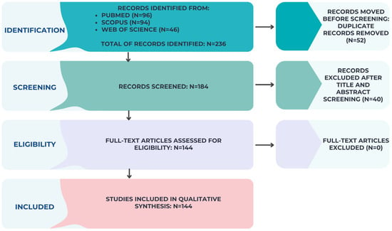
Background-Objectives: Edentulism remains a major global health problem, and implant-supported overdentures (ISODs) are widely used to restore oral function and improve quality of life in edentulous patients. Among the available attachment systems, bar configurations play an important role in determining biomechanical behaviour, retention, stability, and maintenance requirements. This scoping review aimed to map and evaluate the influence of key bar attachment parameters—such as cross-sectional geometry, material, splinting configuration, and distal extension—on the clinical performance of overdenture therapy. Methods: The review followed the Preferred Reporting Items for Systematic Reviews and Meta-Analyses Extension for Scoping Reviews (PRISMA-ScR) framework. A comprehensive search was conducted in PubMed, Scopus, and the Cochrane Library. Eligible studies included clinical investigations, in vitro mechanical studies, and finite element analyses addressing bar-retained implant-supported overdentures. Extracted data included bar configuration characteristics, implant distribution, and reported outcomes such as retention forces, stress distribution, prosthetic complications, and patient-reported measures. Results: The available evidence indicated a recurring balance between increased retention and higher peri-implant stress, particularly in association with Hader bar designs. Material selection also appeared to influence performance. CAD/CAM-milled titanium bars demonstrated favourable mechanical durability, whereas alternative materials such as PEEK and zirconia were associated with improved stress distribution and potential biological advantages, although concerns regarding long-term durability remain. Differences related to arch type were also observed, with splinted bars supported by four implants generally favoured in the maxilla, while two-implant bar overdentures appear to provide satisfactory outcomes in the mandible. Conclusions: Bar selection should be individualised according to anatomical conditions, biomechanical demands, and patient-specific factors. Longer-term clinical studies and more standardised testing protocols are still required, particularly for newer materials and digitally fabricated bar systems, to support more consistent evidence-based decision-making.
Full article


