Review

Transmembrane Mucin-1 Facilitates Oral Microbial Colonization in Oral Cancer

1
Oral Pathology Department, Agartala Government Dental College, Agartala 799001, India
2
Institute of Dentistry, School of Medicine, University of Eastern Finland, 70211 Kuopio, Finland
*
Author to whom correspondence should be addressed.
Submission received: 25 July 2025 / Revised: 26 August 2025 / Accepted: 10 September 2025 / Published: 9 October 2025

Abstract

Mucins are a family of heavily glycosylated proteins that form the main organic component of the oral mucosal barrier complex. Transmembrane mucin 1 (tMUC1) is anchored at the superficial epithelial surface to provide a protective function. The interaction of tMUC1 with oral microbes provides nutrients and physicochemical protection, promotes adhesion, and increases the microbe residence time in the oral cavity. Mucin-degrading microorganisms in the consortia also offer some advantages to oral microbes. The high molecular weight of mucin glycoproteins is hard to study because of their size, complexity, and heterogeneity. This review discusses how mucin facilitates oral microbiome colonization and how mucin–microbial interactions influence the development of oral cancer, mainly oral squamous cell carcinoma.

1. Introduction

Oral cancer is a serious condition affecting various regions of the world and rapidly increasing in incidence. Worldwide, 354,864 new cases and 177,384 estimated oral cancer deaths per year are reported, based on recent GLOBOCON studies [1]. Oral cancer, with its various histopathological entities, has become a global health problem. The histopathological type, such as oral squamous cell carcinoma (OSCC), verrucous carcinoma, basal cell carcinoma, melanoma, salivary gland carcinoma, lymphomas, and sarcomas, influences the survival rate and prognosis [2]. OSCC is the most common oral cancer arising from the mucosal surfaces, showing high rates of recurrence and lymph node metastasis. The etiopathogenesis of OSCC includes diverse factors, such as a genetic predisposition, immunologic disturbances, viral and bacterial infections, food allergies, vitamin and microelement deficiencies, hormonal imbalances, mechanical injuries, and stress [3]. Several oral bacteria may be related to the development of OSCC [4,5,6].
The oral cavity contains the most diverse communities of microbes in the human body after the gut. According to the Human Oral Microbiome Database (HOMD), the oral cavity harbors around 774 species [7]. Most human oral microbiome studies use a 16S rRNA-based next-generation sequencing (NGS) method to study functional and structural aspects of bacterial communities in healthy and diseased conditions [7]. In past decades, research has been focused on oral microflora and the oral microbiome. It has been established that oral epithelial cells absorb and oral fluids contain end products produced by the oral microbiome through bacterial metabolism [8]. However, several factors can disrupt the diversity and composition of the commensal oral microbiota. Alterations in the oral microbiome cause oral mucosal inflammation that accelerates OSCC by causing the direct metabolism of carcinogenic substances [9]. Oral microbial dysbiosis may cause diseases through several molecular mechanisms. Hence, synergistic crosstalk between the oral microbiota and the oral epithelium is crucial for maintaining mucosal homeostasis.
Following a comprehensive search of the English language literature, we present a narrative review focusing on the interactions between the oral mucosal barrier, mainly mucin, and oral microbes. We also attempt to highlight how their interactions influence the progression to oral cancer, mainly OSCC.

2. Oral Mucosal Defense

The oral epithelium forms the first line of defense in the oral cavity, forming a barrier between the external environment and the rest of the body. The oral mucosal barrier complex (OMBC) is formed at the superficial layer of the oral epithelium by bioadhesion between salivary components and the apical portion of superficial epithelial cells. It performs multiple functions to protect the oral mucosa from microorganisms and other toxic materials [10]. Mucins are some of the components of the OMBC and help to stabilize the mucosal pellicle. MUC1 is a transmembrane glycoprotein that normally protects and lubricates epithelial cells [11,12]. Among several mucins, mucin 1 (MUC1) is produced by the oral epithelium and acts as an anchoring protein for the mucosal pellicle. MUC1 can crosslink with other salivary mucins (MUC5B and MUC7) and provide a signaling pathway between saliva and mucosal cells while stabilizing the mucosal pellicle [13]. Factors such as a weak immune system, nutritional deficiencies, metabolic diseases, diet, poor oral hygiene, and habits can contribute to oral dysbiosis, facilitating a breach in the epithelial barrier and incorporation of microorganisms into different oral niches [14].
It has been shown that the mucosal barrier and microbiota can modulate immune responses. Altered immune responses can cause inflammation that disturbs the symbiotic relationships between microorganisms, thereby increasing the number of pathogenic microorganisms [15]. Microbes regulate immunity by controlling regulatory T cells (Tregs) and T helper 17 (Th17) cells [16]. MUC1 is a known T cell regulator [17]. The interplay between the gut microbiota, immune system, and epithelial barrier has been well studied [18,19], but limited literature is available about the oral cavity.

3. Oral Mucosal Epithelium and Oral Microbiome

The oral mucosal epithelium forms a barrier that allows for numerous structural and functional protein interactions in response to various exogenous, possibly toxic, influences [20]. Due to the oral mucosa’s integrity and its continuous renewal, the oral mucosa prevents microorganisms from entering deeper tissues. However, the defensive function of the mucosa is linked with the presence of mucosal defense cells and the host immune system [21]. Oral mucosal inflammation occurs due to an imbalance in symbiotic microorganisms and/or pathogenic microorganisms. Several studies have shown that the mucosal microbiota can modulate innate and adaptive immune responses [15,22]. It has been suggested that microbes regulate immunity by controlling regulatory T cells (Tregs) and T helper 17 (Th17) cells [16].
The mucosal pellicle is formed through interactions between MUC1 and mucins secreted in the saliva, namely MUC5B and MUC7. MUC1 lubricates the epithelial surface owing to its glycosylation [23]. MUC1 has an extracellular and cytoplasmic domain. It has been suggested that the dissociation of the extracellular domain of MUC1 is a part of a defense mechanism against epithelial aggression. Such dissociation potentially stimulates the MUC1’s cytoplasmic domain and activates intracellular pathways [24]. The oral mucosal epithelium anchors Toll-like receptors (TLRs), transmembrane proteins that also act as immunodefenses against fungal, bacterial, and viral pathogens. TLRs recognize molecular structures classified as “pathogen-associated molecular patterns” (PAMPs) and activate a downstream signaling pathway that has an important role in innate and adaptive immune responses [25]. The activation of TLRs is suppressed by the anti-inflammatory activity of MUC1 on the oral epithelium. MUC1 prevents the release of pro-inflammatory products and cytokines induced by TLRs [13].
Evidence supports the fact that mucins are involved in complex biological processes like epithelial differentiation, cell adhesion, and cell signaling [26]. The constant exposure of the oral mucosa to oral microbes and their secreted products can release pro-inflammatory mediators that cause increased transcription of MUC1 [27]. Alteration in the expression of MUC1 can only protect oral epithelial surfaces from various noxious, pathogenic, and non-pathogenic microbes to an extent [13]. Changes in the expression of MUC1 by oral mucosal epithelial cells are indicative of an altered salivary composition and mucosal immune defense response. Hence, the balanced immune–inflammatory state of the host’s oral mucosal pellicle/biofilm is disturbed by dysbiotic microbiota. Growing evidence has emerged to support the idea that oral microorganisms play a key role in the development and progression of certain disease entities [28]. Figure 1 summarizes the most important information relating to the changes in the oral microbial composition during the development of oral and systemic diseases.

4. Saliva Regulates Oral Microbiota

Saliva is a complex biological fluid secreted by the major and minor salivary glands that contains a broad spectrum of biomarkers of health and the disease status [14]. In the oral cavity, an optimal growth environment is always available for oral microbes due to the maintenance of a stable temperature and humidity [29]. The end products produced by oral microorganisms in the saliva, as salivary metabolites, represent a change in the oral metabolic pathway. These salivary metabolites have become a potential source of biomarkers to assess oral diseases using various advanced methods such as nuclear magnetic resonance (NMR) spectroscopy, mass spectroscopy (MS), gas chromatography (GS), capillary electrophoresis (CE), and high-performance liquid chromatography (HPLC) [8].
The HOMD lists site-specific bacteria in a healthy oral cavity and in the saliva that can proliferate differently in various ecological niches [30]. Streptococcus, Uncl. Pasteurellaceae, Neisseria flavescens, Rhodotorulla mucilaginosa, Streptococcus salivarius, Prevotella histicola, Veillonella parvula, Veillonella atypica, Streptococcus parasanguinis, Actinomycetales, Fusobacterium, Neisseria, Prevotella, Tannerella, and Veillonella are observed in healthy human saliva and coexist in balance [9,31]. However, when this balance is disrupted, harmful bacteria, such as Porphyromonas gingivalis, Tannerella forsythia, and Treponema denticola, can proliferate and invade gingiva tissue, causing inflammation and potentially leading to tooth loss and/or the onset of periodontal diseases [32]. Due to a lack of salivary flushing action, pathogenic bacteria produce more diverse bacterial waste products that either function as signaling molecules or affect intracellular pathways or activate oncogenic pathways [33]. Streptococcus mutans (S. mutans) causes dental caries by fermenting dietary sugars into lactic acid, which can demineralize tooth enamel. Also, a reduction in the salivary pH and an increase in porosity have been observed in the dental plaque matrix [34].
Saliva has several antimicrobial components, such as agglutinins, lyzosomes, lactoferrins, and peroxidases, which prevent bacterial aggregation through phagocytosis and cell lysis, suppress biofilm formation, and inhibit the bacterial glycolytic system [35]. In addition, saliva contains secretory IgA (sIgA) and the secreted mucins MUC5B and MUC7, which inhibit bacterial attachment to the oral mucosa [35]. Certain factors, like stress, aging, Sjögren’s syndrome, and the adverse effects of radiation therapy for head and neck cancer, can disrupt the functionality of salivary components. This alters the homeostasis between the oral mucosa and a vast number of oral bacteria [36]. The metabolic products produced by the oral microbiota identified in saliva metabolomic studies of several oral diseases are outlined in our previous article [8].

5. Oral Biofilm and Oral Microbiota

Oral biofilms are complex structures present on the surfaces of the gums, teeth, and mucosal lining of the oral cavity. They consists of several microbial species, proteins, lipids, carbohydrates, and salivary and host components. The complex symbiotic interactions (involving coagulation, metabolic exchange, communication, and exchange of genetic material) of different microbes result in the formation, development, and maturation of oral biofilms [37]. The matrix scaffold of biofilms consists of biological macromolecules such as proteins, carbohydrates, and nucleic acids, which are observed on humid surfaces, including in the oral cavity. The development of a biofilm matrix supports the existence of microbes by reducing their susceptibility to unfavorable environmental conditions or antifungal or antibacterial treatment. It also constitutes a mode of survival for microorganisms [38].
The oral microbiota is highly diverse, including more than 700 types of bacteria, and the main species include Streptococcus, Neisseria, Veillonella, Prevotella, and Haemophilus. Different bacterial species in the oral cavity, such as Streoptocoocus species, produce bacteriocins through quorum sensing and regulate biofilm formation [39]. Bacteriocin produced by Streptococcus gordanii (hydrogen peroxide) prevents the growth of invading bacteria and minimizes plaque formation. On the other hand, it is lethal for Actinomyces naeslundii growth. Actinomyces naeslundii is an important species in oral biofilms that removes hydrogen peroxide and aids in the growth of Streptococcus gordonii [40]. Apart from bacteria, some diverse non-bacterial forms of oral microbes, such as protozoa, fungi, and viruses, are also involved in biofilm formation. Such interactions result in polymicrobial biofilm formation. In the oral cavity, polymicrobial biofilms enhance interaction, cross-feeding, and environmental changes that facilitate the development of several pathogenic oral microbiomes. This results in oral microbial dysbiosis [41]. In a healthy individual, oral bacterial communities possess pathogenic properties; however, due to host tolerance, symptoms are not observed (Table 1).
The adhesion of oral microbes to host tissue is required for invasion and infection. The bacterial communities in oral biofilms produce metabolites. The metabolites produced are fatty acids (branched or short-chain), amines, and gases. These metabolites cause modifications in the oral environment and biochemical pathways. The inflow of metabolites increases with increased bacterial activity and growth [56]. The early colonizers in oral biofilms include Actinomyces subspecies and oral Streptococci, whereas Porphyromonas gingivalis, Aggregatibacter actinomycetemcomitans, Prevotella intermedia, Eubacterium subspecies, Tannerella forsythia, Selenomonas flueggei, and Treponema denticola are late colonizers. Between the early and late colonization periods in oral biofilms, Fusobacterium nucleatum emerges [57]. Such colonization of polymicrobial biofilms forms a basis for several oral diseases such as dental caries, periodontitis, oral premalignancy, and oral cancers.

6. Interactions Between Mucin and Oral Microbiota

The oral cavity contains a complex, viscous mucus secretion of water, mucins, and other proteins. Mucins are mainly gel-forming and transmembrane glycoproteins, which are heavily O-glycosylated molecules and have a peptide backbone densely covered with distinct branching sugar chains (glycans) [13]. Mucins interact with microbes in many ways: (1) they can be a nutrient source for bacteria that encode machinery to degrade complex carbohydrates [58], (2) mucin binds to microbes to mediate their spatial organization [59], and (3) mucins and their O-linked glycans can influence microbial behaviors that affect biofilm formation, communication, and competition [60].
A recent study has confirmed that mucins and their glycans significantly modulate the oral community composition, promoting diversity and retaining health-associated bacteria, while resisting microbial outgrowth in the presence of simple dietary sugars [61]. Mucin glycans have shown diversity in their cooperative degradation and cross-feeding mechanisms. Some dual-species models have demonstrated that mucin glycans support diverse communities beyond providing nutrition, shaping cooperative and antagonistic microbial interactions through their effects on spatial organization [62,63]. In the oral cavity, the glycosylation of the transmembrane mucin MUC1 and salivary mucins MUC5B and MUC7 is heterogeneous and can differ between individuals. The production and presence of these mucins play a role in the adhesion of various oral bacteria that show a preference for localization in various niches in the oral cavity [64]. MUC5B and MUC7 are gel-forming mucins with cysteine-rich sequences, located in the N- and C-terminal regions. This allows for the formation of disulfide bridges, forming filamentous multimers or complex covalent networks [65]. On the other hand, membrane-bound mucin, MUC1, does not form covalent multimers or gels; instead, it contains a C-terminal transmembrane anchor. Due to this, MUC1 plays a role as a cell surface receptor and sensor [66].
Mucins can be differentiated bio- and histochemically, and they can show differences in their glycosylation patterns. The extensive glycosylation of mucins strongly affects their physical properties. This glycosylation facilitates specific binding of immune cells and pathogenic and commensal microbes [67]. Alterations in mucins’ structure change their protective properties. The carbohydrate structures on mucins provide an initial attachment site for microbes, facilitating further access to epithelial cells. This was confirmed in a study where the pathogenic bacterium H. pylori, responsible for gastric ulcers, achieved motility by modifying the rheological properties of the mucus layer in the gastrointestinal tract [68]. Also, carbohydrate structures account for approximately 80% of mucin molecules, and they constitute a significant endogenous carbon and energy source for microbes. Accumulation of more microbes at the epithelial surface, mucin degradation, and disturbance of the protection of host mucosal surfaces are regarded as initial stages in disease pathogenesis.
Mucin degradation provides ecological advantages to certain bacteria. Mucin degradation has been recognized as a normal process of mucus turnover in the GI tract [69]. An experimental study investigating dental plaque microbiota on saliva-containing culture media showed cell-bound microbial enzymes’ degradation of salivary proteins, including mucins [70]. It was concluded that the complete degradation of mucin is achieved through the action of a specific consortium. For example, Bacteroides species have been shown to ferment mucins [71]. Many saccharolytic and proteolytic enzymes produced by the host and bacteria cause mucin degradation. Due to bacterial complexity and diversity, a panel of various enzymes, including proteases, glycosidases, sialidases, and sulfatases, called ‘mucinases’, completely degrade mucins [72]. Table 2 presents a list of bacteria and the enzymes produced that degrade mucin in the oral cavity.

7. Involvement of Mucins and Microbes in Several Oral Diseases

7.1. Oral Infectious Diseases

Oral candidal infection is caused by Candida species, mainly C. albicans, a common, highly versatile commensal organism and an important component of the oral microbiota. C. albicans is an important player in communication in the oral microbiome. When the microbial equilibrium is altered in the oral cavity, many bacterial species may overgrow and form mutualistic relationships with C. albicans. C. albicans may cause well-coordinated dysbiosis, which amplifies mucosal damage. An article we recently published reviewed how C. albicans facilitates oral dysbiosis [14,38]. Mucins have a role in tempering the virulent traits of many microorganisms, including C. albicans, in cross-kingdom interactions. At the epithelial surface, mucin can suppress the virulence of C. albicans and its hyphae, which penetrate the host cells [74]. It has been shown that mucin O-glycans can inhibit the virulent behaviors of microorganisms. The complexity and diversity of mucin glycans and their glycosylation are based on the cell type, developmental stage, and disease state. Structural changes in the host’s mucin O-glycans act as a signal to activate or inhibit the function of mucins [62]. Mucin glycans might function as ligands to stimulate nutrient signaling pathways or modulate morphogenesis by binding directly to C. albicans adhesins. However, the mechanisms by which mucins reduce the virulence of C. albicans remain unknown.
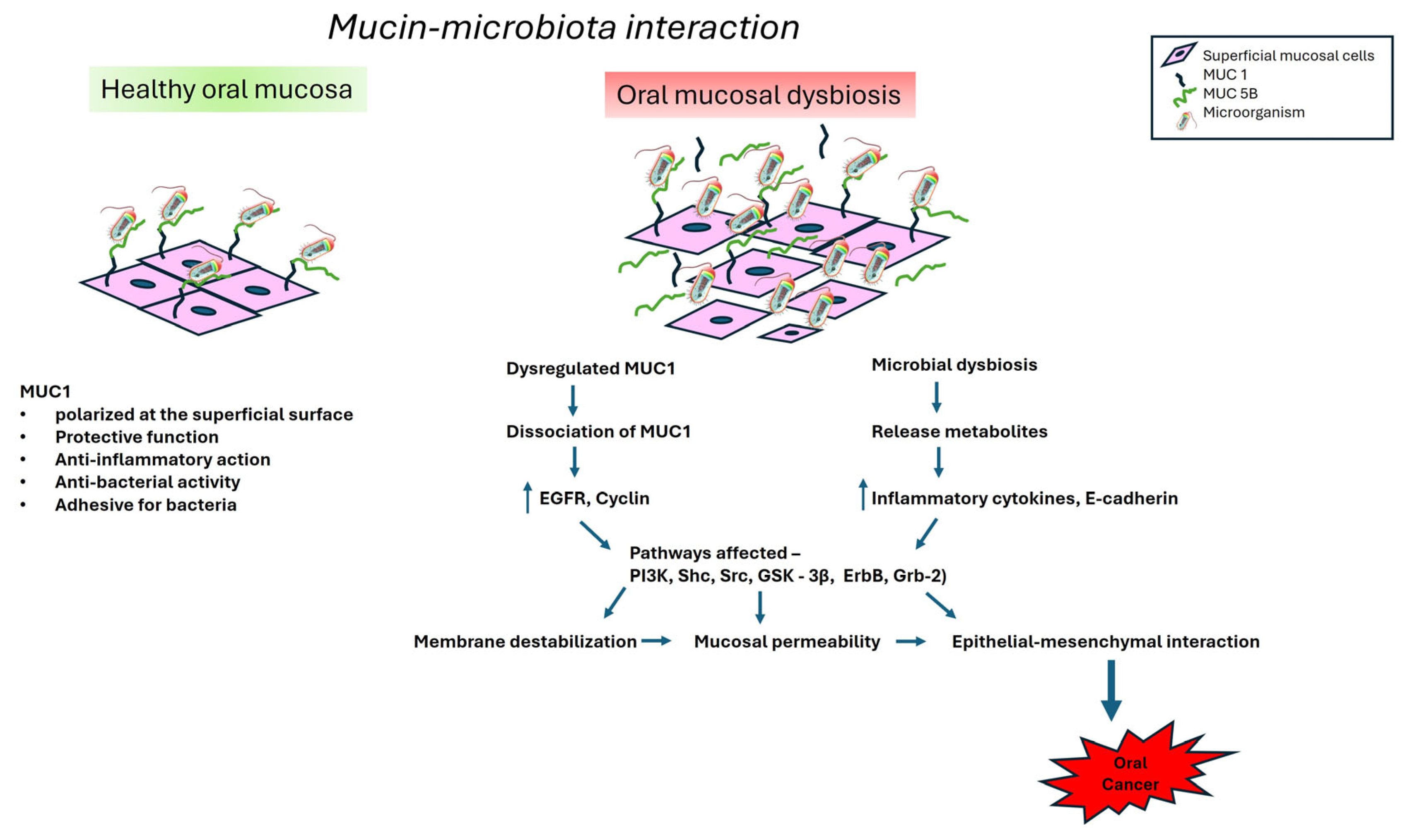
The oral mucosal barrier complex, containing mucins, is the first to sense the commensal transition from a benign to a damaging, pathogenic state, causing dysbiosis. Such early recognition is partially mediated by epidermal growth factor receptor (EGFR), the nuclear activity of which is regulated by the mucin MUC1. EGFR stimulation takes place at the cell surface in the presence of MUC1 [13]. The cross-kingdom interaction of oral microbes and their released products activates mitogen-activated protein kinase (MAPK) signal transduction induced by EGFR and modulated by the cytoplasmic domain of MUC1. This triggers increased production of inflammatory cytokines and tumor necrosis factor alpha (TNF-α), which affects the production and turnover of MUC1 at the transcriptional level [75,76]. The cytoplasmic domain of MUC1 is also involved in several cell signaling processes, such as those involving phosphoinosotide 3-kinase (PI3K), Shc adaptor protein (Shc), proto-oncogene tyrosine protein kinase (Src), β-catenin, Glycogen Synthase Kinase 3 beta (GSK-3β), the erythroblastic leukemia viral oncogene homolog (ErbB) receptor family, and growth factor receptor bound protein 2 (Grb-2) [13].

7.2. Oral Precancer

The commensals and pathogens interact directly with mucin glycans by expressing their cell surface lectins. This induces variations in the mucosal glycosylation patterns that influence microbial colonization and the invasion susceptibility. Recent advances in microbiome sequencing studies have demonstrated the role of microbial dysbiosis in oral precancer and cancer development. The oral microflora changes are also significant and unique in precancer [14]. Decreased glycosylation or short mucin glycans have been shown to cause increased gut permeability, potentially contributing to increased microbial translocation [77]. Oral lichen planus (OLP), a chronic inflammatory and precancerous condition, is associated with an elevated salivary concentration of interleukin 17 (IL-17). The high concentration of IL-17 indicated a shift in the oral bacterial community composition [16].
Oral leukoplakia (OLK) is the most common oral potentially malignant disorder (OPMD), with a malignant transformation rate that ranges from 1 to 20% [78]. The change in the MUC1 defensive barrier in oral precancer is well reported [13]. In recent years, several microbiome studies have been carried out to identify changes in the microbiota and metabolites in OLK. Patients with OLK have shown variable microbial colonization with an increased abundance of Fusobacteria and reduced levels of Firmicutes. The cooccurrence of Fusobacterium, Leptotrichia, and Campylobacter species in OLK is strikingly like the microbial cooccurrence patterns observed in colorectal cancers [79]. Mucins are hypothesized to shape microbiological behavior within the mucus gel/oral biofilm and the three-dimensional mucin network. In addition, mucin-associated glycans may serve as chemical signals that attenuate microbial virulence. An acetaldehydogenic microorganism, Rothia mucilaginosa, is observed in OLK at lingual sites, and Leptotrichia spp. and Campylobacter have been shown to be associated with epithelial dysplasia in OLK [80]. R. mucilaginosa has been shown to form acetaldehyde (ACH) from ethanol in vitro, which may result in the development and/or the malignant transformation of OLK [81].
Several factors, such as smoking, have been shown to alter oral epithelial homeostasis. Smoking can transform oral epithelial cells and promote oral cancer progression through multiple events and pathways. It influences immunity and regulates it either by aggravating pathogenic immune responses or by diminishing defensive immunity [82]. As reported previously, smoking causes depolarization and increased expression of MUC1 in oral epithelial cells in oral precancer patients [83]. This implies that MUC1’s protective properties are hampered, which allows for bacterial accumulation in the oral cavity and, hence, causes continuous oral inflammation. An animal study has shown that MUC1 serves as a binding site for microbes through an interaction between bacterial cell components and the MUC1 ectodomain [84]. Bacterial compositional differences have been demonstrated among healthy individuals and precancer and cancer patients [85]. The mucin–microbe interactions are multifaceted, with both physicochemical properties and glycosylation patterns playing important roles. The altered protection offered by mucins in oral precancers and cancers could be due to changes in their gene expression, production, degradation, and dehydration at the cell surface mucus layer, allowing for multispecies microbial colonization [86].

7.3. Oral Cancer

Oral cancers, mainly oral squamous cell carcinoma (OSCC), are the most frequently occurring cancer arising from the mucosal surfaces of the oral cavity. They represent a heterogeneous disease group with different levels of differentiation and aggressiveness. They can be preceded by OPMDs [87]. Transmembrane MUC1 (tMUC1) is usually found to be overexpressed in a variety of epithelial cancers, including oral cancer [83,88]. The structure and distribution of cell surface MUC1 glycoproteins are thought to influence the biologic behavior of OSCC tumor cells during malignant transformation and tumor progression. The involvement of tMUC1 accelerates the survival, escape, and invasion of malignant cells [13]. Promoting metastasis, the epithelial–mesenchymal transition occurs via cell signaling in the cytoplasmic tail of MUC1. Also, the sialylation of MUC1 at the cell surface imparts a strong negative charge that further affects invasion and metastasis [89]. Glycosylation of MUC1 represents a hallmark of cancers, including OSCC. It has been suggested that the glycosylation process elevates the complexity of the MUC1 protein’s regulatory function. MUC1 functions to regulate the nuclear activity of EGFR at the subcellular level, and this function changes in OSCC and other epithelial-derived malignancies [13].
The cytoplasmic domain of MUC1 functions as an oncoprotein which confers anchorage-independent growth and tumorigenicity [90]. In OSCC, the cytoplasmic domain of MUC1 undergoes diverse post-translational modifications and interacts with multiple effectors of diverse signaling pathways [12]. One such pathway is the PI3K-Akt pathway, which stimulates the activity of glycolytic enzymes. The cytoplasmic domain of MUC1 has shown an association with lipid metabolism and regulates cholesterol and fatty acid synthesis [91]. Previously, it was demonstrated that bacterial growth in the oral cavity is accompanied by the production of degrading enzymes and the utilization of glycoproteins [92]. The diverse role of MUC1 implies that, on one hand, MUC1 favors potentially beneficial bacteria and resists dysbiosis; on the other hand, MUC1 glycans serve as a nutritive substrate to retain native genera and high diversity.
Current investigations are focused on the oral microbiota’s involvement in carcinogenesis, primarily through chronic inflammation, the synthesis of carcinogens/bacterial metabolites, and changes in the mucosal integrity. 16S rRNA amplicon sequencing studies have comprehensively provided relationships between OSCC and oral bacteria [93]. Periodontal pathogenic bacteria, namely Fusobacterium, Peptostreptococcus, Filifactor, Parvimonas, Pseudomonas, Campylobacter, and Capnocytophaga, have been reported in high numbers in OSCC patients. Such pathogenic periodontal bacteria contribute to an inflammatory state and induce DNA damage in epithelial cells [94]. A difference in the oral microbial profile in oral precancerous conditions and cancerous lesions has been reported, where Megasphaera micronuciformis, Prevotella melaninogenica, and Prevotella veroralis were abundant in oral precancer. M. micronuciformis was reported to be a specific biomarker for this precancer [95]. Another study showed that a shift in the Streptococcus and Solobacterium levels is a clinical indicator of potential malignancy in a precancerous lesion [96]. Other bacterial genera, including Bacillus, Enterococcus, Parvimonas, Peptostreptococcus, and Slackia, have also displayed distinct differences in precancer and cancer [94].
Dysfunction of the epithelial barrier facilitates oral pathogen infiltration. Oral C. albicans infection has been observed in OLK and promoted disease progression [97]. Abnormalities in epithelial tight junctions allow for pathogen penetration and infections that may promote tumor growth. Such changes are noted to be early events in tumor metastasis [98]. For the conversion of OPMDs to oral cancer, epithelial–mesenchymal interaction (EMT) is critical. To trigger EMT, several specific pathogens act through different mechanisms to affect the p53, PI3K, and cyclin pathways [99]. E-cadherins on the epithelial cells are deactivated to promote mucosal permeability [100]. P. gingivalis and F. nucleatum have been shown to accelerate cell proliferation by triggering Toll-like receptor (TLR) signaling, increasing IL-6 production, and then activating STAT3/Akt and other pathways that affect cyclin D1 [101]. We believe that the dysregulation of MUC1 and specific pathogens combinedly affect host immunity and promote disease progression, invasion, and metastasis. The combined effects are presented in Figure 2.

8. Mucin–Oral Microbiome—A Potential Biomarkers

It has been well noted that both secreted and membrane-bound mucins at the oral mucosal surface are overexpressed, underglycosylated, and associated with a poor prognosis in many carcinomas. They have been well established as a tumor marker [89]. In cancer cells, MUC1 loses its normal restriction to the apical cell surface and is distributed over the entire cell surface (basal, lateral, and apical surfaces). This allows for interactions with several growth factor receptors and activates multiple signal transduction pathways in the MUC1 cytoplasmic tail in oral cancers [83]. Also, abnormal mucin expression and/or glycosylation are observed in other cancers, including lung, colon, pancreatic, ovarian, and breast cancer [102,103]. Elevated levels of aberrantly glycosylated proteins are a predictor of a poor chemotherapeutic response and a prognostic marker [104]. Due to an awareness of its overexpression and post-translational alteration, MUC1 has been investigated as a potential target for therapies. Several MUC1-targeted therapies are in clinical trials, although no mucin-targeted therapies are currently in clinical use.
The oral microbiome has shown the potential to induce chronic inflammation and produce carcinogenic metabolites that promote oral diseases, including OSCC. Most microbiota studies have analyzed the microbiota composition via 16S rRNA gene sequencing of the salivary or oral mucosa and confirmed the microbiota’s involvement in tumor proliferation, invasion, and metastasis [105,106]. P. gingivalis was highly expressed in cancerous tissue and was positively correlated with the metastasis of malignant tumors [107]. As a diagnostic marker, the oral microbiome showed 80% sensitivity and 83% specificity in differentiating between the OSCC and control groups [108]. Hence, monitoring and modulating the oral microbiome and its metabolites might enhance treatment outcomes. Probiotics and dietary modifications that restore the microbial balance and maintain the epithelial integrity might support overall oral health [14].
Mucins in the oral epithelium and bacteria maintain intimate relationships. The findings brought together in this review have brought new insight into oral mucosal diseases. Oral commensal bacteria can degrade mucins and their substrates can promote the selective growth of certain pathogenic bacteria. Identification of such mucin-degrading consortia necessitates further studies using in vitro models. The mucin–microbiome signatures studied in gastric cancer have been shown to affect the tumor microenvironment and are associated with a poor outcome. This study also emphasized that oral pathogenic taxa, such as Neisseria, Prevotella, and Veillonella, are potential drivers of mucin-mediated signaling in gastric cancer [109]. Bacterial end products tend to stimulate mucin expression in some mucin phenotype tumors [110]. However, the effects of factors such as diverse oral microbial activity and mucin glycosylation on oral disease pathogenesis remain poorly understood.

9. Conclusions and Future Directions

Recent research has discovered growing evidence that interactions between mucins and oral microbes are specific and critical to the pathogenesis of oral diseases. We believe that the metabolic interactions between oral biofilm commensals and host factors’ modulatory effects are potentials driver of dysbiosis. It is essential to note that various host factors, including sex, ethnicity, smoking, alcohol consumption, betel nut chewing, and individual oral health conditions, can cause fluctuations in oral bacterial communities. Hence, future research must focus on understanding how variations in mucin glycosylation patterns due to such factors influence an individual’s susceptibility to oral infection, as well as disease outcomes, and how mucin–microbiome interaction shapes the tumor microenvironment in OSCC.
Knowing the complexity of the oral cavity, several challenges remain to be addressed. (1) It is important to demonstrate through in vivo animal and human model studies the causes or consequences of changes in oral microbiomes. (2) The development of in vitro microbial communities using a top-down approach is needed to allow for examination of microbial interactions in mucin degradation consortia that are host-relevant and difficult to replicate. (3) The identification of the contributing components of the complex microbial community responsible for several oral pathologies is another challenge. (4) The oral cavity has several niches that harbor different microbial species. Such site-specific microbial modulation in each mucosal etiological niche needs exploration. (5) An examination of inter-individual differences to assess shifts in oral microbiome populations is essential to identify early-stage tumorigenesis. We believe that the constant development of mucosal microbiology could enhance our understanding of several oral diseases, and in the future, the use of microbial and metabolic markers will potentially enhance early diagnosis in clinical settings.

Author Contributions

B.K.: conceptualization, methodology, formal analysis, resources, data curation, writing—original draft preparation, review, and editing; A.M.K.: visualization, supervision, writing—review and editing. All authors have read and agreed to the published version of the manuscript.

Funding

This research received no external funding.

Institutional Review Board Statement

Not applicable.

Informed Consent Statement

Not applicable.

Data Availability Statement

No new data were created or analyzed in this study. Data sharing is not applicable.

Conflicts of Interest

The authors declare no conflicts of interest.

References

  1. Bray, F.; Laversanne, M.; Sung, H.; Ferlay, J.; Siegel, R.L.; Soerjomataram, I.; Jemal, A. Global cancer statistics 2022: GLOBOCAN estimates of incidence and mortality worldwide for 36 cancers in 185 countries. CA A Cancer J. Clin. 2024, 74, 229–263. [Google Scholar] [CrossRef]
  2. Roi, A.; Roi, C.I.; Andreescu, N.I.; Riviş, M.; Badea, I.D.; Meszaros, N.; Rusu, L.C.; Iurciuc, S. Oral cancer histopathological subtypes in association with risk factors: A 5-year retrospective study. Rom. J. Morphol. Embryol. 2020, 61, 1213–1220. [Google Scholar] [CrossRef]
  3. Tarle, M.; Lukšić, I. Pathogenesis and Therapy of Oral Carcinogenesis. Int. J. Mol. Sci. 2024, 25, 6343. [Google Scholar] [CrossRef] [PubMed]
  4. Metsäniitty, M.; Hasnat, S.; Salo, T.; Salem, A. Oral Microbiota-A New Frontier in the Pathogenesis and Management of Head and Neck Cancers. Cancers 2021, 14, 46. [Google Scholar] [CrossRef] [PubMed]
  5. Irfan, M.; Delgado, R.Z.R.; Frias-Lopez, J. The Oral Microbiome and Cancer. Front. Immunol. 2020, 11, 591088. [Google Scholar] [CrossRef]
  6. Xiao, L.; Zhang, Q.; Peng, Y.; Wang, D.; Liu, Y. The effect of periodontal bacteria infection on incidence and prognosis of cancer: A systematic review and meta-analysis. Medicine 2020, 99, e19698. [Google Scholar] [CrossRef]
  7. HOMD. Human Oral Microbiome Database. Available online: http://www.homd.org/ (accessed on 10 April 2025).
  8. Hyvärinen, E.; Kashyap, B.; Kullaa, A.M. Oral Sources of Salivary Metabolites. Metabolites 2023, 13, 498. [Google Scholar] [CrossRef]
  9. Tuominen, H.; Rautava, J. Oral Microbiota and Cancer Development. Pathobiology 2021, 88, 116–126. [Google Scholar] [CrossRef]
  10. Asikainen, P.; Ruotsalainen, T.J.; Mikkonen, J.J.W.; Koistinen, A.P.; Bruggenkate, T.C.; Kullaa, A.M. The defence architecture of the superficial cells of oral mucosa. Med. Hypothesis 2012, 78, 790–792. [Google Scholar] [CrossRef] [PubMed]
  11. Lan, Y.; Ni, W.; Tai, G. Expression of MUC1 in different tumours and its clinical significance (Review). Mol. Clin. Oncol. 2022, 17, 161. [Google Scholar] [CrossRef]
  12. Thakur, A.; Tupkari, J.V.; Joy, T.; Kende, P.P.; Siwach, P.; Ahire, M.S. Expression of mucin-1 in oral squamous cell carcinoma and normal oral mucosa: An immunohistochemical study. J. Oral Maxillofac. Pathol. 2018, 22, 210–215. [Google Scholar] [CrossRef] [PubMed]
  13. Kashyap, B.; Kullaa, A.M. Regulation of mucin 1 expression and its relationship with oral diseases. Arch. Oral Biol. 2020, 117, 104791. [Google Scholar] [CrossRef] [PubMed]
  14. Kashyap, B.; Kullaa, A. Salivary Metabolites Produced by Oral Microbes in Oral Diseases and Oral Squamous Cell Carcinoma: A Review. Metabolites 2024, 14, 277. [Google Scholar] [CrossRef] [PubMed]
  15. Su, S.C.; Chang, L.C.; Huang, H.D.; Peng, C.Y.; Chuang, C.Y.; Chen, Y.T.; Lu, M.Y.; Chiu, Y.W.; Chen, P.Y.; Yang, S.F. Oral microbial dysbiosis and its performance in predicting oral cancer. Carcinogenesis 2021, 42, 127–135. [Google Scholar] [CrossRef]
  16. Wang, K.; Miao, T.; Lu, W.; He, J.; Cui, B.; Li, J.; Li, Y.; Xiao, L. Analysis of oral microbial community and Th17-associated cytokines in saliva of patients with oral lichen planus. Microbiol. Immunol. 2015, 59, 105–113. [Google Scholar] [CrossRef]
  17. Agrawal, B.; Gupta, N.; Konowalchuk, J.D. MUC1 Mucin: A Putative Regulatory (Checkpoint) Molecule of T Cells. Front. Immunol. 2018, 9, 2391. [Google Scholar] [CrossRef]
  18. Soderholm, A.T.; Pedicord, V.A. Intestinal epithelial cells: At the interface of the microbiota and mucosal immunity. Immunology 2019, 158, 267–280. [Google Scholar] [CrossRef]
  19. Allaire, J.M.; Crowley, S.M.; Law, H.T.; Chang, S.Y.; Ko, H.J.; Vallance, B.A. The Intestinal Epithelium: Central Coordinator of Mucosal Immunity. Trends Immunol. 2018, 39, 677–696. [Google Scholar] [CrossRef]
  20. Groeger, S.; Meyle, J. Oral Mucosal Epithelial Cells. Front. Immunol. 2019, 10, 208. [Google Scholar] [CrossRef]
  21. Yamashita, Y.; Takeshita, T. The oral microbiome and human health. J. Oral Sci. 2017, 59, 201–206. [Google Scholar] [CrossRef]
  22. Sultan, A.S.; Kong, E.F.; Rizk, A.M.; Jabra-Rizk, M.A. The oral microbiome: A lesson in coexistence. PLoS Pathog. 2018, 14, e1006719. [Google Scholar] [CrossRef]
  23. Ammam, I.; Pailler-Mattéi, C.; Ouillon, L.; Nivet, C.; Vargiolu, R.; Neiers, F.; Canon, F.; Zahouani, H. Exploring the role of the MUC1 mucin in human oral lubrication by tribological in vitro studies. Sci. Rep. 2024, 14, 31019. [Google Scholar] [CrossRef]
  24. Lindén, S.K.; Sheng, Y.H.; Every, A.L.; Miles, K.M.; Skoog, E.C.; Florin, T.H.; Sutton, P.; McGuckin, M.A. MUC1 limits Helicobacter pylori infection both by steric hindrance and by acting as a releasable decoy. PLoS Pathog. 2009, 5, e1000617. [Google Scholar] [CrossRef]
  25. Sasai, M.; Yamamoto, M. Pathogen recognition receptors: Ligands and signaling pathways by Toll-like receptors. Int. Rev. Immunol. 2013, 32, 116–133. [Google Scholar] [CrossRef]
  26. Carraway, K.I.; Ramsauer, V.P.; Haq, B.; Carraway, C.A.C. Cell signaling through membrane mucins. Bioessays 2003, 25, 66–71. [Google Scholar] [CrossRef] [PubMed]
  27. Li, X.; Wang, L.; Nunes, D.P.; Troxler, R.F.; Offner, G.D. Pro-inflammatory cytokines up-regulate MUC1 gene expression in oral epithelial cells. J. Dent. Res. 2003, 82, 883–887. [Google Scholar] [CrossRef]
  28. Ptasiewicz, M.; Grywalska, E.; Mertowska, P.; Korona-Głowniak, I.; Poniewierska-Baran, A.; Niedźwiedzka-Rystwej, P.; Chałas, R. Armed to the Teeth-The Oral Mucosa Immunity System and Microbiota. Int. J. Mol. Sci. 2022, 23, 882. [Google Scholar] [CrossRef]
  29. Pathak, J.L.; Yan, Y.; Zhang, Q.; Wang, L.; Ge, L. The role of oral microbiome in respiratory health and diseases. Respir. Med. 2021, 185, 106475. [Google Scholar] [CrossRef]
  30. Sharma, N.; Bhatia, S.; Sodhi, A.S.; Batra, N. Oral microbiome and health. AIMS Microbiol. 2018, 4, 42–66. [Google Scholar] [CrossRef] [PubMed]
  31. Takeshita, T.; Kageyama, S.; Furuta, M.; Tsuboi, H.; Takeuchi, K.; Shibata, Y.; Shimazaki, Y.; Akifusa, S.; Ninomiya, T.; Kiyohara, Y.; et al. Bacterial diversity in saliva and oral health-related conditions: The Hisayama Study. Sci. Rep. 2016, 6, 22164. [Google Scholar] [CrossRef] [PubMed]
  32. Kozak, M.; Pawlik, A. The Role of the Oral Microbiome in the Development of Diseases. Int. J. Mol. Sci. 2023, 24, 5231. [Google Scholar] [CrossRef]
  33. Hoare, A.; Soto, C.; Rojas-Celis, V.; Bravo, D. Chronic Inflammation as a Link between Periodontitis and Carcinogenesis. Mediators Inflamm. 2019, 2019, 1029857. [Google Scholar] [CrossRef]
  34. van Houte, J. Role of micro-organisms in caries etiology. J. Dent. Res. 1994, 73, 672–681. [Google Scholar] [CrossRef]
  35. van ’t Hof, W.; Veerman, E.C.; Nieuw Amerongen, A.V.; Ligtenberg, A.J. Antimicrobial defense systems in saliva. Monogr. Oral Sci. 2014, 24, 40–51. [Google Scholar] [PubMed]
  36. Marsh, P.D.; Do, T.; Beighton, D.; Devine, D.A. Influence of Saliva on the Oral Microbiota. Periodontology 2000 2016, 70, 80–92. [Google Scholar] [CrossRef]
  37. Jakubovics, N.S.; Kolenbrander, P.E. The road to ruin: The formation of disease-associated oral biofilms. Oral Dis. 2010, 16, 729–739. [Google Scholar] [CrossRef] [PubMed]
  38. Kashyap, B.; Padala, S.R.; Kaur, G.; Kullaa, A. Candida albicans Induces Oral Microbial Dysbiosis and Promotes Oral Diseases. Microorganisms 2024, 12, 2138. [Google Scholar] [CrossRef]
  39. García-Curiel, L.; Del Rocío López-Cuellar, M.; Rodríguez-Hernández, A.I.; Chavarría-Hernández, N. Toward understanding the signals of bacteriocin production by Streptococcus spp. and their importance in current applications. World J. Microbiol. Biotechnol. 2021, 37, 15. [Google Scholar] [CrossRef] [PubMed]
  40. Jakubovics, N.S.; Gill, S.R.; Vickerman, M.M.; Kolenbrander, P.E. Role of hydrogen peroxide in competition and cooperation between Streptococcus gordonii and Actinomyces naeslundii. FEMS Microbiol. Ecol. 2008, 66, 637–644. [Google Scholar] [CrossRef]
  41. Kim, D.; Koo, H. Spatial Design of Polymicrobial Oral Biofilm in Its Native Disease State. J. Dent. Res. 2020, 99, 597–603. [Google Scholar] [CrossRef]
  42. Huang, C.B.; Alimova, Y.; Myers, T.M.; Ebersole, J.L. Short- and medium-chain fatty acids exhibit antimicrobial activity for oral microorganisms. Arch. Oral Biol. 2011, 56, 650–654. [Google Scholar] [CrossRef]
  43. Rudin, A.D.; Khamzeh, A.; Venkatakrishnan, V.; Basic, A.; Christenson, K.; Bylund, J. Short chain fatty acids released by Fusobacterium nucleatum are neutrophil chemoattractants acting via free fatty acid receptor 2 (FFAR2). Cell Microbiol. 2021, 23, e13348. [Google Scholar]
  44. Rudin, A.D.; Khamzeh, A.; Venkatakrishnan, V.; Persson, T.; Gabl, M.; Savolainen, O.; Forsman, H.; Dahlgren, C.; Christenson, K.; Bylund, J. Porphyromonas gingivalis Produce Neutrophil Specific Chemoattractants Including Short Chain Fatty Acids. Front. Cell. Infect. Microbiol. 2021, 10, 620681. [Google Scholar] [CrossRef]
  45. Nguyen, D.; Nguyen, T.K.; Rice, S.A.; Boyer, C. CO-Releasing Polymers Exert Antimicrobial Activity. Biomacromolecules 2015, 16, 2776–2786. [Google Scholar] [CrossRef]
  46. Takahashi, N.; Saito, K.; Schachtele, C.F.; Yamada, T. Acid tolerance and acid-neutralizing activity of Porphyromonas gingivalis, Prevotella intermedia and Fusobacterium nucleatum. Oral Microbiol. Immunol. 1997, 12, 323–328. [Google Scholar] [CrossRef]
  47. Erttmann, S.F.; Gekara, N.O. Hydrogen peroxide release by bacteria suppresses inflammasome-dependent innate immunity. Nat. Commun. 2019, 10, 3493. [Google Scholar] [CrossRef] [PubMed]
  48. Siracusa, R.; Voltarelli, V.A.; Salinaro, A.T.; Modafferi, S.; Cuzzocrea, S.; Calabrese, E.J.; Di Paola, R.; Otterbein, L.E.; Calabrese, V. NO, CO and H2S: A trinacrium of bioactive gases in the brain. Biochem. Pharmacol. 2022, 202, 115122. [Google Scholar] [CrossRef]
  49. Jaffe, F.A. Pathogenicity of carbon monoxide. Am. J. Forensic Med. Pathol. 1997, 18, 406–410. [Google Scholar] [CrossRef] [PubMed]
  50. Johnson, P.; Yaegaki, K.; Tonzetich, J. Effect of methyl mercaptan on synthesis and degradation of collagen. J. Periodontal. Res. 1996, 31, 323–329. [Google Scholar] [CrossRef] [PubMed]
  51. Wójcik, W.; Łukasiewicz, M.; Puppel, K. Biogenic amines: Formation, action and toxicity—A review. J. Sci. Food Agric. 2021, 101, 2634–2640. [Google Scholar] [CrossRef]
  52. Shatalin, K.; Shatalina, E.; Mironov, A.; Nudler, E. H2S: A universal defense against antibiotics in bacteria. Science 2011, 334, 986–990. [Google Scholar] [CrossRef]
  53. Dilek, N.; Papapetropoulos, A.; Toliver-Kinsky, T.; Szabo, C. Hydrogen sulfide: An endogenous regulator of the immune system. Pharmacol. Res. 2020, 161, 105119. [Google Scholar] [CrossRef]
  54. Uematsu, H.; Sato, N.; Hossain, M.Z.; Ikeda, T.; Hoshino, E. Degradation of arginine and other amino acids by butyrate-producing asaccharolytic anaerobic Gram-positive rods in periodontal pockets. Arch. Oral Biol. 2003, 48, 423–429. [Google Scholar] [CrossRef]
  55. Niederman, R.; Brunkhorst, B.; Smith, S.; Weinreb, R.N.; Ryder, M.I. Ammonia as a potential mediator of adult human periodontal infection: Inhibition of neutrophil function. Arch. Oral Biol. 1990, 35, 205S–209S. [Google Scholar] [CrossRef]
  56. Aruni, A.W.; Dou, Y.; Mishra, A.; Fletcher, H.M. The Biofilm Community-Rebels with a Cause. Curr. Oral Health Rep. 2015, 2, 48–56. [Google Scholar] [CrossRef]
  57. Bowen, W.H.; Burne, R.A.; Wu, H.; Koo, H. Oral Biofilms: Pathogens, Matrix, and Polymicrobial Interactions in Microenvironments. Trends Microbiol. 2018, 26, 229–242. [Google Scholar] [CrossRef]
  58. Zhou, Y.; Yang, J.; Zhang, L.; Zhou, X.; Cisar, J.O.; Palmer, R.J., Jr. Differential Utilization of Basic Proline-Rich Glycoproteins during Growth of Oral Bacteria in Saliva. Appl. Environ. Microbiol. 2016, 82, 5249–5258. [Google Scholar] [CrossRef] [PubMed]
  59. Wang, B.X.; Wu, C.M.; Ribbeck, K. Home, sweet home: How mucus accommodates our microbiota. FEBS J. 2021, 288, 1789–1799. [Google Scholar] [CrossRef] [PubMed]
  60. Frenkel, E.S.; Ribbeck, K. Salivary mucins promote the coexistence of competing oral bacterial species. ISME J. 2017, 11, 1286–1290. [Google Scholar] [CrossRef]
  61. Wu, C.M.; Wheeler, K.M.; Cárcamo-Oyarce, G.; Aoki, K.; McShane, A.; Datta, S.S.; Welch, J.L.M.; Tiemeyer, M.; Griffen, A.L.; Ribbeck, K. Mucin glycans drive oral microbial community composition and function. npj Biofilms Microbiomes 2023, 9, 11. [Google Scholar] [CrossRef] [PubMed]
  62. Takagi, J.; Aoki, K.; Turner, B.S.; Lamont, S.; Lehoux, S.; Kavanaugh, N.; Gulati, M.; Valle Arevalo, A.; Lawrence, T.J.; Kim, C.Y.; et al. Mucin O-glycans are natural inhibitors of Candida albicans pathogenicity. Nat. Chem. Biol. 2022, 18, 762–773. [Google Scholar] [CrossRef]
  63. Wang, B.X.; Wheeler, K.M.; Cady, K.C.; Lehoux, S.; Cummings, R.D.; Laub, M.T.; Ribbeck, K. Mucin Glycans Signal through the Sensor Kinase RetS to Inhibit Virulence-Associated Traits in Pseudomonas aeruginosa. Curr. Biol. 2021, 31, 90–102.e7. [Google Scholar] [CrossRef]
  64. Thornton, D.J.; Khan, N.; Mehrotra, R.; Howard, M.; Veerman, E.; Packer, N.H.; Sheehan, J.K. Salivary mucin MG1 is comprised almost entirely of different glycosylated forms of the MUC5B gene product. Glycobiology 1999, 9, 293. [Google Scholar] [CrossRef]
  65. Dekker, J.; Strous, G.J. Covalent oligomerization of rat gastric mucin occurs in the rough endoplasmic reticulum, is N-glycosylation-dependent and precedes initial O-glycosylation. J. Biol. Chem. 1990, 265, 18116–18122. [Google Scholar] [CrossRef]
  66. Corfield, A.P. Mucins: A biologically relevant glycan barrier in mucosal protection. Biochim. Biophys. Acta. 2015, 1850, 236–252. [Google Scholar] [CrossRef]
  67. Hollingsworth, M.A.; Swanson, B.J. Mucins in cancer: Protection and control of the cell surface. Nat. Rev. Cancer 2004, 4, 45–60. [Google Scholar] [CrossRef] [PubMed]
  68. Celli, J.P.; Turner, B.S.; Afdhal, N.H.; Keates, S.; Ghiran, I.; Kelly, C.P.; Ewoldt, R.H.; McKinley, G.H.; So, P.; Erramilli, S.; et al. Helicobacter pylori moves through mucus by reducing mucin viscoelasticity. Proc. Natl. Acad. Sci. USA 2009, 106, 14321–14326. [Google Scholar] [CrossRef] [PubMed]
  69. Norin, K.; Gustafsson, B.E.; Lindblad, B.; Midtvedt, T. The establishment of some microflora associated biochemical characteristics in feces from children during the first years of life. Acta Paediatr. Scand. 1985, 74, 207–212. [Google Scholar] [CrossRef]
  70. De Jong, M.H.; Van der Hoeven, J.S. The growth of oral bacteria on saliva. J. Dent. Res. 1987, 66, 498–505. [Google Scholar] [CrossRef] [PubMed]
  71. Willis, C.; Cummings, J.H.; Neale, G.; Gibson, G.R. In vitro effects of mucin fermentation on the growth of human colonic sulphate-reducing bacteria. Anaerobe 1996, 2, 117–122. [Google Scholar] [CrossRef]
  72. Corfield, A.P.; Wagner, S.A.; Clamp, J.R.; Kriaris, M.S.; Hoskins, L.C. Mucin degradation in the human colon: Production of sialidase, sialate O-acetylesterase, N-acetylneuraminate lyase, arylesterase and glycosulfatase activities by strains of fecal bacteria. Infect. Immun. 1992, 60, 3971–3978. [Google Scholar] [CrossRef]
  73. Van der Hoeven, J.S.; Van den Kieboom, C.W.; Camp, P.J. Utilization of mucin by oral Streptococcus species. Antonie Van Leeuwenhoek 1990, 57, 165–172. [Google Scholar] [CrossRef]
  74. Kavanaugh, N.L.; Zhang, A.Q.; Nobile, C.J.; Johnson, A.D.; Ribbeck, K. Mucins Suppress Virulence Traits of Candida albicans. mBio 2014, 5, e01911–e01914. [Google Scholar] [CrossRef]
  75. Naglik, J.R.; Konig, A.; Hube, B.; Gaffen, S.L. Candida albicans-epithelial interactions and induction of mucosal innate immunity. Curr. Opin. Microbiol. 2017, 40, 104–112. [Google Scholar] [CrossRef]
  76. Hori, Y.; Sugiyama, H.; Soma, T.; Nishida, K. Expression of membrane-associated mucins in cultivated human oral mucosal epithelial cells. Cornea 2007, 26, S65–S69. [Google Scholar] [CrossRef]
  77. Fekete, E.; Buret, A.G. The role of mucin O-glycans in microbiota dysbiosis, intestinal homeostasis, and host-pathogen interactions. Am. J. Physiol. Gastrointest. Liver Physiol. 2023, 324, G452–G465. [Google Scholar] [CrossRef]
  78. Ojeda, D.; Huber, M.A.; Kerr, A.R. Oral potentially malignant disorders and oral cavity cancer. Dermatol. Clin. 2020, 38, 507–521. [Google Scholar] [CrossRef]
  79. Warren, R.L.; Freeman, D.J.; Pleasance, S.; Watson, P.; Moore, R.A.; Cochrane, K.; Allen-Vercoe, E.; Holt, R.A. Co-occurrence of anaerobic bacteria in colorectal carcinomas. Microbiome 2013, 1, 16. [Google Scholar] [CrossRef] [PubMed]
  80. Amer, A.; Galvin, S.; Healy, C.M.; Moran, G.P. The microbiome of potentially malignant oral leukoplakia exhibits enrichment for Fusobacterium, Leptotrichia, Campylobacter, and Rothia Species. Front. Microbiol. 2017, 8, 2391. [Google Scholar] [CrossRef] [PubMed]
  81. Amer, A.; Whelan, A.; Al-Hebshi, N.N.; Healy, C.M.; Moran, G.P. Acetaldehyde production by Rothia mucilaginosa isolates from patients with oral leukoplakia. J. Oral Microbiol. 2020, 12, 1743066. [Google Scholar] [CrossRef] [PubMed]
  82. Sarkar, R.; Das, A.; Paul, R.R.; Barui, A. Cigarette smoking promotes cancer related transformation of oral epithelial cells through activation of Wnt and MAPK pathway. Future Oncol. 2019, 15, 3619–3631. [Google Scholar] [CrossRef]
  83. Kashyap, B.; Mikkonen, J.J.W.; Bhardwaj, T.; Dekker, H.; Schulten, E.A.J.M.; Bloemena, E.; Kullaa, A.M. Effect of smoking on MUC1 expression in oral epithelial dysplasia, oral cancer, and irradiated oral epithelium. Arch. Oral Biol. 2022, 142, 105525. [Google Scholar] [CrossRef]
  84. Kato, K.; Lillehoj, E.P.; Kai, H.; Kim, K.C. MUC1 expression by human airway epithelial cells mediates Pseudomonas aeruginosa adhesion. Front. Biosci. 2010, 2, 68–77. [Google Scholar]
  85. Zhao, H.; Chu, M.; Huang, Z.; Yang, X.; Ran, S.; Hu, B.; Zhang, C.; Liang, J. Variations in oral microbiota associated with oral cancer. Sci. Rep. 2017, 7, 11773. [Google Scholar] [CrossRef] [PubMed]
  86. Agner, C.E.; Wheeler, K.M.; Ribbeck, K. Mucins and Their Role in Shaping the Functions of Mucus Barriers. Annu. Rev. Cell Dev. Biol. 2018, 34, 189–215. [Google Scholar] [CrossRef]
  87. Yagyuu, T.; Funayama, N.; Imada, M.; Kirita, T. Effect of smoking status and programmed death-ligand 1 expression on the microenvironment and malignant transformation of oral leukoplakia: A retrospective cohort study. PLoS ONE 2021, 16, e0250359. [Google Scholar] [CrossRef] [PubMed]
  88. Nath, S.; Mukherjee, P. MUC1: A multifaceted oncoprotein with a key role in cancer progression. Trends Mol. Med. 2014, 20, 332–342. [Google Scholar] [CrossRef]
  89. Nitta, T.; Sugihara, K.; Tsuyama, S.; Murata, F. Immunohistochemical study of MUC1 mucin in premalignant oral lesions and oral squamous cell carcinoma. Association with disease progression, Mode of invasion and Lymph node metastasis. Cancer 2000, 88, 245–254. [Google Scholar] [CrossRef]
  90. Raina, D.; Agarwal, P.; Lee, J.; Bharti, A.; McKnight, C.J.; Sharma, P.; Kharbanda, S.; Kufe, D. Characterization of the MUC1-C Cytoplasmic Domain as a Cancer Target. PLoS ONE 2015, 10, e0135156. [Google Scholar] [CrossRef] [PubMed]
  91. Kosugi, M.; Ahmad, R.; Alam, M.; Uchida, Y.; Kufe, D. MUC1-C oncoprotein regulates glycolysis and pyruvate kinase M2 activity in cancer cells. PLoS ONE 2011, 6, e28234. [Google Scholar] [CrossRef]
  92. Bradshaw, D.J.; Homer, K.A.; Marsh, P.D.; Beighton, D. Metabolic cooperation in oral microbial communities during growth on mucin. Microbiology 1994, 140, 3407–3412. [Google Scholar] [CrossRef] [PubMed]
  93. Guerrero-Preston, R.; Godoy-Vitorino, F.; Jedlicka, A.; Rodríguez-Hilario, A.; González, H.; Bondy, J.; Lawson, F.; Folawiyo, O.; Michailidi, C.; Dziedzic, A.; et al. 16S rRNA amplicon sequencing identifies microbiota associated with oral cancer, human papilloma virus infection and surgical treatment. Oncotarget 2016, 7, 51320–51334. [Google Scholar] [CrossRef]
  94. Lee, W.H.; Chen, H.M.; Yang, S.F.; Liang, C.; Peng, C.Y.; Lin, F.M.; Tsai, L.L.; Wu, B.C.; Hsin, C.H.; Chuang, C.Y. Bacterial alterations in salivary microbiota and their association in oral cancer. Sci. Rep. 2017, 7, 16540. [Google Scholar] [CrossRef] [PubMed]
  95. Mok, S.F.; Karuthan, C.; Cheah, Y.K.; Ngeow, W.C.; Rosnah, Z.; Yap, S.F.; Ong, H.K.A. The oral microbiome community variations associated with normal, potentially malignant disorders and malignant lesions of the oral cavity. Malays. J. Pathol. 2017, 39, 1–15. [Google Scholar]
  96. Lim, Y.; Fukuma, N.; Totsika, M.; Kenny, L.; Morrison, M.; Punyadeera, C. The Performance of an Oral Microbiome Biomarker Panel in Predicting Oral Cavity and Oropharyngeal Cancers. Front. Cell. Infect. Microbiol. 2018, 8, 267. [Google Scholar] [CrossRef]
  97. Heng, R.; Li, D.; Shi, X.; Gao, Q.; Wei, C.; Li, X.; Li, Y.; Zhou, H. Reduced CX3CL1 Secretion Contributes to the Susceptibility of Oral Leukoplakia-Associated Fibroblasts to Candida albicans. Front. Cell. Infect. Microbiol. 2016, 6, 150. [Google Scholar]
  98. Dhawan, P.; Singh, A.B.; Deane, N.G.; No, Y.; Shiou, S.R.; Schmidt, C.; Neff, J.; Washington, M.K.; Beauchamp, R.D. Claudin-1 regulates cellular transformation and metastatic behavior in colon cancer. J. Clin. Investig. 2005, 115, 1765–1776. [Google Scholar] [CrossRef]
  99. Kuboniwa, M.; Hasegawa, Y.; Mao, S.; Shizukuishi, S.; Amano, A.; Lamont, R.J.; Yilmaz, O. P. gingivalis accelerates gingival epithelial cell progression through the cell cycle. Microbes Infect. 2008, 10, 122–128. [Google Scholar] [CrossRef]
  100. Al-Hebshi, N.N.; Nasher, A.T.; Maryoud, M.Y.; Homeida, H.E.; Chen, T.; Idris, A.M.; Johnson, N.W. Inflammatory bacteriome featuring Fusobacterium nucleatum and Pseudomonas aeruginosa identified in association with oral squamous cell carcinoma. Sci. Rep. 2017, 7, 1834. [Google Scholar] [CrossRef]
  101. Gallimidi, A.B.; Fischman, S.; Revach, B.; Bulvik, R.; Maliutina, A.; Rubinstein, A.M.; Nussbaum, G.; Elkin, M. Periodontal pathogens Porphyromonas gingivalis and Fusobacterium nucleatum promote tumor progression in an oral-specific chemical carcinogenesis model. Oncotarget 2015, 6, 22613–22623. [Google Scholar] [CrossRef] [PubMed]
  102. Baldus, S.E.; Engelmann, K.; Hanisch, F.G. MUC1 and the MUCs: A family of human mucins with impact in cancer biology. Crit. Rev. Clin. Lab. Sci. 2004, 41, 189–231. [Google Scholar] [CrossRef]
  103. Gendler, S.J. MUC1, the renaissance molecule. J. Mammary Gland. Biol. 2001, 6, 339–353. [Google Scholar] [CrossRef]
  104. Al-Azawi, D.; Kelly, G.; Myers, E.; McDermott, E.W.; Hill, A.D.; Duffy, M.J.; Higgins, N.O. CA 15-3 is predictive of response and disease recurrence following treatment in locally advanced breast cancer. BMC Cancer 2006, 6, 220. [Google Scholar] [CrossRef]
  105. Zhong, X.; Lu, Q.; Zhang, Q.; He, Y.; Wei, W.; Wang, Y. Oral microbiota alteration associated with oral cancer and areca chewing. Oral Dis. 2021, 27, 226–239. [Google Scholar]
  106. Sarkar, P.; Malik, S.; Laha, S.; Das, S.; Bunk, S.; Ray, J.G.; Chatterjee, R.; Saha, A. Dysbiosis of Oral Microbiota During Oral Squamous Cell Carcinoma Development. Front. Oncol. 2021, 11, 614448. [Google Scholar] [CrossRef]
  107. Torralba, M.G.; Aleti, G.; Li, W.; Moncera, K.J.; Lin, Y.H.; Yu, Y.; Masternak, M.M.; Golusinski, W.; Golusinski, P.; Lamperska, K.; et al. Oral Microbial Species and Virulence Factors Associated with Oral Squamous Cell Carcinoma. Microb. Ecol. 2021, 82, 1030–1046. [Google Scholar] [CrossRef]
  108. Zhou, X.; Hao, Y.; Peng, X.; Li, B.; Han, Q.; Ren, B.; Li, M.; Li, L.; Li, Y.; Cheng, G.; et al. The Clinical Potential of Oral Microbiota as a Screening Tool for Oral Squamous Cell Carcinomas. Front. Cell. Infect. Microbiol. 2021, 11, 728933. [Google Scholar] [CrossRef] [PubMed]
  109. Oosterlinck, B.; Ceuleers, H.; Arras, W.; De Man, J.G.; Geboes, K.; De Schepper, H.; Peeters, M.; Lebeer, S.; Skieceviciene, J.; Hold, G.L.; et al. Mucin-microbiome signatures shape the tumor microenvironment in gastric cancer. Microbiome 2023, 11, 86. [Google Scholar] [CrossRef] [PubMed]
  110. Breugelmans, T.; Oosterlinck, B.; Arras, W.; Ceuleers, H.; De Man, J.; Hold, G.L.; De Winter, B.Y.; Smet, A. The role of mucins in gastrointestinal barrier function during health and disease. Lancet Gastroenterol. Hepatol. 2022, 7, 455–471. [Google Scholar] [CrossRef] [PubMed]
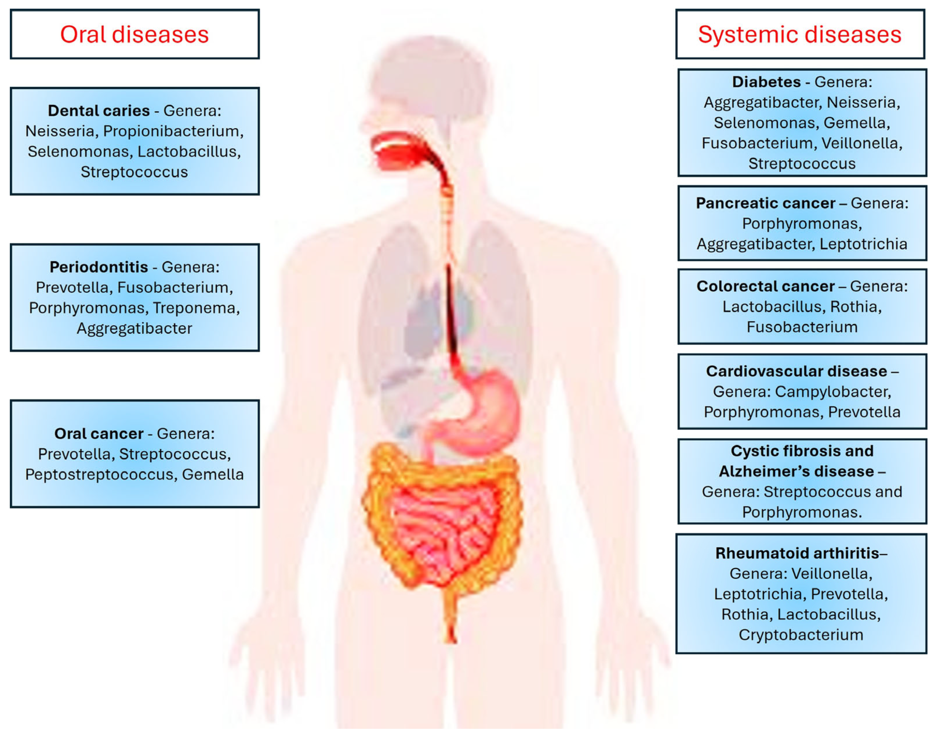
Figure 1. Oral microbiota involved in oral and systemic diseases.
Oral 05 00075 g001
Figure 2. Mucin–microbiota interactions—combined effect on healthy and dysbiotic oral mucosal epithelia. (Note: Biomolecules involved include EGFR—epithelial growth factor receptor; phosphoinosotide 3-kinase (PI3K); Shc adaptor protein (Shc); proto-oncogene tyrosine protein kinase (Src); β-catenin; Glycogen Synthase Kinase 3 beta (GSK-3β); erythroblastic leukemia viral oncogene homolog (ErbB) receptor family; and growth factor receptor bound protein 2 (Grb-2)).
Oral 05 00075 g002
Table 1. Positive and negative effects of oral microbes on oral biofilms and host.
Oral MicrobiomeOral BiofilmHost ResponseReferences
Actinomyces spp., Bacteroides spp., Corynebacteria spp., Eubacterium spp., Fusobacterium spp., Haemophilus spp., Megasphaera spp., Neisseria spp., Propionibacterium, Prevotella spp., Porphyromonas spp., Rothia spp.Antibacterial activityPro-inflammatory
Anti-inflammatory
Chemoattractant
Gut–brain interaction
[42,43,44]
Streptococcus mitisAntimicrobialGasotransmitter[45]
Porphyromonas gingivalis, Prevotella intermediaAntibacterial activityChemoattractant[46]
Streptococcus spp.Regulatory functionInhibition of
inflammasomes
[47]
Campylobacter spp.Bacterial survival and growthAnti-inflammatory[48]
Streptococcus spp., Lactobacillus spp.Stimulus for the growth of most anaerobesToxic[49]
Fusobacterium spp.Altered biofilm compositionDecreases collagen synthesis
Pro-inflammatory
[50]
Fusobacterium spp., Lactobacillus spp., Prevotella spp., Porphyromonas spp., Streptococcus spp., Treponema denticolaIncreased resistance to antibiotics
Formation of biofilms
Affects cell metabolism,
cell differentiation, plasmid stability,
drug resistance, and signaling
Affects bacterial virulence
Toxic
Affects cell physiology
[51]
Fusobacterium spp., Parvimonas micra, Porphyromonas spp., Prevotella intermedia, Treponema denticola, Streptococcus anginosus, Desulfobacter spp., Desulfovibrio spp., Desulfomicrobium
orale
Harmful at high concentrations
Increased resistance to antibiotics
Increased resistance to immune-mediated killing
Protection from oxidative stress
Toxic at high concentrations
Pro-inflammatory
Anti-inflammatory
Gasotransmitter
[52,53]
Fusobacterium spp., Porphyromonas spp., Prevotella spp., Tannerella spp., Treponema spp., Lactobacillus spp.,
Peptostreptococcus spp., Helicobacter pylori, Campylobacter ureolyticus, Haemophilus parainfluenzae, Streptococcus spp., Actinomyces spp., Staphylococcus spp., Rothia dentocariosa
Antibiotic resistance
Inhibits neutrophil
function
Toxic and impairs function of
neutrophils
[54,55]
Table 2. Mucin-degrading enzymes produced by bacteria in the oral cavity [70,73].
Oral Cavity LocationBacterial SpeciesEnzymes Produced
Saliva, buccal mucosa, tongue, hard palate,
gingiva, throat
palatine tonsils,
Streptococcus anginosusβ-N-acetyl-D-glucosaminidase
α- and β-D-galactosidase
Streptococcus mitisβ-N-acetyl-D-galactosaminidase, β-N-acetyl-D-glucosaminidase
α- and β-D-galactosidase, α-L-fucosidase, neuraminidase
Streptococcus mutantsβ-N-acetyl-D-glucosaminidase,
α- and β-D-galactosidase
Streptococcus oralisβ-N-acetyl-D-galactosaminidase, β-N-acetyl-D-glucosaminidase, α- and β-D-galactosidase,
α-L-fucosidase, neuraminidase, protease
Streptococcus sanguinisβ-N-acetyl-D-galactosaminidase, β-N-acetyl-D-glucosaminidase, α- and β-D-galactosidase,
α-L-fucosidase, protease
Streptococcus sobrinusβ-N-acetyl-D-glucosaminidase, β-D-galactosidase
Disclaimer/Publisher’s Note: The statements, opinions and data contained in all publications are solely those of the individual author(s) and contributor(s) and not of MDPI and/or the editor(s). MDPI and/or the editor(s) disclaim responsibility for any injury to people or property resulting from any ideas, methods, instructions or products referred to in the content.