

Oral, Volume 5, Issue 3 (September 2025) – 26 articles
Cover Story (view full-size image):
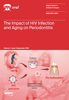
Periodontal disease is a significant oral health concern worldwide, particularly amongst individuals living with the human immunodeficiency virus (HIV). Biological aging is associated with a natural decline in the immune system, which can also affect the severity of periodontitis. In people living with HIV (PLWH), the contribution of both the HIV infection and the aging process can lead to increased susceptibility to periodontal disease. This paper reviewed recent literature about the relationships between HIV infection and early aging and their impact on periodontitis. This exploration through molecular and cellular mechanisms underlying the relationships between aging, HIV, and periodontitis can provide therapeutic implications for dental clinicians to prevent and treat their affected patients. View this paper
- Issues are regarded as officially published after their release is announced to the table of contents alert mailing list.
- You may sign up for e-mail alerts to receive table of contents of newly released issues.
- PDF is the official format for papers published in both, html and pdf forms. To view the papers in pdf format, click on the "PDF Full-text" link, and use the free Adobe Reader to open them.
Previous Issue
Next Issue