- Review
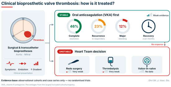
Clinically significant thrombosis of surgical and transcatheter biological valves is increasingly recognised, yet most reviews have concentrated on subclinical leaflet thrombosis after transcatheter aortic valve implantation. This narrative review examines treatment and outcomes after diagnosis of clinical bioprosthetic valve thrombosis in the aortic or mitral position, keeping asymptomatic hypoattenuated leaflet thickening separate. The evidence consists of 17 observational cohorts and case series, several of which overlap, include right-sided valves or use treatment response as part of the diagnosis; no randomised treatment trial has been reported. After anticoagulation, mainly with vitamin K antagonists, most studies reported haemodynamic improvement in most patients assessed, but endpoints and denominators varied; in one transcatheter registry, complete resolution was reported in 12 of 26 patients (46%), a figure that included two patients who were not anticoagulated. Recovery often took several months. Recurrence and bleeding estimates come mainly from one mixed-valve cohort that included right-sided prostheses: thrombosis recurred in 23% of initial responders, and major bleeding occurred in 12% of treated patients versus 2% of matched controls, an observational difference that cannot be attributed wholly to anticoagulation. Evidence for thrombolysis, redo surgery and transcatheter reintervention is limited to small series and case reports. The evidence supports confirming suspected thrombosis with transoesophageal echocardiography or four-dimensional computed tomography, starting a vitamin K antagonist in haemodynamically stable patients, referring unstable patients or those with persistent obstruction for a Heart Team decision, and individualising extended anticoagulation against bleeding risk. Direct oral anticoagulants are not established for clinical thrombosis, and the optimal duration of treatment is unknown.
12 November 2026

![The proposed management framework for suspected bioprosthetic valve thrombosis, derived from the reviewed evidence and current guidelines. The boxes with a solid blue outline and the label ‘ESC/EACTS 2025’ indicate steps supported by the 2025 ESC/EACTS guidelines [12]; the boxes with a dashed orange outline and the label ‘Author-proposed’ indicate review intervals, response thresholds and treatment durations proposed by the author, which are not guideline criteria and have not been validated. Haemodynamic valve deterioration, defined as a rise in mean gradient, with a fall in effective orifice area or Doppler velocity index, from the post-implantation baseline [6], places a patient with leaflet thickening or reduced motion in the clinical branch even without symptoms. An elevated gradient without a documented rise from baseline, as with patient–prosthesis mismatch or a high-flow state, does not, and such a patient is managed in the subclinical branch unless symptoms or embolism are present. The asterisk (*) in the subclinical branch denotes an elevated gradient without a documented rise from the post-implant baseline, which is explained in the box marked * below it. When no baseline study is available, classification relies on valve-specific reference values, assessment of flow, TEE or CT and serial echocardiography. Response: a fall in the mean gradient of at least 50% or a return towards the post-implant baseline, with improved leaflet mobility on echocardiography or CT. Partial response: a fall of 25% to 49%, for which anticoagulation is continued, anticoagulation control and adherence are reviewed, and imaging is repeated. Persistent obstruction: a fall of less than 25% or unchanged leaflet immobility. Options for unstable patients and for persistent obstruction are supported by very limited evidence in biological valves, and the suggested minimum duration of anticoagulation is not based on comparative evidence. 4D, four-dimensional; CT, computed tomography; DOAC, direct oral anticoagulant; HALT, hypoattenuated leaflet thickening; INR, international normalised ratio; RLM, reduced leaflet motion; TAV, transcatheter aortic valve; TAVI, transcatheter aortic valve implantation; TEE, transoesophageal echocardiography; TTE, transthoracic echocardiography; VKA, vitamin K antagonist.](https://mdpi-res.com/jvd/jvd-05-00050/article_deploy/html/images/jvd-05-00050-ag-550.jpg)