


Abstract
Background: Retinal microvascular assessment has emerged as a promising non-invasive approach for evaluating vascular health. Recent advances in retinal imaging technologies have enabled detailed characterization of retinal vascular structure and function, raising interest in their potential role as biomarkers of exercise-related vascular adaptation. This systematic review with narrative synthesis aimed to summarize current evidence regarding the relationship between exercise, physical fitness, and retinal microvascular biomarkers. Methods: A systematic literature search was conducted in PubMed, Scopus, and Web of Science for studies published from January 2021 to June 2026, followed by a supplementary update search to identify eligible publications available through 31 July 2026. Search terms combined retinal microvascular assessment techniques with terms related to exercise, physical activity, physical fitness, and exercise training. Following screening and eligibility assessment, 42 publications were included in the final qualitative synthesis, comprising 20 publications identified through the initial search and 22 additional publications identified through the supplementary update search. Several publications represented complementary analyses of the same or overlapping study cohorts and were therefore not considered independent replications. Results: The included studies comprised observational studies and exercise intervention trials involving healthy individuals and clinical populations. Retinal biomarkers assessed included vessel diameters, arteriovenous ratio (AVR), vessel density, perfusion density, foveal avascular zone (FAZ), retinal oxygen extraction, wall-to-lumen ratio (WLR), and choriocapillaris perfusion parameters. Several studies reported measurable acute and chronic retinal vascular responses to exercise, although the direction, magnitude, and clinical meaning of these changes varied according to population, exercise modality, imaging technique, and biomarker. Observational studies generally reported associations between higher physical activity or cardiorespiratory fitness and a more favorable retinal vascular profile, but causal interpretation remains limited. Conclusions: Retinal microvascular biomarkers appear sensitive to acute physiological stress and may reflect selected adaptations associated with longer-term exercise exposure. However, retinal responses are heterogeneous and biomarker-specific, and their biological and clinical significance remains incompletely established. Methodological heterogeneity, overlapping study cohorts, and the limited number of adequately powered randomized and longitudinal studies constrain causal interpretation and generalizability. Retinal vascular measures should therefore currently be regarded as candidate biomarkers and research tools rather than validated surrogate outcomes or routine clinical measures.
1. Introduction
Microvascular dysfunction is increasingly recognized as a key contributor to the development of cardiovascular, metabolic, renal, and neurological disorders and may precede the onset of overt clinical disease [1,2,3,4,5]. Structural and functional alterations of the microcirculation impair tissue perfusion, oxygen delivery, nutrient exchange, and metabolic homeostasis, thereby contributing to disease progression across multiple organ systems. Consequently, there is growing interest in identifying sensitive, reproducible, and non-invasive biomarkers capable of detecting early microvascular alterations before irreversible vascular damage becomes clinically apparent [1,2,3,4,5,6].
Among the various vascular beds available for clinical assessment, the retinal circulation provides a unique opportunity for direct, non-invasive visualization of the human microvasculature in vivo. Because retinal vessels share anatomical, physiological, and embryological characteristics with the cerebral and systemic microcirculation, retinal vascular biomarkers are increasingly regarded as surrogate indicators of generalized vascular health [2,3,4,5,7,8,9]. Large population-based studies have further demonstrated that retinal vascular measurements are independently associated with long-term cardiovascular outcomes and all-cause mortality, underscoring their prognostic value beyond ophthalmology [8,9]. Moreover, retinal microvascular alterations have also been associated with cognitive decline, further supporting the concept that the retina reflects vascular and neurological health beyond the eye [4,10]. This accessibility distinguishes the retina from other microvascular territories and has established retinal imaging as an attractive tool for investigating systemic vascular function [2,3,6].
The retinal circulation is tightly regulated through autoregulatory and neurovascular mechanisms that maintain adequate tissue perfusion despite fluctuations in systemic blood pressure, metabolic demand, and neuronal activity [11,12,13,14]. Recent advances in retinal imaging, including optical coherence tomography angiography (OCTA), static and dynamic retinal vessel analysis, retinal oximetry, and adaptive optics imaging, enable quantitative assessment of multiple structural and functional biomarkers, including retinal vessel caliber, capillary density, perfusion, vascular reactivity, oxygen extraction, wall-to-lumen ratio, and choriocapillaris characteristics [2,3,4,15]. These techniques allow the detection of subtle microvascular alterations that may precede clinically overt cardiovascular and metabolic disease [2,3,4,5,7,8,9].
Regular physical activity and structured exercise are well-established non-pharmacological strategies for improving cardiovascular health and reducing the risk of chronic disease [16,17,18,19]. Exercise promotes vascular adaptation through several complementary mechanisms, including improved endothelial function, increased nitric oxide bioavailability, enhanced vascular responsiveness, attenuation of chronic low-grade inflammation and oxidative stress, and favorable vascular remodeling [16,17,18,19]. Because the retinal circulation is highly sensitive to systemic hemodynamic and metabolic changes, retinal biomarkers may provide a practical means of capturing both acute vascular responses to exercise and long-term adaptations to exercise training. Emerging evidence from observational studies and exercise interventions indicates that retinal microvascular parameters are responsive to physical activity across different populations, including children, older adults, healthy individuals, athletes, and patients with cardiovascular or metabolic disease [7,8,9,15,20,21,22,23,24,25,26,27,28,29,30,31,32,33,34,35]. Long-term lifestyle interventions and improvements in cardiorespiratory fitness have likewise been associated with favorable retinal microvascular profiles, supporting the potential utility of retinal biomarkers in preventive medicine and exercise physiology [21,23,24,25,26,27,28,29,30,31,32,33,36].
Despite growing interest in this field, the available evidence remains heterogeneous. Existing studies differ substantially with respect to participant characteristics, exercise modalities, intervention duration, retinal imaging devices, scan protocols, image analysis methods, and the retinal biomarkers evaluated [7,8,9,15,20,21,22,23,24,25,26,27,28,29,30,31,32,33,34,35]. Furthermore, findings have not always been consistent, and the physiological and clinical significance of changes in individual OCTA- and retinal vessel analysis-derived parameters remains incompletely understood [2,3,6,37]. These methodological differences currently limit direct comparison between studies and hinder the translation of retinal biomarkers into routine clinical and research practice [6,37].
A previous systematic review by Streese et al. synthesized evidence available up to January 2020 regarding the associations of physical activity and cardiorespiratory fitness, as well as the effects of exercise interventions, with retinal microvascular health [36]. That review focused primarily on static and dynamic retinal vessel analysis parameters, including central retinal arteriolar equivalent (CRAE), central retinal venular equivalent (CRVE), arteriovenous ratio (AVR), and flicker light-induced dilatation (FID), and excluded acute exercise studies and interventions lasting less than two weeks [36]. Since its publication, the field has expanded considerably through the emergence of new observational and interventional studies involving diverse populations and exercise modalities, together with the broader application of contemporary retinal imaging technologies, particularly OCT angiography, retinal oximetry, and adaptive optics imaging [7,8,9,15,20,21,22,23,24,25,26,27,28,29,30,31,32,33,34,35]. The present review therefore provides an updated and expanded syn-thesis of studies published between January 2021 and June 2026, encompassing both acute and chronic exercise responses and a wider range of retinal microvascular outcomes assessed using optical coherence tomography angiography, retinal oximetry, adaptive optics imaging, static retinal vessel analysis, and related techniques [7,8,9,15,20,21,22,23,24,25,26,27,28,29,30,31,32,33,34,35,37].
Beyond updating the available evidence, this review extends previous work by integrating findings from both acute exercise responses and chronic exercise interventions while also incorporating newer retinal imaging modalities, including OCT angiography, retinal oximetry, adaptive optics imaging, and choriocapillaris assessment [20,24,27,28,29,30,35,37]. Furthermore, rather than focusing on individual retinal parameters, the present review provides an integrated overview of structural and functional retinal microvascular biomarkers, highlighting their potential translational relevance as non-invasive indicators of exercise-induced vascular adaptation and vascular health [1,2,3,6,36].
Therefore, the aim of this systematic review with narrative synthesis was to synthesize current evidence published between January 2021 and June 2026 regarding the relationships between acute exercise, structured exercise training, physical activity, physical fitness, and retinal microvascular biomarkers. Particular emphasis was placed on retinal biomarkers assessed using contemporary retinal imaging techniques, the methodological quality of the available evidence, conflicting findings across studies, and current limitations affecting the interpretation and clinical applicability of retinal vascular outcomes.
2. Materials and Methods
This study was conducted as a systematic review with narrative synthesis. A predefined literature search strategy, explicit eligibility criteria, independent study selection, standardized data extraction, methodological quality assessment using the Joanna Briggs Institute (JBI) Critical Appraisal Tools, and reporting in accordance with the Preferred Reporting Items for Systematic Reviews and Meta-Analyses (PRISMA) 2020 statement were employed to ensure methodological rigor and transparency. Owing to substantial clinical and methodological heterogeneity among the included studies, quantitative meta-analysis was not appropriate; therefore, the findings were synthesized narratively. The review was not prospectively registered.
An initial comprehensive literature search was performed in the PubMed, Scopus, and Web of Science databases for studies published between January 2021 and June 2026. The initial search was last conducted on 27 June 2026. Search strategies were adapted to the indexing systems and search functionalities of each database while maintaining the same conceptual framework across all databases. Searches combined terms related to retinal microvascular assessment with terms describing exercise, physical activity, physical fitness, and exercise training.
In PubMed, the following search strategy was used: (“retinal vessel analysis” OR “retinal microvasculature” OR “retinal microcirculation” OR “retinal vessel diameter” OR “retinal vessel diameters” OR “optical coherence tomography angiography”) AND (exercise OR “physical activity” OR training OR fitness OR “aerobic exercise” OR “high-intensity interval training” OR HIIT OR “resistance training”). Filters were applied to restrict results to studies published between January 2021 and June 2026, involving human participants, and available in English.
In Scopus, the search strategy was: TITLE-ABS-KEY ((“retinal vessel analysis” OR “retinal vessel diameter*” OR “retinal microvasculature” OR “retinal microcirculation” OR “retinal capillary density”) AND (exercise OR “physical activity” OR HIIT OR “high-intensity interval training” OR “aerobic training” OR “resistance training”)). Results were limited to publications published between January 2021 and June 2026, article and review article document types, English-language studies, and human research.
In Web of Science, the search strategy was: TS = ((“retinal vessel analysis” OR “retinal microvasculature” OR “retinal microcirculation” OR “retinal vessel diameter*” OR “retinal capillary density” OR “retinal perfusion”) AND (exercise OR “physical activity” OR HIIT OR “high-intensity interval training” OR “aerobic training” OR “resistance training” OR endurance)). Filters were applied to include only articles published between January 2021 and June 2026 and written in English.
Records retrieved from the three databases were manually screened for duplicate entries, and duplicate records were removed before title and abstract screening. Following peer review, a supplementary update search was conducted across PubMed, Scopus, and Web of Science to improve the sensitivity of the original search and identify potentially eligible publications not retrieved by the initial strategy. The supplementary search expanded the retinal microvascular terminology to include choriocapillaris, retinal blood flow, retinal perfusion, perfusion density, vessel density, capillary density, and retinal oximetry, and expanded the exercise-related terminology to include physical activity, exercise, physical fitness, cardiorespiratory fitness, high-intensity interval training (HIIT), resistance training, circuit training, yoga, and meditation. The updated search covered publications from 1 January 2021 through 31 July 2026. The full database-specific supplementary search strategies are provided in Supplementary Table S1.
The supplementary update search identified 591 records across the three databases (PubMed, n = 84; Scopus, n = 205; Web of Science, n = 302). After removal of 256 duplicate records, 335 unique records remained for screening. These records were evaluated using the same predefined eligibility criteria as in the initial search. The supplementary search identified 22 additional eligible publications for inclusion in the qualitative synthesis. Publications already represented in the original review were not counted as newly included reports. All newly identified eligible publications were incorporated into the study characteristics tables, methodological quality assessment, and narrative synthesis.
To ensure comprehensive coverage of the available literature, the reference lists of eligible studies and relevant review articles were additionally screened for potentially relevant publications. Review articles identified during the search process were used solely for reference screening and were not included in the final qualitative synthesis. No restrictions regarding study design were applied during the database search; eligibility was determined during the screening process according to the predefined inclusion and exclusion criteria. The review considered retinal vascular biomarkers broadly to include retinal and closely related ocular microvascular imaging parameters that may reflect retinal or systemic microvascular physiology. Consequently, studies evaluating retinal vessel morphology and function, retinal oxygenation, optic nerve head perfusion, and choriocapillaris perfusion in the context of exercise, physical activity, or physical fitness were considered eligible for inclusion. Retinal structural measures, such as retinal nerve fiber layer (RNFL) or macular thickness, were extracted only when reported alongside eligible retinal microvascular outcomes and were not considered sufficient as the sole basis for study inclusion.
Studies were considered eligible if they investigated the relationship between exercise, physical activity, physical fitness, or exercise training and retinal microvascular outcomes in human participants. Studies employing retinal vascular assessment techniques, including OCTA, retinal vessel analysis, retinal oximetry, fundus-based vascular measurements, adaptive optics retinal imaging, or related methods, were considered for inclusion. Both observational and interventional study designs were eligible, including cross-sectional studies, cohort studies, longitudinal investigations, randomized controlled trials, and other exercise intervention studies. Only articles published in peer-reviewed journals and written in English were included.
Studies were excluded if they focused exclusively on retinal diseases without evaluating exercise-, fitness-, or physical activity-related associations, if they reported only ophthalmic structural outcomes unrelated to retinal microvascular parameters, or if they consisted of conference abstracts, editorials, letters, expert opinions, case reports, or animal studies. Studies lacking sufficient information regarding retinal vascular outcomes were also excluded.
Study selection and eligibility assessment were performed independently by E.K. and I.M. The same study selection procedure and predefined eligibility criteria were applied to records identified through both the initial and supplementary searches. Disagreements were resolved through discussion and consensus. The study selection process is presented in a PRISMA 2020 flow diagram (Figure 1). Reasons for full-text exclusion are summarized in the PRISMA 2020 flow diagram (Figure 1), whereas a complete study-specific list of excluded full-text articles and the corresponding reasons for exclusion are provided in Supplementary Table S2. The completed PRISMA 2020 Checklist is provided as Supplementary Table S3. Detailed characteristics of the included studies are provided in Supplementary Table S4. Potential overlap of study populations was assessed by comparing study characteristics, recruitment periods, study settings, trial registration numbers, and author groups. Publications arising from the same study cohort were treated as complementary reports rather than independent sources of evidence during data synthesis and interpretation.
Figure 1.
Literature Search and Study Selection Process.
The initial database search identified 236 records (165 from PubMed, 35 from Scopus, and 36 from Web of Science). Following title and abstract screening, 47 articles were selected for full-text assessment, and 20 publications met the predefined eligibility criteria. The subsequent supplementary update search identified 591 records (84 from PubMed, 205 from Scopus, and 302 from Web of Science), of which 335 unique records remained after duplicate removal. Screening of the supplementary search results identified 22 additional eligible publications. Consequently, a total of 42 publications were included in the final qualitative synthesis. Several publications represented complementary or overlapping analyses of the same underlying study populations and were therefore not interpreted as independent replications.
For data extraction, information regarding study design, participant characteristics, exercise modality, duration of intervention, retinal assessment method, evaluated retinal biomarkers, and principal findings was collected from each study using a standardized data extraction form developed for this review. Data extraction was performed independently by M.O. and P.Š. using the standardized data extraction form. Any discrepancies were resolved through discussion and consensus among the research team. For intervention studies, within-group changes were distinguished from between-group or group-by-time effects whenever these were reported, and within-group statistical significance alone was not interpreted as evidence of a significant intervention effect. No study investigators were contacted for additional information, and no automation tools were used during the data extraction process. For observational studies, findings were interpreted as associations rather than causal effects.
Owing to substantial heterogeneity in study populations, exercise modalities, intervention duration, retinal imaging techniques, and outcome measures, a quantitative meta-analysis was not considered appropriate. Instead, findings were synthesized narratively.
Methodological quality was independently assessed by two reviewers using the design-specific Joanna Briggs Institute (JBI) Critical Appraisal Tools for randomized controlled trials, quasi-experimental studies, cohort studies, and analytical cross-sectional studies. Any disagreements were resolved through discussion until consensus was reached. Each publication was evaluated item by item using the response categories Yes, No, Unclear, and Not applicable, as appropriate. The appraisal was used to identify methodological strengths and potential sources of bias and to inform the qualitative interpretation of the evidence. Rather than calculating an aggregate numerical score, methodological strengths, potential sources of bias, and their implications for the interpretation of the findings were considered qualitatively. In accordance with JBI guidance, no aggregate numerical quality score was calculated, and studies were not excluded solely on the basis of the appraisal findings. The complete item-level JBI critical appraisals are provided in Supplementary File S5, whereas Supplementary Table S6 summarizes the principal methodological strengths, limitations, and implications for the interpretation of each included publication.
Studies were assigned to the qualitative syntheses according to their primary research focus, based on study design, participant characteristics, exercise modality, and the retinal microvascular outcomes evaluated. Studies investigating immediate physiological responses to a single exercise session were grouped under acute retinal responses, whereas intervention studies assessing repeated exercise exposure were included in the chronic exercise-induced adaptation synthesis. Observational studies examining habitual physical activity or cardiorespiratory fitness were synthesized separately within the physical activity and physical fitness section. Particular attention was given to retinal vascular parameters proposed as indicators of systemic vascular function, including vessel diameter, vascular density, perfusion metrics, oxygen saturation, wall-to-lumen ratio, neurovascular coupling, choriocapillaris characteristics, and other measures of retinal microvascular structure and function. When multiple publications originated from the same or overlapping study cohort, their findings were synthesized as complementary analyses and were not treated as independent confirmation of the same association or intervention effect.
3. Results
3.1. Overview of Included Studies
Following the initial literature search and the supplementary update search, a total of 42 publications were included in the final qualitative synthesis. Of these, 20 publications were identified through the initial search and 22 additional eligible publications through the supplementary update search. The included publications were published between 2021 and July 2026 and encompassed randomized controlled trials, prospective and quasi-experimental intervention studies, repeated-measures and crossover studies, longitudinal and prospective cohort studies, cross-sectional investigations, and secondary analyses of prospectively collected data.
The included populations were heterogeneous and ranged from children and healthy young or older adults to athletes and clinical populations, including individuals with hypertension, type 1 or type 2 diabetes mellitus, primary open-angle glaucoma, Parkinson’s disease, cardiovascular disease or elevated cardiovascular risk, and patients receiving active anticancer treatment. Exercise-related exposures likewise varied substantially and included acute dynamic and isometric exercise, endurance exercise, high-intensity interval training (HIIT), high-speed circuit resistance training (HSCT), habitual physical activity, objectively measured daily activity, cardiorespiratory or physical fitness, yoga, and other structured exercise interventions.
Importantly, the 42 publications do not represent 42 independent study populations, because several reports were derived from overlapping or complementary cohorts. Twerenbold et al., Streese et al., and Müller et al. reported complementary outcomes from the HyperVasc cohort. Multiple publications also originated from the Miami HSCT program, including complementary analyses of the 8-week and 24-week training cohorts, while several publications evaluating yoga in Parkinson’s disease arose from the same underlying intervention program. These publications were retained because they reported distinct retinal vascular or neurovascular outcomes but were treated as complementary analyses rather than independent replications.
Retinal vascular outcomes were evaluated using a broad range of imaging and functional assessment techniques, including optical coherence tomography angiography (OCTA), static retinal vessel analysis, retinal oximetry, adaptive optics retinal imaging, Retinal Function Imager–based measurements, laser speckle flowgraphy, Doppler OCT, and multimodal retinal neurovascular imaging. The principal outcomes included retinal vessel caliber, retinal vessel and capillary density, retinal blood flow and tissue perfusion, foveal avascular zone (FAZ) characteristics, choriocapillaris perfusion and flow-deficit metrics, retinal oxygen saturation and extraction, vascular reactivity and vasomotion, optic nerve head and peripapillary perfusion, and arteriolar wall-to-lumen ratio.
For qualitative synthesis, the evidence was organized into three principal domains: acute retinal responses to exercise, chronic exercise-induced retinal adaptations, and associations of habitual physical activity or physical fitness with retinal vascular health. Because of substantial heterogeneity in study populations, exercise protocols, imaging modalities, retinal vascular outcomes, and analytical approaches, findings were synthesized narratively rather than quantitatively. The main characteristics of the studies included in the qualitative synthesis are summarized in Table 1, while detailed study characteristics are provided in Supplementary Table S4. A summary of the methodological appraisal of the included studies is presented in Supplementary Table S6.
Table 1.
Characteristics of studies included in the qualitative synthesis.
3.2. Acute Retinal Responses to Exercise
Several studies investigated the immediate effects of physical exertion on retinal microvascular function. Collectively, these studies demonstrated that the retinal circulation responds rapidly to acute physiological stress and exhibits dynamic autoregulatory adaptations.
Using OCTA, Brinkmann et al. found that retinal perfusion remained unchanged after both isometric and dynamic exercise, whereas dynamic exercise significantly increased perfusion across the assessed choroidal sublayers. Similar findings were reported by Hua et al., who observed transient reductions in retinal vessel density immediately following exercise, followed by gradual recovery toward baseline values. The use of face masks modified the magnitude of these vascular responses, suggesting an interaction between exercise-induced physiological stress and retinal autoregulation. Liang et al. further demonstrated transient reductions in selected optic nerve head functional vessel-density measures immediately after exercise under different face-mask conditions, with recovery toward baseline within 30 min [56].
Additional evidence for acute retinal vascular responsiveness was provided by studies utilizing alternative imaging modalities. Żmijewska et al. demonstrated significant exercise-induced alterations in retinal vessel caliber and microvascular morphology following endurance exercise. Similarly, Krobot Čutura et al. reported acute changes in optic nerve head and macular perfusion during isometric exercise that were associated with exercise-induced fluctuations in intraocular pressure [40]. Vieversytė-Dvylienė et al. further showed that retinal vascular responses to sustained isometric exercise differed between men and women, suggesting potential sex-related differences in retinal vascular regulation. Petersen et al. further investigated dynamic retinal vascular regulation during isometric exercise and demonstrated significant arteriolar constriction accompanied by increased very-low-frequency vasomotion [48]. These findings provide additional evidence that acute exercise-induced increases in arterial blood pressure modify retinal autoregulatory activity and vascular tone.
Additional acute-exercise studies identified through the supplementary search further demonstrated that retinal vascular responses were dependent on the vascular compartment, exercise stimulus, and study population. Nie et al. observed regional increases in macular superficial vessel density during recovery after moderate running in individuals with primary open-angle glaucoma, whereas optic nerve head perfusion remained largely unchanged [52]. Li et al. reported transient, layer-specific changes in retinal vessel density following cycling, with response patterns differing between myopic and emmetropic children [53]. Drachmann et al. demonstrated region-specific alterations in retinal oxygenation and blood flow during isometric exercise, including arteriolar contraction and increased venular oxygen saturation, while Bek and Jeppesen similarly observed a reduction in peripheral retinal oxygen extraction without a corresponding significant change in the macular region [55,60]. Together, these findings further support substantial spatial and physiological heterogeneity in the acute retinal response to exercise.
The magnitude of the acute response also appeared to depend on exercise intensity, duration, and underlying health status. Mauget-Faÿsse et al. reported a significant reduction in retinal vascular plexus density following prolonged endurance exercise in marathon runners, accompanied by changes in retinal and choroidal structural parameters [58]. Ikemura et al. found no significant acute change in retinal arteriolar stiffness indices following moderate cycling, despite reductions in retinal blood-flow measures, suggesting that different vascular properties may respond differently to the same exercise stimulus [57]. In patients with effort angina, Ben Aoun et al. observed a significant post-exercise reduction in deep capillary plexus vessel density, whereas superficial retinal and choriocapillaris parameters remained largely unchanged [61]. Wefelnberg et al. further demonstrated differential acute retinal microvascular responses to HIIT between patients receiving active anticancer treatment and healthy controls, with significant group-by-time interactions across several superficial and deep retinal vascular parameters [45].
Exercise-related retinal adaptations were also observed in studies investigating neurovascular function. Yu et al. reported that both moderate- and vigorous-intensity exercise breaks increased retinal arteriolar and venular diameters compared with prolonged sitting [38]. Notably, improvements in retinal vascular parameters were accompanied by enhanced executive function, highlighting a potential link between retinal microvascular responses and cognitive performance.
3.3. Chronic Exercise-Induced Retinal Adaptations
Intervention studies provided direct but still limited evidence regarding exercise-related retinal adaptations. Findings varied across populations, exercise modalities, intervention durations, and retinal biomarkers, with some studies reporting favorable changes and others showing neutral or difficult-to-interpret findings.
Complementary publications originating from the same HyperVasc cohort reported changes in complementary aspects of retinal microvascular health following high-intensity interval training (HIIT) in patients with hypertension. Müller et al. reported that eight weeks of HIIT significantly reduced CRVE and increased AVR compared with the control condition, whereas CRAE and retinal oxygen-saturation measures did not change significantly. These findings suggest a favorable change in retinal vessel caliber, primarily driven by venular narrowing, but do not provide evidence of an exercise-induced change in retinal oxygen saturation. Twerenbold et al. demonstrated significant improvements in retinal microvascular endothelial function, primarily reflected by increased arteriolar flicker light-induced dilatation. Streese et al. further reported reductions in the retinal arteriolar wall-to-lumen ratio, suggesting favorable microvascular remodeling following exercise training. Together, these complementary analyses suggest potentially favorable effects of HIIT across several retinal outcomes within the same underlying cohort. However, the publication of multiple outcomes from one cohort does not constitute independent replication or increase the number of independently studied populations. Therefore, these findings should be interpreted cautiously, and confidence in their generalizability remains limited.
Studies involving healthy older adults yielded inconclusive findings. In the 24-week HSCT study by Simms et al., OCTA-derived retinal vessel densities of the retinal vascular network, superficial vascular plexus, and deep vascular plexus decreased significantly within the training group. However, these changes were not significantly different from those observed in the control group, and therefore a statistically significant intervention effect was not demonstrated. The biological significance of the within-group reductions remains uncertain. In a separate 8-week randomized controlled trial, Simms et al. found no statistically significant changes in choriocapillaris density in either the HSCT or control group, and the between-group difference in change was also not significant. Taken together, these studies do not provide clear evidence of an exercise-induced OCTA-derived retinal or choriocapillaris adaptation in healthy older adults.
Additional publications examining longer-term exercise interventions further demonstrated heterogeneous retinal microvascular responses. Alten et al., Jiang et al., Zhang et al., Gameiro et al., and Wan et al. evaluated exercise-related changes in retinal perfusion and microvascular parameters across different populations and training protocols [44,46,49,50,54]. While selected retinal vascular measures showed significant changes following training, other OCTA-derived parameters remained unchanged, reinforcing the variability of chronic retinal adaptations according to the vascular compartment examined, exercise modality, and characteristics of the study population.
Not all intervention studies demonstrated measurable retinal vascular adaptations. In a recent pilot randomized controlled trial, Saloň et al. investigated the effects of four weeks of standard cardiac rehabilitation supplemented with either yoga or transcendental meditation on retinal microcirculation in patients after cardiac surgery [41]. Retinal vascular parameters (CRAE, CRVE, and AVR), assessed using digital fundus imaging and semi-automated retinal vessel analysis, did not differ significantly between the intervention groups and the standard rehabilitation group at discharge or follow-up. These findings suggest that short-term complementary interventions may be insufficient to induce detectable changes in static retinal vascular parameters.
Further evidence from yoga-based interventions was provided by Jiang et al., Wan et al., and Hoover et al., who evaluated complementary retinal microvascular and neurovascular outcomes following structured yoga training in patients with Parkinson’s disease [42,43,46]. These studies evaluated retinal and choriocapillaris microvascular outcomes following structured yoga training and reported predominantly modest or context-dependent changes, including differences according to disease-related characteristics in exploratory analyses. As several of these publications originated from the same or overlapping intervention cohort, their findings should be regarded as complementary analyses rather than independent evidence of a training effect.
Exercise-induced improvements in retinal vascular structure were also observed in athletes. Karaküçük et al. demonstrated that six weeks of HIIT significantly increased superficial and deep retinal vessel density while reducing FAZ area in young football players, suggesting enhanced retinal perfusion and microvascular integrity. In contrast, the VascuFit trial reported significant improvements in systemic endothelial function following aerobic exercise training but did not observe corresponding changes in retinal vessel parameters, indicating that retinal and systemic vascular adaptations may not always occur in parallel.
Additional intervention studies identified through the supplementary search, including Zhou et al., further expanded the evidence across different exercise modalities and retinal vascular outcomes [59]. Overall, these studies demonstrated that chronic exercise-related retinal adaptations are not uniformly directional: significant changes were observed in selected measures of vessel density, perfusion, vascular function, and oxygen-related parameters, whereas other retinal and choriocapillaris outcomes remained unchanged. The additional evidence therefore supports a heterogeneous and biomarker-specific response to exercise training rather than a consistent improvement across all retinal microvascular parameters.
3.4. Physical Activity, Physical Fitness, and Retinal Vascular Health
A substantial body of observational evidence demonstrated associations between higher levels of physical activity, greater cardiorespiratory fitness, and more favorable retinal vascular profiles.
In older adults, Cui et al. reported that higher physical activity levels were associated with a smaller foveal avascular zone (FAZ), greater central vessel density and perfusion density, increased retinal nerve fiber layer thickness, and greater foveal thickness. Because of the cross-sectional study design, these findings demonstrate associations rather than causal effects. Recreational physical activity showed the strongest favorable associations, whereas non-leisure physical activity demonstrated less consistent and, for some vascular parameters, opposite associations.
Additional cross-sectional evidence further supports associations between habitual physical activity and retinal microvascular characteristics in adult populations. Additional cross-sectional evidence further supports associations between habitual physical activity or physical fitness and retinal microvascular characteristics. Uğurlu and İçel found that athletes exhibited higher superficial, deep, and radial peripapillary capillary vessel densities and more compact FAZ morphology than nonathletes [51]. In contrast, Fang et al. found no significant association between physical fitness and OCTA-derived retinal vessel density in cognitively normal older adults, illustrating that favorable associations between fitness and retinal microvascular parameters have not been observed consistently across populations [10]. These findings extend the observational evidence beyond conventional retinal vessel caliber measures, although their cross-sectional nature precludes conclusions regarding the direction or causality of these relationships.
Similarly, Xiang et al. found that physical activity was associated with wider retinal vessel calibers and lower odds of moderate and severe non-proliferative diabetic retinopathy in the cross-sectional analysis. In the three-year longitudinal cohort, physical activity was associated with a lower risk of incident diabetic retinopathy and favorable changes in selected peripheral retinal vascular parameters. However, the observational nature of these analyses precludes causal inference.
Evidence from pediatric populations was largely consistent. Hauser et al. reported that higher cardiorespiratory fitness was associated with healthier retinal vessel diameters and lower cardiovascular risk during childhood and adolescence. Likewise, Ludyga et al. found that greater physical fitness was associated with a more favorable retinal microvascular profile and improved cognitive development. Köchli et al. reported that higher parental physical activity was associated with a higher AVR in school-aged children; however, this association was not independent of parental smoking. However, these findings are derived predominantly from observational cohort studies and therefore should not be interpreted as evidence that higher physical activity or cardiorespiratory fitness causally improves retinal vascular structure.
More recent studies identified through the supplementary search further examined the relationship between physical or cardiorespiratory fitness and retinal microvascular health. Liang et al. reported that individuals with primary open-angle glaucoma who accumulated more than 10,000 steps per day had greater optic-disc whole-image and capillary vessel density, with the associations persisting after multivariable adjustment [62]. Similarly, Luo et al. found that greater moderate-to-vigorous physical activity was independently associated with greater choroidal vascular volume and higher choriocapillaris flow density in individuals with primary open-angle glaucoma [47]. These findings extend the observational evidence to optic nerve head and choroidal vascular compartments; however, their cross-sectional design precludes causal interpretation.
Additional support for the relationship between retinal vascular health and systemic physiological function was provided by Triantafyllou et al., who found that healthier retinal vascular profiles were associated with more favorable cerebral oxygenation responses during exercise and lower levels of target-organ damage [39].
Taken together, the expanded observational evidence indicates consistent associations between physical activity or physical fitness and selected retinal vascular characteristics across different age groups and clinical populations, while the predominantly observational nature of these studies limits causal interpretation.
3.5. Retinal Microvascular Biomarkers Evaluated Across Studies
Considerable heterogeneity was observed regarding retinal outcome measures. Structural biomarkers included retinal vessel diameters, retinal capillary density, vessel density, perfusion density, FAZ area, retinal nerve fiber layer thickness, choriocapillaris perfusion, and wall-to-lumen ratio. Functional biomarkers included retinal oxygen extraction, retinal vascular reactivity, neurovascular responses, and exercise-induced perfusion changes.
OCTA represented the most frequently employed imaging modality and was primarily used to assess vessel density, perfusion metrics, FAZ characteristics, and choriocapillaris perfusion. Retinal vessel analysis techniques were most commonly utilized for the evaluation of retinal vessel diameters and vascular remodeling, while adaptive optics imaging provided detailed assessment of exercise-induced microvascular changes.
Overall, the available evidence suggests that retinal microvascular biomarkers are sensitive to both acute physiological stress and chronic exercise exposure, although the magnitude and direction of observed responses varied considerably across biomarkers, exercise modalities, study populations, and imaging techniques. Higher levels of physical activity and cardiorespiratory fitness were generally associated with more favorable retinal vascular phenotypes; however, these findings were derived predominantly from observational studies and therefore do not establish causal relationships. Structured exercise interventions produced measurable changes in selected retinal microvascular structural and functional parameters, whereas other studies reported neutral or inconsistent findings. Collectively, the evidence supports a heterogeneous and biomarker-specific retinal microvascular response to exercise rather than a uniform improvement across vascular compartments. A summary of the principal retinal biomarkers evaluated across studies, their overall direction of change or association, supporting evidence, and potential physiological interpretation is presented in Table 2.
Table 2.
Summary of retinal microvascular biomarkers associated with exercise and physical fitness.
4. Discussion
4.1. Acute and Chronic Retinal Adaptations to Exercise
The present review demonstrates that retinal microvascular biomarkers are responsive to both acute physiological stress and long-term exercise exposure. Across the included studies, exercise elicited measurable alterations in retinal perfusion, vessel diameter, oxygen utilization, vascular density, and microvascular remodeling, supporting the concept that the retinal circulation is highly responsive to systemic physiological demands.
Acute exercise studies generally reported transient changes in retinal vascular parameters immediately following physical exertion [15,20,30,35]. These findings likely reflect the unique autoregulatory capacity of the retinal circulation, which maintains relatively stable tissue oxygenation despite substantial fluctuations in systemic hemodynamics. During exercise, increases in cardiac output, blood pressure, sympathetic activation, and metabolic demand challenge vascular homeostasis, requiring rapid adjustment of retinal blood flow. Consequently, acute changes in retinal perfusion and vessel caliber should not necessarily be interpreted as pathological phenomena but rather as indicators of dynamic vascular adaptation.
The additional studies identified through the supplementary search reinforce this interpretation while also demonstrating substantial heterogeneity in the direction and magnitude of acute retinal responses. Changes in retinal vessel caliber, blood flow, OCTA-derived vessel density and perfusion, retinal oxygenation, and vascular reactivity differed according to exercise modality and intensity, retinal vascular compartment, participant characteristics, and timing of post-exercise assessment. This variability suggests that acute retinal responses are better interpreted as dynamic manifestations of vascular regulation and autoregulation than as uniformly favorable or unfavorable changes.
The methodological appraisal demonstrated that the overall body of evidence was strengthened by the widespread use of standardized retinal imaging protocols and generally appropriate analytical methods. However, important methodological concerns were also identified, particularly the inherent limitations of observational study designs, the inability to blind participants in exercise interventions, and the potential for residual confounding. These factors should be considered when interpreting the available evidence (Supplementary Table S6).
The reviewed intervention studies further suggest that repeated exposure to exercise-induced physiological stress results in longer-term retinal adaptations. Aerobic exercise, high-intensity interval training, and resistance training were associated with measurable changes in retinal vessel structure and function [24,25,26,27,28]. Notably, several publications included in this evidence base originated from the same underlying study cohort and evaluated complementary retinal microvascular outcomes rather than independent study populations. Collectively, these complementary analyses suggest potentially favorable changes across selected retinal biomarkers, including venular caliber, arteriovenous ratio, endothelial function, and vascular remodeling following structured exercise interventions. However, retinal oxygen-saturation measures did not change significantly in the Müller et al. trial. However, the expanded evidence base also includes studies reporting no significant between-group intervention effects or changes restricted to selected retinal outcomes. In particular, the HSCT studies in healthy older adults did not demonstrate clear between-group effects on OCTA-derived retinal vessel density or choriocapillaris density, while other interventions produced neutral findings in static retinal vascular parameters. Thus, although selected exercise interventions appear capable of modifying retinal vascular structure or function, the available evidence does not support a uniform retinal microvascular adaptation to exercise training.
The supplementary evidence further broadens the range of exercise modalities and populations in which retinal microvascular outcomes have been investigated. Studies of aerobic training, HIIT, resistance or circuit-based training, yoga, and other structured interventions reported a mixture of favorable, neutral, and biomarker-specific responses. Importantly, several publications originated from the same or overlapping intervention cohorts, including the HyperVasc, Miami HSCT, and Parkinson/yoga programs. These reports provide complementary information across different retinal outcomes but should not be interpreted as independent replication. Consequently, the apparent volume of evidence is greater than the number of independently studied populations, and confidence in the generalizability of specific training effects remains limited.
Interestingly, not all retinal biomarkers demonstrated identical responses to exercise interventions. While several studies reported increases in retinal vessel diameter, vascular density, or perfusion, others observed reductions in specific vascular parameters, such as retinal capillary density [24,25,28,29]. These apparently contradictory findings likely reflect differences in retinal vascular physiology, exercise modality, participant characteristics, and biomarker interpretation [20,26]. The biological interpretation of reductions in OCTA-derived retinal vessel density remains uncertain and should therefore be interpreted cautiously. Therefore, changes in retinal biomarkers should be interpreted within the context of physiological adaptation rather than assuming that increases in all vascular parameters represent universally favorable outcomes.
Importantly, the direction of change in an isolated retinal biomarker should not automatically be interpreted as beneficial or detrimental. For example, increased vessel density may reflect greater perfused capillary area, but it may also be influenced by vessel dilation, segmentation behavior, or image quality. Conversely, reduced vessel density may reflect physiological adaptation, technical characteristics of OCTA image acquisition and analysis, altered capillary recruitment, or true microvascular change. Current evidence does not permit a definitive interpretation of these findings. Therefore, interpretation of OCTA-derived vascular density metrics requires consideration of accompanying retinal vascular parameters, oxygen extraction, systemic hemodynamics, retinal thickness, image quality, and the overall clinical context. Acute reductions in retinal vessel density or perfusion immediately following exercise should not necessarily be interpreted as adverse effects, as they may reflect transient autoregulatory responses. In contrast, chronic increases in retinal perfusion, favorable arteriolar remodeling, and reductions in wall-to-lumen ratio may be more consistent with long-term vascular adaptation when supported by appropriate between-group comparisons and complementary physiological measures. Accordingly, isolated reductions in retinal capillary density should not automatically be interpreted as evidence of beneficial vascular remodeling. Importantly, the available intervention studies did not consistently demonstrate statistically significant intervention effects between the exercise and control groups for OCTA-derived retinal vessel density, further limiting causal interpretation of these findings.
However, not all intervention studies demonstrated measurable retinal microvascular adaptations. Saloň et al. reported no significant changes in CRAE, CRVE, or AVR following four weeks of cardiac rehabilitation supplemented with yoga or transcendental meditation [48]. These findings contrast with studies reporting retinal vascular adaptations after structured exercise training and suggest that retinal responses may depend on factors such as intervention duration, exercise modality, participant characteristics, and the specific retinal biomarkers assessed. Moreover, the absence of detectable changes in static retinal vessel parameters does not necessarily exclude the presence of functional or systemic vascular adaptations. Collectively, the available evidence suggests that retinal microvascular biomarkers are capable of capturing both immediate vascular responses and chronic exercise-induced adaptations, although the magnitude and consistency of these responses vary across study populations and retinal imaging modalities.
Taken together, the expanded evidence base indicates that retinal microvascular responses to exercise are highly context-dependent. Acute exercise primarily reveals dynamic vascular regulation, whereas chronic training may induce structural, functional, or metabolic adaptations in selected retinal vascular compartments. However, the direction and magnitude of these changes are influenced by exercise modality, intervention duration, baseline vascular health, imaging methodology, and the specific biomarker examined. The current evidence therefore supports retinal microvascular imaging as a sensitive research tool for characterizing exercise-related vascular physiology, while evidence for consistent training-induced improvement across retinal biomarkers remains insufficient.
4.2. Potential Physiological Mechanisms
Several biological mechanisms may contribute to the observed associations between exercise and retinal microvascular health. Among these, improved endothelial function represents a plausible pathway. Repeated increases in blood flow during exercise expose the vascular endothelium to elevated shear stress, stimulating endothelial nitric oxide synthase activity and enhancing nitric oxide bioavailability [16,17]. Nitric oxide is a key regulator of vascular tone and contributes to vasodilation, improved blood flow distribution, and maintenance of vascular homeostasis.
Exercise is also known to reduce oxidative stress and chronic low-grade inflammation, two major contributors to endothelial dysfunction and microvascular damage [18,19]. Excessive production of reactive oxygen species and inflammatory mediators has been implicated in the pathogenesis of hypertension, diabetes mellitus, obesity, and atherosclerosis, all of which have been associated with retinal microvascular abnormalities. By attenuating these processes, exercise may contribute to the preservation of retinal vascular integrity and microvascular function.
Another important mechanism involves neurovascular coupling. The retina is among the most metabolically active tissues in the human body and requires precise matching between neuronal activity and blood flow. Several retinal biomarkers, particularly those assessing vascular reactivity and dynamic vessel responses, may therefore reflect not only vascular function but also the interaction between neural and vascular systems. The observed associations between retinal vascular parameters, cognitive performance, and physical fitness further support the hypothesis that retinal and cerebral microvascular function share common regulatory pathways [4,10,33].
Exercise-induced changes in metabolic health may provide an additional explanation for some of the observed retinal vascular associations and adaptations. Enhanced insulin sensitivity, improved glucose regulation, reduced adiposity, and lower blood pressure collectively reduce the burden of cardiometabolic risk factors known to negatively influence retinal microvascular health. Consequently, retinal vascular changes may represent both direct vascular adaptations and indirect effects mediated through improved systemic health.
However, these proposed mechanisms remain largely inferential within the current retinal exercise literature. Most included studies did not simultaneously assess retinal vascular changes alongside mechanistic markers of endothelial function, inflammation, oxidative stress, metabolic regulation, or neurovascular coupling. Therefore, although these pathways provide biologically plausible explanations for the observed retinal responses, their direct contribution to exercise-related retinal microvascular adaptations remains to be established.
4.3. Clinical and Translational Implications of Retinal Biomarkers
The findings of this review highlight the potential of retinal vascular assessment as a non-invasive research approach for evaluating vascular adaptation and its relationship with cardiometabolic health. Unlike many other microvascular territories, the retinal circulation can be directly visualized in vivo without invasive procedures, providing a unique opportunity to assess microvascular structure and function.
Retinal biomarkers have demonstrated associations with hypertension, diabetes mellitus, obesity, metabolic syndrome, cardiovascular disease, cognitive decline, and cerebrovascular pathology [2,4,5,8,22]. In addition, the evidence synthesized in this review suggests that several retinal microvascular parameters may be responsive to acute exercise and longer-term exercise interventions, although the magnitude and direction of these responses vary across retinal biomarkers, study populations, and exercise modalities. These findings support the potential of retinal imaging as a complementary tool for investigating exercise-related vascular adaptations while emphasizing that further validation is required before clinical implementation.
Recent advances in retinal imaging technologies, including optical coherence tomography angiography, dynamic retinal vessel analysis, adaptive optics imaging, and retinal oximetry, have considerably expanded the range of measurable retinal biomarkers. These techniques enable the assessment of retinal vessel morphology, perfusion, vascular reactivity, oxygen extraction, and microvascular remodeling, thereby providing complementary measures of microvascular structure and function alongside conventional cardiovascular assessments.
As these technologies become increasingly available, retinal biomarkers may eventually complement established cardiovascular assessment tools. Potential future applications include research monitoring of vascular responses to exercise and exploration of risk phenotypes; however, these are not currently validated clinical indications. Clinically meaningful thresholds, test–retest reliability, responsiveness, incremental prognostic value beyond standard measures (including blood pressure, lipid profile, glycemia, arterial stiffness, and cardiorespiratory fitness), and cost-effectiveness remain to be established. Larger prospective studies and randomized controlled trials are therefore needed before retinal biomarkers can be used for individualized exercise prescription, cardiovascular risk stratification, or routine vascular health monitoring.
From a translational perspective, retinal imaging offers a non-invasive and directly accessible window into the microcirculation. At present, however, retinal biomarkers should be regarded as research measures and candidate biomarkers rather than validated tools for early diagnosis, monitoring of exercise adaptation, or individualized cardiovascular risk assessment. Their clinical utility will depend on standardized acquisition and analysis, demonstration of reproducibility and responsiveness, and evidence that they provide meaningful information beyond established cardiovascular risk markers.
4.4. Limitations of the Current Evidence
Several limitations within the current body of evidence should be acknowledged. First, substantial heterogeneity exists across studies regarding participant populations, exercise modalities, intervention duration, retinal imaging techniques, and reported outcome measures [22,24,25,27,28,30]. The included studies investigated healthy individuals, athletes, children, older adults, hypertensive patients, diabetic populations, and individuals at elevated cardiovascular risk, making direct comparisons challenging. Furthermore, although studies included participants ranging from children to older adults, relatively few directly compared retinal microvascular responses across different age groups. Consequently, the available evidence remains insufficient to determine how age may modify retinal adaptations to exercise and physical fitness. Moreover, although several studies included both male and female participants, sex-stratified analyses were rarely performed. Consequently, the current evidence does not allow firm conclusions regarding potential sex-specific retinal microvascular responses to exercise and physical fitness.
Second, retinal vascular health was assessed using a broad range of structural and functional biomarkers. While some studies focused on vessel diameters and vascular remodeling, others evaluated vessel density, perfusion metrics, oxygen saturation, choriocapillaris characteristics, or neurovascular function [20,24,26,27,28,29,30]. Differences in biomarker selection may contribute to variability in reported findings and complicate efforts to establish unified conclusions.
Third, methodological differences among retinal imaging devices and analysis protocols may influence study outcomes. OCTA platforms, segmentation algorithms, image acquisition procedures, and quality control criteria vary substantially across studies, potentially affecting the reproducibility and comparability of results [37]. Different OCTA devices employ proprietary segmentation algorithms, image processing pipelines, and projection-artifact correction methods, which may influence vessel density and perfusion measurements. Likewise, differences in scan size, signal strength thresholds, and retinal vessel analysis software may further reduce comparability across studies and contribute to inconsistent findings. Similar challenges exist for retinal vessel analysis and other retinal imaging techniques.
Furthermore, many intervention studies included relatively small sample sizes and short follow-up periods, limiting the ability to evaluate long-term retinal adaptations [24,26,27,28]. The predominance of cross-sectional and observational designs also restricts causal inference regarding the relationship between exercise and retinal microvascular health [22,32,33,34].
Another important limitation is the presence of multiple publications derived from the same or potentially overlapping study cohorts. Although these publications evaluated distinct or complementary retinal outcomes and were therefore relevant to the scope of this review, they do not represent independent study populations. Consequently, the number of included publications should not be interpreted as equivalent to the number of independent cohorts, and multiple reports from the same underlying population may increase the apparent volume of evidence without providing independent replication.
The narrative nature of the synthesis and the methodological heterogeneity of the included studies also precluded quantitative pooling of results and formal meta-analysis, despite the use of a structured and transparent search strategy.
Additionally, this review was limited to English-language publications and was not based on a prospectively registered protocol. Methodological quality assessment was independently performed by E.K. and I.M. using the design-specific Joanna Briggs Institute (JBI) Critical Appraisal Tools. Disagreements were resolved through discussion and consensus. The appraisal identified recurrent concerns, including the predominance of observational designs, limited blinding in exercise interventions, small samples, short follow-up, and residual confounding. These limitations reduce confidence in the magnitude, generalizability, and causal interpretation of the observed associations, even though several publications reported findings in a broadly favorable direction (Supplementary Table S6). A supplementary update search was conducted through 31 July 2026 to identify recently published eligible studies; however, publication and database-indexing delays may have resulted in the omission of very recent or not yet indexed studies.
4.5. Future Directions
Future research should focus on establishing standardized methodologies for retinal vascular assessment within exercise and cardiovascular research. Harmonization of imaging protocols, outcome definitions, and reporting standards would substantially improve comparability between studies and facilitate future quantitative syntheses [2].
Large-scale longitudinal studies are needed to determine the long-term responsiveness of retinal biomarkers to different exercise modalities and training intensities. Particular attention should be directed toward identifying which retinal biomarkers are most sensitive to exercise-induced adaptation and whether retinal vascular changes predict clinically meaningful improvements in cardiovascular outcomes. Future intervention studies should therefore include adequately powered control groups, prespecified retinal outcomes, and standardized assessment time points to distinguish true training effects from acute physiological variability, within-group fluctuations, and measurement-related changes. Independent replication across geographically and clinically diverse cohorts will also be essential, particularly because several currently available publications originate from the same or overlapping study populations.
Future studies should examine sex-related differences in retinal microvascular responses to exercise. Although several included publications enrolled both women and men, sex-stratified analyses were uncommon, and some studies included only men, athletes, or narrowly defined clinical or age groups. Potential differences in vascular regulation, endothelial function, autonomic responses, and hormonal influences may affect retinal responses. Adequately powered sex-stratified analyses are therefore needed to determine whether biomarker responses and reference ranges differ by sex. Age-related differences should be investigated separately. Because aging is accompanied by structural and functional vascular changes, retinal biomarkers may show different acute and chronic response patterns across the lifespan. Well-designed studies including diverse age groups are needed to establish whether age modifies responsiveness and whether age-specific reference values are warranted.
Future investigations should also explore the relationship between retinal biomarkers and established measures of vascular health, including endothelial function, arterial stiffness, blood pressure regulation, and cardiometabolic risk profiles. Integrating retinal imaging with systemic physiological assessments may improve understanding of the mechanisms linking exercise to vascular adaptation. Simultaneous assessment of retinal and systemic vascular or mechanistic biomarkers may also help determine whether observed retinal changes reflect local microvascular adaptation, broader systemic vascular improvement, or both.
Finally, emerging technologies such as artificial intelligence-assisted retinal image analysis and automated vascular quantification may further enhance the research utility of retinal biomarkers [4]. These approaches could facilitate large-scale image analysis, improve reproducibility and automated quantification, and enable more standardized assessment of retinal vascular phenotypes. Future studies should determine whether AI-derived retinal biomarkers provide incremental predictive or prognostic value beyond established cardiovascular risk markers before their potential integration into clinical or preventive medicine can be considered.
5. Conclusions
Retinal microvascular biomarkers have emerged as promising non-invasive candidate indicators of vascular health and may provide insight into physiological adaptation to exercise. The evidence synthesized in this systematic review with narrative synthesis suggests that both acute exercise and longer-term exercise training are associated with measurable changes in retinal microvascular structure and function. Furthermore, higher levels of physical activity and cardiorespiratory fitness are generally associated with more favorable retinal vascular profiles across diverse populations, including children, healthy adults, older individuals, and patients with cardiometabolic disease. Advances in retinal imaging technologies, particularly optical coherence tomography angiography and retinal vessel analysis, enable detailed assessment of retinal vascular morphology, perfusion, oxygenation, and reactivity. Several studies reported measurable acute and chronic retinal vascular responses to exercise, but the direction, magnitude, and clinical meaning of these changes varied by population, exercise modality, imaging technique, and biomarker. Observational associations between higher physical activity or fitness and more favorable retinal profiles are encouraging but do not establish causality.
Retinal microvascular parameters may serve as candidate markers or potential indicators of systemic vascular physiology in future research. Their possible applications in monitoring exercise interventions or complementing cardiovascular assessment remain unvalidated. Methodological heterogeneity, variability in imaging and analysis protocols, overlapping study cohorts, and the limited number of adequately powered randomized and longitudinal studies substantially constrain interpretation.
Future research should prioritize standardized retinal acquisition and analysis protocols, independent replication across diverse populations, and validation against established systemic vascular, metabolic, and clinical outcomes. Studies should determine clinically meaningful thresholds, reproducibility, responsiveness, incremental prognostic value, and cost-effectiveness before retinal biomarkers are considered for routine exercise or cardiovascular medicine.
In conclusion, retinal imaging provides a promising non-invasive research window into exercise-related microvascular physiology. Current evidence is promising but heterogeneous, and retinal vascular measures should presently be regarded as candidate biomarkers rather than validated surrogate outcomes or routine clinical tools.
Supplementary Materials
The following supporting information can be downloaded at: https://www.mdpi.com/article/10.3390/ijtm6030034/s1, Table S1: Database-specific search strategies used in the supplementary update search; Table S2: Full-Text Articles Excluded, with Study-Specific Reasons; Table S3: PRISMA 2020 Checklist; Table S4: Characteristics of studies included in the qualitative synthesis; File S5: Complete Item-Level Joanna Briggs Institute Critical Appraisals of the Included Studies; Table S6: Summary of the design-specific Joanna Briggs Institute (JBI) methodological quality appraisal of the included studies.
Author Contributions
Conceptualization, I.M., M.J.V., E.K. and Z.T.; methodology, I.M., M.J.V., S.M. (Suzana Matić), E.K. and D.B.; study selection and data extraction, E.K., S.V., M.O. and P.Š.; data synthesis and interpretation, I.M., M.J.V., S.M. (Suzana Matić), D.B., S.M. (Sanja Masnec) and Z.T.; writing—original draft preparation, I.M., M.J.V. and E.K.; writing—review and editing, S.M. (Suzana Matić), S.V., M.O., P.Š., D.B., S.M. (Sanja Masnec) and Z.T.; visualization, E.K., M.O. and S.M. (Sanja Masnec); supervision, I.M., M.J.V. and Z.T.; project administration, D.B. and S.M. (Sanja Masnec). All authors have read and agreed to the published version of the manuscript.
Funding
This research received no external funding.
Institutional Review Board Statement
Not applicable.
Informed Consent Statement
Not applicable.
Data Availability Statement
No new datasets were generated during the current study. All data analyzed in this review are available in the published articles cited within the manuscript.
Acknowledgments
We thank Mia Edl and Marta Bolješić Dumančić for their contributions to manuscript review and editing.
Conflicts of Interest
M.O. is employed by Ophthalmology Polyclinic Dr. Balog. The remaining authors declare that the research was conducted in the absence of any commercial or financial relationships that could be construed as a potential conflict of interest.
Abbreviations
The following abbreviations are used in this manuscript:
| AVO2D | Arteriovenous Oxygen Difference |
| AVR | Arteriovenous Ratio |
| CRAE | Central Retinal Arteriolar Equivalent |
| CRVE | Central Retinal Venular Equivalent |
| DCP | Deep Capillary Plexus |
| FAZ | Foveal Avascular Zone |
| FMD | Flow-Mediated Dilation |
| HIIT | High-Intensity Interval Training |
| HSCT | High-Speed Circuit Resistance Training |
| OCTA | Optical Coherence Tomography Angiography |
| PA | Physical Activity |
| PD | Perfusion Density |
| RNFL | Retinal Nerve Fiber Layer |
| SCP | Superficial Capillary Plexus |
| SS-OCTA | Swept-Source Optical Coherence Tomography Angiography |
| SVA | Static Vessel Analysis |
| VD | Vessel Density |
| WLR | Wall-to-Lumen Ratio |
References
- Hanssen, H. Vascular biomarkers in the prevention of childhood cardiovascular risk: From concept to clinical implementation. Front. Cardiovasc. Med. 2022, 9, 935810. [Google Scholar] [CrossRef] [Scilit] [PubMed]
- Iorga, R.E.; Costin, D.; Munteanu-Dănulescu, R.S.; Rezuș, E.; Moraru, A.D. Non-invasive retinal vessel analysis as a predictor for cardiovascular disease. J. Pers. Med. 2024, 14, 501. [Google Scholar] [CrossRef] [Scilit] [PubMed]
- Danielescu, C.; Dabija, M.G.; Nedelcu, A.H.; Lupu, V.V.; Lupu, A.; Ioniuc, I.; Gîlcă-Blanariu, G.E.; Donica, V.C.; Anton, M.L.; Musat, O. Automated retinal vessel analysis based on fundus photographs as a predictor for non-ophthalmic diseases—Evolution and perspectives. J. Pers. Med. 2024, 14, 45. [Google Scholar] [CrossRef] [Scilit] [PubMed]
- Abraham, A.G.; Guo, X.; Arsiwala, L.T.; Dong, Y.; Sharrett, A.R.; Huang, D.; You, Q.; Liu, L.; Lujan, B.J.; Tomlinson, A.; et al. Cognitive decline in older adults: What can we learn from optical coherence tomography (OCT)-based retinal vascular imaging? J. Am. Geriatr. Soc. 2021, 69, 2524–2535. [Google Scholar] [CrossRef] [Scilit] [PubMed]
- Zhong, P.; Qin, J.; Li, Z.; Jiang, L.; Peng, Q.; Huang, M.; Lin, Y.; Liu, B.; Li, C.; Wu, Q.; et al. Development and validation of retinal vasculature nomogram in suspected angina due to coronary artery disease. J. Atheroscler. Thromb. 2022, 29, 579–596. [Google Scholar] [CrossRef] [Scilit] [PubMed]
- Streese, L.; Lona, G.; Wagner, J.; Knaier, R.; Burri, A.; Nève, G.; Infanger, D.; Vilser, W.; Schmidt-Trucksäss, A.; Hanssen, H. Normative data and standard operating procedures for static and dynamic retinal vessel analysis as biomarker for cardiovascular risk. Sci. Rep. 2021, 11, 14136. [Google Scholar] [CrossRef] [Scilit] [PubMed]
- Streese, L.; Springer, A.M.; Deiseroth, A.; Carrard, J.; Infanger, D.; Schmaderer, C.; Schmidt-Trucksäss, A.; Madl, T.; Hanssen, H. Metabolic profiling links cardiovascular risk and vascular end organ damage. Atherosclerosis 2021, 331, 45–53. [Google Scholar] [CrossRef] [Scilit] [PubMed]
- Seidelmann, S.B.; Claggett, B.; Bravo, P.E.; Gupta, A.; Farhad, H.; Klein, B.E.; Klein, R.; Di Carli, M.; Solomon, S.D. Retinal vessel calibers in predicting long-term cardiovascular outcomes: The Atherosclerosis Risk in Communities Study. Circulation 2016, 134, 1328–1338. [Google Scholar] [PubMed]
- Yusufu, M.; Chen, Y.; Dayimu, A.; Bulloch, G.; Jin, S.; Vingrys, A.J.; Zhang, L.; Shang, X.; Shi, D.; He, M. Retinal vascular measurements and mortality risk: Evidence from the UK Biobank Study. Transl. Vis. Sci. Technol. 2024, 13, 2. [Google Scholar] [CrossRef] [Scilit] [PubMed]
- Fang, M.; Strand, K.; Zhang, J.; Totillo, M.; Signorile, J.F.; Galvin, J.E.; Wang, J.; Jiang, H. Retinal vessel density correlates with cognitive function in older adults. Exp. Gerontol. 2021, 152, 111433. [Google Scholar] [CrossRef] [Scilit] [PubMed]
- Kur, J.; Newman, E.A.; Chan-Ling, T. Cellular and physiological mechanisms underlying blood flow regulation in the retina and choroid in health and disease. Prog. Retin. Eye Res. 2012, 31, 377–406. [Google Scholar] [CrossRef] [Scilit] [PubMed]
- Pournaras, C.J.; Riva, C.E.; Tsacopoulos, M.; Strommer, K. Regulation of retinal blood flow in health and disease. Prog. Retin. Eye Res. 2008, 27, 284–330. [Google Scholar] [CrossRef] [Scilit] [PubMed]
- Newman, E.A. Functional hyperemia and neurovascular coupling in the retinal vasculature. J. Cereb. Blood Flow Metab. 2013, 33, 1685–1695. [Google Scholar] [CrossRef] [Scilit] [PubMed]
- Garhöfer, G.; Zawinka, C.; Resch, H.; Huemer, K.H.; Schmetterer, L.; Dorner, G.T. Response of retinal vessel diameters to flicker stimulation in humans: Influence of baseline diameter and vascular reactivity. Investig. Ophthalmol. Vis. Sci. 2004, 45, 2289–2294. [Google Scholar]
- Vieversytė-Dvylienė, M.; Streckis, V.; Streckis, M.; Solianik, R. Sex differences in intraocular pressure and retinal vessel responses after sustained isometric knee extension in young adults: A quasi-experimental study. J. Clin. Med. 2026, 15, 2858. [Google Scholar] [CrossRef] [Scilit] [PubMed]
- Green, D.J.; Hopman, M.T.; Padilla, J.; Laughlin, M.H.; Thijssen, D.H. Vascular adaptation to exercise in humans: Role of hemodynamic stimuli. Physiol. Rev. 2017, 97, 495–528. [Google Scholar] [CrossRef] [Scilit] [PubMed]
- Joyner, M.J.; Green, D.J. Exercise protects the cardiovascular system: Effects beyond traditional risk factors. J. Physiol. 2009, 587, 5551–5558. [Google Scholar] [CrossRef] [Scilit] [PubMed]
- Gleeson, M.; Bishop, N.C.; Stensel, D.J.; Lindley, M.R.; Mastana, S.S.; Nimmo, M.A. The anti-inflammatory effects of exercise: Mechanisms and implications for the prevention and treatment of disease. Nat. Rev. Immunol. 2011, 11, 607–615. [Google Scholar] [CrossRef] [Scilit] [PubMed]
- Powers, S.K.; Radak, Z.; Ji, L.L. Exercise-induced oxidative stress: Past, present and future. J. Physiol. 2016, 594, 5081–5092. [Google Scholar] [CrossRef] [Scilit] [PubMed]
- Brinkmann, M.P.; Kibele, N.X.; Prasuhn, M.; Kakkassery, V.; Toro, M.D.; Ranjbar, M.; Grisanti, S.; Becker, M.; Rommel, F. Evaluating retinal and choroidal perfusion changes after isometric and dynamic activity using optical coherence tomography angiography. Diagnostics 2021, 11, 808. [Google Scholar] [CrossRef] [Scilit] [PubMed]
- Repo, O.; Juonala, M.; Niinikoski, H.; Rovio, S.; Mykkänen, J.; Lagström, H.; Cheung, C.Y.; Yang, D.; Vaahtoranta-Lehtonen, H.; Jula, A.; et al. Randomized 20-year infancy-onset dietary intervention, life-long cardiovascular risk factors and retinal microvasculature. Eur. Heart J. 2024, 45, 3072–3085. [Google Scholar] [CrossRef] [Scilit] [PubMed]
- Xiang, Z.; Lin, S.; Xu, Y.; Lu, L.; Shi, Y.; Wang, Y.; Yang, Q.; Ling, S.; Zhou, D.; Qin, X.; et al. The effects of physical activity on diabetic retinopathy in type 2 diabetes using automated vascular analysis: A cohort study. J. Glob. Health 2025, 15, 04319. [Google Scholar] [CrossRef] [Scilit] [PubMed]
- Goeder, D.; Kröpfl, J.M.; Angst, T.; Hanssen, H.; Hauser, C.; Infanger, D.; Maurer, D.J.; Oberhoffer-Fritz, R.; Schmidt-Trucksäss, A.; Königstein, K. VascuFit: Aerobic exercise improves endothelial function independent of cardiovascular risk: A randomized-controlled trial. Atherosclerosis 2024, 399, 118631. [Google Scholar] [CrossRef] [Scilit] [PubMed]
- Müller, C.; Hauser, C.; Carrard, J.; Gugleta, K.; Hinrichs, T.; Schmidt-Trucksäss, A.; Hanssen, H.; Streese, L. Effects of high-intensity interval training on retinal vessel diameters and oxygen saturation in patients with hypertension: A cross-sectional and randomized controlled trial. Microvasc. Res. 2024, 151, 104616. [Google Scholar] [CrossRef] [Scilit] [PubMed]
- Twerenbold, S.; Hauser, C.; Gander, J.; Carrard, J.; Gugleta, K.; Hinrichs, T.; Schmidt-Trucksäss, A.; Hanssen, H.; Streese, L. Short-term high-intensity interval training improves micro- but not macrovascular function in hypertensive patients. Scand. J. Med. Sci. Sports 2023, 33, 1231–1241. [Google Scholar] [CrossRef] [Scilit] [PubMed]
- Streese, L.; Carrard, J.; Gugleta, K.; Hinrichs, T.; Schmidt-Trucksäss, A.; Hanssen, H. Microvascular wall-to-lumen ratio in patients with arterial hypertension: A randomized controlled exercise trial. Microvasc. Res. 2023, 148, 104527. [Google Scholar] [CrossRef] [Scilit] [PubMed]
- Karaküçük, Y.; Okudan, N.; Bozkurt, B.; Belviranlı, M.; Tobakçal, F. Evaluation of the effect of high-intensity interval training on macular microcirculation via swept-source optical coherence tomography angiography in young football players. Indian. J. Ophthalmol. 2021, 69, 2334–2339. [Google Scholar] [CrossRef] [Scilit] [PubMed]
- Simms, A.G.; Parrino, R.; Gameiro, G.R.; Cipolla, J.; Wang, J.; Jiang, H.; Signorile, J.F. Decreased retinal capillary density as a beneficial response to 24-week high-speed circuit resistant training in healthy older adults. Microvasc. Res. 2024, 153, 104668. [Google Scholar] [CrossRef] [Scilit] [PubMed]
- Simms, A.G.; Signorile, J.F.; Gameiro, G.R.; Allaf, A.M.; Wang, J.; Jiang, H. Choriocapillaris perfusion after 8 weeks of high-speed circuit training in older healthy adults. Curr. Eye Res. 2024, 49, 888–894. [Google Scholar] [CrossRef] [Scilit] [PubMed]
- Hua, D.; Xu, Y.; Heiduschka, P.; Zhang, W.; Zhang, X.; Zeng, X.; Zhu, X.; He, T.; Zheng, H.; Xiao, X.; et al. Retina vascular perfusion dynamics during exercise with and without face masks in healthy young adults: An OCT angiography study. Transl. Vis. Sci. Technol. 2021, 10, 23. [Google Scholar] [CrossRef] [Scilit] [PubMed]
- Hauser, C.; Lichtenstein, E.; Nebiker, L.; Streese, L.; Köchli, S.; Infanger, D.; Faude, O.; Hanssen, H. Cardiorespiratory fitness and development of childhood cardiovascular risk: The EXAMIN YOUTH follow-up study. Front. Physiol. 2023, 14, 1243434. [Google Scholar] [CrossRef] [Scilit] [PubMed]
- Köchli, S.; Endes, K.; Grenacher, J.; Streese, L.; Lona, G.; Hauser, C.; Deiseroth, A.; Zahner, L.; Hanssen, H. Socioeconomic status and parental lifestyle are associated with vascular phenotype in children. Front. Public Health 2021, 9, 610268. [Google Scholar] [CrossRef] [Scilit] [PubMed]
- Ludyga, S.; Streese, L.; Köchli, S.; Endes, K.; Hanssen, H. Influence of physical fitness and retinal microcirculation on the development of cognitive abilities from childhood to preadolescence. Ment. Health Phys. Act. 2023, 25, 100552. [Google Scholar] [CrossRef] [Scilit]
- Cui, B.; Zhu, Y.; Zhang, X.; He, K.; Shi, Y.; Yu, J.; Zhou, W.; Zhu, Y.; Yan, H. Association of physical activity with retinal thickness and vascular structure in elderly Chinese population. Ophthalmic Res. 2023, 66, 281–292. [Google Scholar] [CrossRef] [Scilit] [PubMed]
- Żmijewska, M.A.; Wawrzyniak, Z.M.; Janiszewski, M.; Zaleska-Żmijewska, A. Retinal microcirculation measurements in response to endurance exercises analysed by adaptive optics retinal camera. Diagnostics 2024, 14, 710. [Google Scholar] [CrossRef] [Scilit] [PubMed]
- Streese, L.; Guerini, C.; Bühlmayer, L.; Lona, G.; Hauser, C.; Bade, S.; Deiseroth, A.; Hanssen, H. Physical activity and exercise improve retinal microvascular health as a biomarker of cardiovascular risk: A systematic review. Atherosclerosis 2020, 315, 33–42. [Google Scholar] [CrossRef] [Scilit] [PubMed]
- Spaide, R.F.; Fujimoto, J.G.; Waheed, N.K.; Sadda, S.R.; Staurenghi, G. Optical coherence tomography angiography. Prog. Retin. Eye Res. 2018, 64, 1–55. [Google Scholar] [CrossRef] [Scilit] [PubMed]
- Yu, Q.; Zhang, Z.; Ludyga, S.; Erickson, K.I.; Cheval, B.; Hou, M.; Pindus, D.M.; Hillman, C.H.; Kramer, A.F.; Falck, R.S.; et al. Effects of physical exercise breaks on executive function in a simulated classroom setting: Uncovering a window into the brain. Adv. Sci. 2025, 12, 2406631. [Google Scholar] [CrossRef] [Scilit] [PubMed]
- Triantafyllou, G.A.; Triantafyllou, A.; Zafeiridis, A.S.; Koletsos, N.; Zafeiridis, A.; Gkaliagkousi, E.; Douma, S.; Dipla, K. Association of cerebral oxygenation during exercise with target organ damage in middle-aged hypertensive and normotensive individuals. Am. J. Hypertens. 2022, 35, 664–672. [Google Scholar] [CrossRef] [Scilit] [PubMed]
- Krobot Čutura, N.; Čutura, D.M.; Mrak, M.; Petric Vicković, I.; Ružić, L. Association between intraocular pressure changes and optic nerve head and macular perfusion parameters during isometric exercise: OCTA study. Diagnostics 2026, 16, 374. [Google Scholar] [CrossRef] [Scilit] [PubMed]
- Saloň, A.; Schmid-Zalaudek, K.; Steuber, B.; Rudlof, M.E.; Bartel, T.O.; Mächler, P.; Dorr, A.; Picha, R.; Fredriksen, P.M.; Nkeh-Chungag, B.N.; et al. Randomized trial: A pilot study investigating the effects of transcendental meditation and yoga through retinal microcirculation in cardiac rehabilitation. J. Clin. Med. 2025, 14, 232. [Google Scholar] [CrossRef] [Scilit] [PubMed]
- Jiang, H.; Stremousov, K.; Dai, Y.; Levin, B.; Virgets, A.; Hoover, A.; Martinez, K.J.; Signorile, J.; Wang, J. Retinal microvascular and microcirculatory responses related to cognition improvement following yoga training in patients with Parkinson’s disease. Clin. Park. Relat. Disord. 2025, 12, 100329. [Google Scholar] [CrossRef] [Scilit] [PubMed]
- Hoover, A.; Virgets, A.; Martinez, K.J.; Levin, B.; Lam, B.L.; Haq, I.; Navia, F.; Signorile, J.F.; Wang, J.; Jiang, H. Retinal neurovascular signatures in response to 6-month yoga exercise in patients with Parkinson’s disease. Park. Relat. Disord. 2026, 150, 108906. [Google Scholar] [CrossRef] [Scilit] [PubMed]
- Alten, F.; Eter, N.; Schmitz, B. Differential effects of high-intensity interval training (HIIT) on choriocapillaris perfusion in healthy adults and patients with type 1 diabetes mellitus (T1DM). Microvasc. Res. 2021, 135, 104128. [Google Scholar] [CrossRef] [Scilit] [PubMed]
- Wefelnberg, M.M.; Hubert, J.; Baumann, F.T.; Zubac, D. Acute effects of high-intensity interval training on retinal microvascular circulation in cancer patients and healthy controls: Insight from optical coherence tomography angiography. Eur. J. Appl. Physiol. 2026, 126, 2285–2298. [Google Scholar] [CrossRef] [Scilit] [PubMed]
- Wan, L.; Hoover, A.; Gameiro, G.R.; Virgets, A.; Martinez, K.J.; Signorile, J.; Jiang, H.; Wang, J. Effects of a 24-week yoga intervention on choriocapillaris density in Parkinson’s disease. Clin. Park. Relat. Disord. 2025, 13, 100360. [Google Scholar] [CrossRef] [Scilit] [PubMed]
- Luo, Z.; Lin, Z.; Yu, Y.; Du, Y.; Xu, Y.; Yang, Y.; Huang, S.; Han, Y.; Huang, J. Physical activity and macular choroidal circulation in older adults with primary open-angle glaucoma: A swept-source optical coherence tomography angiography study. Transl. Vis. Sci. Technol. 2026, 15, 33. [Google Scholar] [CrossRef] [Scilit] [PubMed]
- Petersen, L.; Aalkjaer, C.; Bek, T. Vasomotion in retinal arterioles is modified by exercise and flicker stimulation. Investig. Ophthalmol. Vis. Sci. 2022, 63, 7. [Google Scholar] [CrossRef] [Scilit] [PubMed]
- Jiang, H.; Signorile, J.F.; Simms, A.G.; Wang, J. Improvement of retinal capillary function after high-speed circuit resistance training in healthy older adults. J. Neuro-Ophthalmol. 2023, 43, 180–184. [Google Scholar] [CrossRef] [Scilit] [PubMed]
- Gameiro, G.R.; Moarefi, M.; Nguyen, B.; Hoover, A.; Virgets, A.; Martinez, K.J.; Rich, C.; Signorile, J.; Jiang, H.; Wang, J. Retinal neurovascular and mitochondrial function signatures in response to a 24-week yoga intervention in healthy adults. Investig. Ophthalmol. Vis. Sci. 2025, 66, 62. [Google Scholar] [CrossRef] [Scilit] [PubMed]
- Uğurlu, A.; İçel, E. Retinal microvascular vessel density differences between adult athletes and nonathletes. Optom. Vis. Sci. 2021, 98, 839–845. [Google Scholar] [CrossRef] [Scilit] [PubMed]
- Nie, L.; Cheng, D.; Cen, J.; Ye, Y.; Qiao, Y.; Fang, J.; Zhu, X.; Wu, M.; Xu, J.; Liang, Y.; et al. Effects of exercise on optic nerve and macular perfusion in glaucoma and normal subjects. J. Glaucoma 2022, 31, 804–811. [Google Scholar] [CrossRef] [Scilit] [PubMed]
- Li, S.; Pan, Y.; Xu, J.; Li, X.; Spiegel, D.P.; Bao, J.; Chen, H. Effects of physical exercise on macular vessel density and choroidal thickness in children. Sci. Rep. 2021, 11, 2015. [Google Scholar] [CrossRef] [Scilit] [PubMed]
- Zhang, J.; Strand, K.; Totillo, M.; Chen, Q.; Signorile, J.F.; Jiang, H.; Wang, J. Improvement of retinal tissue perfusion after circuit resistance training in healthy older adults. Exp. Gerontol. 2021, 146, 111210. [Google Scholar] [CrossRef] [Scilit] [PubMed]
- Drachmann, J.; Jeppesen, S.K.; Bek, T. Increased oxygen saturation in retinal venules during isometric exercise is accompanied with increased peripheral blood flow in normal persons. Investig. Ophthalmol. Vis. Sci. 2023, 64, 25. [Google Scholar] [CrossRef] [Scilit] [PubMed]
- Liang, X.; Yan, Y.; Wu, X.; Li, S.; Hu, A. OCTA quantitative assessment of exercise-induced variations and recovery in retinal microvasculature of healthy subjects with or without masks. Microvasc. Res. 2024, 155, 104719. [Google Scholar] [CrossRef] [Scilit] [PubMed]
- Ikemura, T.; Nakamura, N.; Hayashi, N. Impact of acute dynamic exercise on vascular stiffness in the retinal arteriole in healthy subjects. J. Appl. Physiol. 2022, 132, 459–468. [Google Scholar] [CrossRef] [Scilit] [PubMed]
- Mauget-Faÿsse, M.; Arej, N.; Paternoster, M.; Zuber, K.; Derrien, S.; Thevenin, S.; Alonso, A.-S.; Salviat, F.; Lafolie, J.; Vasseur, V. Retinal and choroidal blood flow variations after an endurance exercise: A real-life pilot study at the Paris Marathon. J. Sci. Med. Sport 2021, 24, 1100–1104. [Google Scholar] [CrossRef] [Scilit] [PubMed]
- Zhou, K.K.; Simms, A.G.; Gregori, A.; Parrino, R.; Allaf, A.M.; Signorile, J.F.; Wang, J.; Jiang, H. Choriocapillaris density remains constant in healthy older adults after 24 weeks of high-speed circuit training. Curr. Eye Res. 2025, 50, 624–630. [Google Scholar] [CrossRef] [Scilit] [PubMed]
- Bek, T.; Jeppesen, S.K. Reduced oxygen extraction in the retinal periphery when the arterial blood pressure is increased by isometric exercise in normal persons. Investig. Ophthalmol. Vis. Sci. 2021, 62, 11. [Google Scholar] [CrossRef] [Scilit] [PubMed]
- Ben Aoun, S.; Sassi, H.; Mzoughi, K.; Saidane, S.; Ferchichi, M.; Fekih, K.; Cheour, M. Variation of retinal and choroidal microvascularization after an exercise stress test in patients with effort angina. Eur. J. Ophthalmol. 2025, 35, 314–323. [Google Scholar] [CrossRef] [Scilit] [PubMed]
- Liang, Y.; Yu, Y.; Wen, X.; Wang, M.; Lan, Y.; Pan, X. Walking more than 10,000 steps per day may be associated with increased optic disc vessel density in patients with primary open-angle glaucoma. J. Glaucoma 2025, 34, 249–255. [Google Scholar] [CrossRef] [Scilit] [PubMed]
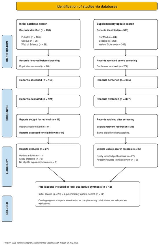
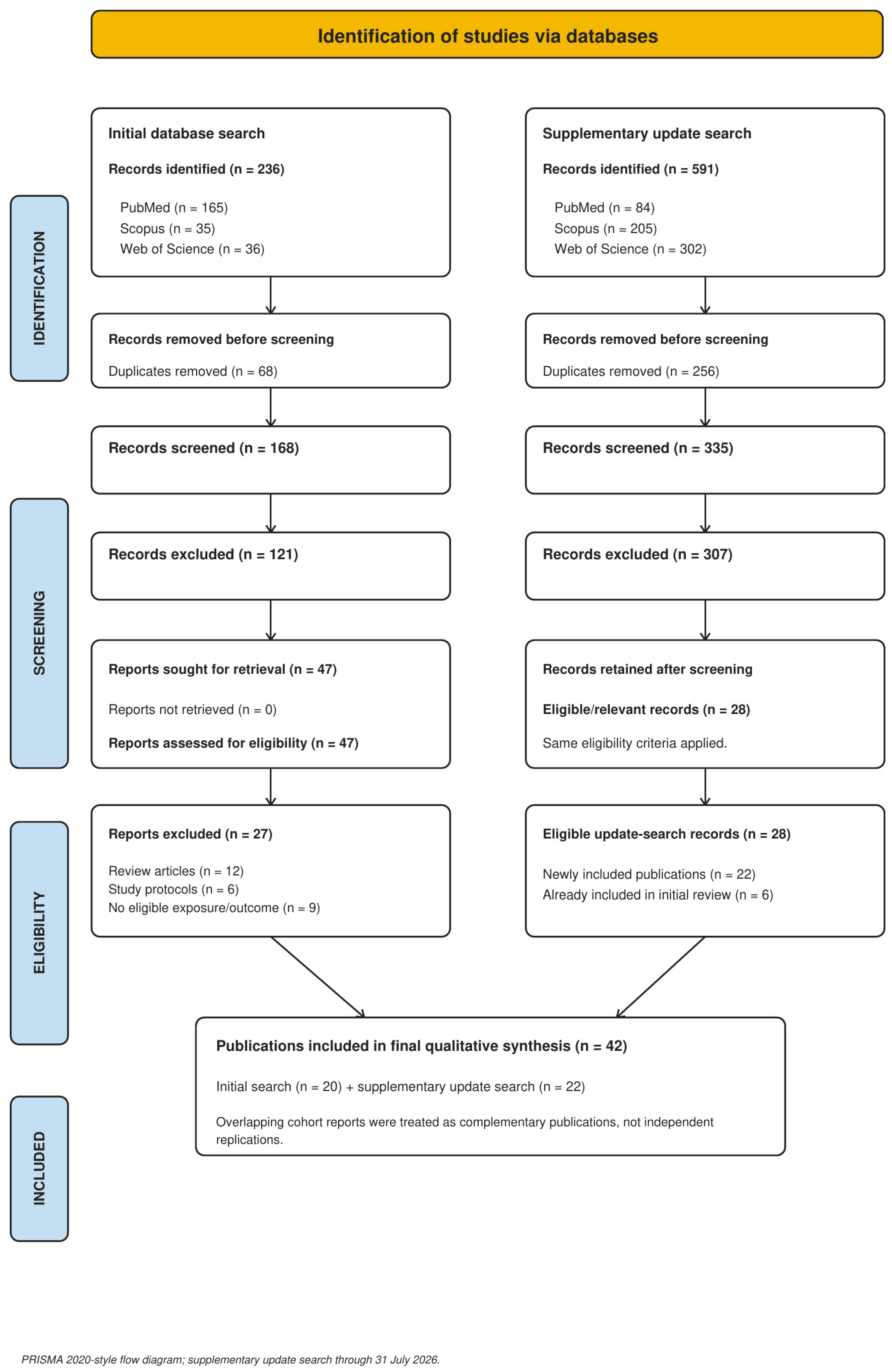

Disclaimer/Publisher’s Note: The statements, opinions and data contained in all publications are solely those of the individual author(s) and contributor(s) and not of MDPI and/or the editor(s). MDPI and/or the editor(s) disclaim responsibility for any injury to people or property resulting from any ideas, methods, instructions or products referred to in the content. |
© 2026 by the authors. Licensee MDPI, Basel, Switzerland. This article is an open access article distributed under the terms and conditions of the Creative Commons Attribution (CC BY) license.