1. Introduction
Psoriasis is a chronic, immune-mediated inflammatory disease characterized by dysregulated interactions between innate and adaptive immune responses, with a central role of proinflammatory cytokines such as tumour necrosis factor alpha (TNF-α) in disease pathogenesis [
1]. The advent of biologic therapies targeting TNF-α has significantly improved clinical outcomes and remains a widely used treatment option for moderate-to-severe psoriasis [
2].
Despite their efficacy, TNF-α inhibitors have been associated with immunogenicity, including the induction of autoantibodies such as antinuclear antibodies (ANA) and anti-double-stranded DNA (anti-dsDNA) antibodies [
3]. Several studies have demonstrated a positive correlation between prolonged exposure to TNF-α inhibitors and increased titres of these autoantibodies [
3,
4]. The clinical significance of this phenomenon, however, remains controversial. On one hand, the emergence of ANA and anti-dsDNA antibodies has been linked to the development of anti-drug antibodies (ADAs), which may reduce therapeutic efficacy and contribute to secondary treatment failure [
4]. Furthermore, the presence of these autoantibodies has been associated with drug-induced lupus-like syndromes, raising concerns about long-term safety [
5]. On the other hand, some evidence suggests that seroconversion to ANA or anti-dsDNA positivity does not necessarily correlate with clinically significant adverse events or diminished treatment response, indicating that these immunological changes may be epiphenomenal in certain patients [
3,
5].
In recent years, increasing attention has been directed toward the immunological consequences of long-term biologic therapy beyond clinical efficacy alone. Although TNF-α inhibitors are designed to suppress inflammatory pathways, their interaction with the immune system is complex and may result in the induction of humoral immune responses. Immunogenicity remains a major challenge during biologic treatment and encompasses the development of both anti-drug antibodies (ADAs) and autoantibodies. The generation of ADAs may alter drug pharmacokinetics through the formation of immune complexes, accelerated drug clearance, and neutralization of therapeutic activity, potentially leading to diminished clinical response and treatment discontinuation [
4]. Recent evidence suggests that the prevalence of immunogenic responses to adalimumab may be higher than previously recognized when highly sensitive detection methods are employed [
6].
Among patients receiving biologic therapy, adalimumab has been associated with one of the highest rates of ADA formation. A recent systematic review reported a pooled ADA incidence exceeding 30% in psoriasis patients treated with adalimumab, with a substantial proportion of antibodies demonstrating neutralizing capacity [
7]. Importantly, immunogenicity may not only affect treatment persistence but may also contribute to broader alterations in immune regulation. The emergence of autoantibodies during TNF-α inhibitor therapy has therefore been proposed as a potential marker of treatment-related immune activation [
3,
4].
Furthermore, the clinical importance of autoantibody monitoring remains an area of active investigation. While routine screening for ANA and anti-dsDNA antibodies is not universally recommended in patients receiving biologic therapy, several studies have suggested that changes in autoantibody profiles may precede alterations in therapeutic response or signal the adverse events [
4,
5]. Consequently, a better understanding of the temporal relationship between TNF-α inhibition and autoantibody induction may provide valuable insights into treatment monitoring strategies and facilitate the identification of patients at increased risk of immunogenicity-related complications.
Given these conflicting data, further investigation into the immunogenic effects of TNF-α inhibitors in psoriasis is warranted. In particular, understanding the dynamics of ANA and anti-dsDNA antibody development may help clarify their role as potential biomarkers for treatment response, immunogenicity, and autoimmune complications.
3. Results
3.1. Research Participants
A total of 92 patients with psoriasis receiving adalimumab therapy for at least 12 months were initially identified. Of the 71 included patients, 49 (69.0%) were male, and 22 (31.0%) were female. The mean age was 50.5 ± 13.1 years, with a median age of 49 years (IQR 19; range 17–77 years). A total of 30 patients (42%) were younger than 45 years, while 41 patients (58%) were aged 45 years or older. Psoriatic arthritis was present in 20 (28.2%) patients, while concomitant autoimmune diseases were identified in 14 (19.7%). Baseline PASI scores were available for 56 patients, with a median PASI of 16.2 (IQR 8.4). Baseline BSA values were also available for 56 patients, with a median BSA of 16.0% (IQR 27.3). None of the patients received concomitant systemic corticosteroid therapy during adalimumab treatment, 65 (91.5%) patients received methotrexate before the therapy with adalimumab, and 2 (2.8%) patients were receiving it during the therapy. Previous biologic therapy was documented in 4 of the 71 patients (5.6%). However, because these treatments had been administered as part of previous clinical trials and the specific biologic agents could not be reliably identified from the available medical records, previous biologic exposure was not included in the statistical analyses.
3.2. Descriptive Statistics for Study Variables
Descriptive statistics were calculated for demographic, clinical, and laboratory variables. Baseline clinical characteristics of the study population are presented in
Table 1. Continuous variables are reported as mean ± standard deviation (SD) or median and interquartile range (IQR), as appropriate. The distribution of continuous variables was assessed using the Shapiro–Wilk test. Because anti-dsDNA IgG levels, PASI, and BSA values were not normally distributed, these variables are primarily presented as median (IQR), whereas mean ± SD values are additionally provided for descriptive purposes. Categorical variables are presented as counts and percentages.
3.3. Comparison of Baseline and Post-Treatment Anti-dsDNA Levels
At baseline, median anti-dsDNA IgG levels were low across all subgroups, with slightly higher values observed in males than in females and in patients aged <45 years than in those aged ≥45 years. After 12 months of adalimumab therapy, anti-dsDNA IgG levels increased in the overall cohort.
Because anti-dsDNA values were not normally distributed according to the Shapiro–Wilk test, comparisons between baseline and post-treatment measurements were performed using the Wilcoxon signed-rank test. A statistically significant increase in anti-dsDNA levels was observed in the overall cohort (W = 272, p < 0.001), with a large effect size (rank-biserial correlation = 0.634).
Median anti-dsDNA levels increased from 0.9 IU/mL (IQR 9.4) to 15.0 IU/mL (IQR 31.4) in females and from 0 IU/mL (IQR 2.9) to 3.1 IU/mL (IQR 13.6) in males. Similarly, median anti-dsDNA levels increased from 0 IU/mL (IQR 3.8) to 2.5 IU/mL (IQR 11.3) in patients aged <45 years and from 0.5 IU/mL (IQR 3.3) to 9.6 IU/mL (IQR 27.2) in patients aged ≥45 years.
Wilcoxon signed-rank analyses demonstrated statistically significant increases in anti-dsDNA levels in both females (W = 18, p = 0.004; rank-biserial correlation = 0.789) and males (W = 153, p = 0.005; rank-biserial correlation = 0.541). A significant increase was also observed in patients aged ≥45 years (W = 80, p < 0.001; rank-biserial correlation = 0.746), whereas the increase in patients aged <45 years did not reach statistical significance (W = 57, p = 0.131; rank-biserial correlation = 0.400). These analyses evaluated changes in anti-dsDNA levels as a continuous variable.
Mann–Whitney U tests showed no statistically significant differences in median anti-dsDNA levels between females and males either before therapy (U = 458, p = 0.274) or after therapy (U = 398, p = 0.076). Likewise, no statistically significant differences were observed between patients aged <45 and ≥45 years either before therapy (U = 512, p = 0.347) or after therapy (U = 478, p = 0.196).
Although a statistically significant increase in anti-dsDNA levels was observed only in patients aged ≥45 years, a greater proportion of younger patients exceeded the laboratory threshold for anti-dsDNA positivity after treatment. These findings reflect different analytical approaches, as the present analyses evaluated changes in continuous anti-dsDNA concentrations, whereas subsequent analyses assessed anti-dsDNA increase as a binary outcome. The results are summarized in
Table 2.
3.4. Factors Associated with Anti-dsDNA Autoantibody Development
An increase in anti-dsDNA levels after adalimumab therapy was not associated with the presence of psoriatic arthritis (Pearson’s χ2(1) = 0.455, p = 0.500) or concomitant autoimmune diseases (Pearson’s χ2(1) = 0.285, p = 0.594). Likewise, no significant differences in baseline PASI scores were observed between patients with and without an increase in anti-dsDNA levels (median 15.6 vs. 16.3, Mann–Whitney U = 366.5, p = 0.729). Categorization of patients according to PASI severity (<10 vs. ≥10) also showed no significant association with anti-dsDNA increase (Fisher’s exact test, p = 0.501). The strength of the association was weak (Phi = −0.164). Similarly, baseline BSA values did not differ significantly between patients with and without anti-dsDNA elevation after treatment (median 14.0% vs. 18.0%, U = 32.5, p = 0.361).
3.5. Predictors of Anti-dsDNA Level Increase After Adalimumab Therapy
To identify possible predictors of anti-dsDNA increase after therapy, an exploratory multivariable logistic regression analysis was performed. In contrast to the Wilcoxon analyses, this model evaluated anti-dsDNA increase as a binary outcome. Age (OR 0.998, 95% CI 0.96–1.04,
p = 0.920), sex (OR 1.46, 95% CI 0.50–4.29,
p = 0.488), baseline anti-dsDNA level (OR 1.04, 95% CI 0.98–1.11,
p = 0.170), and PASI score (OR 0.998, 95% CI 0.99–1.00,
p = 0.515) were not independently associated with anti-dsDNA increase. These results should be interpreted as exploratory because of the limited number of anti-dsDNA increase events. Obtained results can be observed in
Table 3.
3.6. ANA/ENA Autoantibody Development
At baseline, ANA/ENA positivity was observed in 4 of 71 patients (5.6%), whereas 67 (94.4%) were ANA/ENA-negative. After 12 months of adalimumab therapy, ANA/ENA positivity was detected in 16 patients (23%). McNemar’s test demonstrated a statistically significant increase in ANA/ENA positivity after therapy (χ2 = 12.0, p < 0.001). Of the 71 patients, 12 (16.9%) converted from ANA/ENA-negative to ANA/ENA-positive, whereas no patients converted from positive to negative.
ANA/ENA seroconversion was more frequent in females (32%) than in males (18%), although this difference did not reach statistical significance (p = 0.210). Most patients remained ANA/ENA-negative after therapy (77% overall).
When stratified by age, ANA/ENA seroconversion occurred in both groups without significant differences between patients aged <45 and ≥45 years (
p = 0.662). Results are presented in
Table 4.
3.7. Factors Associated with ANA/ENA Seroconversion
ANA/ENA seroconversion was observed in 20% of patients with psoriatic arthritis and in 15.7% of patients without psoriatic arthritis; however, this difference was not statistically significant (Fisher’s exact test, p = 0. 729). Likewise, no significant association was found between concomitant autoimmune diseases and ANA/ENA seroconversion (p = 0.106). Patients who developed ANA/ENA seroconversion had slightly higher baseline PASI scores (median 18.3 vs. 15.3) and BSA values (median 27.5% vs. 15.5%) than those without seroconversion, although neither difference reached statistical significance (U = 186.0, p = 0.346 and U = 209.5, p = 0.659).
3.8. Predictors of ANA/ENA Seroconversion
To identify possible predictors of ANA/ENA seroconversion after therapy, an exploratory multivariable logistic regression analysis was performed. Age (OR 1.01, 95% CI 0.96–1.06,
p = 0.747), sex (OR 0.40, 95% CI 0.10–1.56,
p = 0.189), and PASI score (OR 1.01, 95% CI 0.999–1.01,
p = 0.079) were not independently associated with ANA/ENA seroconversion. Given the limited number of seroconversion events, these findings should be interpreted as exploratory and require confirmation in larger prospective cohorts. Detailed results are presented in
Table 5.
3.9. Impact of Concomitant Methotrexate Use on Anti-dsDNA Antibody Development and ANA/ENA Seroconversion
To assess the association between methotrexate use, anti-dsDNA development and ANA/ENA seroconversion, Fisher’s exact test was performed. Overall, 65 of 71 patients (91.5%) had received methotrexate before initiating adalimumab therapy. Anti-dsDNA increase was observed in 38 of 65 patients (58.5%) with prior methotrexate exposure and in 2 of 6 patients (33.3%) without prior methotrexate exposure. No significant association was found between prior methotrexate use and anti-dsDNA increase (Fisher’s exact test, p = 0.393). Similarly, ANA/ENA seroconversion occurred in 11 of 65 patients (16.9%) with prior methotrexate exposure and in 1 of 6 patients (16.7%) without prior methotrexate exposure, with no significant association observed (Fisher’s exact test, p = 1.000).
Only two patients received concomitant methotrexate during adalimumab therapy. Anti-dsDNA increase was not associated with concomitant methotrexate use (Fisher’s exact test, p = 1.000). ANA/ENA seroconversion was observed in both patients receiving concomitant methotrexate (2/2, 100%) and in 10 of 69 patients (14.5%) not receiving concomitant methotrexate. Although Fisher’s exact test indicated a statistically significant association (p = 0.027), this finding should be interpreted with considerable caution because it was based on an extremely small subgroup.
3.10. Distribution of Autoantibody Levels Before and After 12 Months of Therapy
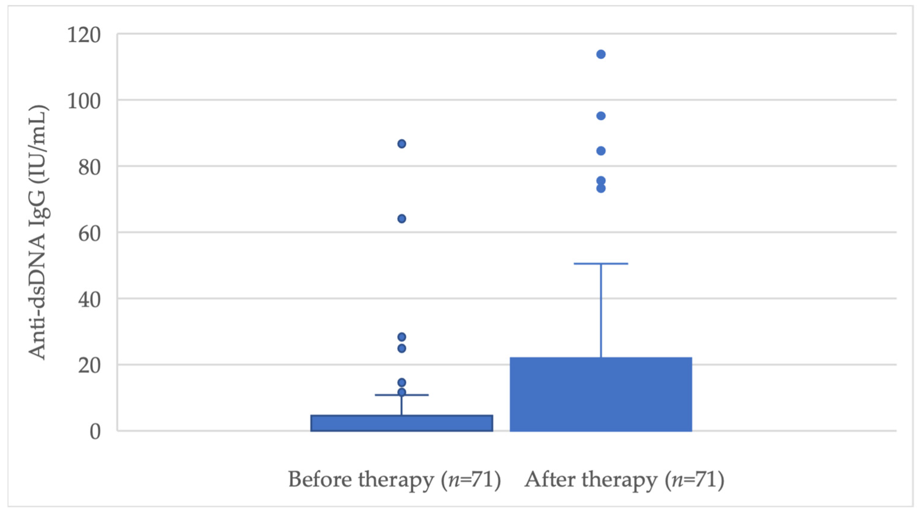
ANA/ENA positivity increased during the 12-month treatment period, reaching 23% after therapy. Baseline anti-dsDNA IgG levels were generally low, with only a few patients showing elevated titres before treatment initiation. After 12 months of adalimumab therapy, anti-dsDNA IgG levels increased in a substantial proportion of patients. As illustrated in
Figure 1, the distribution of anti-dsDNA titres became broader and shifted toward higher values after treatment, with several patients developing markedly elevated titres. These findings indicate an increase in anti-dsDNA antibody levels during adalimumab therapy.
3.11. Biologic Medication Change
A total of 17 patients (24%) were switched to another biologic therapy during follow-up. On average, treatment switching occurred 23 months after initiation of adalimumab therapy, with the earliest switch performed after 14 months. The primary reason for treatment modification was inadequate clinical response, defined according to national treatment criteria as a body surface area (BSA) involvement of ≥10%. No treatment changes were made because of autoimmune manifestations attributable to ANA/ENA or anti-dsDNA positivity.
Elevated autoantibody levels were observed in 65% of patients who required treatment modification. However, exploratory logistic regression analyses demonstrated that neither ANA/ENA seroconversion (OR 1.02, 95% CI 0.24–4.28,
p = 0.975), changes in anti-dsDNA levels (OR 0.998, 95% CI 0.978–1.02,
p = 0.876), nor baseline PASI score (OR 0.996, 95% CI 0.990–1.00,
p = 0.137) were significantly associated with subsequent biologic treatment switching (
Table 6).
4. Discussion
In this single-centre retrospective study, we demonstrated that 12 months of adalimumab therapy in patients with psoriasis was associated with a significant increase in serum anti-dsDNA IgG levels and a higher frequency of ANA/ENA positivity. Anti-dsDNA titres increased significantly in both female and male patients and in individuals aged ≥45 years, while ANA/ENA seroconversion was observed in 23% of the cohort. Although patients with newly developed autoantibodies were more likely to switch to another biologic agent, this association did not reach statistical significance. These findings support the concept that TNF-α inhibition can induce humoral autoimmune responses, while the clinical relevance of these serological changes remains uncertain.
Although TNF-α inhibition is well known to impair protective humoral immunity, this does not necessarily contradict the increased ANA and anti-dsDNA levels observed in the present study. Recent studies have demonstrated that TNF inhibitors reduce germinal centre-dependent immune responses, resulting in impaired affinity maturation, diminished antigen-specific memory B-cell generation, reduced long-lived plasma cell output, and lower vaccine-induced IgG responses [
8,
9,
10,
11,
12]. These observations primarily reflect impaired protective immune responses against foreign antigens rather than autoreactive immunity.
In contrast, ANA and anti-dsDNA antibodies represent a loss of immune tolerance rather than enhanced protective humoral immunity. Therefore, the increased autoantibody levels observed after adalimumab treatment should not be interpreted as evidence of generalized B-cell activation or increased antibody production. Instead, they likely reflect qualitative immune dysregulation, whereby protective antigen-specific germinal centre responses are diminished while immune tolerance mechanisms become altered, allowing autoreactive B-cell clones to emerge.
The induction of autoantibodies during TNF-α inhibitor therapy is a well-recognized phenomenon. Despite being fully human monoclonal antibodies, agents such as adalimumab can trigger immune responses directed both against the therapeutic molecule and against endogenous nuclear antigens. Several mechanisms have been proposed, including increased apoptosis with release of nucleosome material, impaired clearance of apoptotic debris, and cytokine shifts favouring autoreactive B-cell activation [
5,
13]. These immunological effects may explain the frequent emergence of ANA and anti-dsDNA antibodies in patients treated with TNF-α antagonists. The development of ANA and anti-dsDNA autoantibodies during TNF-α inhibitor therapy may reflect broader treatment-induced immune dysregulation rather than biologic immunogenicity. TNF-α plays a central role in maintaining immune homeostasis, and its inhibition may alter cytokine signalling, apoptotic cell clearance, and B-cell tolerance, thereby promoting autoreactive immune responses. Emerging evidence suggests that activation of type I interferon pathways may represent a key mechanism linking TNF-α blockade with autoantibody production. Type I interferons enhance dendritic cell activation, antigen presentation, and autoreactive B-cell differentiation, contributing to sustained autoantibody generation and amplification of autoimmune responses [
14,
15]. These findings are consistent with the hypothesis that treatment-induced autoantibodies may reflect underlying immune dysregulation associated with long-term TNF-α inhibition, although the clinical significance of these serological changes remains uncertain.
Our findings are consistent with prior studies in psoriasis and other immune-mediated inflammatory diseases. A single-centre study evaluating patients with psoriasis treated with biologic agents reported a significant increase in ANA positivity after treatment with adalimumab and etanercept but found no clear association with reduced clinical efficacy or adverse events [
16]. Similarly, a systematic review by Hsu et al. reported that anti-adalimumab antibodies develop in 6–45% of patients with psoriasis and may be associated with lower serum drug concentrations and reduced treatment response [
17]. However, the development of conventional autoantibodies, such as ANA and anti-dsDNA, does not necessarily parallel the formation of anti-drug antibodies (ADAs) and should be interpreted separately. Because anti-drug antibodies and adalimumab trough concentrations were not assessed in the present study, no conclusions regarding biologic immunogenicity can be drawn.
In our study, ANA/ENA positivity increased from 6% at baseline to 23% after one year of therapy. This seroconversion rate is comparable to previously reported frequencies and confirms that ANA induction is common during TNF-α inhibitor treatment [
16,
18]. Importantly, no statistically significant differences were observed between sexes or age groups, suggesting that autoantibody induction is a broadly distributed effect rather than being restricted to specific demographic subgroups.
Anti-dsDNA antibody levels also increased significantly after treatment. The increase was statistically significant in both sexes and in patients aged ≥45 years, although patients younger than 45 years did not demonstrate a statistically significant rise in median titres. These findings may reflect differences in baseline variability and sample size rather than distinct biological mechanisms. The broad dispersion of anti-dsDNA values and the presence of several high-titre outliers indicate considerable interindividual heterogeneity in humoral response to adalimumab.
The clinical implications of these autoantibodies remain controversial. Several studies have shown that ANA and anti-dsDNA seroconversion often occurs in the absence of lupus-related symptoms and does not require discontinuation of treatment [
16,
18]. Conversely, elevated autoantibody titres have been associated with anti-drug antibody formation and secondary loss of response in some cohorts. In patients with rheumatoid arthritis treated with infliximab, baseline ANA positivity was associated with an increased risk of developing ADAs and treatment failure [
19]. In psoriasis, baseline anti-dsDNA antibodies have also been proposed as potential predictors of reduced adalimumab effectiveness [
20]. Our results do not allow conclusions regarding the relationship between treatment-induced autoantibody development and anti-drug antibody formation, as ADAs were not measured. Although patients with increased autoantibody levels more frequently switched to another biologic therapy, this association was not statistically significant and should be interpreted cautiously.
Another clinically relevant concern is the development of TNF inhibitor-induced lupus (ATIL). This rare but well-documented adverse event is typically characterized by ANA and anti-dsDNA positivity accompanied by clinical manifestations such as arthralgia, serositis, or cutaneous lupus lesions [
21]. A recent case report described systemic lupus erythematosus arising during adalimumab therapy in a patient with psoriasis and psoriatic arthritis [
22]. In the present study, we did not systematically evaluate clinical manifestations of lupus; therefore, we cannot determine whether serological conversion was associated with clinically significant autoimmunity. Nevertheless, our data suggest that isolated autoantibody positivity is substantially more common than overt autoimmune disease. Although no patient in our cohort developed clinically apparent lupus during the observation period, the significant increase in anti-dsDNA levels observed after 12 months of adalimumab therapy may reflect immune alterations similar to those implicated in lupus pathogenesis. Experimental studies suggest that TNF-α blockade may impair the clearance of apoptotic cells, resulting in increased exposure of nuclear antigens and activation of autoreactive B cells. In addition, reduced IL-2 and IL-10 production together with a shift toward a Th1-dominant immune response have been proposed as mechanisms contributing to the loss of self-tolerance and autoantibody formation [
14]. Long-term follow-up studies are needed to determine whether treatment-induced seroconversion has prognostic significance for future autoimmune complications.
The relationship between treatment-induced autoantibodies and true anti-drug antibodies deserves further investigation. ADAs against adalimumab can reduce circulating drug concentrations, increase drug clearance, and contribute to loss of therapeutic response [
23,
24,
25]. Recent high-sensitivity studies have demonstrated that ADA formation may occur in a substantial proportion of patients within the first months of treatment and is strongly associated with lower trough concentrations [
6]. Whether ANA or anti-dsDNA positivity is associated with anti-drug antibody formation remains unknown. Because anti-drug antibodies and serum drug concentrations were not evaluated in the present study, ANA and anti-dsDNA should not be considered surrogate markers of biologic immunogenicity based on our findings. Recent studies employing highly sensitive and drug-tolerant immunoassays suggest that ADA prevalence may be substantially underestimated by conventional detection methods. Persistent ADA development has been demonstrated in a large proportion of adalimumab-treated patients, with higher ADA titres associated with accelerated drug clearance, reduced drug exposure, and an increased likelihood of disease relapse [
6]. In addition, anti-adalimumab antibodies have been shown to exhibit high-affinity binding to both originator and biosimilar formulations, supporting their biological relevance and potential impact on treatment persistence [
25]. Together, these findings from previous studies indicate that biologic immunogenicity is a dynamic process that may substantially influence long-term treatment outcomes. However, the present study did not directly evaluate immunogenicity because anti-drug antibodies and serum drug concentrations were not assessed.
Methotrexate (MTX) may partially mitigate these effects. Concomitant MTX therapy has been associated with reduced ADA formation, improved biologic drug survival, and higher adalimumab trough concentrations. Furthermore, MTX has been shown to reduce anti-adalimumab antibody formation in a dose-dependent manner, thereby maintaining therapeutic drug concentrations and preserving clinical response [
26,
27,
28]. Collectively, these findings suggest that concomitant MTX therapy may attenuate biologic immunogenicity and improve long-term treatment persistence in selected patients. However, these observations are derived from previous studies and could not be confirmed in the present cohort. In the present cohort, prior methotrexate exposure was not associated with either anti-dsDNA increase or ANA/ENA seroconversion. Only two patients received concomitant methotrexate during adalimumab therapy. Although both developed ANA/ENA seroconversion, this finding should be interpreted with considerable caution because it was based on an extremely small subgroup, and no association was observed with anti-dsDNA increase. Therefore, our data do not allow conclusions regarding the effect of concomitant methotrexate on treatment-associated autoantibody development. The clinical significance of treatment-associated ANA and anti-dsDNA seroconversion remains uncertain. In the absence of compatible clinical manifestations, isolated autoantibody positivity should be interpreted cautiously and should not be regarded as evidence of biologic immunogenicity or an indication for treatment modification. Therapeutic drug monitoring, including anti-drug antibody assessment, remains the preferred approach when immunogenic loss of response is clinically suspected. Future prospective studies incorporating both autoantibody measurements and anti-drug antibody testing are needed to clarify whether any relationship exists between these distinct immunological phenomena.
The practical implications of our findings are twofold. First, routine development of ANA or anti-dsDNA antibodies during adalimumab therapy should be interpreted cautiously in the absence of compatible clinical symptoms. Second, the clinical significance of treatment-associated ANA and anti-dsDNA seroconversion remains uncertain. Therapeutic drug monitoring, including anti-drug antibody assessment, should be considered on the basis of clinical suspicion of treatment failure rather than autoantibody positivity alone.
This study has several limitations. Its retrospective design limits control over confounding factors, and the sample size was modest. Data were derived from a single centre, which may limit generalizability. The apparent discrepancy between age-group analyses likely reflects the different statistical approaches used. Continuous analyses demonstrated a greater overall increase in anti-dsDNA concentrations among older patients, whereas categorical analyses identified a higher proportion of younger patients exceeding the laboratory positivity threshold. In addition, the limited sample size may have reduced the statistical power of subgroup analyses, particularly after stratification by both age and sex. Furthermore, the exploratory regression analyses should be interpreted cautiously because of the limited sample size and number of outcome events. Accordingly, these findings should be regarded as hypothesis-generating rather than as evidence of independent predictors of treatment-associated autoantibody development. Information regarding previous biologic exposure was incomplete because detailed records of biologic agents administered as part of previous clinical trials were not consistently available in the retrospective medical records. Consequently, the potential influence of previous biologic therapy on treatment-associated autoantibody development could not be evaluated. Other immune suppressants, including systemic corticosteroids, were not used during the therapy with adalimumab; therefore, they could not have influenced the development of autoantibodies. Although methotrexate use before and during adalimumab therapy was evaluated, the very small number of patients receiving concomitant methotrexate limited the interpretation of these findings. ANA and anti-dsDNA were evaluated as markers of treatment-associated serological changes rather than direct measures of biologic immunogenicity. Anti-adalimumab antibodies and serum drug concentrations were not assessed. Therefore, no conclusions regarding biologic immunogenicity or its relationship to treatment-associated autoantibody development can be drawn. Although the present study focused on the clinically relevant 12-month monitoring interval, longer follow-up studies are needed to evaluate the long-term dynamics and clinical significance of treatment-induced autoantibody development. Another limitation of this study is the lack of disease activity assessment and cytokine profiling. Owing to the retrospective design and the absence of routine cytokine measurements in clinical practice, these associations could not be evaluated.
Future prospective studies should address their potential role in treatment-induced autoantibody development. Clinical disease activity scores and detailed reasons for treatment discontinuation were not analysed systematically. Nonetheless, the study reflects real-world clinical practice and provides longitudinal data on serological changes in a homogeneous cohort of patients treated with adalimumab.
5. Conclusions
Adalimumab therapy in patients with psoriasis was associated with a significant increase in anti-dsDNA IgG levels and a higher frequency of ANA/ENA positivity after 12 months of treatment. These findings indicate that TNF-α inhibitor therapy may be accompanied by treatment-induced autoantibody formation across different age and sex groups. However, the clinical significance of these serological changes remains uncertain, as neither anti-dsDNA level increase nor ANA/ENA seroconversion was significantly associated with biologic treatment switching in the present cohort.
The observed autoantibody development may reflect broader treatment-induced immune dysregulation rather than biologic immunogenicity, as anti-drug antibodies and serum adalimumab concentrations were not assessed. Therefore, the present findings should not be interpreted as evidence of biologic immunogenicity or as support for the clinical use of ANA or anti-dsDNA as biomarkers of treatment response. Although previous studies have demonstrated that concomitant methotrexate may reduce anti-drug antibody formation and improve biologic drug persistence, our cohort did not allow meaningful evaluation of this effect because only two patients received concomitant methotrexate during adalimumab therapy. Consequently, no conclusions regarding the influence of methotrexate on treatment-associated autoantibody development can be drawn from the present study.
Taken together, our findings support the concept that treatment-induced autoantibody formation and biologic immunogenicity represent distinct immunological processes that should not be considered interchangeable. Further research is required to determine whether these phenomena are mechanistically related or occur independently during TNF-α inhibitor therapy.
Future prospective studies incorporating anti-drug antibody assays, therapeutic drug monitoring, serum drug concentrations, longitudinal clinical outcome measures, and larger patient cohorts are warranted to clarify the mechanisms underlying treatment-induced autoantibody development and to determine its potential clinical relevance during long-term TNF-α inhibitor therapy.