Article

The Relationship Between Coronary Artery Ectasia and PAI-1 4G/5G Gene Polymorphisms

1
Department of Cardiology, Faculty of Medicine, Recep Tayyip Erdoğan University, Rize 53200, Turkey
2
Department of Medical Biology, Faculty of Medicine, Recep Tayyip Erdoğan University, Rize 53200, Turkey
*
Author to whom correspondence should be addressed.
Int. J. Transl. Med. 2026, 6(3), 33; https://doi.org/10.3390/ijtm6030033
Submission received: 12 June 2026 / Revised: 14 August 2026 / Accepted: 18 August 2026 / Published: 21 August 2026

Abstract

Background: Coronary artery ectasia (CAE) is characterized by either diffuse or segmental enlargement of the coronary artery, with a diameter that exceeds 1.5 times that of the adjacent normal vessel. The underlying causes of CAE remain inadequately understood. Higher plasminogen activator inhibitor-1 (PAI-1) concentrations have been related to aortic aneurysms, possibly resulting from occlusion of vasa vasorum, followed by diminished vessel wall supply. Whether the same process has a role in CAE is not known. This research seeks to ascertain if a correlation exists between polymorphisms of the PAI-1 gene and the occurrence of CAE. Methods: Fifty-three patients with CAE and 52 patients with angiographically normal coronary arteries were included. The PAI-1 4G/5G polymorphism was genotyped by real-time PCR, using melting curve analysis of LightCycler 480 II. Results: Genotype distributions in both groups were consistent with Hardy–Weinberg equilibrium (p = 1.00). The frequencies of the 4G/4G, 4G/5G, and 5G/5G genotypes did not differ significantly between patients with CAE and controls. Similarly, allele frequencies of the 4G and 5G variants were comparable between the two groups, and no statistically significant association was observed between the PAI-1 −675 4G/5G polymorphism and the presence of CAE (all p > 0.05). In the allele-dose sensitivity analysis, the number of 4G alleles was not independently associated with CAE (adjusted OR per 4G allele: 0.82, 95% CI: 0.37–1.82; p = 0.626). After adjustment for age, sex, hypertension, diabetes mellitus, body mass index, and smoking status, the PAI-1 genotype was not independently associated with CAE (overall likelihood-ratio test: p = 0.867). Conclusions: No independent association was detected between the PAI-1 −675 4G/5G polymorphism and CAE; however, smaller genetic effects cannot be excluded because of the modest sample size and wide confidence intervals.

1. Introduction

Coronary artery ectasia (CAE) is characterized by either diffuse or segmental enlargement of the coronary artery, surpassing 1.5 times the diameter of the adjacent healthy vessel [1]. The etiology of CAE encompasses concurrent atherosclerotic conditions in approximately 50% of cases, while congenital factors account for 20–30% of the patient population [2,3,4]. The occurrence of isolated CAE has been documented to range from 1.2% to 4.9% across diverse research investigations [1,2,3,4,5]. The underlying causes, or etiology, as well as the progression and clinical manifestations of CAE have not yet been elucidated with sufficient clarity or comprehensiveness to provide a definitive understanding of this complex condition. Nevertheless, a comprehensive analysis of histopathological studies has unveiled remarkably similar characteristics pertaining to atherosclerotic plaques, which include widespread degeneration of both the intimal and medial layers, as well as the occurrence of hyalinization, observed in patients suffering from CAE [6]. Thrombus, vasospasm, and slow flow have also been documented in this condition [7].
Plasminogen activator inhibitor-1, commonly referred to as PAI-1, serves as a principal inhibitor of the physiological process known as fibrinolysis, which is fundamental for the breakdown of fibrin in blood clots, and this important protein is secreted by a variety of cell types, including but not limited to endothelial cells that line the blood vessels, vascular smooth muscle cells that contribute to the structural integrity of the vasculature, and macrophages that play a crucial role in immune responses and tissue homeostasis [8,9]. A compelling body of evidence indicates that elevated concentrations of PAI-1 are frequently observed in patients diagnosed with coronary artery disease, suggesting a potential link between increased levels of this inhibitor and the pathophysiological mechanisms underlying this common cardiovascular condition [10,11,12]. In a previous investigation, we disclosed that patients suffering from CAE exhibited significantly higher levels of PAI-1 when compared to those without this condition, thereby highlighting the potential importance of PAI-1 as a biomarker or therapeutic target in such cardiovascular disorders [13].
The promoter region of the PAI-1 gene possesses an insertion or deletion of a G nucleotide (−675 4G/5G) as a polymorphism [14]. Homozygous individuals for the 4G/4G deletion have high plasma PAI-1 levels, while the subjects genotyped as 5G/5G present low PAI-1 levels [15,16,17]. The effects of 4G/5G polymorphism on PAI-1 activity have been demonstrated in healthy subjects [18], in non-insulin-dependent diabetes mellitus (NIDDM) patients [19], and in patients with coronary heart disease [17].
In the present study, we aimed to determine whether there was a difference between the CAE group and the control group in terms of the PAI-1 gene polymorphism.

2. Materials and Methods

2.1. Study Population

Among 2881 patients who underwent coronary angiography during a one-year period, 53 patients with CAE (44 men and nine women; mean age, 61.5 ± 10.9 years) were included. The control group comprised 52 patients (22 men and 30 women; mean age, 55.1 ± 11.0 years) who underwent coronary angiography for suspected coronary artery disease but had angiographically normal coronary arteries. All participants were aged ≥18 years, and cases and controls were recruited from the same institution.
Coronary artery disease (CAD) was defined as an atherosclerotic plaque or luminal irregularity in at least one major epicardial coronary artery, regardless of stenosis severity. Patients with CAE and non-obstructive CAD were included, while those with >50% luminal stenosis capable of causing clinically relevant ischemia were excluded. The control group had no angiographically visible CAE, plaque, luminal irregularity, or coronary narrowing. Angiographically verified controls were selected to minimize misclassification of CAE phenotype.
Informed consent was secured by all participants before the commencement of the study. The research was conducted in accordance with the Helsinki Declaration, as approved by the Recep Tayyip Erdoğan University Non-Interventional Clinical Research Ethics Committee No. 2019/120 on 9 September 2019.
The manifestation of classic angina pectoris alongside positive or ambiguous findings from noninvasive stress testing served as the criteria for proceeding with coronary angiography. Hypertension was deemed present if the patient was receiving antihypertensive treatment or if systolic blood pressure exceeded 140 mmHg, diastolic blood pressure surpassed 90 mmHg, or both conditions were noted during the clinical assessment. Diabetes mellitus was identified if fasting glucose levels were greater than 126 mg/dL, or if the individual was undergoing antidiabetic treatment or adhering to a specialized diet. Current smokers and people who had continued smoking up to 6 months ago were considered to be smokers.
Patients with heart failure with reduced ejection fraction (LVEF ≤ 40%), left ventricular hypertrophy, atrial fibrillation, moderate-to-severe valvular heart disease, or myocardial or pericardial disease were excluded. Other exclusion criteria were renal or hepatic dysfunction, anemia, thrombocytopenia, malignancy, recent major surgery, inflammatory or autoimmune disease, hemolytic disorders, hematological disease, and chromosomal abnormalities. The same exclusion criteria were applied to both groups.

2.2. Coronary Angiography and Definition of CAE

Coronary angiography was conducted utilizing 6 French left and right coronary catheters, employing the conventional Judkins method without the administration of nitroglycerin [20]. Two experts independently evaluated the coronary angiograms while blinded to the clinical and genetic data. Coronary diameters and percent stenosis were measured by using computerized quantitative angiography (Siemens-Artis, Erlangen, Germany). Coronary artery ectasia is characterized as the expansion of the coronary artery lumen to a degree that surpasses 1.5 times the diameter of an adjacent normal vessel.

2.3. Genotyping

Genomic DNA was isolated from leukocytes in peripheral blood, which was collected in ethylenediaminetetraacetic acid (EDTA) tubes using a previously reported salting-out method [21]. The concentration and purity of the DNA samples were evaluated by the absorbance method using the µDrop™ plate (Thermo Fisher Scientific Inc., Wilmington, DE, USA) in the spectrophotometer device. The PAI-1 4G/5G polymorphism was genotyped by real-time polymerase chain reaction (PCR) using the commercially available kit, PAI-1 (4G/5G) Real Time HYB-FRT Kit (Diagen Biyoteknolojik Sistemleri A.Ş, Ankara, Turkey), according to the manufacturer’s instructions. After the PAI-1 gene is amplified with specific primers, PCR fragment analysis is performed with specific hybridization probes in this protocol. Subsequently, the 5G/5G, 4G/5G, and 4G/4G genotypes are determined by the formation of specific melting points (Tm) of the 4G and 5G alleles in the melting curve. The human PAI-1 4G DNA shows a Tm of 51 °C, and the 5G DNA shows a Tm of 58 °C according to kit protocol. The real-time PCR protocol completed with melting curve analysis was carried out on a Roche Applied Science LightCycler® 480 II (Roche Diagnostics GmbH, Mannheim, Germany) device.

2.4. Statistical Analysis

Statistical analysis was carried out with the IBM SPSS Statistics 21 (Statistical Package for Social Sciences; SPSS Inc., Chicago, IL, USA) package program. Relationships between categorical variables were evaluated using the Chi-square test. The distribution characteristics of continuous variables were assessed utilizing the Kolmogorov–Smirnov Test, while the t-test was employed for analyzing variables exhibiting a normal distribution, and the Mann–Whitney U test was applied for the evaluation of variables that did not conform to a normal distribution. Hardy–Weinberg equilibrium was assessed using an exact test. A two-sided p value of <0.05 was considered statistically significant.

2.5. Sample-Size Considerations

Sample-size adequacy was evaluated using G*Power software (version 3.1), with a two-sided significance level of α = 0.05 and a target power of 80%. The original calculation assumed moderate effect sizes. However, genetic association studies frequently identify smaller effects, and the available sample of 53 patients with CAE and 52 controls may not provide sufficient statistical power to detect odds ratios in the range of approximately 1.2–1.5. Accordingly, effect estimates are presented together with their 95% confidence intervals to demonstrate the precision of the observed associations.
A multivariable binary logistic regression analysis was performed to determine whether the association between the PAI-1 −675 4G/5G polymorphism and CAE was independent of potential confounding factors. The presence of CAE was entered as the dependent variable. The PAI-1 genotype was included as a categorical variable, with the 5G/5G genotype used as the reference category. Age, sex, hypertension, diabetes mellitus, body mass index, and smoking status were included as clinically relevant covariates. Diabetes mellitus was analyzed as a binary variable regardless of treatment modality. Adjusted odds ratios (ORs) and 95% confidence intervals (CIs) were calculated. The overall contribution of the genotype variable was assessed by a likelihood-ratio test comparing models with and without the genotype terms. A two-sided p value of <0.05 was considered statistically significant.

3. Results

Genotype distributions of the PAI-1 −675 4G/5G polymorphism were consistent with Hardy–Weinberg equilibrium in both the CAE and control groups (p = 1.00). The frequencies of the 4G/4G, 4G/5G, and 5G/5G genotypes were 17.0%, 50.9%, and 32.1% in the CAE group and 23.1%, 51.9%, and 25.0% in the control group, respectively. Genotype distribution did not differ significantly between the groups (χ2 = 0.952, df = 2, p = 0.33). The 4G and 5G allele frequencies were also comparable between the CAE and control groups (p = 0.27). In the allele-dose sensitivity analysis, the number of 4G alleles was not independently associated with CAE (adjusted OR per 4G allele: 0.82, 95% CI: 0.37–1.82; p = 0.626).
The two groups were comparable in terms of most baseline clinical characteristics and cardiovascular risk factors. However, patients in the CAE group were significantly older than those in the control group (61.5 ± 10.9 vs. 55.1 ± 11.0 years; p = 0.004), and the proportion of male participants was higher in the CAE group. Detailed demographic characteristics, cardiovascular risk factors, and laboratory findings are presented in Table 1 and Table 2. Age and sex were therefore included as covariates in the multivariable logistic regression analysis.
Significant differences were observed in lipid parameters. Total cholesterol levels were significantly lower in the CAE group compared with the control group (188.3 ± 39.5 vs. 229.0 ± 49.4 mg/dL, respectively; p < 0.001). Similarly, LDL cholesterol levels were significantly lower in the CAE group (115.2 ± 32.5 vs. 144.1 ± 40.1 mg/dL; p < 0.001). Statin use was more common among patients with CAE than among controls (49.1% vs. 28.8%, respectively; p = 0.034).
Based on the results of the PCR analysis, both the patient cohort and the control group were categorized into three distinct genotypes of the promoter region of the PAI-1 gene: 4G/4G, 4G/5G, and 5G/5G. Table 3 shows the genotype distribution between CAE patients and the control group.
In an exploratory analysis, genotype distribution did not differ significantly between patients with multivessel CAE and controls. Among the 23 patients with multivessel CAE, the 4G/4G, 4G/5G, and 5G/5G genotypes were present in 3, 12, and 8 patients, respectively, compared with 12, 27, and 13 controls (χ2 = 1.348, df = 2, p = 0.510). Allele frequencies were also not significantly different (p = 0.262).
The frequencies of vascular risk factors in patients with PAI-1 gene 4G/4G, 5G/5G, and 4G/5G genotypes were compared. A significant relationship was observed between hypertension and genotype distribution (p = 0.027). A significantly higher prevalence of hypertension was observed in individuals with the 4G/5G genotype.
There was no statistical difference between other vascular risk factors and PAI-1 genotypes. The distribution and percentages of vascular risk factors in PAI-1 gene polymorphism are shown in Table 4.
Multivariable binary logistic regression analysis was performed in all 105 participants. After adjustment for age, sex, hypertension, diabetes mellitus, body mass index, and smoking status, the PAI-1 genotype was not independently associated with CAE (overall likelihood-ratio test: χ2 = 0.284, df = 2, p = 0.867). Using the 5G/5G genotype as the reference category, the adjusted OR was 0.69 (95% CI: 0.14–3.47; p = 0.651) for the 4G/4G genotype and 0.73 (95% CI: 0.20–2.73; p = 0.642) for the 4G/5G genotype.
Advanced age (adjusted OR per year: 1.12, 95% CI: 1.06–1.19; p < 0.001), male sex (adjusted OR: 25.00, 95% CI: 5.29–118.12; p < 0.001), hypertension (adjusted OR: 4.58, 95% CI: 1.30–16.15; p = 0.018), and body mass index (adjusted OR per kg/m2: 1.22, 95% CI: 1.06–1.40; p = 0.006) were independently associated with CAE. Diabetes mellitus and smoking status were not statistically significant in the adjusted model.
In coronary angiography evaluation, eight patients (15.1%) had ectasia in their left main coronary artery, 29 patients (54.7%) had ectasia in their left anterior descending artery, 23 patients (43.2%) had ectasia in their circumflex artery, and 23 patients (43.2%) had ectasia in their right coronary artery. Thirty patients had single-vessel CAE, whereas 23 had multivessel involvement: 18 had two-vessel, three had three-vessel, and two had four-vessel CAE.

4. Discussion

The present study evaluated the association between the PAI-1 −675 4G/5G promoter polymorphism and CAE. No statistically significant association was detected after adjustment for age, sex, hypertension, diabetes mellitus, body mass index, and smoking status. However, the wide confidence intervals reflect the modest sample size, and smaller genetic effects cannot be excluded.
CAE is a complex form of vascular remodeling whose pathogenesis remains incompletely understood. Histopathological studies have demonstrated degeneration of elastin, collagen, and the extracellular matrix, together with inflammatory-cell accumulation in ectatic arteries [22,23,24]. These findings suggest that inflammation, endothelial dysfunction, thrombosis, and extracellular matrix remodeling may interact in the development of CAE.
PAI-1 is a principal physiological inhibitor of fibrinolysis. Increased PAI-1 activity may impair fibrin clearance, promote fibrin deposition and thrombosis, and disrupt hemostatic balance [25,26,27]. The 4G allele is more transcriptionally active than the 5G allele because the additional nucleotide in the 5G allele provides a binding site for a transcriptional repressor. Consequently, 4G/4G carriers generally have higher circulating PAI-1 concentrations than 5G/5G carriers [25].
Previous studies have reported associations between CAE and variants in genes involved in vascular remodeling. Gulec et al. identified the ACE deletion polymorphism as a potential risk factor for CAE [28], whereas Lamblin et al. reported an association between the matrix metalloproteinase-3 5A allele and aneurysmal coronary disease [29]. Yalım et al. evaluated several thrombophilic gene variants, including the PAI-1 4G/5G polymorphism, and reported broadly similar variant frequencies in patients with CAE and controls [30]. The present study provides independent, hypothesis-driven evidence consistent with the absence of a detectable association between this PAI-1 variant and CAE. This study was designed as a focused, hypothesis-driven investigation based on our previous observation of increased circulating PAI-1 levels in patients with CAE [13]. We therefore aimed to determine whether the functional PAI-1 −675 4G/5G promoter polymorphism was independently associated with CAE using adjusted genotype models, an allele-dose sensitivity analysis, and an exploratory analysis according to multivessel involvement.
In a previous study, we found significantly higher plasma PAI-1 levels in patients with CAE than in individuals with normal coronary arteries. We hypothesized that elevated PAI-1 might promote obstruction of the vasa vasorum supplying the coronary arterial wall, thereby impairing wall integrity and contributing to ectatic remodeling [13]. However, plasma PAI-1 levels are influenced by circadian variation and preanalytical factors, while commonly used assays predominantly measure total rather than biologically active PAI-1 [31,32]. Genetic analysis was therefore employed as a complementary approach because genetic variants are stable and less susceptible to these influences. The PAI-1 −675 4G/5G polymorphism was selected based on the established involvement of PAI-1 in fibrinolysis, atherothrombosis, and vascular-wall remodeling.
The absence of an association between the PAI-1 −675 4G/5G polymorphism and CAE does not contradict our previous finding of elevated circulating PAI-1 levels in patients with CAE. Circulating PAI-1 is influenced not only by genetic predisposition but also by inflammatory, endothelial, metabolic, and environmental factors and therefore cannot be explained by a single promoter variant. Inflammation, endothelial dysfunction, oxidative stress, tissue hypoxia, renin–angiotensin system activation, and metabolic disturbances may all increase PAI-1 expression.
Epigenetic mechanisms, variants in other genes, and gene–environment interactions may also regulate PAI-1 independently of the −675 4G/5G polymorphism. Thus, elevated PAI-1 levels in CAE may primarily reflect acquired factors associated with vascular remodeling rather than a direct effect of the investigated genotype. Future studies should evaluate circulating PAI-1 levels and functional activity together with relevant genetic variants.
Patients with CAE had significantly lower total cholesterol and LDL cholesterol levels than controls, which may be partly explained by the higher frequency of statin use in the CAE group (49.1% vs. 28.8%; p = 0.034). Therefore, lipid levels measured at enrollment may not reflect pretreatment profiles. Because data on baseline lipid levels, statin indication, type, dose, treatment duration, and adherence were unavailable, the effect of statin therapy could not be fully assessed and should be considered when interpreting these findings.

4.1. Potential Translational Mechanisms Linking PAI-1 and Coronary Artery Ectasia

Although no significant association was found between the PAI-1 4G/5G polymorphism and coronary artery ectasia (CAE), PAI-1 may still contribute to its pathogenesis through effects on fibrinolysis, thrombosis, extracellular matrix turnover, vascular remodeling, and inflammation [33,34,35,36].
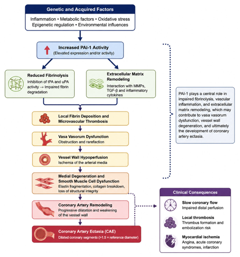
Increased PAI-1 activity may promote local fibrin deposition and microthrombosis, potentially impairing vasa vasorum perfusion. Resulting vascular wall ischemia, together with PAI-1-associated endothelial dysfunction, oxidative stress, and inflammation, may weaken the medial layer and favor ectatic remodeling [33,34,35].
PAI-1 also interacts with transforming growth factor-β, pro-inflammatory cytokines, and matrix metalloproteinases. Dysregulation of these pathways may disrupt extracellular matrix homeostasis, promote elastin fragmentation, and impair vascular wall integrity—mechanisms considered relevant to CAE [34,35,36,37].
The lack of association with the 4G/5G polymorphism does not exclude a pathogenic role for PAI-1. Environmental and epigenetic factors, gene–gene interactions, and other functional variants may influence PAI-1 independently of this polymorphism. Circulating concentrations and functional activity may therefore better reflect its biological relevance than a single genetic marker [36,37,38,39,40].
The proposed pathways are summarized in Figure 1. Overall, dysregulated fibrinolysis, endothelial dysfunction, inflammation, and extracellular matrix remodeling may collectively promote coronary dilatation. Prospective studies integrating genetic, molecular, and circulating biomarker data are needed to clarify the potential value of PAI-1 as a biomarker or therapeutic target.

4.2. Translational Relevance and Future Directions

The present findings support the view that CAE is a multifactorial vascular disorder that is unlikely to be explained by a single common promoter polymorphism. The biological relevance of the PAI-1 pathway may depend more on functional activity and acquired inflammatory, metabolic, environmental, and epigenetic regulation than on the isolated presence of the −675 4G/5G variant [13,33,34,35].
Future studies should move beyond isolated candidate-gene analyses and integrate multigene or genome-wide approaches with epigenomic, transcriptomic, proteomic, functional biomarker, environmental, and advanced vascular-imaging data. Such integrated approaches may improve the biological characterization and risk stratification of CAE [38].

4.3. Study Limitations

This study has several limitations. First, the modest sample size may have limited the ability to detect small but clinically relevant genetic effects. Although the initial sample-size calculation assumed moderate effect sizes, common genetic variants often have smaller effects. This uncertainty is reflected in the wide confidence interval for the adjusted association per 4G allele (OR: 0.82, 95% CI: 0.37–1.82). Therefore, the findings indicate an absence of a detectable association in the present sample rather than definitive evidence of no association. Larger, adequately powered, multicenter studies are required to estimate smaller genetic effects more precisely. Normality was assessed using the Kolmogorov–Smirnov test; although this test was prespecified, the Shapiro–Wilk test may have provided greater sensitivity in the present sample size.
Second, the groups were not matched for age, sex, or cardiovascular risk factors, and significant differences were present in several baseline characteristics. Although age, sex, hypertension, diabetes mellitus, body mass index, and smoking status were included in the multivariable analysis, residual confounding from measured or unmeasured factors cannot be excluded. Plasma PAI-1 levels and functional activity were not measured; therefore, their relationships with PAI-1 genotype and CAE could not be directly assessed. Furthermore, only the PAI-1 −675 4G/5G polymorphism was evaluated, and the potential contributions of other genetic variants, epigenetic mechanisms, and gene–environment interactions remain unknown.
Finally, the controls were not healthy community-based individuals but patients referred for coronary angiography because of suspected coronary artery disease. Although all controls had angiographically normal coronary arteries and no CAE, this referral process may have introduced selection bias and may limit the generalizability of the findings. Conventional coronary angiography also evaluates the vessel lumen and cannot completely exclude early intramural plaque or positive vascular remodeling without visible luminal abnormalities. Because intravascular imaging was not performed, the controls should be regarded as angiographically normal rather than definitively free of all subclinical atherosclerosis. Nevertheless, coronary angiography objectively confirmed the absence of CAE in all controls and reduced the risk of phenotype misclassification.

5. Conclusions

The present study did not detect an independent association between the PAI-1 −675 4G/5G polymorphism and CAE after adjustment for relevant clinical covariates. However, the modest sample size and wide confidence intervals do not exclude smaller but potentially clinically relevant genetic effects.
These findings suggest that CAE susceptibility is unlikely to be determined by this single promoter variant and should instead be considered within a broader framework involving multiple genetic, inflammatory, metabolic, environmental, and vascular-remodeling mechanisms. Larger multicenter studies integrating genetic data with circulating PAI-1 levels, functional biomarkers, multi-omics analyses, and vascular imaging are needed to clarify the biological and clinical relevance of the PAI-1 pathway in CAE.

Author Contributions

M.E. and Y.Ç. conceived and designed the analysis; M.E. and E.E. collected the data; Y.Ç. and F.S. contributed data or analysis tools; Y.Ç. performed the analysis; M.E. and A.G.Ö. wrote the paper; and M.E., Y.Ç., A.G.Ö., F.S. and E.E. reviewed the manuscript. All authors have read and agreed to the published version of the manuscript.

Funding

This research received no external funding.

Institutional Review Board Statement

The research was conducted in accordance with the Helsinki Declaration, as approved by the Recep Tayyip Erdoğan University Non-Interventional Clinical Research Ethics Committee No. 2019/120 on 9 September 2019.

Informed Consent Statement

Informed consent was obtained from all subjects involved in the study.

Data Availability Statement

The data that support the findings of this study are not openly available and are available from the corresponding author upon reasonable request.

Conflicts of Interest

The authors declare no conflict of interest.

References

  1. Swaye, P.S.; Fisher, L.D.; Litwin, P.; Vignola, P.A.; Judkins, M.P.; Kemp, H.G.; Mudd, J.G.; Gosselin, A.J. Aneurysmal coronary artery disease. Circulation 1983, 67, 134–138. [Google Scholar] [CrossRef] [Scilit] [PubMed]
  2. Devabhaktuni, S.; Mercedes, A.; Diep, J.; Ahsan, C. Coronary artery ectasia—A review of current literature. Curr. Cardiol. Rev. 2016, 12, 318–323. [Google Scholar] [CrossRef] [Scilit] [PubMed]
  3. Ozturk, S.; Yetkin, E.; Waltenberger, J. Molecular and cellular insights into the pathogenesis of coronary artery ectasia. Cardiovasc. Pathol. 2018, 35, 37–47. [Google Scholar] [CrossRef] [Scilit] [PubMed]
  4. Boles, U.; Johansson, A.; Wiklund, U.; Sharif, Z.; David, S.; McGrory, S.; Henein, M.Y. Cytokine disturbances in coronary artery ectasia do not support atherosclerosis pathogenesis. Int. J. Mol. Sci. 2018, 19, 260. [Google Scholar] [CrossRef] [Scilit] [PubMed]
  5. Sorrell, V.L.; Davis, M.J.; Bove, A.A. Current knowledge and significance of coronary artery ectasia: A chronologic review of the literature, recommendations for treatment, possible etiologies, and future considerations. Clin. Cardiol. 1998, 21, 157–160. [Google Scholar] [CrossRef] [Scilit] [PubMed]
  6. Markis, J.E.; Joffe, C.D.; Cohn, P.F.; Feen, D.J.; Herman, M.V.; Gorlin, R. Clinical significance of coronary arterial ectasia. Am. J. Cardiol. 1976, 37, 217–222. [Google Scholar] [CrossRef] [Scilit] [PubMed]
  7. Papadakis, M.C.; Manginas, A.; Cotileas, P.; Demopoulos, V.; Voudris, V.; Pavlides, G.; Foussas, S.G.; Cokkinos, D.V. Documentation of slow coronary flow by the TIMI frame count in patients with coronary ectasia. Am. J. Cardiol. 2001, 88, 1030–1032. [Google Scholar] [CrossRef] [Scilit] [PubMed]
  8. Lupu, F.; Bergonzelli, G.E.; Heim, D.A.; Cousin, E.; Genton, C.Y.; Bachmann, F.; Kruithof, E.K. Localization and production of plasminogen activator inhibitor-1 in human healthy and atherosclerotic arteries. Arterioscler. Thromb. 1993, 13, 1090–1100. [Google Scholar] [CrossRef] [Scilit] [PubMed]
  9. Lijnen, H.R. Pleotropic functions of plasminogen activator inhibitor-1. J. Thromb. Haemost. 2005, 3, 35–45. [Google Scholar] [CrossRef] [Scilit] [PubMed]
  10. Nakamura, Y.; Tomura, S.; Tachibana, K.; Chida, Y.; Marumo, F. Enhanced fibrinolytic activity during the course of hemodialysis. Clin. Nephrol. 1992, 38, 90–96. [Google Scholar] [PubMed]
  11. Segarra, A.; Chacon, P.; Martinez-Eyarre, C.; Argelaguer, X.; Vila, J.; Ruiz, P.; Fort, J.; Bartolomé, J.; Camps, J.; Moliner, E.; et al. Circulating levels of plasminogen activator inhibitor type-1, tissue plasminogen activator, and thrombomodulin in hemodialysis patients: Biochemical correlations and role as independent predictors of coronary artery stenosis. J. Am. Soc. Nephrol. 2001, 12, 1255–1263. [Google Scholar] [PubMed]
  12. Tomura, S.; Nakamura, Y.; Doi, M.; Ando, R.; Ida, T.; Chida, Y.; Ootsuka, S.; Shinoda, T.; Yanagi, H.; Tsuchiya, S.; et al. Fibrinogen, coagulation factor VII, tissue plasminogen activator, plasminogen activator inhibitor-1, and lipid as cardiovascular risk factors in chronic hemodialysis and continuous ambulatory peritoneal dialysis patients. Am. J. Kidney Dis. 1996, 27, 848–854. [Google Scholar] [CrossRef] [Scilit] [PubMed]
  13. Cicek, Y.; Durakoglugil, M.E.; Erdogan, T.; Yilmaz, A.; Uydu, H.A.; Saglam, H.; Cetin, M.; Satiroglu, O.; Bostan, M.; Canga, A.; et al. Increased plasminogen activator inhibitor-1 levels in patients with isolated coronary artery ectasia. J. Thromb. Thrombolysis 2012, 33, 120–123. [Google Scholar] [CrossRef] [Scilit] [PubMed]
  14. Mansfield, M.W.; Stickland, M.H.; Grant, P.J. Plasminogen activator inhibitor-1 (PAI-1) promoter polymorphism and coronary artery disease in non-insulin-dependent diabetes. Thromb. Haemost. 1995, 74, 1032–1034. [Google Scholar] [CrossRef] [Scilit]
  15. Nauck, M.; Wieland, H.; Marz, W. Rapid, homogeneous genotyping of the 4G/5G polymorphism in the promoter region of the PAI1 gene by fluorescence resonance energy transfer and probe melting curves. Clin. Chem. 1999, 45, 1141–1147. [Google Scholar] [CrossRef] [Scilit]
  16. Rallidis, L.S.; Gialeraki, A.; Merkouri, E.; Liakos, G.; Dagres, N.; Sionis, D.; Travlou, A.; Lekakis, J.; Kremastinos, D.T. Reduced carriership of 4G allele of plasminogen activator inhibitor-1 4G/5G polymorphism in very young survivors of myocardial infarction. J. Thromb. Thrombolysis 2010, 29, 497–502. [Google Scholar] [CrossRef] [Scilit] [PubMed]
  17. Eriksson, P.; Kallin, B.; van ’t Hooft, F.M.; Bavenholm, P.; Hamsten, A. Allele-specific increase in basal transcription of the plasminogen-activator inhibitor 1 gene is associated with myocardial infarction. Proc. Natl. Acad. Sci. USA 1995, 92, 1851–1855. [Google Scholar] [CrossRef] [Scilit] [PubMed]
  18. Dawson, S.J.; Wiman, B.; Hamsten, A.; Green, F.; Humphries, S.; Henney, A.M. The two allele sequences of a common polymorphism in the promoter of the plasminogen activator inhibitor-1 (PAI-1) gene respond differently to interleukin-1 in HepG2 cells. J. Biol. Chem. 1993, 268, 10739–10745. [Google Scholar] [CrossRef] [Scilit]
  19. Panahloo, A.; Mohamed-Ali, V.; Lane, A.; Green, F.; Humphries, S.E.; Yudkin, J.S. Determinants of plasminogen activator inhibitor-1 activity in treated NIDDM and its relation to a polymorphism in the plasminogen activator inhibitor 1 gene. Diabetes 1995, 44, 37–42. [Google Scholar] [CrossRef] [Scilit] [PubMed]
  20. Judkins, M.P. Selective coronary arteriography. Radiology 1967, 89, 815–824. [Google Scholar] [CrossRef] [Scilit] [PubMed]
  21. Miller, S.A.; Dykes, D.D.; Polesky, H.F. A simple salting out procedure for extracting DNA from human nucleated cells. Nucleic Acids Res. 1988, 16, 1215. [Google Scholar] [CrossRef] [Scilit] [PubMed]
  22. Diet, F.; Pratt, R.E.; Berry, G.J.; Momose, N.; Gibbons, G.H.; Dzau, V.J. Increased accumulation of tissue ACE in human atherosclerotic coronary artery disease. Circulation 1996, 94, 2756–2767. [Google Scholar] [CrossRef] [Scilit] [PubMed]
  23. Tamarina, N.A.; McMillan, W.D.; Shively, V.P.; Pearce, W.H. Expression of matrix metalloproteinases and their inhibitors in aneurysms and normal aorta. Surgery 1997, 122, 264–272. [Google Scholar] [CrossRef] [Scilit] [PubMed]
  24. Knox, J.B.; Sukhova, G.K.; Whittemore, A.D.; Libby, P. Evidence for altered balance between matrix metalloproteinases and their inhibitors in human aortic diseases. Circulation 1997, 95, 205–212. [Google Scholar] [CrossRef] [Scilit] [PubMed]
  25. Wang, J.; Wang, C.; Chen, N.; Shu, N.; Guo, X.; He, Y.; Zhou, Y. Association between the plasminogen activator inhibitor-1 4G/5G polymorphism and risk of venous thromboembolism: A meta-analysis. Thromb. Res. 2014, 134, 1241–1248. [Google Scholar] [CrossRef] [Scilit] [PubMed]
  26. Akhter, M.S.; Biswas, A.; Ranjan, R.; Meena, A.; Yadav, B.K.; Sharma, A.; Saxena, R. Plasminogen activator inhibitor-1 (PAI-1) gene 4G/5G promoter polymorphism is seen in higher frequency in the Indian patients with deep vein thrombosis. Clin. Appl. Thromb. Hemost. 2010, 16, 184–188. [Google Scholar] [CrossRef] [Scilit] [PubMed]
  27. Incalcaterra, E.; Meli, F.; Muratori, I.; Corrado, E.; Amato, C.; Canino, B.; Ferrara, F. Residual vein thrombosis and onset of post-thrombotic syndrome: Influence of the 4G/5G polymorphism of plasminogen activator inhibitor-1 gene. Thromb. Res. 2014, 133, 371–374. [Google Scholar] [CrossRef] [Scilit] [PubMed]
  28. Gulec, S.; Aras, O.; Atmaca, Y.; Akyurek, O.; Hanson, N.Q.; Sayin, T.; Tsai, M.Y.; Akar, N.; Oral, D. Deletion polymorphism of the angiotensin I converting enzyme gene is a potent risk factor for coronary artery ectasia. Heart 2003, 89, 213–214. [Google Scholar] [CrossRef] [Scilit] [PubMed]
  29. Lamblin, N.; Bauters, C.; Hermant, X.; Lablanche, J.M.; Helbecque, N.; Amouyel, P. Polymorphisms in the promoter regions of MMP-2, MMP-3, MMP-9, and MMP-12 genes as determinants of aneurysmal coronary artery disease. J. Am. Coll. Cardiol. 2002, 40, 43–48. [Google Scholar] [CrossRef] [Scilit] [PubMed]
  30. Yalim, Z.; Onrat, S.T.; Emren, S.V.; Dural, I.E.; Avsar, A.; Onrat, E. Analysis of thrombophilic gene mutations in coronary artery ectasia. Turk. Kardiyol. Dern. Ars. 2020, 48, 368–373. [Google Scholar] [CrossRef] [Scilit] [PubMed]
  31. Scheer, F.A.J.L.; Shea, S.A. Human circadian system causes a morning peak in prothrombotic plasminogen activator inhibitor-1 (PAI-1) independent of the sleep/wake cycle. Blood 2013, 123, 590–593. [Google Scholar] [CrossRef] [Scilit] [PubMed]
  32. Juhan-Vague, I.; Alessi, M.C.; Raccah, D.; Aillaud, M.F.; Billerey, M.; Ansaldi, J.; Philip-Joet, C.; Vague, P. Daytime fluctuations of plasminogen activator inhibitor 1 (PAI-1) in populations with high PAI-1 levels. Thromb. Haemost. 1992, 67, 76–82. [Google Scholar] [CrossRef] [Scilit]
  33. Vaughan, D.E. PAI-1 and atherothrombosis. J. Thromb. Haemost. 2005, 3, 1879–1883. [Google Scholar] [CrossRef] [Scilit] [PubMed]
  34. Cesari, M.; Pahor, M.; Incalzi, R.A. Plasminogen activator inhibitor-1 (PAI-1): A key factor linking fibrinolysis and age-related subclinical and clinical conditions. Cardiovasc. Ther. 2010, 28, e72–e91. [Google Scholar] [CrossRef] [Scilit] [PubMed]
  35. Gils, A.; Declerck, P.J. Three decades of research on plasminogen activator inhibitor-1: A multifaceted serpin. Semin. Thromb. Hemost. 2013, 39, 356–364. [Google Scholar] [CrossRef] [Scilit] [PubMed]
  36. Rabieian, R.; Boshtam, M.; Zareei, M.; Kouhpayeh, S.; Masoudifar, A.; Mirzaei, H. Plasminogen activator inhibitor type-1 as a regulator of fibrosis. J. Cell. Biochem. 2018, 119, 17–27. [Google Scholar] [CrossRef] [Scilit] [PubMed]
  37. Kaikita, K.; Fogo, A.B.; Ma, L.; Schoenhard, J.A.; Brown, N.J.; Vaughan, D.E. Plasminogen activator inhibitor-1 deficiency prevents hypertension and vascular fibrosis in response to long-term nitric oxide synthase inhibition. Circulation 2001, 104, 839–844. [Google Scholar] [CrossRef] [Scilit] [PubMed]
  38. Iwaki, T.; Urano, T.; Umemura, K. PAI-1, progress in understanding the clinical problem and its aetiology. Br. J. Haematol. 2012, 157, 291–298. [Google Scholar] [CrossRef] [Scilit] [PubMed]
  39. Björnsson, T.D.; Schneider, D.E.; Berger, H., Jr. PAI-1: Physiological and pathological aspects. Cardiovasc. Drug Rev. 2003, 21, 293–337. [Google Scholar]
  40. Ghosh, A.K.; Vaughan, D.E. PAI-1 in tissue fibrosis. J. Cell. Physiol. 2012, 227, 493–507. [Google Scholar] [CrossRef] [Scilit] [PubMed]
Figure 1. Proposed pathophysiological pathway linking plasminogenactivator inhibitor-1 (PAI-1) and coronary artery ectasia (CAE). Increased PAI-1 activity, influenced by genetic and acquired factors, may promote impaired fibrinolysis, vascular remodeling, vasa vasorum dysfunction, and vascular wall degeneration, ultimately contributing to CAE development and its clinical consequences. tPA: tissue plasminogen activator; uPA: urokinase plasminogen activator; MMPs: matrix metalloproteinases; TGF-β: transforming growth factor-β.
Ijtm 06 00033 g001
Table 1. Demographic characteristics of study groups.
Patients (n = 53)Control (n = 52)p
Age (years)61.5 ± 10.955.1 ± 11.00.004
Male (n, %)44 (83.1)22 (42.3)0.01
BMI (kg/m2)29.5 ± 6.727.7 ± 3.70.49
HT (n, %)42 (79)27 (51)0.03
DM (n, %)15 (28)11 (20)0.18
HPL (n, %)18 (33)13 (24)0.28
FH of CAD (n, %)16 (30)10 (19)0.20
BMI: body mass index, DM: diabetes mellitus, FH of CAD: family history of coronary artery disease, HPL: hyperlipidemia, HT: hypertension.
Table 2. Laboratory characteristics of study groups.
CAE Group
(n = 53)
Control Group
(n = 52)
p
Creatinine (mg/dL)0.94 ± 0.220.77 ± 0.19<0.001
Leukocyte (103)7.64 ± 2.057.78 ± 2.640.689
Platelet (103)232 ± 55.6266 ± 81.90.060
Hemoglobin (gr/dL)16.6 ± 1.916.1 ± 1.60.405
Glucose (mg/dL)131.2 ± 61123.9 ± 47.40.635
Alanine aminotransferase (IU/L)25.0 ± 1426.1 ± 190.610
Aspartate aminotransferase (IU/L)25.5 ± 1028.6 ± 150.908
Total cholesterol (mg/dL)188.3 ± 39.5229 ± 49.4<0.001
LDL (mg/dL)115.2 ± 32.5144.1 ± 40.1<0.001
HDL (mg/dL)42.6 ± 1149.6 ± 10<0.001
Triglyceride (mg/dL)175.5 ± 127201 ± 1710.317
Data are presented as mean ± SD. HDL, high-density lipoprotein; LDL, low-density lipoprotein.
Table 3. Genotype and allele frequencies of PAI-1 4G/5G polymorphism in study groups.
Frequency HWE (Hardy–Weinberg Equilibrium)
CAE Patients
(n = 53)
Control Group
(n = 52)
p
Genotypep = 1.00
4G/4G n (%)9 (17)12 (23.1)0.33
4G/5G n (%)27 (50.9)27 (51.9)
5G/5G n (%)17 (32.1)13 (25.0)
Allele
4G n (%)45 (42.5)51 (49.1)0.27
5G n (%)61 (57.5)53 (50.9)
Table 4. Vascular disease risk factors—genotype relationship.
Vascular Risk Factors4G/4G4G/5G5G/5Gp Value
Smoking (n, %)9 (19.1)24 (51.1)14 (29.8)0.962
Alcohol (n, %)1 (6.3)10 (62.5)5 (31.3)0.320
Obesity (n, %)7 (21.2)18 (54.5)8 (24.2)0.802
Hypertension (n, %)17 (24.6)29 (42.0)23 (33.3)0.027
Hyperlipidemia (n, %)9 (28.1)13 (40.6)10 (31.3)0.262
Diabetes mellitus (n, %)7 (25.9)12 (44.4)8 (29.6)0.607
Coronary artery disease (n, %)5 (16.1)13 (41.9)13 (41.9)0.146
Valvular heart disease (n, %)4 (13.8)19 (65.5)6 (20.7)0.203
Family history (n, %)6 (23.1)15 (57.7)5 (19.2)0.447
Disclaimer/Publisher’s Note: The statements, opinions and data contained in all publications are solely those of the individual author(s) and contributor(s) and not of MDPI and/or the editor(s). MDPI and/or the editor(s) disclaim responsibility for any injury to people or property resulting from any ideas, methods, instructions or products referred to in the content.