Review

Topical Oxygen Therapy (blue®m) for Post-Surgical Care Protocols to Promote Wound Healing in Periodontology and Dental Implants: A Case-Based Literature Review

by
Cristian Scognamiglio
1,2,†,
Alessandro Perucchi
1,2,†,
Chalini Sundar
3,
Tatiana Miranda Deliberador
2 and
Hamdan Alghamdi
3,*
1
Private Practice, Studio Odontoiatrico, 6850 Mendrisio, Switzerland
2
Latin American Institute of Dental Research and Education—ILAPEO, Curitiba 80710-150, PR, Brazil
3
College of Dentistry, King Saud University, Riyadh 11545, Saudi Arabia
*
Author to whom correspondence should be addressed.
These authors contributed equally to this work.
Submission received: 27 June 2025 / Revised: 21 July 2025 / Accepted: 24 July 2025 / Published: 29 July 2025

Abstract

Background: Stable post-surgical wound healing surrounding teeth and dental implants is essential for achieving excellent clinical outcomes, both during the initial phases of treatment and over the long term. Objectives: This work follows the new emerging trend of case-based literature reviews. The aim of this review includes providing clinical findings from case series that demonstrate the efficacy of using blue®m oxygen treatment to promote post-surgical wound healing in patients that underwent periodontal and dental implant surgeries. In addition, a systematic review of the literature aimed to answer the focused research question: “In periodontal and implant surgeries, what are the aftercare protocols used to maintain optimal wound healing?” Case Presentation: One clinical case report involved the presentation of complex periodontal surgery. The other two cases focused on advanced implant surgeries. All patients were treated post-surgically with the local application of an oxygen-based therapy (blue®m) gel. This therapy was further emphasized during the wound-healing phase by instructing patients to maintain thorough dental hygiene using toothpaste and mouthwash containing a similar oxygen-release formulation (blue®m). Patients achieved satisfactory treatment outcomes. Systematic Review: PubMed and EMBASE were used in order to search for relevant studies in the scientific literature published up until June 2025. Only human clinical studies that used a specific protocol in regard to aftercare wound healing after periodontal or dental implant surgeries were included. As a result, 27 clinical studies were included. The outcome data were categorized and summarized. Conclusions: The use of local oxygen-based therapy showed a positive effect as a conventionally used aftercare modality in maintaining optimal post-surgical wound healing, following periodontal and implant surgeries. Further clinical studies are needed.

1. Introduction

In dentistry, post-surgical aftercare management differs among practitioners and encompasses mechanical cleaning techniques, antiseptic application, systemic or localized antibiotics, and supplementary therapies, such as laser treatment [1,2]. Recently, numerous aftercare protocols have been established in regard to post-surgical wound healing in periodontology, as well as for dental implants, to facilitate the maintenance and healing of hard and soft oral tissues [1,3]. Wound healing after oral surgery encompasses complex biological reactions that are affected by numerous local circumstances, including the properties of the tissue and biomaterials involved [3]. For instance, the postoperative phase after periodontal surgery or implant placement is crucial, since adequate wound healing ensures tissue stability [1,4]. The formation of an early soft tissue barrier is crucial for efficient periodontal wound healing [1,4]. In addition, integrating soft tissue with the supra-structures in dental implants is essential to avert bacterial infiltration, which may result in peri-implant illness or implant failure.

Necessity of Surgical Wound Aftercare

Nowadays, regenerative interventions involving periodontology and dental implants are often necessary. Therefore, aftercare in terms of wound healing is crucial for the success of these surgeries [5]. Indeed, wound healing is a complex biological process, involving various cellular and molecular factors, in numerous stages. The inflammatory stage (0–7 days) involves the formation of a blood clot, which works against disturbing forces or bacterial challenges that occur in regard to the initial healing of the wound. In particular, the oral cavity is a remarkable environment in which wound healing occurs in the presence of saliva that contains millions of microorganisms [6]. Neutrophils, macrophages, and inflammatory cells remove the damaged tissue, leading to the secretion of cytokines and growth factors that initiate the healing response. During the proliferation stage (1–3 weeks), mesenchymal progenitor cells migrate to the defected site and differentiate into mature cells to form soft/hard tissues, which remodel into more organized tissues. However, certain healed bone tissues are not capable of regeneration and possess the same functionality or morphology as the lost tissue. In addition, surgical wounds can be closed and stitched successfully with non-absorbable or absorbable sutures, which must remain strong and stable in the wound bed to facilitate optimal healing [7]. An open or infected wound may slow the healing process, potentially leading to greater problems and procedural failures. Therefore, post-surgical monitoring for optimal wound healing is a necessity. Clinically, several post-surgical protocols have been suggested and utilized to promote early wound healing and reduce the patient’s symptoms. Indeed, the common end goals are complete soft and hard tissue healing, the absence of abnormal clinical symptoms, and no post-surgical complications [5]. As many clinicians are facing problems in regard to the healing of soft and hard tissues in oral wounds in daily practice, researchers have been inspired to explore alternative treatments and potential autotherapies to enhance wound healing. By leveraging advancements in biomaterials, there is great potential for the use of translational (adjunctive) therapy in oral tissue wound healing and regeneration.
In recent times, an oxygen-based local adjunct formulation (blue®m) was developed [8,9]. It is increasingly studied for its potential to enhance wound healing and reduce complications post-surgery [9]. Such local deliveries of oxygen products can directly enable intermittent oxygen exposure to the surgical area. Accordingly, these oxygen-based products are clinically available in different forms (e.g., oral gel, oxygen fluid, mouthwash, foam, and toothpaste) with considerably better treatment outcomes. The clinical benefits of these aftercare protocols on wound healing results have not been fully investigated.
This work follows the new emerging trend of a case-based review literature. Please refer to the published recommendations/guidelines specifically for composing case-based reviews [10]. First, we present a series of case reports aimed at clinically evaluating the effectiveness of the topical oxygen therapy (blue®m) as an aftercare protocol for post-surgical wound healing. Second, a comprehensive search and synthesis of the existing literature was conducted to summarize current evidence on how different aftercare strategies influence wound healing. Finally, an effort was made to propose a protocol for the use of local oxygen-based formulations as the preferred treatment modality for post-surgical wound aftercare in periodontology and dental implants.

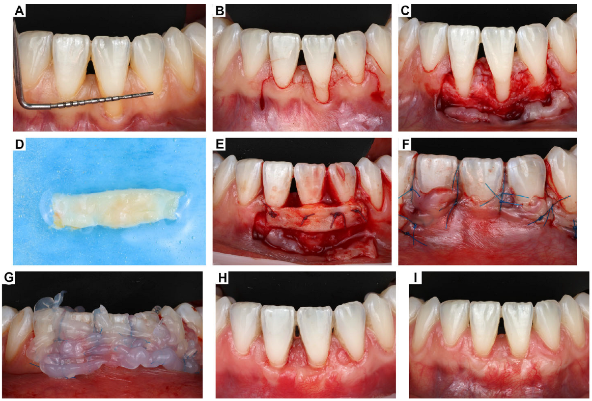
2. Clinical Case Presentation of Complex Periodontal Surgery

Case Report-1

A healthy 35-year-old female patient, a non-smoker, who had previously received orthodontic treatment, reported to the periodontal clinic with significant recession on tooth #31. The recommended treatment was periodontal plastic surgery (i.e., root coverage) using a partial-thickness flap in conjunction with a palatal connective tissue graft. The flap incision involved teeth #41 and #32 to maintain optimal vascularization. Before the therapy, the patient underwent oral hygiene procedures to ensure a sterile environment was preserved. A metallic template was employed to ascertain the size of the graft. The connective tissue graft was harvested from the palate, with no epithelial collar. The palate wound was occluded with Spongostan (Ethicon Biosurgery, Johnson and Johnson, New Brunswick, NJ, USA) and secured with non-resorbable pseudomonofilament Seralon® sutures (SERAG-WIESSNER Co., Naila, Germany). At the main surgical site, the root surface was meticulously cleansed once more. Subsequently, antifungal gel by Straumann was administered and irrigated with saline. Then, the CT graft was positioned over the root surface and sutured to the periosteum with absorbable sutures (5.0). The grafted tissue was then covered with the primary flap, which was then sutured in place with non-resorbable sutures.
As post-surgical protocol, the applications of Emdogain® (Straumann, Basel, Switzerland) and blue®m oxygen therapy (BlueM Europe, Zwolle, The Netherlands) were utilized to promote healing. This was followed by the reinforcement of meticulous oral hygiene using local oxygen therapy (blue®m) toothpaste and mouthwash and topical application of blue®m oral gel twice a day after cleaning. The patient was instructed not to spit or rinse for one hour after gel application.
Subsequent evaluations demonstrated adequate healing after eighteen months, signifying effective root coverage. The tissues exhibited vitality and health, with discernible blood vessels, deemed an outstanding result (Figure 1).

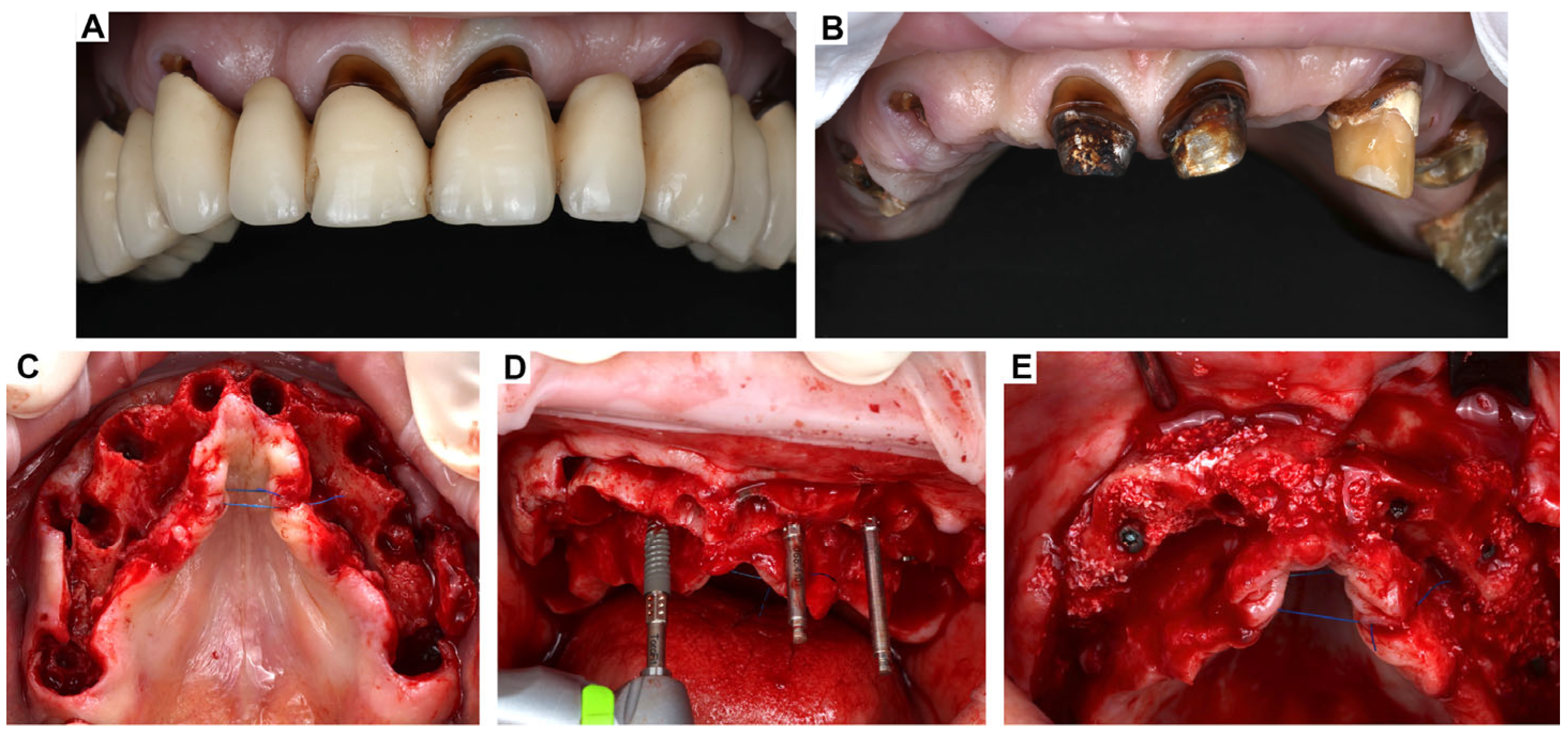
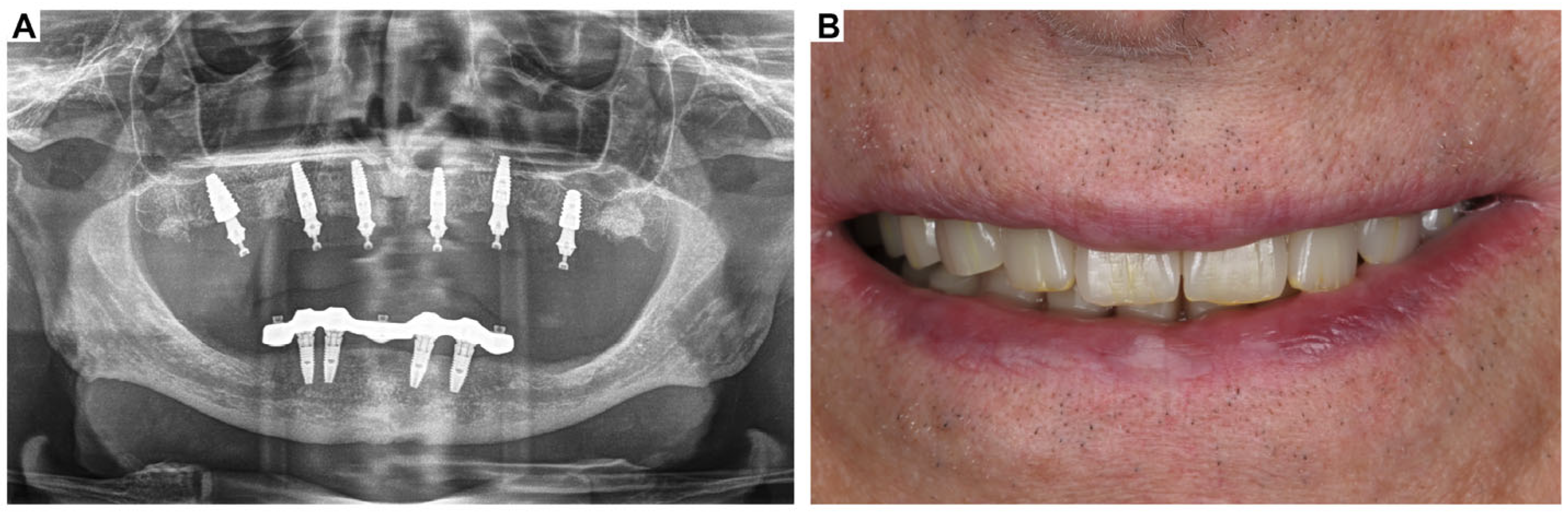
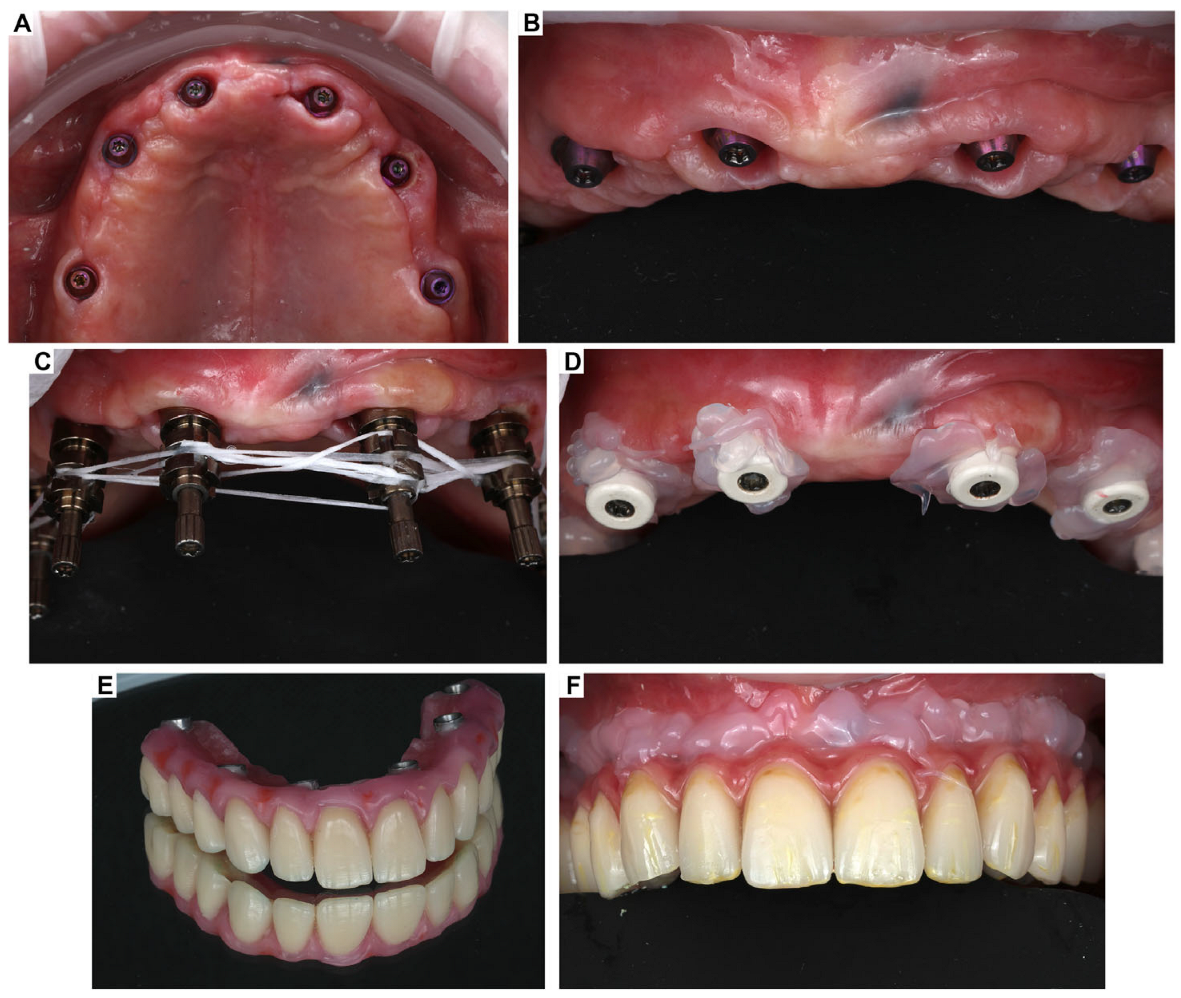
3. Clinical Cases Presentation of Advanced Implant Surgeries

3.1. Case Report-1

The first case was composed of a 67-year-old male patient with no medical conditions, presenting at the oral implantology clinic for replacing an old long-span bridge with dental implants on the upper arch (Figure 2).
A clinical/radiographic examination and a 3D digital image analysis were performed to conduct initial treatment planning. On the day of surgery, the old bridge was removed. Atraumatic extractions of the remaining teeth were carried out to preserve the bone. Following the teeth extractions, six Straumann® iEXCEL BLC implants (i.e., bone-level implants with torque-fit) were immediately placed in the assigned positions. All implants featured an SLActive® surface and were inserted according to the proper manufacturers’ instructions and the surgical insertion protocol provided by Institute Straumann AG, Basel, Switzerland. After placing the implant fixtures, guided bone regeneration (GBR) was conducted using bone xenograft mixed with the patient’s bone, then covered with resorbable membranes (Figure 3).
After suturing, a provisional restoration was delivered on the six implants using SRA (Submerged Root Abutments) by Straumann®, which are secondary components that sit between the implant and the restoration (Figure 4).
Post-surgically, the patient was instructed to apply blue®m gel three times a day for the first month as well as to rinse with blue®m mouthwash twice a day. Clinical follow-ups were scheduled at 1 week, and the sutures were removed after 14 days. The patient was advised to practice oral hygiene twice a day using blue®m toothpaste and mouthwash and apply blue®m oral gel around the peri-implant mucosa using an interdental brush three times a day.
As shown in Figure 5, following clinical and radiological assessment, the implants were determined to be osseointegrated and prepared for prosthetic placement. Digital scanning was conducted for the top and lower implants with Mini Conical Scan-bodies. The final restoration was attained by the placement of a screw-retained prosthesis, consisting of a titanium laser-melted bar with composite material, atop the Mini Conical Abutments (Figure 6). Between each step, blue®m gel was applied around the peri-implant mucosa.
At the following appointments (1, 3, 6, and 12 months), the prostheses were firstly checked for cleanability and occlusion in a digital dynamic manner. Then, specific oral hygiene instructions based on the application of local oxygen therapy (blue®m protocol and formulations) were given to the patient, adapting the interdental brush to apply blue®m oral gel in the gap between the mucosa and the prostheses due to soft tissue healing.

3.2. Case Report-2

The second case pertained to a complex single implant operation for a 35-year-old female patient. The patient was a non-smoker and in satisfactory overall condition. Cone beam computed tomography (CBCT), periapical radiographs, and digital photos were acquired for diagnostic and planning objectives (Figure 7). The treatment approach included atraumatic extraction of tooth #14, followed by immediate implant insertion with little bone augmentation at the buccal site. The site preparation was performed in accordance with the manufacturer’s recommendations. An yttria-stabilized zirconia implant of 4.3 × 11.5 mm (Neodent®, Curitiba, Brazil) was inserted utilizing the transfer piece with a final insertion torque of 60 N.cm, facilitating immediate loading. A periapical X-ray was acquired to verify the accurate location of the implant. The PEEK abutment was then chosen and positioned to support the provisional restoration. Sutures were employed to seal the wound and were removed ten days later. Again, the patient was post-surgically instructed to apply blue®m gel three times a day for the first month as well as to rinse with blue®m mouthwash twice a day.
Following a three-month period, the PEEK abutment was removed, and intraoral scanning was conducted with the suitable scan bodies. A zirconia base abutment was selected, and a lithium disilicate ceramic crown was designed using Dental System software (3Shape, Copenhagen K, Denmark), produced in a CAD/CAM milling machine, and crystallized in a ceramic furnace (M series by Amann Girrbach North America, Charlotte, NC, USA). The crown was cemented extraorally to the zirconia base utilizing RelyX U200 (3M), which was subsequently secured to the implant following appropriate occlusal adjustment (Figure 8).
The patient was advised to practice oral hygiene twice a day using blue®m toothpaste and mouthwash and apply blue®m oral gel around the peri-implant mucosa using an interdental brush three times a day. The patient was monitored clinically and radiographically every three months, with no problems detected or reported during the follow-up period. At the 12-month follow-up, the patient exhibited clinical and radiographic success of the implant, characterized by implant stability, absence of peri-implantitis, complete osseointegration, satisfactory marginal bone-level maintenance (<2 mm at one year), and exceptional soft tissue aesthetics, including harmonious interdental papillae shape, attached gingival appearance, and precise margin form (Figure 9).

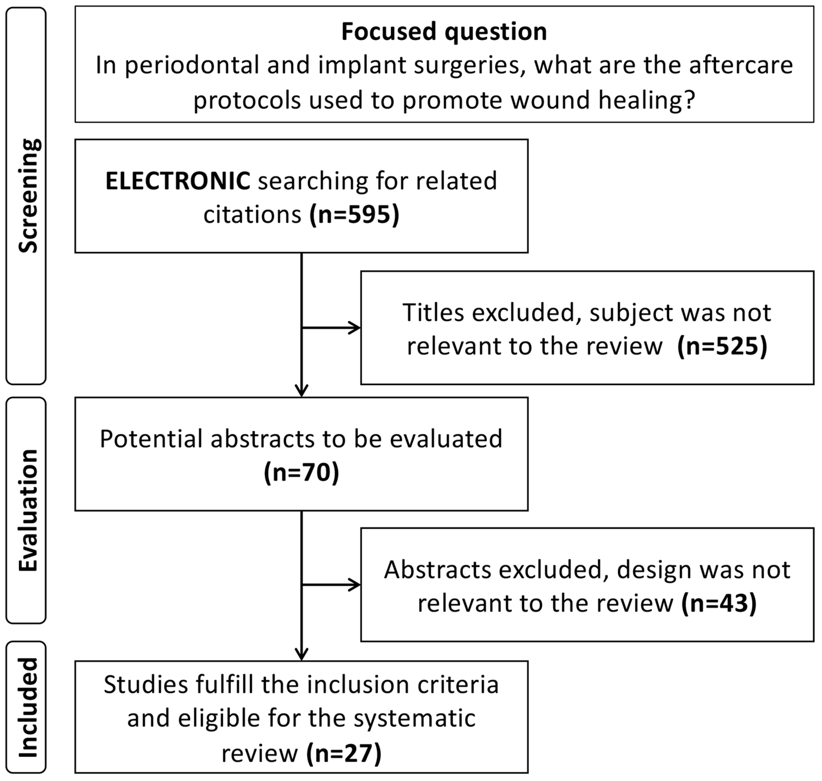
4. Systematic Review of the Literature: Methodology and Results

The Preferred Reporting Items for Systematic Reviews and Meta-Analyses (PRISMA) guidelines/checklist were followed during the present systematic review. An electronic search was conducted by two reviewers (C.S. and H.A.) on the following databases: PubMed, Embase, and Cochrane Central Register of Controlled Trials, until June 2025. The focused question was, “In periodontal and implant surgeries, what are the aftercare protocols used to maintain optimal wound healing?” The search strategy/terms were used as follows: (wound healing[mh] OR wound*[ti]) AND (dental implants/surgery[mh] OR periodontal diseases/surgery[mh] OR gingiv*[ti] OR dental implant*[ti] OR periodontal*[ti] OR soft tissue[ti]) AND (monitoring, physiologic[mh] OR postoperative care[mh] OR applicat*[ti] OR assess*[ti] OR examinat*[ti] OR maint*[ti] OR monitor*[ti] OR observat*[ti] OR predict*[ti] OR progno*[ti]). Only human clinical studies that used or proposed a specific protocol for aftercare wound healing of periodontal or dental implant surgeries were included. We excluded studies with designs that were not relevant to the review. Review articles, workshop consensus, and articles not written in English were also excluded. Figure 10 displays the flowchart that explains the literature review process.
As a result, the electronic search returned 595 references. Titles that were not relevant to the review were removed, resulting in 70 unique titles/abstracts for screening. Based on the inclusion and exclusion criteria, 27 articles were finally subjected to evaluation and data extraction. The complete list of the included studies and their characteristics is summarized in Table 1.

5. Discussion

This article details clinical examples of periodontal and dental implant surgeries that were treated post-surgically with local application of oxygen-based therapy (blue®m) gel. The importance of aftercare was further emphasized during the wound healing phase by instructing patients to maintain thorough dental hygiene using toothpaste and mouthwash of a similar oxygen-release formulation (blue®m). A systematic literature review was conducted to compare outcomes and elucidate the necessity of surgical wound aftercare using different local adjunct therapies. However, the results showed variations in the 27 included studies by conducting different aftercare modalities, such as metronidazole gel, chlorhexidine, hyaluronic acid, platelet-rich fibrin, Emdogain, and photobiomodulation therapy, in maintaining optimal post-surgical wound healing following periodontal and implant surgeries.
There was no predominant modality of post-surgical therapy in the included studies. Some studies reported using either chlorhexidine (CHX), Coe-Pak dressing, or metronidazole gel for 1 to 2 weeks after surgery [27]. The procedure of utilizing leukocyte-platelet-rich fibrin (L-PRF) as post-surgical aftercare protocol was also reported in some clinical studies, although it did not produce significant advantages for the patients with advanced periodontal surgeries [15,18,22,30]. Among the studies reporting the use of hyaluronic acid (HA) and Emdogain in the present review [13,14,20,31], none of them showed superior wound healing outcomes. The further claim of the use of soft-laser treatment post-surgically does not significantly enhance clinical outcomes in patients who undergo periodontal or implant surgeries [16,17,33,34].
Another important aspect of immediate implant placement after tooth extraction is the role of immediate prosthetic rehabilitation. In particular, the provisional prosthesis not only contributes to esthetics and function but also plays a fundamental role in soft tissue stabilization and wound closure. As highlighted by Menchini-Fabris et al. [38], the use of a customized healing abutment and immediate prosthetic sealing contributes significantly to the early closure of the wound, especially in periodontally compromised sockets, thereby directly influencing healing outcomes and peri-implant tissue stability [38]. This emphasizes that, while adjunctive therapies like blue®m may support healing, they should be considered in a broader clinical context where surgical technique and prosthetic design have a primary role in immediate tissue management.
Many clinical reports have indicated that inadequate postoperative care may be associated with early wound complications [1,3,4]. It is believed that the optimal healing process is contingent upon the resolution of the inflammation phase and then the transition to the regeneration phase. Swelling, suppuration, and altered tissue color are evidently undesirable indicators that must be avoided [1,4]. The degree of healing can also be assessed by the patient’s discomfort and pain perception. The intensity and duration of pain can increase due to prolonged inflammation or underlying infection. In practice, the timing and frequency of postoperative care are primarily determined by arbitrary factors.
Aftercare protocols are highly reliant on patient compliance with hygiene instructions, resulting in improved soft tissue healing and reduced inflammation [39]. Topical antiseptics, particularly chlorhexidine, are commonly used, although there are concerns about their side effects [40,41]. Povidone–iodine and hydrogen peroxide are used as alternatives with varying antimicrobial effectiveness [41]. Microbial biofilms can trigger inflammation in the wound tissues associated with oral surgeries. Therefore, preventing wound disease is essential for promoting optimal healing after periodontal/implant surgeries. This care also involves the patient’s daily commitment to performing self-care procedures for controlling biofilm [41]. For clinicians, achieving optimal long-term outcomes in periodontology and implant dentistry requires not only appropriate surgical and prosthetic techniques but also effective aftercare protocols.
The local oxygen-release formulation with antimicrobial properties may offer protection against early bacterial colonization, potentially reducing the risk of wound complications after periodontal and implant surgeries [8,42]. For instance, blue®m formulation is composed primarily of hydrocarbon-oxo-borate complex (HCOBc) and lactoferrin and is capable of slow and sustained oxygen release when applied topically [43]. This helps in wound bio-modulation, reduced inflammation, enhanced healing, and neovascularization [44,45]. Nevertheless, more evidence demonstrating the clinical benefits of such an oxygen-release formulation on oral wound healing remains in significant demand.
According to our review, different conventionally used aftercare modalities, such as metronidazole gel, chlorhexidine, hyaluronic acid, platelet-rich fibrin, Emdogain, and photobiomodulation therapy, were used to maintain optimal post-surgical wound healing following periodontal and implant surgeries [2,13,18,23,46]. With respect to the findings reported, the included clinical studies significantly contributed to inconclusive results and revealed a great heterogeneity due to variability in their data, particularly for the assessed and reported outcomes, as well as discrepancies in follow-up durations. For instance, Hagenaars et al. [13] examined the effect of Emdogain (enamel matrix derivative (EMD)) on the healing of soft-tissue wounds following periodontal surgery. The EMD group exhibited a higher gingival swelling score and revealed no significant difference from control sites in terms of early wound healing following periodontal flap surgeries. Moreover, Mutallibli and Sağlam [18] studied the effects of platelet-rich fibrin (PRF) on the healing of palatal wounds after free gingival graft harvesting in 36 patients. Complete epithelialization was higher in the control group on day 7 than in the test groups (p < 0.05).
Researchers evaluated the effect of periodontal wound healing associated with the local application of metronidazole gel [23]. Twelve patients participated in the study. During the early phase of healing, post-surgically, no statistically significant differences between the test and control sites were noted.
Heitz et al. [2] conducted a randomized controlled trials (RCT) to evaluate early wound healing following a specific post-surgical care protocol: rinsing twice daily for 1 min with 0.1% of chlorhexidine (CHX) for 4 weeks. As a result, the use of post-surgical protocol, including the local application of CHX in addition to daily rinsing with CHX, may be recommended. While CHX is regarded as the gold standard for antiseptic treatment in the oral cavity, numerous in vitro studies have indicated adverse effects on oral tissue repair. In a recent study by Pilloni et al. [40], gingival biopsies were collected from three patients 24 h post-surgery following the recommendation of a 0.12% CHX mouth rinse. The alterations in collagen synthesis, cellular proliferation, and cell apoptosis were analyzed. Quantitative real-time PCR analysis was conducted to assess the expression of fibrotic scar markers (COL1A1, αSMA) in wound healing. The application of CHX mouth rinse following surgery results in fibrotic transformation, producing a “scar wound healing response” pattern. This underscores the necessity for novel post-surgical care regimens predicated on a purposeful absence of CHX.
Finally, photobiomodulation therapy (PBMT) has attracted the attention of many researchers in recent years [47]. For instance, one clinical study showed that application of HA gel and PBMT improved wound healing following surgical gingivectomy as well as decreased postoperative pain perception by patients [46]. On the contrary, Damante et al. [48] showed that PBMT using a diode laser did not accelerate the healing of oral mucosa after gingival surgery. In spite of the conflicting results from the studies mentioned above, further research is necessary to optimize the truth-versus-fiction effectiveness of photobiomodulation lasers in clinical settings.
Given the limitations of this study, clear clinical recommendations for an effective post-surgical aftercare protocol to promote wound healing in periodontology and dental implants could not be established. Moreover, the nature of clinical case reporting appears to be restricted due to the need for definitive objective measurements and outcomes. Therefore, well-structured prospective clinical studies should evaluate the present proposed aftercare protocol using topical oxygen-release formulation (e.g., blue®m) across different surgical interventions.

Clinical Implications

Oral surgical interventions, particularly those related to periodontal and peri-implant soft and hard tissue regeneration, require meticulous tissue management during treatment and ongoing assessment of wound healing. The prompt identification of adverse events necessitates enhanced monitoring and intervention as required. An assessment of the primary indicators of inflammation, wound margin condition, and patient perception is essential. Optimal healing for regenerative purposes is defined by complete wound closure, a smooth progression through the healing phases, and a sufficient increase in tissue volume thereafter. Therefore, a clear and standardized post-surgical care protocol is crucial for effective clinical practice among dental professionals, as it enhances wound healing across different surgical procedures and emphasizes regenerative and aesthetic factors.

6. Conclusions

Aftercare protocols have a substantial impact on the outcomes of periodontal and peri-implant wound healing. The selection of protocol must be customized according to the specific risk profiles of patients, the complexity of the surgical procedure, and the likelihood of adherence. Additional high-quality randomized controlled trials (RCT) are necessary to standardize evidence-based protocols. Nonetheless, topical oxygen-release formulations (e.g., blue®m) may promote oral wound healing post-surgically. However, more clinical evidence is needed.

Author Contributions

Conceptualization, C.S. (Cristian Scognamiglio) and H.A.; methodology, C.S. (Chalini Sunder); software, A.P.; validation, T.M.D., C.S. (Cristian Scognamiglio) and A.P.; formal analysis, H.A.; investigation, C.S. (Cristian Scognamiglio); resources, A.P.; data curation, C.S. (Chalini Sunder); writing—original draft preparation, H.A.; writing—review and editing, T.M.D.; visualization, C.S. (Cristian Scognamiglio); supervision, T.M.D. All authors have read and agreed to the published version of the manuscript.

Funding

This research received no external funding.

Institutional Review Board Statement

Since the case report describes one or two patients and is purely descriptive, according to the institution regulation, it is typically considered not human subject research and does not require IRB approval.

Informed Consent Statement

Written informed consent was obtained from the participants whose clinical data were shared, without any personal identifying information.

Data Availability Statement

All data are contained within the article.

Conflicts of Interest

The authors declare no conflicts of interest.

References

  1. Mani, A.; Mani, S.; Sachdeva, S.; Sodhi, J.K.; Vora, H.R.; Gholap, S. Post-surgical care in surgical periodontics. Int. J. Periodontol. Implantol. 2021, 6, 74–78. [Google Scholar]
  2. Sanz, M.; Newman, M.G.; Anderson, L.; Matoska, W.; Otomo—Corgel, J.; Saltini, C. Clinical Enhancement of Post-Periodontal Surgical Therapy by a 0.12% Chlorhexidine Gluconate Mouthrinse. J. Periodontol. 1989, 60, 570–576. [Google Scholar] [CrossRef]
  3. Pippi, R. Post-surgical clinical monitoring of soft tissue wound healing in periodontal and implant surgery. Int. J. Med. Sci. 2017, 14, 721. [Google Scholar] [CrossRef] [PubMed]
  4. Powell, C.A.; Mealey, B.L.; Deas, D.E.; McDonnell, H.T.; Moritz, A.J. Post-surgical infections: Prevalence associated with various periodontal surgical procedures. J. Periodontol. 2005, 76, 329–333. [Google Scholar] [CrossRef] [PubMed]
  5. Cho, Y.-D.; Kim, K.-H.; Lee, Y.-M.; Ku, Y.; Seol, Y.-J. Periodontal wound healing and tissue regeneration: A narrative review. Pharmaceuticals 2021, 14, 456. [Google Scholar] [CrossRef] [PubMed]
  6. Brand, H.S.; Ligtenberg, A.; Veerman, E. Saliva and wound healing. Monogr. Oral Sci. 2014, 24, 52–60. [Google Scholar]
  7. Byrne, M.; Aly, A. The surgical suture. Aesthetic Surg. J. 2019, 39, S67–S72. [Google Scholar] [CrossRef]
  8. Leventis, M.; Deliberador, T.; Alshehri, F.; Alghamdi, H. Topical oxygen therapy as a novel strategy to promote wound healing and control the bacteria in implantology, oral surgery and periodontology: A review. Saudi Dent. J. 2024, 36, 841–854. [Google Scholar] [CrossRef]
  9. Shaheen, M.Y.; Abas, I.; Basudan, A.M.; Alghamdi, H.S. Local Oxygen-Based Therapy (blue® m) for Treatment of Peri-Implant Disease: Clinical Case Presentation and Review of Literature about Conventional Local Adjunct Therapies. Medicina 2024, 60, 447. [Google Scholar] [CrossRef]
  10. Benlidayi, I.C.; Gupta, L. CAse-BAsed REview sTandards (CABARET): Considerations for Authors, Reviewers, and Editors. J. Korean Med. Sci. 2024, 39, 225. [Google Scholar] [CrossRef]
  11. Maffei, S.H.; Fernandes, G.V.O.; Fernandes, J.C.H.; Orth, C.; Joly, J.C. Clinical and histomorphometric soft tissue assessment comparing free gingival graft and a collagen matrix as alveolar-sealer materials: A randomized controlled pilot clinical trial. Quintessence Int. 2023, 54, 756–769. [Google Scholar] [CrossRef]
  12. Palled, V.; Rao, J.; Singh, R.D.; Tripathi, S.; Singh, K.; Radav, R.; Verma, U.; Chand, P. Assessment of the Healing of Dental Implant Surgical Site Following Low-Level Laser Therapy Using Bioclinical Parameters: An Exploratory Study. J. Oral Implantol. 2021, 47, 230–235. [Google Scholar] [CrossRef] [PubMed]
  13. Hagenaars, S.; Louwerse, P.H.; Timmerman, M.F.; Van der Velden, U.; Van der Weijden, G.A. Soft-tissue wound healing following periodontal surgery and Emdogain application. J. Clin. Periodontol. 2004, 31, 850–856. [Google Scholar] [CrossRef] [PubMed]
  14. Vela, O.C.; Boariu, M.; Rusu, D.; Iorio-Siciliano, V.; Ramaglia, L.; Boia, S.; Radulescu, V.; Ilyes, I.; Stratul, S.I. Healing of Periodontal Suprabony Defects following Treatment with Open Flap Debridement with or without Hyaluronic Acid (HA) Application. Medicina 2024, 60, 829. [Google Scholar] [CrossRef] [PubMed]
  15. Bozkurt, E.; Uslu, M. Evaluation of the effects of platelet-rich fibrin, concentrated growth factors, and autologous fibrin glue application on wound healing following gingivectomy and gingivoplasty operations: A randomized controlled clinical trial. Quintessence Int. 2022, 53, 328–341. [Google Scholar] [CrossRef]
  16. Masse, J.F.; Landry, R.G.; Rochette, C.; Dufour, L.; Morency, R.; D’Aoust, P. Effectiveness of soft laser treatment in periodontal surgery. Int. Dent. J. 1993, 43, 121–127. [Google Scholar]
  17. Watanabe, H.; Ishikawa, I.; Suzuki, M.; Hasegawa, K. Clinical assessments of the erbium: YAG laser for soft tissue surgery and scaling. J. Clin. Laser Med. Surg. 1996, 14, 67–75. [Google Scholar] [CrossRef]
  18. Mutallibli, A.; Sağlam, M. Comparison of the effect of A-PRF and L-PRF application to palatal donor sites on quality of life and wound healing after free gingival graft surgery. Quintessence Int. 2024, 55, 472–481. [Google Scholar] [CrossRef]
  19. Heitz, F.; Heitz-Mayfield, L.J.; Lang, N.P. Effects of post-surgical cleansing protocols on early plaque control in periodontal and/or periimplant wound healing. J. Clin. Periodontol. 2004, 31, 1012–1018. [Google Scholar] [CrossRef]
  20. de Araújo Nobre, M.; Cintra, N.; Maló, P. Peri-implant maintenance of immediate function implants: A pilot study comparing hyaluronic acid and chlorhexidine. Int. J. Dent. Hyg. 2007, 5, 87–94. [Google Scholar] [CrossRef]
  21. Kumar, M.B.V.; Narayanan, V.; Jalaluddin, M.; Almalki, S.A.; Dey, S.M.; Sathe, S. Assessment of Clinical Efficacy of Different Periodontal Dressing Materials on Wound Healing: A Comparative Study. J. Contemp. Dent. Pract. 2019, 20, 896–900. [Google Scholar]
  22. Gatti, F.; Iorio-Siciliano, V.; Scaramuzza, E.; Tallarico, M.; Vaia, E.; Ramaglia, L.; Chiapasco, M. Patient-reported outcome measures of leucocyte- and platelet-rich fibrin (L-PRF) or hemostatic agent application at palatal donor sites after free gingival graft harvesting: A randomized controlled clinical trial. Quintessence Int. 2023, 54, 408–417. [Google Scholar] [CrossRef]
  23. Sander, L.; Frandsen, E.V.; Arnbjerg, D.; Warrer, K.; Karring, T. Effect of local metronidazole application on periodontal healing following guided tissue regeneration. Clinical findings. J. Periodontol. 1994, 65, 914–920. [Google Scholar] [CrossRef]
  24. Uslu, M.; Akgül, S. Evaluation of the effects of photobiomodulation therapy and ozone applications after gingivectomy and gingivoplasty on postoperative pain and patients’ oral health-related quality of life. Lasers Med. Sci. 2020, 35, 1637–1647. [Google Scholar] [CrossRef]
  25. Trombelli, L.; Scabbia, A.; Scapoli, C.; Calura, G. Clinical effect of tetracycline demineralization and fibrin-fibronectin sealing system application on healing response following flap debridement surgery. J. Periodontol. 1996, 67, 688–693. [Google Scholar] [CrossRef]
  26. Yaghobee, S.; Rouzmeh, N.; Taheri, M.; Aslroosta, H.; Mahmoodi, S.; Mohammadnejad Hardoroodi, M.; Soleimanzadeh Azar, P.; Khorsand, A. Evaluation of topical erythropoietin application on the healing outcome of gingival graft recipient site; A randomized controlled clinical trial. BMC Oral Health 2021, 21, 578. [Google Scholar] [CrossRef]
  27. Langebaek, J.; Bay, L. The effect of chlorhexidine mouthrinse on healing after gingivectomy. Eur. J. Oral Sci. 1976, 84, 224–228. [Google Scholar] [CrossRef]
  28. Frandsen, E.V.; Sander, L.; Arnbjerg, D.; Theilade, E. Effect of local metronidazole application on periodontal healing following guided tissue regeneration. Microbiological findings. J. Periodontol. 1994, 65, 921–928. [Google Scholar] [CrossRef]
  29. Palombo, D.; Dobos, A.; Duran, M.L.; Esporrin, J.S.; Sanz, M. Harvest of epithelialized gingival grafts without application of hemostatic sutures: A randomized clinical trial using laser speckle contrast imaging. J. Periodontol. 2024, 95, 1160–1170. [Google Scholar] [CrossRef]
  30. Ibrahim, S.S.A.; Mandil, I.A.; Ezzatt, O.M. Injectable platelet rich fibrin effect on laser depigmented gingiva: A clinical randomized controlled split mouth trial with histological assessment. J. Appl. Oral Sci. 2024, 32, e20230307. [Google Scholar] [CrossRef]
  31. Çankaya, Z.T.; Gürbüz, S.; Bakirarar, B.; Kurtiş, B. Evaluation of the Effect of Hyaluronic Acid Application on the Vascularization of Free Gingival Graft for Both Donor and Recipient Sites with Laser Doppler Flowmetry: A Randomized, Examiner-Blinded, Controlled Clinical Trial. Int. J. Periodontics Restor. Dent. 2020, 40, 233–243. [Google Scholar] [CrossRef] [PubMed]
  32. Peñarrocha-Diago, M.; Menéndez-Nieto, I.; Cervera-Ballester, J.; Maestre-Ferrín, L.; Blaya-Tárraga, J.A.; Peñarrocha-Oltra, D. Influence of Hemostatic Agents in the Prognosis of Periapical Surgery: A Randomized Study of Epinephrine versus Aluminum Chloride. J. Endod. 2018, 44, 1205–1209. [Google Scholar] [CrossRef] [PubMed]
  33. Santamaria, M.P.; Fernandes-Dias, S.B.; Araújo, C.F.; Lucas da Silva Neves, F.; Mathias, I.F.; Rebelato Bechara Andere, N.M.; Neves Jardini, M.A. 2-Year Assessment of Tissue Biostimulation With Low-Level Laser on the Outcomes of Connective Tissue Graft in the Treatment of Single Gingival Recession: A Randomized Clinical Trial. J. Periodontol. 2017, 88, 320–328. [Google Scholar] [CrossRef] [PubMed]
  34. Monea, A.; Beresescu, G.; Boeriu, S.; Tibor, M.; Popsor, S.; Antonescu, D.M. Bone healing after low-level laser application in extraction sockets grafted with allograft material and covered with a resorbable collagen dressing: A pilot histological evaluation. BMC Oral Health 2015, 15, 134. [Google Scholar] [CrossRef] [PubMed]
  35. Patel, P.V.; Kumar, S.; Vidya, G.D.; Patel, A.; Holmes, J.C.; Kumar, V. Cytological assessment of healing palatal donor site wounds and grafted gingival wounds after application of ozonated oil: An eighteen-month randomized controlled clinical trial. Acta Cytol. 2012, 56, 277–284. [Google Scholar] [CrossRef]
  36. Bokor, M. The effect of hexetidine spray on dental plaque following periodontal surgery. J. Clin. Periodontol. 1996, 23, 1080–1083. [Google Scholar] [CrossRef]
  37. Trombelli, L.; Scabbia, A.; Wikesjö, U.M.; Calura, G. Fibrin glue application in conjunction with tetracycline root conditioning and coronally positioned flap procedure in the treatment of human gingival recession defects. J. Clin. Periodontol. 1996, 23, 861–867. [Google Scholar] [CrossRef]
  38. Menchini-Fabris, G.-B.; Cosola, S.; Toti, P.; Hwan Hwang, M.; Crespi, R.; Covani, U. Immediate Implant and Customized Healing Abutment for a Periodontally Compromised Socket: 1-Year Follow-Up Retrospective Evaluation. J. Clin. Med. 2023, 12, 2783. [Google Scholar] [CrossRef]
  39. Blinder, D.; Rotenberg, L.; Peleg, M.; Taicher, S. Patient compliance to instructions after oral surgical procedures. Int. J. Oral Maxillofac. Surg. 2001, 30, 216–219. [Google Scholar] [CrossRef]
  40. Pilloni, A.; Ceccarelli, S.; Bosco, D.; Gerini, G.; Marchese, C.; Marini, L.; Rojas, M.A. Effect of chlorhexidine digluconate in early wound healing of human gingival tissues. A histological, immunohistochemical and biomolecular analysis. Antibiotics 2021, 10, 1192. [Google Scholar] [CrossRef]
  41. Teixeira, D.D.S.; Figueiredo, M.A.Z.; Cherubini, K.; de Oliveira, S.D.; Salum, F.G. The topical effect of chlorhexidine and povidone-iodine in the repair of oral wounds. A review. Stomatologija 2019, 21, 35–41. [Google Scholar]
  42. Alghamdi, H.; Leventis, M.; Deliberador, T. Management of Infected Tissues Around Dental Implants: A Short Narrative Review. Braz. Dent. J. 2024, 35, e246160. [Google Scholar] [CrossRef] [PubMed]
  43. Shibli, J.A.; Rocha, T.F.; Coelho, F.; de Oliveira Capote, T.S.; Saska, S.; Melo, M.A.; Pingueiro, J.M.S.; de Faveri, M.; Bueno-Silva, B. Metabolic activity of hydro-carbon-oxo-borate on a multispecies subgingival periodontal biofilm: A short communication. Clin. Oral Investig. 2021, 25, 5945–5953. [Google Scholar] [CrossRef] [PubMed]
  44. Grover, M.; Gupta, A.; Dubey, T.; Dua, P.; Arora, G.K. New age oxygen therapy for enhanced recovery: BLUE® M oral gel in post-extraction socket healing. J. Dent. Spec. 2025, 13, 131–137. [Google Scholar] [CrossRef]
  45. Basudan, A.M.; Abas, I.; Shaheen, M.Y.; Alghamdi, H.S. Effectiveness of Topical Oxygen Therapy in Gingivitis and Periodontitis: Clinical Case Reports and Review of the Literature. J. Clin. Med. 2024, 13, 1451. [Google Scholar] [CrossRef]
  46. Yakout, B.K.; Kamel, F.R.; Khadr, M.A.E.-A.A.; Heikal, L.A.H.; El-Kimary, G.I. Efficacy of hyaluronic acid gel and photobiomodulation therapy on wound healing after surgical gingivectomy: A randomized controlled clinical trial. BMC Oral Health 2023, 23, 805. [Google Scholar] [CrossRef]
  47. Sadeghian, A.; Rohani, B.; Salehi-Marzijarani, M.; Fekrazad, R. Radiographical impact of photobiomodulation therapy on bone regeneration in clinical studies: A systematic review. Lasers Med. Sci. 2025, 40, 23. [Google Scholar] [CrossRef]
  48. Damante, C.A.; Greghi, S.L.; Sant’Ana, A.C.; Passanezi, E.; Taga, R. Histomorphometric study of the healing of human oral mucosa after gingivoplasty and low-level laser therapy. Lasers Surg. Med. 2004, 35, 377–384. [Google Scholar] [CrossRef]
Figure 1. Clinical case presentation of complex periodontal surgery, (A) clinical examination showing significant recession on tooth #31; (B,C) the partial-thickness flap involved teeth #41 and #32 to maintain optimal vascularization; (D) the connective tissue graft was harvested from the palate, with no epithelial collar; (E) then, the CT graft was positioned over the root surface and sutured to the periosteum; (F) the flap was advanced over the grafted tissue and sutured using non-resorbable sutures; (G) as post-surgical protocol, the applications of Emdogain® and blue®m oxygen gel were utilized to promote healing; (H) follow-up after 3 months signifying effective root coverage; and (I) at 18 months, the tissues exhibited vitality and health, with discernible blood vessels, deemed an outstanding result.
Oral 05 00053 g001
Figure 2. Clinical case composed of a 67-year-old male patient for replacing an old long-span bridge with dental implants on the upper arch. (A) Clinical and (B) radiographic examination as well as a 3D digital image analysis for treatment planning.
Oral 05 00053 g002
Figure 3. On the day of surgery, (A,B) the old bridge was removed; (C) atraumatic extractions of the remaining teeth were carried out to preserve the bone; (D) following the teeth extractions, six Straumann® iEXCEL BLC implants were immediately placed following proper manufacturers’ instructions and the surgical insertion protocol (Institute Straumann AG); (E) guided bone regeneration (GBR) using bone-xenograft mixed with the patient’s bone, then covered with resorbable membranes.
Oral 05 00053 g003
Figure 4. After suturing, (A,B) provisional restoration was delivered on six implants using SRA (Submerged Root Abutments) by Straumann®. Post-surgically, the patient was instructed to apply blue®m gel three times a day for the first month as well as to rinse with blue®m mouthwash twice a day.
Oral 05 00053 g004
Figure 5. After 3 months, (A,B) clinical assessment and preparation for prosthesis fabrication. Two types of impressions were then performed: (C) impression transfers were screwed onto the six implants at 5 N.cm; then, a conventional impression was taken using a customized open tray and a polyether material; (D) digital impressions were produced by digital scanning using Mini Conical Scan-bodies. The final restoration (E) consisted of a titanium laser-melted bar with composite material atop the mini conical abutments. Between each step, (F) blue®m gel was applied around the peri-implant mucosa.
Oral 05 00053 g005
Figure 6. OPG X-ray and clinical images (A,B) showing the final restoration in place and attained by the placement of a screw-retained prosthesis.
Oral 05 00053 g006
Figure 7. Cone beam computed tomography (CBCT) was acquired (A) and illustrated (B) treatment planning of a Neodent® zirconia implant (4.3 × 11.5 mm) replacing tooth #14.
Oral 05 00053 g007
Figure 8. The case treatment started with (A,B) atraumatic extraction of tooth #14, followed by (C) immediate insertion of a Neodent® zirconia implant (4.3 × 11.5 mm); (D) a PEEK abutment was used to support (E) provisional restoration; sutures were employed and removed ten days later; (F) blue®m gel was applied around the peri-implant mucosa. Following 3 months, (G) peri-implant mucosa showed excellent healing; then, (H) a lithium disilicate ceramic crown was produced and cemented after appropriate occlusal adjustment.
Oral 05 00053 g008
Figure 9. At 12-month follow-up, the patient exhibited (A) clinical and (B) radiographic success of implant stability, absence of peri-implantitis, marginal bone-level maintenance (<2 mm at one year), and exceptional soft tissue aesthetics and precise margin form.
Oral 05 00053 g009
Figure 10. Flowchart explaining the literature systematic review process.
Oral 05 00053 g010
Table 1. Summary of articles included in the systematic review.
StudyInterventionPost-Surgical CareFollow-UpAssessmentOutcome
Maffei et al. [11]Socket preservationAlveolar sealing: free gingival graft (FGG) vs. porcine collagen membrane (MS)3, 7, 15, 30, 60, 90, and 120 daysVisual analog scaleThe result was superior and more significant for the MS group, with faster wound healing and lower discomfort.
Palled et al. [12]Implant surgical siteLow-level laser therapy2 weeks, 6 weeks, and 3 monthsImplant stability, probing index, bleeding index, and osteoprotegerin levelThe healing of peri-implant soft tissues may be enhanced with the use of low-level laser therapy.
Hagenaars et al. [13]Periodontal flap surgeryEmdogain1, 4, and 8 weeksGingival indexThe early wound-healing using Emdogain exhibited higher gingival swelling.
Vela et al. [14]Suprabony periodontal defectsHyaluronic acid (HA)baseline and 12 monthsProbing pocket depth (PPD), gingival recession (GR), full-mouth plaque score (FMPS), and full-mouth bleeding score (FMBS)Wound healing could benefit from the additional application of HA.
Bozkurt et al. [15]Gingivectomy and gingivoplastyPlatelet-rich fibrin (PRF), concentrated growth factors (CGF), and autologous fibrin glue (AFG)days 0, 7, 14, and 28Wound healing was evaluated with H2O2 test, visual analog scale for pain, and Landry, Turnbull, and Howley (LTH) indexPRF, CGF, and AFG application were found to have positive effects on wound healing.
Masse et al. [16]Mucogingival proceduresSoft-laser treatmentdays 7 and 14Modified McGill pain scale, inflammatory index, and Landry, Turnbull, Howley (LTH) IndexSoft laser (As-Ga and He-Ne) not a useful aftercare treatment for wound healing.
Watanabe et al. [17]Soft tissue surgeryErbium:YAG laserweeks 2, 4, and 6Pain, redness, and swelling of the gingiva and the subjective patient comfort parametersEr:YAG laser is useful for soft tissue surgery and scaling.
Mutallibli and Sağlam, [18]Palatal donor woundsLeukocyte platelet-rich fibrin (L-PRF) and advanced platelet-rich fibrin (A-PRF)days 7 and 14H2O2 test, visual analog scale, and Oral Health Impact Profile-14 (OHIP-14) scoreBoth PRF procedures have similar effects on palatal wound healing and quality of life.
Heitz et al. [19]Periodontal flap surgery0.1% of chlorhexidine (CHX)1, 2, and 4 weeksGingival crevicular fluid (GCF) flow rate, probing depth, probing attachment level, presence of bleeding on probing, and full-mouth plaque scoreThe use of post-surgical cleansing protocols may be recommended.
de Araújo Nobre et al. [20]Dental implant surgeryHyaluronic acid (HA) vs. chlorhexidine (CHX) gels10th day, 2 months, 4 months, and 6 months post-surgeryPlaque and bleeding indexThe findings point out the importance of a maintenance protocol in immediate function implants.
Kumar et al. [21]Periodontal flap surgeryCollagen dressing, light-cure dressing, and non-eugenol-based dressingdays 7 and 14Plaque index, vertical probing depth, pain, gingival index, patient satisfaction, and visual analog scale (VAS)The periodontal wound covered with a collagen dressing provide significant symptomatic relief and better healing.
Gatti et al. [22]Palatal donor woundsLeucocyte- and platelet-rich fibrin (L-PRF) membranes or a hemostatic agent with oxidized and regenerated cellulose1 weekPostoperative pain, postoperative discomfort, inability to chew, postoperative stress, surgical chair time, and thickness of the palatal fibro-mucosaThe application of L-PRF membrane at palatal donor sites after FGG harvesting did not produce significant advantages for the patients.
Sander et al. [23] Guided tissue regeneration (GTR)Metronidazole gel4 and 6 weeksPlaque, bleeding on probing, and inflammation of marginal gingivaApplication of metronidazole gel has a beneficial effect on healing of guided tissue regeneration.
Uslu and Akgül [24]Gingivectomy and gingivoplastyPhotobiomodulation therapy (PBM) and ozone applicationsdays 3, 7, 14, and 28Visual analogue scale (VAS) and Oral Health Impact Profile (OHIP-14) scoreThe PBM and ozone applications after gingivectomy and gingivoplasty reduce the pain levels.
Trombelli et al. [25]Flap debridement surgeryTetracycline (TTC) conditioning and fibrin-fibronectin sealing system (FFSS)0–6 monthsGingival index, plaque control record, clinical attachment level, probing depth, recession, and bleeding on probingThese results suggest there is no additional benefit with TTC demineralization and topical FFSS application on wound healing.
Yaghobee et al. [26]Free gingival graft (FGG)Topical erythropoietindays 7, 14, 21, 28, 60, and 90Blinded observers to compare the healing and inflammation of the areasTopical application of erythropoietin can accelerate the healing of gingival grafts and reduce the inflammation during healing period.
Langebaek and Bay [27]Gingivectomy0.2% chlorhexidine (CHX) and Coe-Pak dressingdays 7, 14, and 21Plaque index and gingival index CHX and Coe-Pak dressing maintained low plaque scores and promoted healing.
Frandsen et al. [28]Guided tissue regeneration (GTR)Local metronidazole gel2 weeksMicrobial colonization of the wound areaThe influence of metronidazole gel on wound healing appears not significant.
Palombo et al. [29]Palatal epithelialized gingival grafts (EGG)Hemostatic sponges7, 14, and 30 daysLaser speckle contrast imaging (LSCI), postoperative bleeding, pain, discomfort, and analgesic consumptionHemostatic sutures provide no relevant differences in microvascular, clinical, and patient-related results.
Ibrahim et al. [30]Diode laser gingival depigmentation (DLGD)Injectable platelet-rich fibrin (i-PRF)1 week and 1 and 3 monthsWound Healing Score (WHS), patient satisfaction, and Pigmentation Index (DOPI)i-PRF demonstrated better clinical and histological healing potential and less patient discomfort.
Çankaya et al. [31]Free gingival graft (FGG)Topical hyaluronic acid (HA)4, 7, 10, 14, and 30 daysLaser doppler flowmetry (LDF)Application of HA on the recipient bed under the FGG at the first week of healing allows the formation of a well-vascularized layer.
Peñarrocha-Diago et al. [32]Periapical surgeryHemostatic agents: epinephrine or aluminum chloride12 monthsHemorrhage control indexThe hemostatic agents showed no relationship with the healing outcome.
Santamaria et al. [33]Periodontal plastic surgeryLow-level laser therapy (LLLT)0, 2, 4, 6, 7, 10, 12, and 14 days and 2 yearsClinical and esthetic evaluationsLLLT showed no additional benefit in wound healing.
Monea et al. [34]Extraction/socket graft and implant placementLow-level laser therapy (LLLT)2 and 3 monthsBiopsy analysisLLLT photobiomodulation can reduce the healing time after grafting the extraction socket.
Patel et al. [35]Free gingival graft (FGG)Topical ozonated oildays 1, 3, 7, 14, and 21;
months 2, 3, 8, and 18
Cytological analysis: keratinization and superficial cell indicesAftercare showed significant improvement in epithelial healing and gingival health.
Bokor [36]Periodontal surgery0.2% hexetidine spraydays 0, 7, 14, 21, and 28Turesky modification of Quigley–Hein index, Löe–Silness, and the papilla bleeding indexSignificant reduction in plaque accumulation and an improvement in wound healing were demonstrated for the test spray.
Trombelli et al. [37]Coronally positioned flap (CPF)Fibrin glue (FG) and tetracycline HCI (TTC)6 monthsRecession depth reduction and attachment gainThere were no clinically significant effects of fibrin glue on wound healing.
Disclaimer/Publisher’s Note: The statements, opinions and data contained in all publications are solely those of the individual author(s) and contributor(s) and not of MDPI and/or the editor(s). MDPI and/or the editor(s) disclaim responsibility for any injury to people or property resulting from any ideas, methods, instructions or products referred to in the content.

