Case Report

Brown Tumor in Jaw Associated with Hyperparathyroidism: A Case Report

by
Verónica Montserrat Silva-Gutiérrez
1,
Judith Berenice Macías-Jiménez
1,
Adriana Molotla-Fragoso
1,
Claudia Patricia Mejía-Velázquez
1,
Gabriel Levi Estévez-González
2 and
Luis Fernando Jacinto-Alemán
1,*
1
Oral Medicine and Pathology Department, Postgraduate and Research Division, Dentistry School, National Autonomous University of Mexico, Mexico City 04510, Mexico
2
Oral and Maxillofacial Surgery Department, Postgraduate Division, Dental School, National Autonomous University of Mexico, Mexico City 04510, Mexico
*
Author to whom correspondence should be addressed.
Submission received: 23 June 2025 / Revised: 12 August 2025 / Accepted: 13 August 2025 / Published: 14 August 2025

Abstract

Background/Objectives: Brown tumors are bone manifestations of hyperparathyroidism, and they are characterized by histologic similarities with Central Giant Cell Granuloma (CGCG). Their diagnosis requires clinical, microscopic, macroscopic, and serologic correlation, as there is usually an elevation in parathormone levels due to the underlying metabolic disorder. Methods: This case describes a patient with a left mandibular lesion and a history of CGCG. Results: Through the joint analysis of clinical, histologic, and serologic findings, the diagnosis of a brown tumor associated with hyperparathyroidism was confirmed. Conclusions: This case highlights the importance of a comprehensive evaluation of oral and systemic features for accurate diagnoses and appropriate patient management.

1. Introduction

Hyperparathyroidism-associated brown tumors are non-neoplastic bone lesions resulting from a metabolic alteration caused by hypersecretion of parathormone (PTH) [1,2]. This condition, first described by Sylvanus Bevan in 1743, develops as part of a reparative process characterized by an increase in osteoclastic activity and proliferation of fibrovascular tissue, resulting in progressive bone resorption [2]. The name “brown tumor” originates from its dark-brown appearance, resulting from vascularization, hemorrhage, and hemosiderin deposits in the affected tissue [2,3].
The diagnosis of these lesions requires identifying the type of hyperparathyroidism involved: primary, secondary, or tertiary. The primary form presents a higher incidence of brown tumors, with a prevalence of approximately 4.5% in these patients, which can be compared to 1.5% in secondary-type cases [1,4]. However, the overall incidence of this bone lesion remains low, reported at approximately 0.1% in the general population. There is a marked predilection for the female gender with an approximate ratio of 4:1, and it is usually diagnosed between the third and sixth decades of life, with a mean age close to 40 years. The most common etiology of primary hyperparathyroidism is parathyroid adenoma [3,4]. In patients with primary hyperparathyroidism, the chronic excess of PTH leads to increased osteoclast proliferation, resulting in thinner and more porous bone cortices. If this condition is severe and prolonged, some patients could develop osteitis fibrosa cystica characterized by more and active osteoclasts, that replace the bone by granulation and fibrous tissue, with brown hemosiderin deposits (brown tumours) [5,6].
These tumors can appear in various skeletal locations, including the ribs, clavicles, pelvis, extremities, and maxillofacial region [7]. The mandible is affected in approximately 4.5% of cases. Radiologically, their presentation could manifest as lytic, solitary, or multifocal lesions, which can be confused with other benign or malignant pathologies, resulting in unnecessarily aggressive treatments [8].
For patients with hyperparathyroidism who develop bone lesions, an early and accurate identification is essential. The diagnostic approach should integrate clinical history, imaging studies, biochemical results (PTH, calcium, phosphorus, alkaline phosphatase, calcitonin, and vitamin D), and histopathological analysis [8,9]. Likewise, in asymptomatic patients, control of hypercalcemia and biochemical monitoring are essential to avoid complications and allow conservative treatment when possible [4,7].
Our aim was to present a clinical brown tumor case report with a previous diagnosis of Central Giant Cell Granuloma (CGCG) related to hyperparathyroidism.

2. Case Report

2.1. Examination

A 51-year-old female patient was received in the Oral Pathology and Medicine Department of Postgraduate Studies Division of the Faculty of Dentistry, UNAM, who was apparently healthy, and presented with a clinical volume increase in the left mandibular body after 4 months of evolution. The patient reported having been diagnosed with a central giant cell granuloma in the right mandibular body 23 years ago, which was completely resected. At the intraoral examination, we observed a volume increase in the left mandibular body extending to the midline from the lower left canine tooth to the lower right central incisor tooth; a smooth surface, firm consistency, normal mucosa coverage, and asymptomatic dental mobility from teeth 33 to 41 (Figure 1A) were observed.
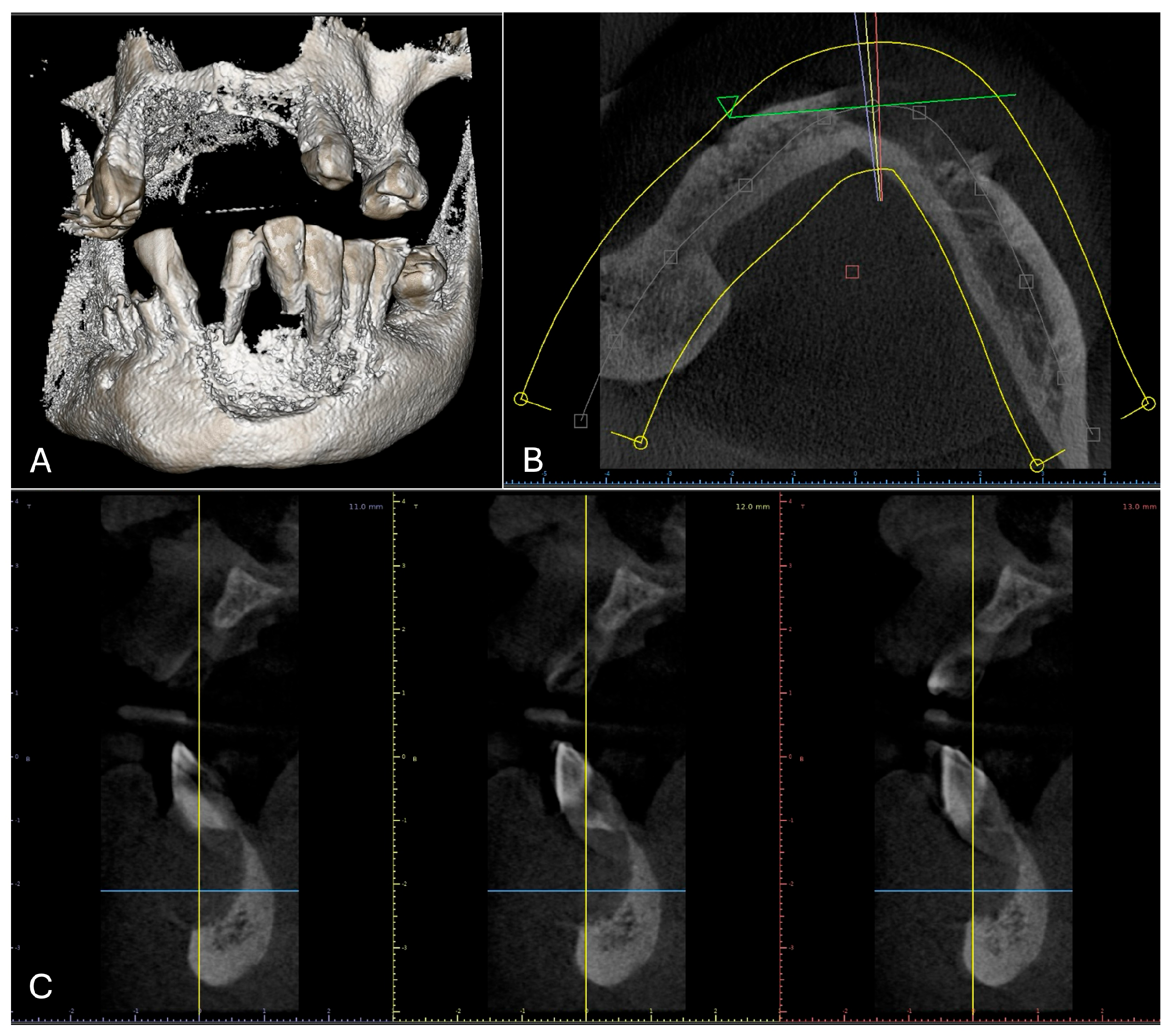
Radiographically, a unilocular radiolucent area with radiomixed portions in the center—surrounding the roots of teeth 34, 33, 32, and 31, with well delimited and diffuse borders related to rhizolysis in tooth 32—was observed (Figure 1B). The tomographic analysis was performed with a NewTom VGI EVO apparatus, at 70 Kv and 10 mA; and confirmed the lesion’s extension and showed that the lingual cortical bone was preserved (Figure 2). Based on the clinical features and past records, a presumptive diagnosis of central or peripheral giant cell granuloma was made.

2.2. Treatment Procedures and Histopathological Results

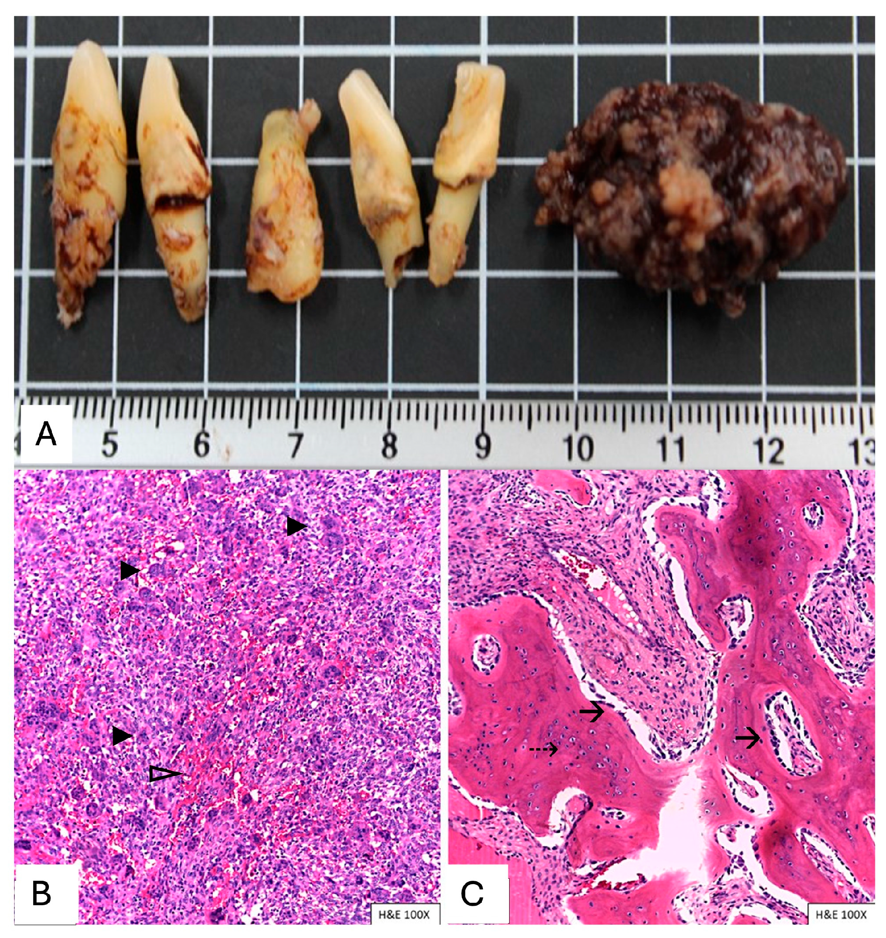
Given the patient’s previous diagnosis of a giant cell granuloma, the maxillofacial surgeon decided to perform an excisional biopsy of the current lesion in the Oral and Maxillofacial Surgery Specialty at the Postgraduate Studies Division. Macroscopically, a fragment of soft tissue measuring 3 × 1.7 × 1.3 cm was observed. It had an irregular shape and surface, dark brown color, and firm consistency. Longitudinal sections revealed a solid, heterogeneous internal appearance, and it was light brown in color with dark brown areas; likewise, 5 teeth (42, 41, 31, 32, and 33) correlated with their radiological features (Figure 3A).
Histologically, hypercellular connective tissue was observed with two types of cell populations: one population consisting of osteoclast-like multinucleated giant cells with 6 to 10 nuclei and the second population comprising mononuclear cells interspersed with areas of erythrocyte extravasation (Figure 3B). In other histological fields, areas of increased collagenization were observed, containing trabeculae of lamellar mature bone tissue with osteocytes, surrounded by bordering osteoblasts (Figure 3C). Considering these data, our histological diagnostic impression was consistent with a new CGCG.
However, based on these findings, mainly due to the history of a completely resected CGCG without recurrence, a probable alteration in the parathyroid gland was suspected. Thus, parathormone, serum calcium, serum phosphorus, alkaline phosphatase, and calcitonin studies were requested, obtaining the results shown in Table 1.
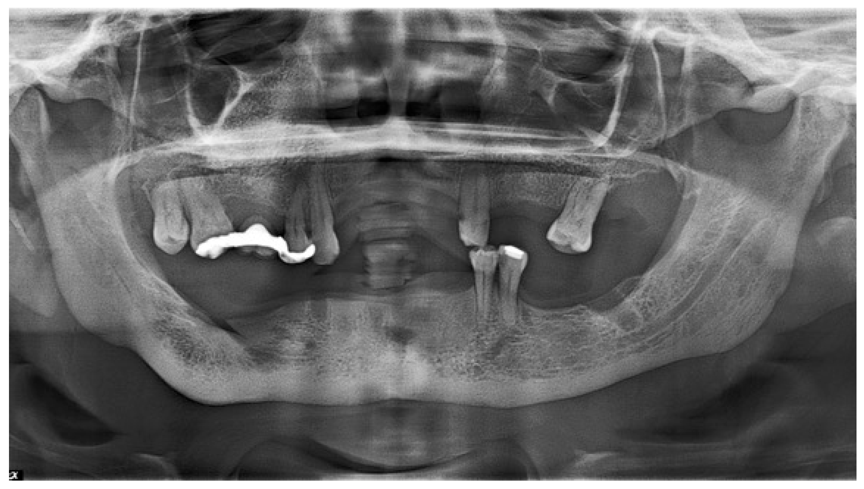
With the above laboratory study results, our final diagnosis was a brown tumor associated with hyperparathyroidism. The patient was referred to the Endocrinology Service for evaluation, diagnosis, and hyperparathyroidism treatment. Three months later, adequate bone regeneration was observed (Figure 4).

3. Discussion

A brown tumor is a rare bone manifestation of hyperparathyroidism, and it is characterized by benign growth associated with increased osteoclastic activity induced by parathyroid hormones. This increase generates bone resorption, proliferation of fibrous tissue, hemorrhage, and hemosiderin deposits, endowing it with its characteristic brown color [5,6]. Histologically, it resembles CGCG; thus, it is essential to perform clinical, histological, and biochemical correlations to establish an accurate diagnosis [10,11].
In the case presented, the patient manifested a lesion in the left mandibular body; the mandible is reported as the most common location for this tumor [12]. Clinically, the lesion manifested as an asymptomatic, slow-growing, expansive radiolucency, but it exhibited signs of resorption of the tooth apex. Histopathological analyses revealed features that were consistent with CGCG: cellular proliferation composed of mononuclear spindle cells and osteoclast-like multinucleated giant cells, accompanied by hemorrhage and the presence of hemosiderin. Lobules separated by fibrous septa, thin osteoid trabeculae, and bone tissue were observed without evidence of mitotic activity [11,13]. Although the patient reported complete removal of the previous CGCG, recurrence of this type of lesion is possible [14]. However, given that another lesion developed at an adjacent site, the recurrence possibility was ruled out, and the lesion was considered a new occurrence, raising suspicion of a possible underlying systemic disease [11,12].
Imaging tests helped to consolidate this diagnosis. Their differential diagnosis with other lesions includes odontogenic cysts and tumors (radicular cyst and ameloblastoma), fibrous dysplasia, or malignant lesions [12,15,16]. With tomographic projections, the lesion’s extension could be confirmed, which is always useful for determining the type of surgical approach.
However, the brown tumor’s definitive diagnosis was confirmed by integrating the clinical, imagenological, and histological findings with biochemical results. The elevated PTH, serum calcium, and alkaline phosphatase levels, together with a decrease in serum phosphorus levels, are characteristic laboratory findings of hyperparathyroidism. Among these, elevated PTH is the central feature guiding a multidisciplinary approach to patient management [17,18].
The clinical management of brown tumors is mainly based on the extent of involvement and the treatment of the underlying hyperparathyroidism [15,19]. In some cases, hormonal disorder corrections result in the spontaneous regression of bone lesions without the need for surgical intervention. However, in situations where the lesion persists, grows, causes disfigurement, or is adjacent to a possible infection area, as observed in the case presented, surgical excision is indicated [16,17,18,19,20].
Finally, although the clinical, histological, and serological features supported early detection of hyperparathyroidism, appropriate management requires a multidisciplinary evaluation to determine the type and extent of the disease [11,12,21]. In this case, an orthopantomography follow-up was carried out three months after the biopsy to assess the healing of the zone posteriorly, and the patient was referred to endocrinology service in a different hospital; however, due to logistical and transportation problems, tomography follow-up was lost. This is an important limitation, since a comprehensive diagnosis and treatment of the patient must consider determining whether there were brown tumors in other skeletal regions and the type of alteration in the parathyroid gland. Therefore, to overcome this limitation, it is necessary for the dentist to establish consultations with various specialists. Metabolic systemic bone scanning and tomographic imaging for osteolytic lesions were necessary to determine whether multiple bone sites were affected [22]. With respect to the parathyroid gland, ultrasound examinations could be the first approach, but according to the results, a histological analysis could be necessary [23].
This case reinforces the importance of a comprehensive initial clinical, radiological, histopathological, and biochemical evaluation in order to arrive at an accurate diagnosis and provide appropriate and early treatment. Although dental surgeons cannot participate in the medical treatment prescribed by endocrinologists, it is our responsibility to refer the patient with all relevant evidence to facilitate appropriate management.

4. Conclusions

The brown tumor, as a manifestation of hyperparathyroidism, presents various features that can be diagnosed through clinical, radiological, histological, and serological integrated tests. Coordinated dental and medical management is necessary in order to improve patients’ quality of life and avoid complications due to a misdiagnosis. For this reason, effective patient communication is essential.

Author Contributions

Conceptualization, V.M.S.-G. and L.F.J.-A.; methodology, A.M.-F., G.L.E.-G. and V.M.S.-G.; formal analysis, C.P.M.-V.; investigation, J.B.M.-J. and G.L.E.-G.; writing—original draft preparation, V.M.S.-G.; writing—review and editing, and L.F.J.-A.; visualization, C.P.M.-V.; supervision, C.P.M.-V. and L.F.J.-A.; project administration, C.P.M.-V. and L.F.J.-A. All authors have read and agreed to the published version of the manuscript.

Funding

This research received no external funding and the APC was funded by all authors.

Institutional Review Board Statement

No ethical approval was required because this is a case report and not an experimental or intervention study.

Informed Consent Statement

Written informed consent was obtained from the patient to publish this study.

Data Availability Statement

The original contributions presented in the study are included in the article; further enquiries can be directed to the corresponding authors.

Conflicts of Interest

The authors declare no conflicts of interest.

References

  1. Shetty, A.D.; Namitha, J.; James, L. Brown tumor of mandible in association with primary hyperparathyroidism: A case report. J. Int. Oral Health 2015, 7, 50–52. [Google Scholar] [PubMed]
  2. Pham, M.; Bhatt, P.; Purvey, T.A. Massive Exophytic Brown’s Tumor of the Bilateral Maxilla with Mandibular Involvement. J. Craniofac. Surg. 2022, 33, 897–900. [Google Scholar] [CrossRef] [PubMed]
  3. Pati, A.R.; Mubeen, K.V.; Singh, C. Diagnosis and clinico-radiological presentation in an aggressive maxillary brown tumour. J. Clin. Diagn. Res. 2014, 8, ZD13–ZD15. [Google Scholar] [CrossRef] [PubMed]
  4. Martínez Díaz-Guerra, G.; Hawkins Carranza, F. Concept, etiology and epidemiology of primary hyperparathyroidism. Endocrinol. Nutr. 2009, 56 (Suppl. S1), 2–7. [Google Scholar] [CrossRef]
  5. Alwani, M.M.; Monaco, G.N.; Harmon, S.M.; Nwosu, O.I.; Vortmeyer, A.O.; Payner, T.D.; Ting, J. A Systematic Review of Sellar and Parasellar Brown Tumors: An Analysis of Clinical, Diagnostic, and Management Profiles. World Neurosurg. 2019, 132, e423–e429. [Google Scholar] [CrossRef]
  6. Pokhrel, B.; Leslie, S.W.; Levine, S.N. Primary Hyperparathyroidism. In StatPearls; StatPearls Publishing: Petersburg, FL, USA, 2024. [Google Scholar]
  7. Gallana Álvarez, S.; Salazar Fernandez, C.I.; Avellá Vecino, F.J.; Torres Gómez, J.; Pérez Sanchez, J.M. Tumor pardo maxilar: Elemento diagnóstico de hiperparatiroidismo primario. Rev. Esp. Cirug. Oral y Maxilofac. 2005, 27, 225–230. [Google Scholar] [CrossRef]
  8. Majic Tengg, A.; Cigrovski Berkovic, M.; Zajc, I.; Salaric, I.; Müller, D.; Markota, I. Expect the unexpected: Brown tumor of the mandible as the first manifestation of primary hyperparathyroidism. World J. Clin. Cases 2024, 12, 1200–1204. [Google Scholar] [CrossRef]
  9. Liu, Z.; Yang, H.; Tan, H.; Song, R.; Zhang, Y.; Zhao, L. Brown tumor of the cervical spine with primary hyperparathyroidism: A case report and literature review. Medicine 2023, 102, e32768. [Google Scholar] [CrossRef]
  10. Antin, F.; Bakhos, D.; Jegoux, F.; Merkouza, M.; Laccourreye, L. Maxillofacial brown tumours: Series of 5 cases. Eur. Ann. Otorhinolaryngol. Head Neck Dis. 2018, 135, 227–230. [Google Scholar] [CrossRef]
  11. WHO Classification of Tumours Editorial Board. Giant Cell Lesions and Bone Cysts, Central Giant Cell Granuloma. In Head and Neck Tumours WHO Classification of Tumours, 5th ed.; IARC: Lyon, France, 2022; Volume 9. [Google Scholar]
  12. Genovesi, E.; Lerena, V.S.; Torres, I.C.; Paes De Lima, A.; Lucas, S.P.; Diaz, A.G. Brown tumors of the jaw: An atypical manifestation of primary hyperparathyroidism. Medicina (B Aires) 2023, 83, 462–466. [Google Scholar]
  13. Flanagan, A.M.; Speight, P.M. Giant cell lesions of the craniofacial bones. Head Neck Pathol. 2014, 8, 445–453. [Google Scholar] [CrossRef] [PubMed]
  14. Boffano, P.; Neirotti, F.; Brucoli, M.; Ruslin, M.; Stathopoulos, P.; Tsekoura, K.; Laco, J.; Mottl, R.; Dediol, E.; Kos, B.; et al. Diagnosis and management of central giant cell granulomas of the jaws: A European and multicenter study. J. Craniomaxillofac. Surg. 2025, 53, 1455–1462. [Google Scholar] [CrossRef]
  15. Khaoula, B.A.; Kaouther, B.A.; Ines, C.; Sami, T.; Zakraoui, L.; Khedher, A. An unusual presentation of primary hyperparathyroidism: Pathological fracture. Case Rep. Orthop. 2011, 2011, 521578. [Google Scholar] [CrossRef] [PubMed]
  16. Reséndiz-Colosia, J.A.; Alvarado-Cabrero, I.; Flores-Díaz, R.; Juan, M.H.; Barroso-Bravo, S.; Gómez-Acosta, F.; Rodríguez-Cuevas, S.A. Multiple maxillofacial brown tumors as primary hyperparathyroidism manifestation. Gac. Med. Mex. 2008, 144, 155–160. [Google Scholar]
  17. Hakkou, F.; Benjelloun, L.; Hallab, L.; Chbicheb, S. Brown tumor of the jaw as a rare manifestation of hyperparathyroidism: Two case reports and literature review. Int. J. Surg. Case Rep. 2023, 111, 108823. [Google Scholar] [CrossRef]
  18. Batra, S.; Srivastava, A.; Shukla, S. Unmasking the Archnemesis: Primary Hyperparathyroidism Presenting As Brown Tumors of the Jaws. Cureus 2025, 17, e84427. [Google Scholar] [CrossRef] [PubMed]
  19. Mastrapasqua, S.; Hendel, I. Maxillary brown tumor in secondary hyperparathyroidism. Nefrologia 2006, 26, 647–648. [Google Scholar]
  20. Fontseré Baldellou, N.; Andrés Santamaría, M.; Bella Cueto, F.; Ramírez de Arellano Serna, M. Brown tumour of the mandible and asymptomatic severe hypercalcemia as atypical forms of presentation of primary hyperparathyroidism. An. Med. Interna 2007, 24, 561–562. [Google Scholar]
  21. Mora-Escudero, I.; Gato-Díez, A.; Blázquez-Cabrera, J.A.; Lozano-Setien, E.; García de la Torre, J.P. Pretibial brown tumors as first manifestation of primary hyperparathyroidism. Rev. Clin. Esp. 2012, 212, e15–e18. [Google Scholar] [CrossRef]
  22. Zhong, Y.; Huang, Y.; Luo, J.; Ye, Y. Misdiagnosis of brown tumour caused by primary hyperparathyroidism: A case report with literature review. BMC Endocr. Disord. 2022, 22, 66. [Google Scholar] [CrossRef]
  23. Dinoi, E.; Prete, A.; Sardella, C.; Pierotti, L.; Della Valentina, S.; Dal Lago, A.; Borsari, S.; Pardi, E.; Caligo, M.A.; Michelucci, A.; et al. The challenge of the differential diagnosis between brown tumors and metastases in parathyroid carcinoma: A case report. Front. Endocrinol. 2024, 15, 1414896. [Google Scholar] [CrossRef] [PubMed]
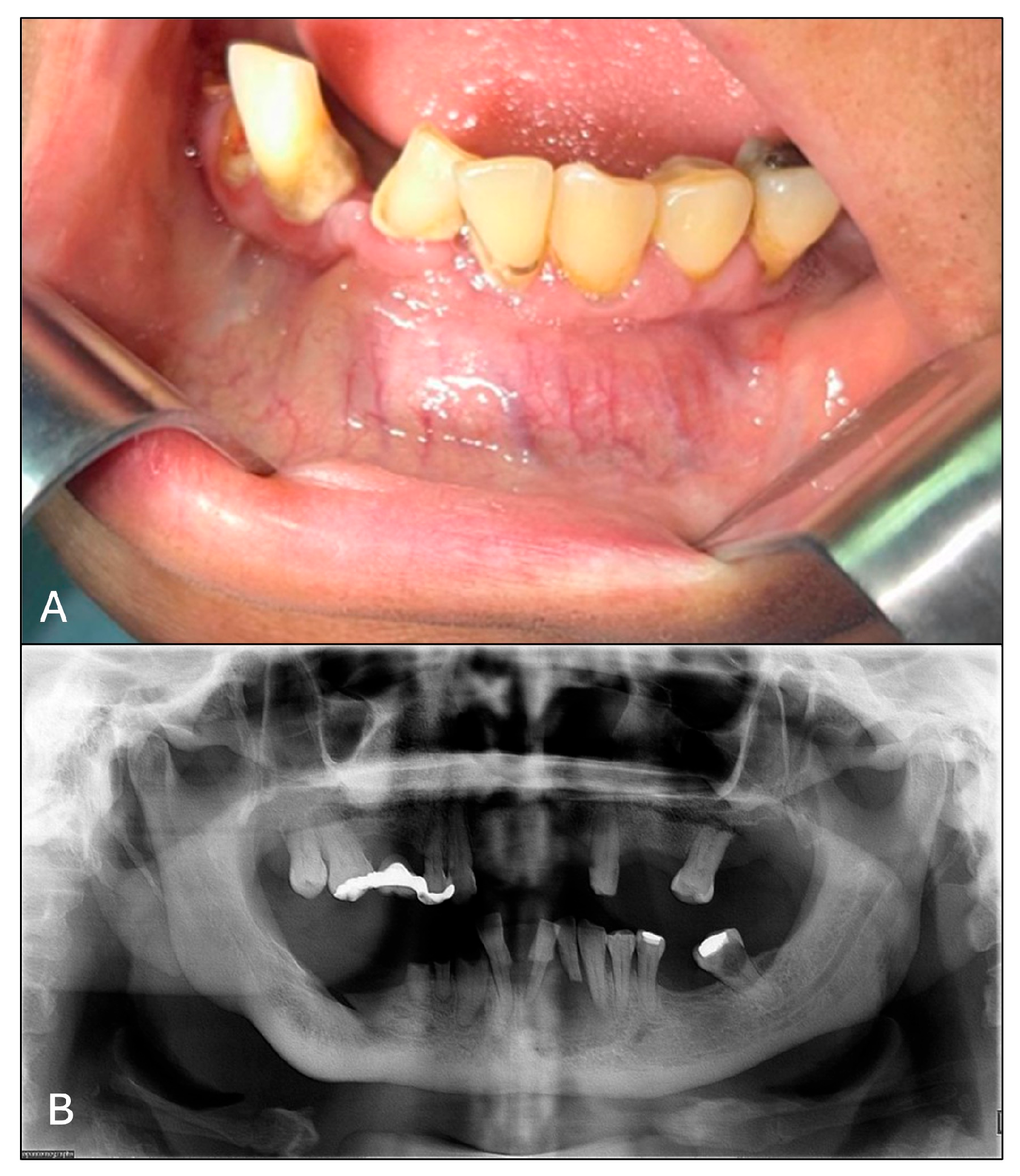
Figure 1. Initial clinical and imagenological features. (A) Initial clinical features of the patient showing increased volume in the body and mandibular symphysis. (B) Unilocular radiolucent area surrounding roots 34, 33, 32, and 31.
Oral 05 00059 g001
Figure 2. Tomography analysis. (A) Frontal view of a tomographic 3D reconstruction, showing anterior lesion extension; (B) cross−sectional image; and (C) sagittal sectional image confirming the preservation of the lingual bone wall.
Oral 05 00059 g002
Figure 3. Macroscopic and microscopic features. (A) Associated teeth with a tumoral specimen showing dark brown areas with light brown areas; root apex resorption corresponding to tooth 32 was observed. (B) Osteoclast-like multinucleated giant cells (arrowhead) in the stroma of mononuclear cells intermingled with areas of erythrocyte extravasation (hollow arrowhead). (C) Lamellar mature bone tissue trabeculae with osteocytes in their lacunae (dashed arrow), surrounded by a rim of osteoblasts in some areas (arrow).
Oral 05 00059 g003
Figure 4. Radiographic follow-up. Orthopantomography 3 months after excisional biopsy showing adequate bone regeneration.
Oral 05 00059 g004
Table 1. PTH and mineral-related metabolite blood test results.
ResultsReference Value
Parathormone120.00 pg/mL *15–65 pg/mL
Calcium8.80 mg/dL8.50–10.40 mg/dL
Phosphorus3.60 mMol/L2.50–4.50 mMol/L
Alkaline phosphatase93.00 U/L40.0–279.00 U/L
Calcitonin<2.0 pg/mL0.00–11.5 pg/mL
* The patient presented elevated parathormone values.
Disclaimer/Publisher’s Note: The statements, opinions and data contained in all publications are solely those of the individual author(s) and contributor(s) and not of MDPI and/or the editor(s). MDPI and/or the editor(s) disclaim responsibility for any injury to people or property resulting from any ideas, methods, instructions or products referred to in the content.