Case Report

Oral Rehabilitation Following Surgical Treatment of Mandibular Ameloblastoma: Case Report and Comprehensive Literature Review

1
Department of Surgical, Medical, Molecular Pathology and of the Critical Area, University of Pisa, 56126 Pisa, Italy
2
Dental Biomaterials Research Unit (d-BRU), University of Liege, 4000 Liège, Belgium
3
Unit of Dentistry and Oral Surgery, University-Hospital of Pisa, 56100 Pisa, Italy
4
Unit of Maxillo-Facial Surgery, University-Hospital of Pisa, 56100 Pisa, Italy
5
Unit of Otorhinolaryngology, University-Hospital of Pisa, 56100 Pisa, Italy
6
Unit of Plastic Surgery, University-Hospital of Pisa, 56100 Pisa, Italy
*
Author to whom correspondence should be addressed.
These authors contributed equally to this work.
Submission received: 30 June 2025 / Revised: 31 July 2025 / Accepted: 4 August 2025 / Published: 8 August 2025

Abstract

Objectives: Ameloblastoma is a locally aggressive odontogenic tumor of the jaws characterized by a high recurrence rate. This work aims to present our clinical experience in managing patient oral rehabilitation following an extensive mandibular ameloblastoma, with a specific focus on mandibular reconstruction using a fibula free flap, followed by dental implant placement and prosthetic rehabilitation in a female patient. Additionally, we provide a comprehensive review of the current evidence on surgical management, reconstruction techniques, and long-term outcomes in ameloblastoma treatment. Methods: A 44-year-old female patient presented with a painless swelling in the left mandible. Orthopantomography (OPG) and computed tomography (CT) demonstrated a well-defined radiolucent lesion extending from the canine to the second premolar. An incisional biopsy was performed, and histopathological examination confirmed the diagnosis of mandibular ameloblastoma. The patient underwent segmental resection of the left mandibular body, followed by immediate reconstruction using a vascularized fibular free flap. Eighteen months postoperatively, four dental implants were placed. One implant failed during the osseointegration phase and was removed. Due to residual hard and soft tissue deficiency, prosthetic rehabilitation was achieved with a metal-reinforced resin overdenture, restoring both function and aesthetics. Results: At the three-year follow-up, clinical and radiographic examinations revealed no evidence of tumor recurrence. The patient remained asymptomatic, reporting neither pain nor functional discomfort. Prosthetic rehabilitation with the metal-reinforced resin overdenture was successfully completed, achieving satisfactory masticatory function and aesthetics. Conclusions: The use of the fibula free flap for mandibular reconstruction after ameloblastoma resection provides excellent flexibility, enabling effective bone integration of dental implants.

1. Introduction

Ameloblastoma is an odontogenic tumor of the jaws, representing approximately 13–58% of all odontogenic tumors. Despite a benign histologic pattern, ameloblastoma is locally aggressive and has a high recurrence rate if not properly managed [1,2]. Ameloblastoma more frequently affects the mandible (80% of all cases), particularly in the angle and ramus, although it can occur in any region of the mandible [3]. The disease shows no gender predilection, and most commonly affects patients aged between 30 and 60 years. The diagnosis of ameloblastoma is often delayed, due to its slow-growing nature, and is frequently identified incidentally on orthopantomography (OPG) [4]. Clinical manifestations often cause non-specific symptoms, including facial swelling, occlusal disturbances, speech difficulties, pain, paresthesia, dental mobility, tooth loss, nasal obstruction, and periodontal disease [5]. According to the World Health Organization (WHO) classification, three distinct clinicopathological variants of ameloblastoma exist: conventional, unicystic, and peripheral ameloblastoma [6]. The conventional type comprises six histological variants: plexiform, follicular, acanthomatous, desmoplastic, granular, and basal cell types. The unicystic type is further divided into luminal, intraluminal, and mural subtypes [7].
Radiographically, ameloblastoma typically appears as a unilocular or multilocular radiolucent lesion, exhibiting a characteristic honeycomb or soap bubble appearance [8,9]. Conventional ameloblastoma is the most common subtype, accounting for approximately 80% of cases, predominantly affecting the posterior regions of the jaw and occurs mainly in the 3rd and 4th decades of life [6,10]. Unicystic variants demonstrate cystic architecture lined by ameloblastic epithelium with potential luminal or mural proliferation [11]. Peripheral ameloblastoma, accounting up for 1% of all cases, primarily affects individuals of approximately 50 years. It is commonly located in the mandibular gingiva, with a low tendency for recurrence, even after a conservative surgical approach [12].
The management of mandibular ameloblastoma presents significant challenges, with ongoing debates in the literature concerning the most effective therapeutic approach [9]. The treatment plan should ensure complete removal of the tumor, minimize the risk of recurrence, enhance aesthetic outcomes, and restore oral functions to improve the overall quality of life of the patients.
Due to its locally aggressive nature and the high risk of recurrence, ameloblastoma requires a surgical approach as the primary form of treatment.
Two surgical approaches are commonly employed based on factors such as bone invasion and adequacy of excision margins:
  • Conservative surgery encompasses surgical enucleation, curettage, and cryotherapy. These techniques are generally used for less aggressive forms of the tumor like unicystic and peripheral ameloblastoma. These methods need required a reduced surgical time but may be associated with higher recurrence rates and the potential need for re-intervention.
  • Radical surgical techniques, such as marginal and segmental mandibulectomy, are indicated for more invasive tumors or in cases of recurrence of the disease. Radical approaches generally result in lower recurrence rates [8,9,13].
The mandible plays a crucial role in speech, mastication, swallowing, and facial aesthetics. Indeed, the disruption of its continuity in case of radical surgery significantly affects function and reduces patients’ quality of life. Consequently, these procedures frequently require complex reconstructive surgeries, to restore both function and structure. For tumors requiring mandibular resection, reconstruction using vascularized free bone grafts has long been considered the gold standard [14]. The fibula free flap is an extremely reliable option for mandibular defect reconstruction, offering several advantages with respect to other vascularized flaps, including sufficient bone length to match the maxillary and mandibular defects, a large vessel caliber for vessel anastomosis, and a sufficient skin flap for reconstruction of intraoral and/or extraoral defects [15,16]. Furthermore, the fibula’s anatomical features make it particularly suitable for the placement of dental implants. Several factors must be considered to achieve implant success, including proper soft tissue management, adequate circumferential bone, and sufficient restorative space. Specifically, when placing dental implants in the jaws, 1.5 mm should be maintained between the implant and natural teeth, and a 3 mm distance should be maintained between adjacent implants [14,17,18]. Peri-implant reactive tissue complications are common postoperative occurrences in implants placed within fibula free flaps. Although these inflammatory complications can present in various forms, the commonly observed manifestations are mucositis, hypertrophic tissue formation, and peri-implantitis. Individual patient risk factors for developing peri-implantitis and reactive tissue responses include poor oral hygiene, smoking, genetic predisposition, gingivitis, diabetes, alcohol abuse, and pre- or postoperative chemotherapy [19].
The aim of this work is to present our experience in the management of an extensive mandibular ameloblastoma—with a particular focus on the use of a fibula bone graft for reconstructing the lower left jaw, followed by placement of implants and an implant-supported prosthetic rehabilitation—and to present a comprehensive review of current evidence on surgical management, reconstruction techniques, and long-term outcomes in ameloblastoma treatment.
This case report has been prepared in accordance with the CARE guidelines for case reports (https://www.care-statement.org/ Access on 4 August 2025).

2. Case Report

A 44-year-old female patient presented to the Oral Surgery department of the University Hospital of Pisa with a swelling in the left lower jaw, seeking specialist medical advice. The intraoral examination revealed a raised, painless, and tender-at-palpation mass in the buccal vestibular area of the lower left mandible, extending into the labial region, with normal color and the appearance of the surrounding mucosa (Figure 1).
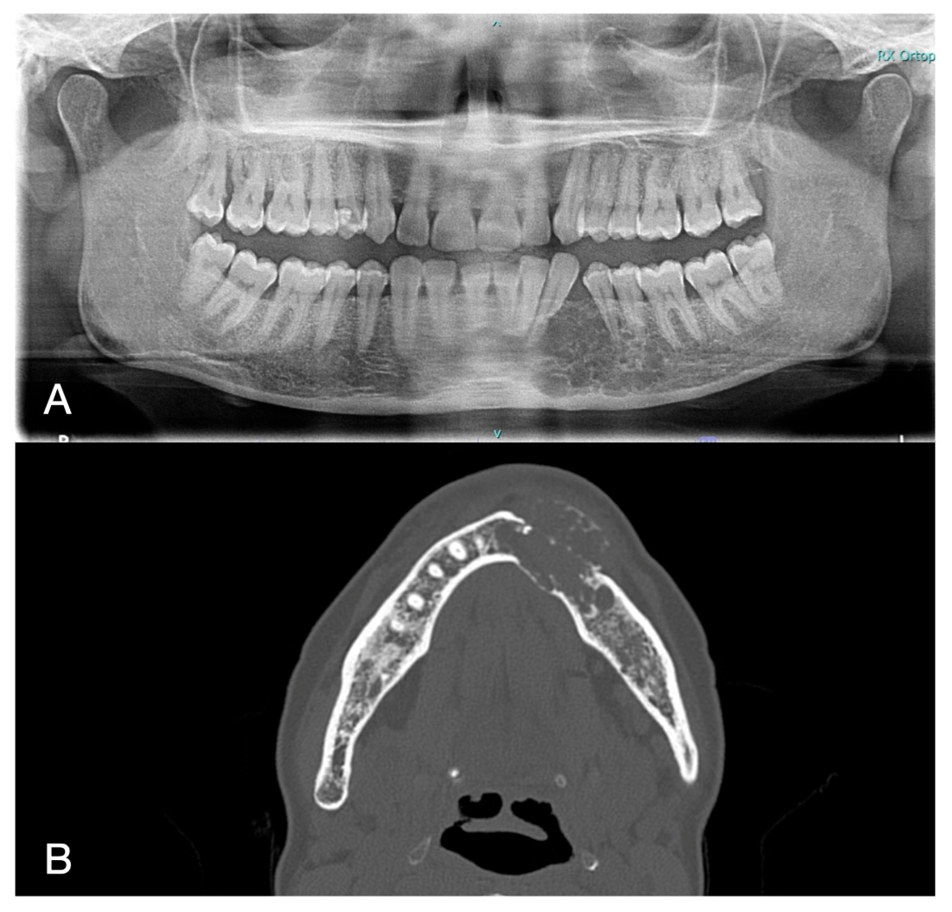
The pulp vitality test (cold test) was positive for the elements 3.2–3.3–3.4–3.5 and 3.6, and no pathological pocket probing depth was detected in any of the involved teeth. The bi-dimensional OPG showed a wide radiolucent lesion, located between the canine and second premolar on the left side of the mandible. The second level tri-dimensional imaging CT scan (axial tomography) showed a radiolucent mass in the left mandibular body, with erosion of the mandibular cortex. The two-dimensional and three-dimensional imaging findings are presented in Figure 2.
A diagnosis of ameloblastoma was suspected based on these imaging findings, considering its typical radiological features and localization.
Accordingly, an incisional biopsy was scheduled to assess the nature of intra-osseous lesion. The procedure was carefully explained to the patient, who then signed an informed consent form.
The biopsy was performed under local anesthesia, and the intra-osseous mass, measuring 10 mm, was sent to the Pathological Anatomy Department for examination. The clinical phases of the surgical intervention are shown in Figure 3.
The histopathological findings confirmed the diagnosis of unicystic ameloblastoma.
The treatment planning was carried out in two phases. The primary objective of the first phase was tumor eradication to prevent recurrence. Bone grafting was performed to ensure sufficient bone volume for future implant placement. The subsequent implant-prosthetic planning was then guided by the available bone and the diagnostic stone cast models.
The patient underwent a complex multidisciplinary mandibular reconstruction after resection of the left body of the mandible, carried out by the Maxillo-facial, Otorhinolaryngology and Plastic Surgery teams of the University Hospital of Pisa. The surgery was performed under general anesthesia. The surgery began with a cervical skin incision on the left side, followed by muscle dissection for releasing the entire surface of the mandible body from mimic muscles and the muscles of the floor of the mouth, and isolation of the facial vessels, including the left superior thyroid artery, to ensure clear visibility and prevent damage to adjacent structures. Secondly, a subperiosteal dissection and oral intrasulcular incisions were performed to expose the mandible, extending from the right mental nerve region to the left mandibular angle. This allowed for realignment of the mandible and proper fitting for the reconstructive procedure. After preparing the site, the affected area of the left mandibular body was excised, and the submandibular gland was removed. The reconstructive phase involved the insertion of a fibular graft from the right leg into the surgical gap. The choice of the right leg was influenced by the presence of varicose veins in the left one. The fibula has been sectioned into two segments: an anterior one of 25 mm length and a posterior one 30 mm length, which was able to fit the gap and was secured in place with a plate fixation system. Previously, a model of the jaw had been created with a 3D printer to prepare a pre-modeled reconstructive plate. The peroneal artery (of the flap) was anastomosed with a termino-terminal technique with the facial artery, while the two comitant veins were anastomosed, using the same termino-terminal technique, with the facial vein and a branch of the external jugular.
The surgical procedure was successfully completed with no immediate complication. One week after surgery a CT scan was performed for post-surgical follow-up (Figure 4).
At three months follow-up, swelling and pain were minimal, there were no signs of infection, and joint function and occlusion were normal. However, mouth opening was limited due to cutting of muscles during the surgery. The face was in an acceptable condition in terms of harmony and aesthetics.
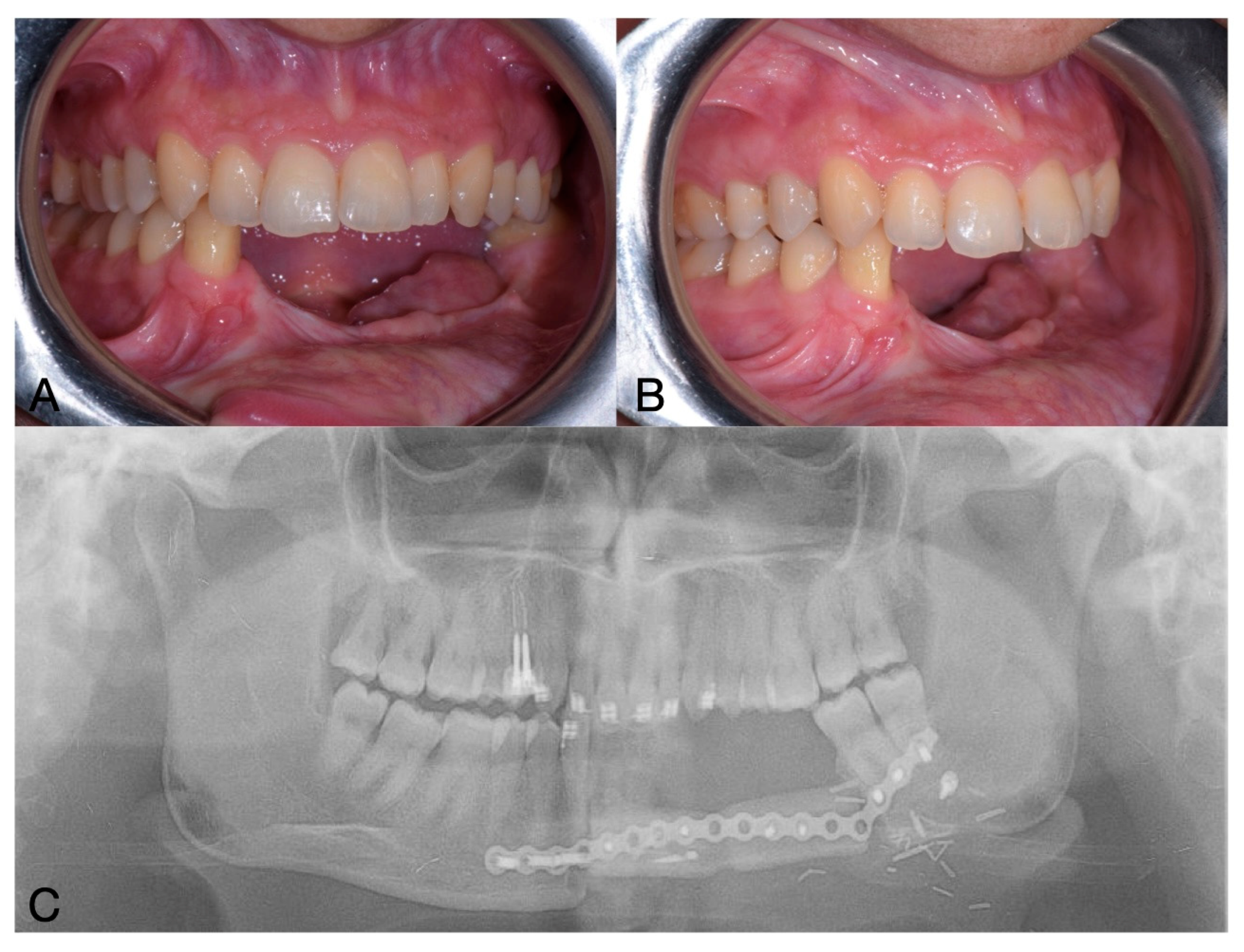
One year after the mandibular surgery, on OPG was performed to plan the subsequent implant-prosthetic rehabilitation (Figure 5).
The procedure involved the removal of the osteosynthesis plate, and placement of four bone-level implants (Straumann, bone-level implants, Basel, Switzerland) with dimensions of 3.3 × 8 mm.
The choice of implant geometry was due to the high amount of cortical bone in the fibula. Implants with aggressive thread designs may generate excessive compressive forces, which could negatively affect osseointegration. Cylindrical implants with a more conservative thread profile allow for a more favorable stress distribution in cortical bone.
The implants were left to heal submerged. Bone-level implants were chosen as they allow for submucosal healing, which promotes more predictable osseointegration and helps prevent potential infections during the healing period [20].
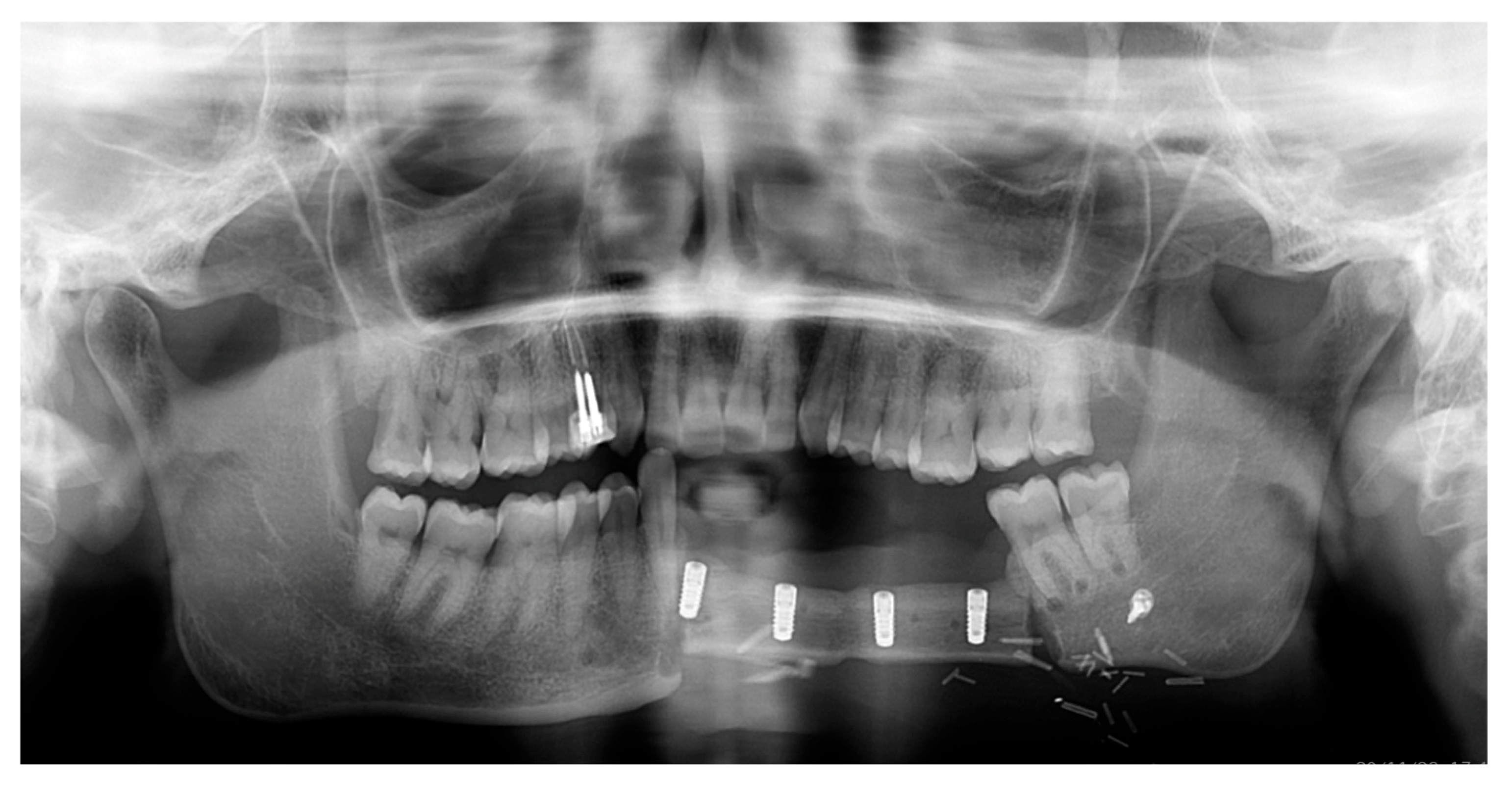
After a 3-month healing period, an OPG was performed, and implant uncovering and soft tissue surgery were planned (Figure 6).
During the second surgery, one implant exhibited absence of osseointegration with no signs of infection and was then removed. Notably, this implant was located near the transition zone between the remaining mandibular bone and the fibula graft, which may have contributed to its failure due to reduced vascularization [21]. An internal connection with a screw-retained abutment was chosen for the prosthetic rehabilitation. Multi-unit abutments (MUAs) were employed to optimize load distribution among the implants. Three MUAs and healing caps were placed, and the flap was apically repositioned to improve buccal fornix depth. The two mesial MUAs had a height of 3.5 mm, whereas the distal one measured 5.5 mm.
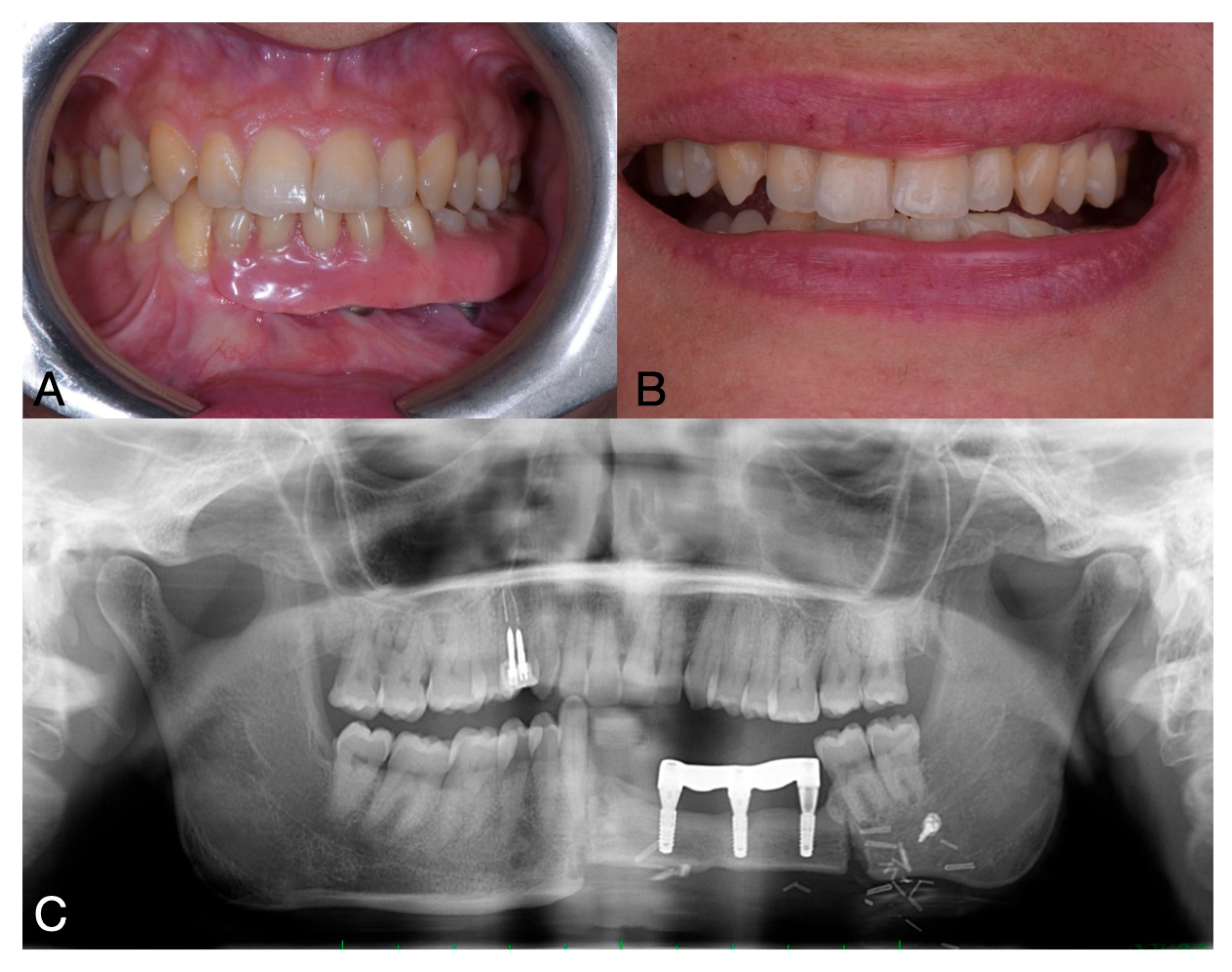
The prosthetic rehabilitation was completed four months after implant placement using a bar-retained, metal reinforced resin overdenture. Due to an unfavorable crown-to-root ratio and the loss of one implant, a removable prosthesis was selected—following discussion with the patient—to avoid overloading the remaining implants and to achieve a more favorable distribution of occlusal forces [22]. Furthermore, the decision was supported by the improved ease of oral hygiene maintenance associated with a removable prosthesis.
Analogic impressions were taken with the use of polyether impression material. Performing this prosthetic rehabilitation was challenging because taking impressions on the implants was difficult due to the patient’s limited mouth opening, as the muscles had been cut during the reconstructive surgery of the mandible. With the prosthesis, restoration of masticatory function and improvement in facial aesthetics were observed. The patient experienced a positive impact on her self-esteem and social interactions (Figure 7).
Patient satisfaction was evaluated using a Visual Analogue Scale (VAS) ranging from 0 to 10, assessing both aesthetic and functional outcomes. The patient reported the highest possible score—10 in both categories—indicating maximum satisfaction. Overall, the patient expressed a high level of satisfaction with the treatment outcomes.

3. Discussion

Ameloblastoma is a rare but locally aggressive benign tumor, best treated by wide resection with 1–1.5 cm margins to ensure local control [23]. Ameloblastomas typically exhibit slow progression; however, they are locally invasive and, if untreated, can lead to substantial morbidity and, in some cases, even exitus [24,25,26,27]. Various etiological factors have been associated with ameloblastoma development, including local trauma, nutritional deficiency, inflammation and molecular or genetic alternations involving multiple signaling pathways. Recent evidence underlined the role of genetic changes in the development of ameloblastomas; these findings could help create less invasive treatment methods, providing more effective care with less harm to the patient [26].
Current management of ameloblastoma primarily involves surgical resection with adequate margins due to the tumor’s high recurrence potential, as conservative approaches like enucleation and curettage demonstrate significantly higher recurrence rates of 50–90% compared to 10–20% with radical resection [27].
Mandibular resection indications extend beyond ameloblastoma to include malignant neoplasms, osteoradionecrosis (ORN), and medication-related osteonecrosis of the jaw (MRONJ) [28].
While complete tumor excision with 1–1.5 cm margins remains oncologically imperative, this approach presents reconstructive challenges including compromised dentition, reduced residual bone volume, and extended treatment timelines, often necessitating staged procedures [29,30]. The advent of vascularized free flaps has transformed reconstruction outcomes, with the fibula flap emerging as the gold standard due to its exceptional bone stock quality for implant placement, reliable vascular pedicle, and dual-layer soft tissue capacity [31]. However, osseous free flap reconstruction demands meticulous three-dimensional planning, expert microsurgical technique, and careful management of the dynamic masticatory environment and of the high bacterial load characteristic of oral cavity procedures [32]. Successful outcomes depend on comprehensive virtual surgical planning, experienced surgical teams, rigorous postoperative monitoring, and strict oral hygiene protocols to balance oncological safety with functional restoration.
The rehabilitation of extensive oral and maxillofacial defects requires a comprehensive, multidisciplinary approach guided by clearly defined therapeutic objectives that address both anatomical restoration and functional recovery [33]. Primary goals include the reestablishment of mandibular continuity through precise bony reconstruction, ensuring adequate bone volume with particular attention to vertical height and buccolingual width of the neo-alveolar ridge, in order to facilitate subsequent prosthetic rehabilitation. Equally critical is the management of soft tissue components, including mucosal coverage and muscle reattachment, which significantly influence both functional outcomes and facial aesthetics. Preventing graft resorption through proper vascularization and load distribution represents another key consideration, particularly in cases requiring dental implant placement. The restoration of facial contour must account for both skeletal support and overlying soft tissue drape, as subtle asymmetries can profoundly impact psychosocial well-being. Functional priorities include the reinstatement of effective masticatory capability with occlusal forces exceeding 150 N for normal diet tolerance, along with the recovery of intelligible speech through proper tongue positioning and oral competence.
Contemporary microvascular techniques, particularly the fibula free flap, have revolutionized this process by allowing simultaneous bony and soft tissue reconstruction while providing adequate bone stock for dental implants [34]. Indeed, the fibula free flap has emerged as the reconstructive gold standard following segmental mandibulectomy, offering unique advantages that address both anatomical and functional requirements [35].
Clinical studies demonstrate 5-year implant survival rates of 88–94% in fibula-reconstructed mandibles, enabling restoration of masticatory function to approximately 80–85% of normal capacity [20,36,37].
The flap’s perforator-based blood supply enhances bone healing, with radiographic union typically occurring within 6–8 weeks postoperatively [38]. When combined with implant-supported prostheses, this approach achieves bite forces exceeding 200 N—sufficient for normal diet tolerance—while simultaneously addressing the aesthetic and functional sequelae of mandibular resection [39,40]. Properly positioned implants in the fibular segment distribute occlusal loads evenly, reducing stress shielding and minimizing the 15–25% risk of plate-related complications seen in non-implant reconstructions [41,42]. Long-term outcomes of ameloblastoma treatment show recurrence rates < 10% when resection margins exceed 1 cm, confirming the oncological safety of this approach [43]. Patient-reported outcomes demonstrate significant improvements in quality-of-life metrics, particularly in speech intelligibility (85% normalization) and social functioning [44]. The fibula’s dual-layer soft tissue paddle also permits simultaneous intraoral lining reconstruction, addressing the common complication of peri-implant soft tissue deficiencies [45].
The positioning of the fibula segment is paramount, requiring careful consideration of occlusal plane orientation and prosthetic-driven planning to optimize both biomechanical loading and aesthetic outcomes. Osteosynthesis plate selection and placement have similarly evolved, with current systems offering improved fatigue resistance and anatomical adaptation through computer-assisted design and manufacturing [32,46,47]. While these advancements have reduced the average operative times by 30–40%, the biological timeline for bone healing remains consistent, with radiographic union typically evident between 6 and 8 weeks postoperatively through the appearance of bridging callus and fading osteotomy lines [32]. However, the complex interplay between biomechanical forces and microbial challenges in the oral cavity contributes to a 15–25% incidence of complications, including plate exposure, hardware failure, and non-union, each capable of derailing the rehabilitation timeline and necessitating additional interventions [48,49]. Plate removal after mandibular reconstruction with a fibula free flap is mainly due to complications such as plate exposure, infection, fistula, with reported rates between 5.4% and 35%. In some cases, removal is also needed to allow implant placement. Approaches for plate removal can be either extraoral or intraoral, and hardware removal may be partial or complete. Early removal before full bone healing may require re-osteosynthesis and delay rehabilitation [27,50].
Following mandibular reconstruction for ameloblastoma, implant-supported prostheses represent the gold standard for functional and aesthetic rehabilitation, particularly in cases involving fibula free flap reconstruction [51]. These prostheses not only restore critical masticatory function (with bite forces approaching 80–90% of natural dentition) but also maintain alveolar ridge contour and facial soft tissue support—crucial considerations given the extensive bone loss characteristic of ameloblastoma resections. Compared to conventional removable options, implant-retained prostheses demonstrate greater stability during function, while simultaneously addressing the common post-surgical challenges of speech articulation and swallowing efficiency that significantly impact patient quality of life [52,53]. Their biomechanical advantages are particularly valuable in ameloblastoma cases, where the reestablished mandibular continuity and occlusal plane following fibula reconstruction require precise prosthetic loading to optimize long-term outcomes.

4. Conclusions

The use of the fibula free flap for mandibular reconstruction after ameloblastoma resection provides excellent flexibility, enabling effective bone integration after dental implant placement. This approach allows for the restoration of oral function, including mastication, swallowing, and speech, with the aid of an implant-supported prosthesis. By combining surgical and prosthodontic approaches, this method ensures both functional and aesthetic outcomes. Long-term success, including the absence of tumor recurrence, further underscores the efficiency of this technique in complex mandibular reconstruction.

Author Contributions

Conceptualization, S.G., C.C., B.C.B., M.N., L.B., F.L. and A.B.; methodology, A.B., R.I., M.N., S.S., L.B. and B.C.B.; software, C.C.; validation, B.C.B. and F.L.; formal analysis, C.C.; investigation, A.B., C.C., M.P. and M.N.; data curation, A.B. and S.S.; writing—original draft preparation, S.G., C.C. and R.I.; writing—review and editing, R.I., M.N., M.P., F.L., B.C.B., L.B., S.S. and A.B.; visualization, A.B.; supervision, A.B.; project administration, A.B. All authors have read and agreed to the published version of the manuscript.

Funding

This research received no external funding.

Institutional Review Board Statement

Ethical review and approval were waived for this study because this a case report, and the patient provided a written consent form for the publication.

Informed Consent Statement

Written informed consent has been obtained from the patient to publish this paper.

Data Availability Statement

Data is unavailable due to privacy reasons.

Acknowledgments

The authors have reviewed and edited the output and take full responsibility for the content of this publication.

Conflicts of Interest

The authors declare no conflicts of interest.

References

  1. Torres-Lagares, D.; Infante-Cossío, P.; Hernández-Guisado, J.M.; Gutiérrez-Pérez, J.L. Mandibular ameloblastoma. A review of the literature and presentation of six cases. Med. Oral Patol. Oral Cir. Bucal 2005, 10, 231–238, (In English, Spanish). [Google Scholar] [PubMed]
  2. Hendra, F.N.; Van Cann, E.M.; Helder, M.N.; Ruslin, M.; de Visscher, J.G.; Forouzanfar, T.; de Vet, H.C.W. Global incidence and profile of ameloblastoma: A systematic review and meta-analysis. Oral Dis. 2020, 26, 12–21. [Google Scholar] [CrossRef] [PubMed]
  3. Shi, H.A.; Ng, C.W.B.; Kwa, C.T.; Sim, Q.X.C. Ameloblastoma: A succinct review of the classification, genetic understanding and novel molecular targeted therapies. Surgeon 2021, 19, 238–243. [Google Scholar] [CrossRef] [PubMed]
  4. Kreppel, M.; Zöller, J. Ameloblastoma—Clinical, radiological, and therapeutic findings. Oral Dis. 2018, 24, 63–66. [Google Scholar] [CrossRef]
  5. Bianchi, B.; Ferri, A.; Ferrari, S.; Leporati, M.; Copelli, C.; Ferri, T.; Sesenna, E. Mandibular resection and reconstruction in the management of extensive ameloblastoma. J. Oral Maxillofac. Surg. 2013, 71, 528–537. [Google Scholar] [CrossRef]
  6. Cadavid, A.M.H.; Araujo, J.P.; Coutinho-Camillo, C.M.; Bologna, S.; Junior, C.A.L.; Lourenço, S.V. Ameloblastomas: Current aspects of the new WHO classification in an analysis of 136 cases. Surg. Exp. Pathol. 2019, 2, 17. [Google Scholar] [CrossRef]
  7. Bwambale, P.; Yahaya, J.J.; Owor, G.; Wabinga, H. Histopathological patterns and biological characteristics of ameloblastoma: A retrospective cross-sectional study. J. Taibah Univ. Med. Sci. 2022, 17, 96–104. [Google Scholar] [CrossRef]
  8. Hendra, F.N.; Natsir Kalla, D.S.; Van Cann, E.M.; de Vet, H.C.W.; Helder, M.N.; Forouzanfar, T. Radical vs conservative treatment of intraosseous ameloblastoma: Systematic review and meta-analysis. Oral Dis. 2019, 25, 1683–1696. [Google Scholar] [CrossRef]
  9. Pappalardo, M.; Tsao, C.K.; Tsang, M.L.; Zheng, J.; Chang, Y.M.; Tsai, C.Y. Long-term outcome of patients with or without osseointegrated implants after resection of mandibular ameloblastoma and reconstruction with vascularized bone graft: Functional assessment and quality of life. J. Plast. Reconstr. Aesthetic Surg. 2018, 71, 1076–1085. [Google Scholar] [CrossRef]
  10. Hertog, D.; Bloemena, E.; Aartman, I.H.A.; van-der-Waal, I. Histopathology of ameloblastoma of the jaws; some critical observations based on a 40 years single institution experience. Med. Oral Patol. Oral Cir. Bucal 2012, 17, e76–e82. [Google Scholar] [CrossRef]
  11. Filizzola, A.I.; Bartholomeu-Dos-Santos, T.C.R.; Pires, F.R. Ameloblastomas: Clinicopathological features from 70 cases diagnosed in a single Oral Pathology service in an 8-year period. Med. Oral Patol. Oral Cir. Bucal 2014, 19, e556–e561. [Google Scholar] [CrossRef]
  12. Sham, E.; Leong, J.; Maher, R.; Schenberg, M.; Leung, M.; Mansour, A.K. Mandibular ameloblastoma: Clinical experience and literature review. ANZ J. Surg. 2009, 79, 739–744. [Google Scholar] [CrossRef]
  13. Effiom, O.A.; Ogundana, O.M.; Akinshipo, A.O.; Akintoye, S.O. Ameloblastoma: Current etiopathological concepts and management. Oral Dis. 2018, 24, 307–316. [Google Scholar] [CrossRef] [PubMed]
  14. Dugast, S.; Longis, J.; Anquetil, M.; Corre, P.; Komarova, S.; Bertin, H. Assessing Dental Implant Success: A Systematic Review and Meta-Analysis of Primary Versus Secondary Implantation in Free Bone Flap Reconstruction for Malignant Tumors. Head Neck 2025, 47, 1277–1290. [Google Scholar] [CrossRef] [PubMed]
  15. Shen, Y.F.; Rodriguez, E.D.; Wei, F.C.; Tsai, C.Y.; Chang, Y.M. Aesthetic and Functional Mandibular Reconstruction with Immediate Dental Implants in a Free Fibular Flap and a Low-Profile Reconstruction Plate: Five-Year Follow-up. Ann. Plast. Surg. 2015, 74, 442–446. [Google Scholar] [CrossRef] [PubMed]
  16. Nguyen, T.T.H.; Eo, M.Y.; Myoung, H.; Kim, M.J.; Kim, S.M. Implant-supported fixed and removable prostheses in the fibular mandible. Int. J. Implant. Dent. 2020, 6, 44. [Google Scholar] [CrossRef]
  17. Neal, T.W.; Williams, F.C.; Carr, B.R.; Pankey, T.; Teigen, K.; Kim, R.Y. Immediate Implants in Fibulas: Does the Implant to Fibula Osteotomy Distance Impact Early Implant Failure? J. Oral Maxillofac. Surg. 2024, 82, 1620–1626. [Google Scholar] [CrossRef]
  18. Wallace, C.G.; Chang, Y.M.; Tsai, C.Y.; Wei, F.C. Harnessing the potential of the free fibula osteoseptocutaneous flap in mandible reconstruction. Plast. Reconstr. Surg. 2010, 125, 305–314. [Google Scholar] [CrossRef]
  19. Raffaelli, S.D.; Neal, T.W.; Jelmini, J.J.; Kim, R.Y.; Williams, F.C. Evaluation of Postoperative Peri-Implant Reactive Tissues Following Implant Supported Prosthetic Rehabilitation in Fibula Free Flaps. Oral Surg. Oral Med. Oral Pathol. Oral Radiol. 2024, 138, e107–e112. [Google Scholar] [CrossRef]
  20. Anne-Gaëlle, B.; Samuel, S.; Julie, B.; Renaud, L.; Pierre, B. Dental implant placement after mandibular reconstruction by microvascular free fibula flap: Current knowledge and remaining questions. Oral Oncol. 2011, 47, 1099–1104. [Google Scholar] [CrossRef] [PubMed]
  21. Jaquiéry, C.; Rohner, D.; Kunz, C.; Bucher, P.; Peters, F.; Schenk, R.K.; Hammer, B. Reconstruction of maxillary and mandibular defects using prefabricated microvascular fibular grafts and osseointegrated dental implants—A prospective study. Clin. Oral Implant. Res. 2004, 15, 598–606. [Google Scholar] [CrossRef] [PubMed]
  22. Lee, J.H.; Kim, M.J.; Choi, W.S.; Yoon, P.Y.; Ahn, K.M.; Myung, H.; Hwang, S.J.; Seo, B.M.; Choi, J.Y.; Choung, P.H.; et al. Concomitant reconstruction of mandibular basal and alveolar bone with a free fibular flap. Int. J. Oral. Maxillofac. Surg. 2004, 33, 150–156. [Google Scholar] [CrossRef] [PubMed]
  23. Mendenhall, W.M.; Werning, J.W.; Fernandes, R.; Malyapa, R.S.; Mendenhall, N.P. Ameloblastoma. Am. J. Clin. Oncol. Cancer Clin. Trials 2007, 30, 645–648. [Google Scholar] [CrossRef] [PubMed]
  24. Gortzak, R.A.T.; Latief, B.S.; Lekkas, C.; Slootweg, P.J. Growth characteristics of large mandibular ameloblastomas: Report of 5 cases with implications for the approach to surgery. Int. J. Oral Maxillofac. Surg. 2006, 35, 691–695. [Google Scholar] [CrossRef]
  25. Zwahlen, R.A.; Grätz, K.W. Maxillary ameloblastomas: A review of the literature and of a 15-year database. J. Cranio-Maxillofac. Surg. 2002, 30, 273–279. [Google Scholar] [CrossRef]
  26. Brown, N.A.; Betz, B.L. Ameloblastoma: A Review of Recent Molecular Pathogenetic Discoveries. Biomark. Cancer 2015, 7, 19–24. [Google Scholar] [CrossRef]
  27. Almeida, R.D.A.C.; Andrade, E.S.D.S.; Barbalho, J.C.; Vajgel, A.; Vasconcelos, B.C.D.E. Recurrence rate following treatment for primary multicystic ameloblastoma: Systematic review and meta-analysis. Int. J. Oral Maxillofac. Surg. 2016, 45, 359–367. [Google Scholar] [CrossRef]
  28. Kreutzer, K.; Steffen, C.; Nahles, S.; Koerdt, S.; Heiland, M.; Rendenbach, C.; Beck-Broichsitter, B. Removal of patient-specific reconstruction plates after mandible reconstruction with a fibula free flap: Is the plate the problem? Int. J. Oral Maxillofac. Surg. 2022, 51, 182–190. [Google Scholar] [CrossRef]
  29. Cuesta-Gil, M.; Ochandiano Caicoya, S.; Riba-García, F.; Ruiz, B.D.; Navarro Cuéllar, C.; Navarro Vila, C. Oral Rehabilitation With Osseointegrated Implants in Oncologic Patients. J. Oral Maxillofac. Surg. 2009, 67, 2485–2496. [Google Scholar] [CrossRef]
  30. McClary, A.C.; West, R.B.; McClary, A.C.; Pollack, J.R.; Fischbein, N.J.; Holsinger, C.F.; Sunwoo, J.; Colevas, A.D.; Sirjani, D. Ameloblastoma: A clinical review and trends in management. Eur. Arch. Oto-Rhino-Laryngol. 2016, 273, 1649–1661. [Google Scholar] [CrossRef]
  31. Kumar, B.P.; Venkatesh, V.; Kumar, K.A.J.; Yadav, B.Y.; Mohan, S.R. Mandibular Reconstruction: Overview. J. Maxillofac. Oral Surg. 2016, 15, 425–441. [Google Scholar] [CrossRef] [PubMed]
  32. Rendenbach, C.; Steffen, C.; Hanken, H.; Schluermann, K.; Henningsen, A.; Beck-Broichsitter, B.; Kreutzer, K.; Heiland, M.; Precht, C. Complication rates and clinical outcomes of osseous free flaps: A retrospective comparison of CAD/CAM versus conventional fixation in 128 patients. Int. J. Oral Maxillofac. Surg. 2019, 48, 1156–1162. [Google Scholar] [CrossRef] [PubMed]
  33. Bucky, L.P. Multi-Specialty Facial Plastic Surgery. Facial Plast. Surg. 2003, 19, 001–002. [Google Scholar] [CrossRef]
  34. Zlotolow, L.M.; Huryn, J.M.; Piro, J.D.; Lenchewski, E.; Hidalgo, D.A. Osseointegrated Implants and Functional Prosthetic Rehabilitation in Microvascular Fibula Free Flap Reconstructed Mandibles. Am. J. Surg. 1992, 164, 677–681. [Google Scholar] [CrossRef]
  35. Hidalgo, D.A. Fibula free flap: A new method of mandible reconstruction. Plast. Reconstr. Surg. 1989, 84, 71–79. [Google Scholar] [CrossRef]
  36. Illand, C.; Destruhaut, F.; Porporatti, A.L.; Wulfman, C.; Naveau, A.; Rignon-Bret, C. Implant Survival Rate in Mandible Reconstructed with Free Fibula Flaps After Oral Tumors: A Systematic Review and Meta-Analysis. Int. J. Oral. Maxillofac. Implant. 2023, 38, 976–985. [Google Scholar] [CrossRef] [PubMed]
  37. Roumanas, E.D.; Garrett, N.; Blackwell, K.E.; Freymiller, E.; Abemayor, E.; Wong, W.K.; Beumer, J.; Fueki, K.; Fueki, W.; Kapur, K.K. Masticatory and swallowing threshold performances with conventional and implant-supported prostheses after mandibular fibula free-flap reconstruction. J. Prosthet. Dent. 2006, 96, 289–297. [Google Scholar] [CrossRef]
  38. Dean, A.; Alamillos, F.; Heredero, S.; Redondo-Camacho, A.; Guler, I.; Sanjuan, A. Fibula free flap in maxillomandibular reconstruction. Factors related to osteosynthesis plates’ complications. J. Cranio-Maxillofac. Surg. 2020, 48, 994–1003. [Google Scholar] [CrossRef]
  39. Urken, M.L.; Buchbinder, D.; Weinberg, H.; Vickery, C.; Sheiner, A.; Parker, R.; Schaefer, J.; Som, P.; Shapiro, A.; Lawson, W.; et al. Functional evaluation following microvascular oromandibular reconstruction of the oral cancer patient: A comparative study of reconstructed and nonreconstructed patients. Laryngoscope 1991, 101, 935–950. [Google Scholar] [CrossRef]
  40. Duyck, J.; Van Oosterwyck, H.; Vander Sloten, J.; De Cooman, M.; Puers, R.; Naert, I. Magnitude and distribution of occlusal forces on oral implants supporting fixed prostheses: An in vivo study. Clin. Oral Implant. Res. 2000, 11, 465–475. [Google Scholar] [CrossRef]
  41. Bede, S.Y.H.; Ismael, W.K.; Hashim, E.A. Reconstruction plate-related complications in mandibular continuity defects. Oral Maxillofac. Surg. 2019, 23, 193–199. [Google Scholar] [CrossRef]
  42. Wijbenga, J.G.; Schepers, R.H.; Werker, P.M.N.; Witjes, M.J.H.; Dijkstra, P.U. A systematic review of functional outcome and quality of life following reconstruction of maxillofacial defects using vascularized free fibula flaps and dental rehabilitation reveals poor data quality. J. Plast. Reconstr. Aesthetic Surg. 2016, 69, 1024–1036. [Google Scholar] [CrossRef]
  43. Qiao, X.; Shi, J.; Liu, J.; Liu, J.; Guo, Y.; Zhong, M. Recurrence Rates of Intraosseous Ameloblastoma Cases with Conservative or Aggressive Treatment: A Systematic Review and Meta-Analysis. Front. Oncol. 2021, 11, 647200. [Google Scholar] [CrossRef] [PubMed]
  44. Rogers, S.N. Quality of life for head and neck cancer patients—Has treatment planning altered? Oral Oncol 2009, 45, 435–439. [Google Scholar] [CrossRef] [PubMed]
  45. Hanasono, M.M.; Zevallos, J.P.; Skoracki, R.J.; Yu, P. A Prospective Analysis of Bony versus Soft-Tissue Reconstruction for Posterior Mandibular Defects. Plast. Reconstr. Surg. 2010, 125, 1413–1421. [Google Scholar] [CrossRef] [PubMed]
  46. Toto, J.M.; Chang, E.I.; Agag, R.; Devarajan, K.; Patel, S.A.; Topham, N.S. Improved operative efficiency of free fibula flap mandible reconstruction with patient-specific, computer-guided preoperative planning. Head Neck 2015, 37, 1660–1664. [Google Scholar] [CrossRef]
  47. Rendenbach, C.; Sellenschloh, K.; Gerbig, L.; Morlock, M.M.; Beck-Broichsitter, B.; Smeets, R.; Heiland, M.; Huber, G.; Hanken, H. CAD–CAM plates versus conventional fixation plates for primary mandibular reconstruction: A biomechanical in vitro analysis. J. Cranio-Maxillofac. Surg. 2017, 45, 1878–1883. [Google Scholar] [CrossRef]
  48. Al-Bustani, S.; Austin, G.K.; Ambrose, E.C.; Miller, J.; Hackman, T.G.; Halvorson, E.G. Miniplates versus reconstruction bars for oncologic free fibula flap mandible reconstruction. Ann. Plast. Surg. 2016, 77, 314–317. [Google Scholar] [CrossRef]
  49. Zavattero, E.; Fasolis, M.; Garzino-Demo, P.; Berrone, S.; Ramieri, G.A. Evaluation of plate-related complications and efficacy in fibula free flap mandibular reconstruction. J. Craniofacial Surg. 2014, 25, 397–399. [Google Scholar] [CrossRef]
  50. Bujtár, P.; Simonovics, J.; Váradi, K.; Sándor, G.K.B.; Avery, C.M.E. The biomechanical aspects of reconstruction for segmental defects of the mandible: A finite element study to assess the optimisation of plate and screw factors. J. Cranio-Maxillofac. Surg. 2014, 42, 855–862. [Google Scholar] [CrossRef]
  51. Heberer, S.; Nelson, K. Clinical Evaluation of a Modified Method of Vestibuloplasty Using an Implant-Retained Splint. J. Oral Maxillofac. Surg. 2009, 67, 624–629. [Google Scholar] [CrossRef]
  52. Schoen, P.J.; Raghoebar, G.M.; Bouma, J.; Reintsema, H.; Burlage, F.R.; Roodenburg, J.L.N.; Vissink, A. Prosthodontic rehabilitation of oral function in head-neck cancer patients with dental implants placed simultaneously during ablative tumour surgery: An assessment of treatment outcomes and quality of life. Int. J. Oral Maxillofac. Surg. 2008, 37, 8–16. [Google Scholar] [CrossRef]
  53. Chodankar, R.N.; Acharya, A.; Patil, R. Oral Rehabilitation of Segmental Mandibulectomy with Extensive Fibrosis- A Daunting Obstacle for the Maxillofacial Prosthodontist. J. Clin. Diagn. Res. 2023, 17, ZD12–ZD14. [Google Scholar] [CrossRef]
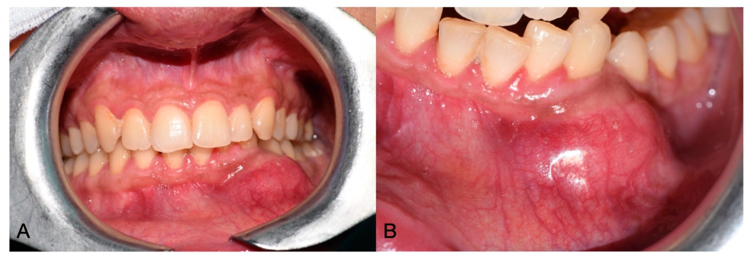
Figure 1. Clinical initial situation in frontal vision (A), and close-up view of the lesion (B).
Oral 05 00057 g001
Figure 2. OPG (A) and CT (B) radiological findings.
Oral 05 00057 g002
Figure 3. Exposed lesion after elevation of a dull-thickness flap (A) and specimen after removal (B).
Oral 05 00057 g003
Figure 4. Radiographic appearance of the fibula flap and buccal osteosynthesis plate.
Oral 05 00057 g004
Figure 5. Pre-operative clinical situation (A,B) and OPG (C).
Oral 05 00057 g005
Figure 6. Postoperative OPG.
Oral 05 00057 g006
Figure 7. Clinical (A,B) and radiographic (C) situation after prosthetic delivery.
Oral 05 00057 g007
Disclaimer/Publisher’s Note: The statements, opinions and data contained in all publications are solely those of the individual author(s) and contributor(s) and not of MDPI and/or the editor(s). MDPI and/or the editor(s) disclaim responsibility for any injury to people or property resulting from any ideas, methods, instructions or products referred to in the content.